Embed Size (px)
Citation preview
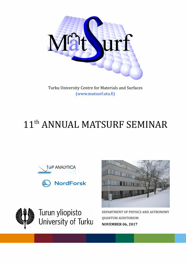
Turku University Centre for Materials and Surfaces(www.matsurf.utu.fi)
11th ANNUAL MATSURF SEMINAR
DEPARTMENT OF PHYSICS AND ASTRONOMY
QUANTUM AUDITORIUM
NOVEMBER 06, 2017
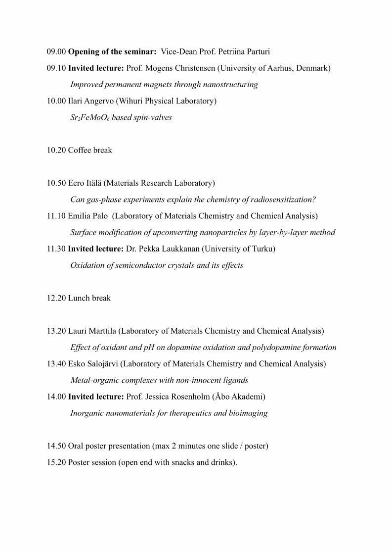
09.00 Opening of the seminar: Vice-Dean Prof. Petriina Parturi
09.10 Invited lecture: Prof. Mogens Christensen (University of Aarhus, Denmark)
Improved permanent magnets through nanostructuring
10.00 Ilari Angervo (Wihuri Physical Laboratory)
Sr2FeMoO6 based spin-valves
10.20 Coffee break
10.50 Eero Itälä (Materials Research Laboratory)
Can gas-phase experiments explain the chemistry of radiosensitization?
11.10 Emilia Palo (Laboratory of Materials Chemistry and Chemical Analysis)
Surface modification of upconverting nanoparticles by layer-by-layer method
11.30 Invited lecture: Dr. Pekka Laukkanan (University of Turku)
Oxidation of semiconductor crystals and its effects
12.20 Lunch break
13.20 Lauri Marttila (Laboratory of Materials Chemistry and Chemical Analysis)
Effect of oxidant and pH on dopamine oxidation and polydopamine formation
13.40 Esko Salojärvi (Laboratory of Materials Chemistry and Chemical Analysis)
Metal-organic complexes with non-innocent ligands
14.00 Invited lecture: Prof. Jessica Rosenholm (Åbo Akademi)
Inorganic nanomaterials for therapeutics and bioimaging
14.50 Oral poster presentation (max 2 minutes one slide / poster)
15.20 Poster session (open end with snacks and drinks).
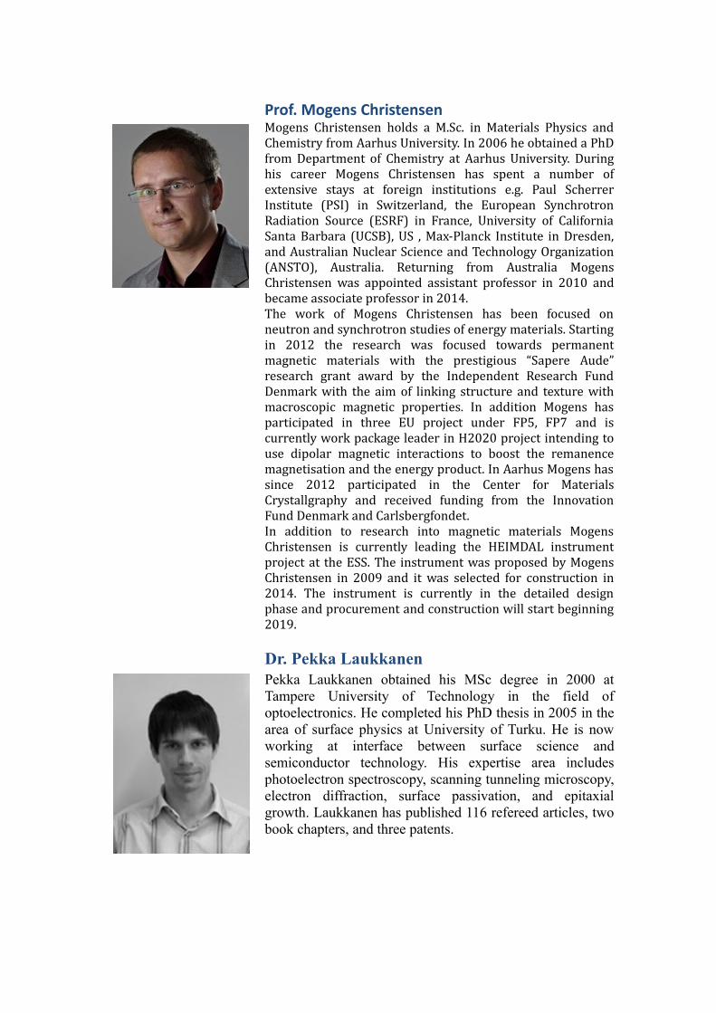
Prof. Mogens ChristensenMogens Christensen holds a M.Sc. in Materials Physics andChemistry from Aarhus University. In 2006 he obtained a PhDfrom Department of Chemistry at Aarhus University. Duringhis career Mogens Christensen has spent a number ofextensive stays at foreign institutions e.g. Paul ScherrerInstitute (PSI) in Switzerland, the European SynchrotronRadiation Source (ESRF) in France, University of CaliforniaSanta Barbara (UCSB), US , Max-Planck Institute in Dresden,and Australian Nuclear Science and Technology Organization(ANSTO), Australia. Returning from Australia MogensChristensen was appointed assistant professor in 2010 andbecame associate professor in 2014.The work of Mogens Christensen has been focused onneutron and synchrotron studies of energy materials. Startingin 2012 the research was focused towards permanentmagnetic materials with the prestigious “Sapere Aude”research grant award by the Independent Research FundDenmark with the aim of linking structure and texture withmacroscopic magnetic properties. In addition Mogens hasparticipated in three EU project under FP5, FP7 and iscurrently work package leader in H2020 project intending touse dipolar magnetic interactions to boost the remanencemagnetisation and the energy product. In Aarhus Mogens hassince 2012 participated in the Center for MaterialsCrystallgraphy and received funding from the InnovationFund Denmark and Carlsbergfondet.In addition to research into magnetic materials MogensChristensen is currently leading the HEIMDAL instrumentproject at the ESS. The instrument was proposed by MogensChristensen in 2009 and it was selected for construction in2014. The instrument is currently in the detailed designphase and procurement and construction will start beginning2019.
Dr. Pekka LaukkanenPekka Laukkanen obtained his MSc degree in 2000 atTampere University of Technology in the field ofoptoelectronics. He completed his PhD thesis in 2005 in thearea of surface physics at University of Turku. He is nowworking at interface between surface science andsemiconductor technology. His expertise area includesphotoelectron spectroscopy, scanning tunneling microscopy,electron diffraction, surface passivation, and epitaxialgrowth. Laukkanen has published 116 refereed articles, twobook chapters, and three patents.
Prof. Jessica RosenholmJessica Rosenholm holds a docentship in biomedicalnanotechnology at Åbo Akademi University, from whereshe also received her MSc(Tech) degree in chemicalengineering in 2002. Her doctorate period included a four-year funded position in the national Biomaterials and TissueEngineering Graduate School, from which she graduatedher DSc(Tech) degree in physical chemistry in 2008. Herthesis work “Modular Design of Mesoporous SilicaMaterials: Towards Multifunctional Drug DeliverySystems” has been awarded national and internationalprizes, e.g. the Akzo Nobel Nordic Research Prize 2009 forbest doctoral thesis and research activity in colloid andsurface science in the Nordic countries. In 2009-2010 shespent a postdoctoral period at the Nano BiomedicalResearch Centre, Med-X Research Institute, Shanghai JiaoTong University in China. Since returning to Finland in2010, she heads her own group, the BioNanoMaterialsgroup. The group’s activities are centered on thedevelopment of nanomedicines for drug delivery and/orimaging, for the enabling of a variety of diagnostic andtherapeutic applications. The designed systems are largelybased on mesoporous silica and its compositenanostructures. Since 1.1.2015 she is appointed professor inpharmaceutical development at the Pharmaceutical SciencesLaboratory, Faculty of Science and Engineering, ÅAU(www.pharmscilab.fi). She has co-authored nearly 100papers with 3839 citations and h-index 29 (GS 13.10.2017).Currently her research group is further involved informulating the already developed nanosystems intodifferent dosage forms including dermal patches,antibacterial coatings and varying 2D/3D printedformulations e.g. for tissue engineering.
ImprovedpermanentmagnetsthroughnanostructuringMogensChristensen
CenterforMaterialsCrystallography,DepartmentofChemistryandiNANO,AarhusUniversityLangelandsgade140,DK-8000AarhusC,Denmark.
Magnetic materials are the foundation for plethora of scientific and technologicalapplications.1,2Controllingthemagneticmaterialsoverlengthscalesfromtheatomicscaleviathe nanoscale to the microscopic scale is paramount for improving the performance ofmagnetic materials and make them useable in a number of applications.3 A promisingcandidateforarare-earthfreemagneticmaterialisthestrontiumhexaferriteSrFe12O19,dueto its pronounced magnetocrystalline anisotropy. By controlling the size and shape ofSrFe12O19nanocrystallites,itispossibletotailorthemagneticproperties.Thecontroloverthestructure and size has been achieved by varying synthesis parameters, such as pH,temperature and the metal concentration upon addition of the base. Subsequently it isnecessarytocontroltheorientationofthenanocrystallitegrainsconstitutingthemagnet.Themicrostructureiscontrolledduringthecompactionprocesses,wheresparkplasmasinteringisusedatelevatedpressureand temperatureresulting indensepelletswithhighlyalignedcrystal grains. In the talk the relation between synthesis condition and the resultingnanocrystalliteswillbedescribedalongwiththeobtainedmagneticproperties.4Thesamplecharacterization includes X-ray and neutron powder diffraction along with texturemeasurementsofthepellets.InadditiontothetalkonmagneticmaterialashortupdateontheplannedneutroninstrumentHEIMDALatESSwillbegiven.5HEIMDAL is auniquemulti-length scale instrument,whichwill covernineordersofmagnitude from0.5·10-10 to50·10-3m.The instrument combineshighresolutionorhighspeedthermalpowderdiffractionwithSANSandimaging.ItuniquelyfeaturesacoldandathermalguidetofulfilthediverserequirementsfordiffractionandSANS.HEIMDALwilltakeadvantageofthehighneutron fluxofthe longpulseatESS.Thepowderdiffractometerhasaq-rangecoverageofupto25Å-1allowingPDFanalysisoftotalscatteringdata.WiththeadditionofSANSwitha10m longcollimationsectionandmatchingdistancefromsampletodetector,HEIMDALwillbeabletocoverauniquelybroadlengthscalewithinasingleinstrumentalset-up.HEIMDALwillbeideallysuitedtocharacterisemagneticmaterialsonmultiplelengthscales,fromtheatomicscaletomagneticdomains.1.I.Matsui,JApplPhys1,2006,45,8302-8307.2.S.TammaruckwattanaandK.Ohyama,IeeeIndElec,2013,7157-7162.3.E.F.KnellerandR.Hawig,IeeeTMagn,1991,27,3588-3600.4.M.Saura-Múzquizetal.Nanoscale,2016,8,2857-28665.S.L.Holm,etal.NuclearInst.andMethodsinPhysicsResearch,A2016,828,229-241.
Sr2FeMoO6 based spin-valves
Ilari Angervo
Wihuri Physical Laboratory, Department of Physics and Astronomy, FIN-20014 University ofTurku, Finland
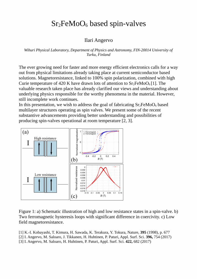
The ever growing need for faster and more energy efficient electronics calls for a wayout from physical limitations already taking place at current semiconductor based solutions. Magnetoresistance, linked to 100% spin polarization, combined with high Curie temperature of 420 K have drawn lots of attention to Sr2FeMoO6 [1]. The valuable research taken place has already clarified our views and understanding aboutunderlying physics responsible for the worthy phenomena in the material. However, still incomplete work continues.In this presentation, we wish to address the goal of fabricating Sr2FeMoO6 based multilayer structures operating as spin valves. We present some of the recent substantive advancements providing better understanding and possibilities of producing spin-valves operational at room temperature [2, 3].
Figure 1: a) Schematic illustration of high and low resistance states in a spin-valve. b)Two ferromagnetic hysteresis loops with significant difference in coercivity. c) Low field magnetoresistance.
[1] K.-I. Kobayashi, T. Kimura, H. Sawada, K. Terakura, Y. Tokura, Nature, 395 (1998), p. 677[2] I. Angervo, M. Saloaro, J. Tikkanen, H. Huhtinen, P. Paturi, Appl. Surf. Sci. 396, 754 (2017)[3] I. Angervo, M. Saloaro, H. Huhtinen, P. Paturi, Appl. Surf. Sci. 422, 682 (2017)
I
I
Low resistance
High resistance
-1
1
0
-0.4 -0.2 0 0.2 0.4Nor
mal
ized
mag
netiz
atio
n
B (T)
1. Ferromagnet2. Ferromagnet
-0.018-0.016-0.014-0.012
-0.01-0.008-0.006-0.004-0.002
0
-0.15 -0.1 -0.05 0 0.05 0.1 0.15
Nor
mal
ized
resi
stan
ce
B (T)
(a)
(c)
(b)
Can gas-phase experiments explain the chemistry of radiosensitization?
Eero Itäläa, Jesús González-Vázquezb, Yang Wangb, Hanna Myllynena, Dang Trinh Haa, Stephan Paul Deniflc,
Edwin Kukka
a Department of Physics and Astronomy, University of Turku b Universidad Autónoma de Madrid
c University of Innsbruck
Nitroimidazoles have recently attracted keen attention from several research groups focusing on molecular fragmentation induced by ionizing radiation.1-4 One reason for such attention is the clinical success in using nitroimidazoles to improve the effects of radiation therapy.5-7 According to the oxygen fixation hypothesi,8,9 the major contribution of the damage inflicted by ionizing radiation is caused indirectly by reactive oxygen species formed via the radiolysis of water molecules. These species form radicals when reacting with DNA. In an environment where free oxygen is present (aerobic environment), the oxygen falsely repairs the damaged DNA, making the repaired DNA function improperly. Under hypoxia, this radical formation is suppressed and DNA crosslink-repair is possible - thus the survival rate of the tumor cells is increased.
To increase the oxygenation using pure oxygen has proven to be very problematic. Actively respiring cells constantly consuming oxygen and the distance to the malignant tumors significantly limit the oxygen diffusion. This has led to the development of “oxygen mimetics”, compounds that match the chemical characteristics of molecular oxygen but have better penetration. Nitroimidazoles are probably the most common and one of the most extensively studied group of oxygen mimetics. However, despite the promising results on nitroimidazolic compounds in radiosensitization, the exact chemistry of how nitroimidazoles act as radiosensitizers is not fully understood. It has been suggested that the nitroimidazole molecules or its fragments mimic the action of oxygen, falsely repairing the DNA damage inflicted by the ionizing radiation (cf. the oxygen fixation hypothesis). Thus the ability to control the fragmentation could be extremely beneficial. References
1 K. Tanzer, et. al. Angew. Chem. Int. Ed., 2014, 53, 12240. 2 L. Feketeová, et. al., Phys. Chem. Chem. Phys., 2015, 17, 12598. 3 L. Feketeová, et. al, .J. Phys. Chem. A 2015, 119, 9986-9995. 4 P. Bolognesi, et. al., J. Chem. Phys. 2016, 145, 191102 5 R. Murata, et. al., Radiother. Oncol. 2008, 87, 331-338. 6 J. Overgaard, et. al., Int, J, Radiat, Oncol, Biol, Phys. 1989, 16, 1065-1068. 7 J. Overgaard, et. al., Radiother. Oncol. 1998, 46, 135-146. 8 P. Howard-Flanders, et. al., Radiat. Res. 1957, 7, 518–540. 9 Radiobiology For The Radiologist 6th edn (E. Hall and A. Giaccia) Lippincott William and Wilkins: Philadelphia, 2006.
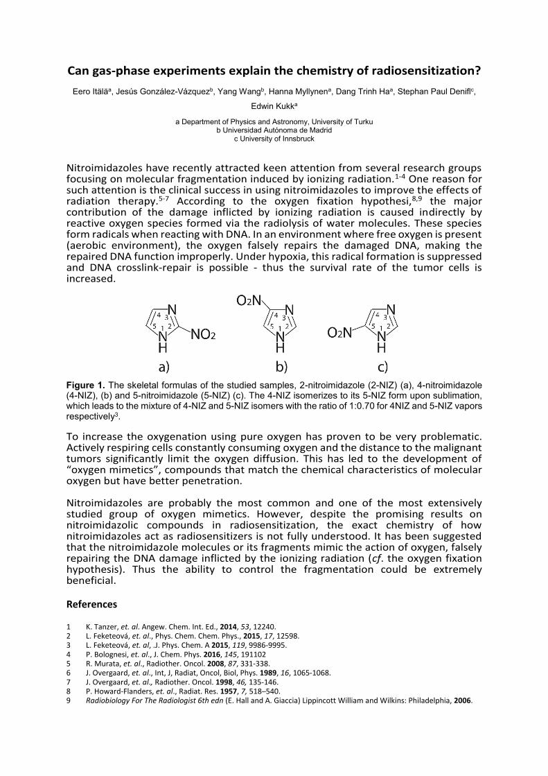
Figure 1. The skeletal formulas of the studied samples, 2-nitroimidazole (2-NIZ) (a), 4-nitroimidazole (4-NIZ), (b) and 5-nitroimidazole (5-NIZ) (c). The 4-NIZ isomerizes to its 5-NIZ form upon sublimation, which leads to the mixture of 4-NIZ and 5-NIZ isomers with the ratio of 1:0.70 for 4NIZ and 5-NIZ vapors respectively3.
SURFACE MODIFICATION OF UPCONVERTING NANOPARTICLESBY LAYER-BY-LAYER METHOD
Emilia Palo1,2, Mikko Salomäki1,3, Mika Lastusaari1,3
1 University of Turku, Department of Chemistry, FI-20014 Turku, Finland2 University of Turku Graduate School (UTUGS), Doctoral Programme in Physical and Chemical
Sciences, Turku, Finland3 Turku University Centre for Materials and Surfaces (MatSurf), Turku, Finland
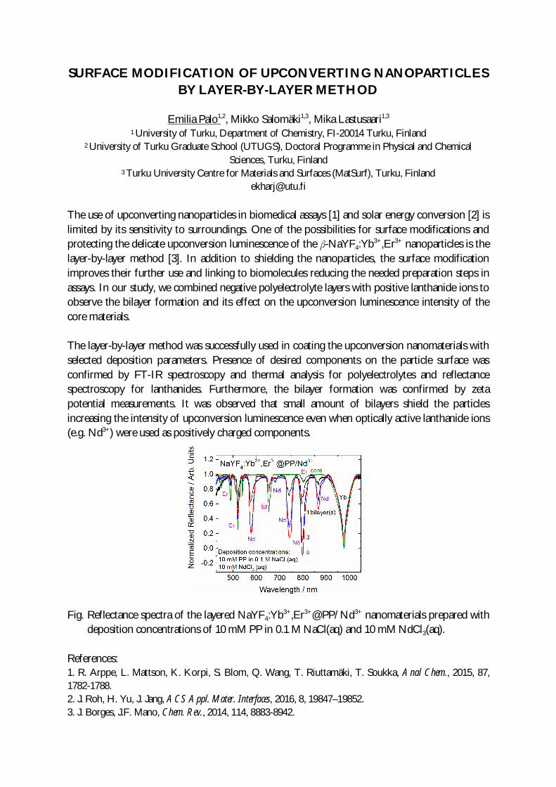
The use of upconverting nanoparticles in biomedical assays [1] and solar energy conversion [2] islimited by its sensitivity to surroundings. One of the possibilities for surface modifications andprotecting the delicate upconversion luminescence of the β-NaYF4:Yb3+,Er3+ nanoparticles is thelayer-by-layer method [3]. In addition to shielding the nanoparticles, the surface modificationimproves their further use and linking to biomolecules reducing the needed preparation steps inassays. In our study, we combined negative polyelectrolyte layers with positive lanthanide ions toobserve the bilayer formation and its effect on the upconversion luminescence intensity of thecore materials.
The layer-by-layer method was successfully used in coating the upconversion nanomaterials withselected deposition parameters. Presence of desired components on the particle surface wasconfirmed by FT-IR spectroscopy and thermal analysis for polyelectrolytes and reflectancespectroscopy for lanthanides. Furthermore, the bilayer formation was confirmed by zetapotential measurements. It was observed that small amount of bilayers shield the particlesincreasing the intensity of upconversion luminescence even when optically active lanthanide ions(e.g. Nd3+) were used as positively charged components.
Fig. Reflectance spectra of the layered NaYF4:Yb3+,Er3+@PP/Nd3+ nanomaterials prepared with deposition concentrations of 10 mM PP in 0.1 M NaCl(aq) and 10 mM NdCl3(aq).
References:1. R. Arppe, L. Mattson, K. Korpi, S. Blom, Q. Wang, T. Riuttamäki, T. Soukka, Anal Chem., 2015, 87,1782-1788.2. J. Roh, H. Yu, J. Jang, ACS Appl. Mater. Interfaces, 2016, 8, 19847–19852.3. J. Borges, J.F. Mano, Chem. Rev., 2014, 114, 8883-8942.
Oxidation of semiconductor crystals and its effectsP. Laukkanen1, J. Mäkelä1, M. Tuominen1, M. Kuzmin1, J.-P. Lehtiö1, J. Dahl1, M. Yasir1, S.Granroth1, M. Punkkinen1, K. Kokko1, M. Lastusaari2, R. Punkkinen3, H.-P. Hedman3, R. Félix4,V. Polojärvi5, J. Lyytikäinen5, A. Tukiainen5, M. Guina5
1 Department of Physics and Astronomy, University of Turku, FI-20014, Finland.2 Department of Chemistry, University of Turku, FI-20014, Finland.3 Department of Information Technology, University of Turku, FI-20014, Finland.4 Helmholtz-Zentrum Berlin für Materialien und Energie GmbH, D-14109, Germany.5 ORC, Tampere University of Technology, FI-33101, Finland.
A semiconductor surface becomes easily oxidized in uncontrolled way when asemiconductor crystal interacts with any oxygen-containing environment (e.g., air or growthconditions of insulating films) because the semiconductor oxidation is energetically veryfavored process, and because it is very difficult in practice to avoid the oxygen contact ofsemiconductor crystals. An oxidized semiconductor layer can be even several nanometersthick, and it is often a weak part of devices because oxidized semiconductor layers areamorphous and include many defects. These defects are harmful and cause non-radiativerecombination and increase leakage currents, for instance. The performance of solar cellsprovides a good example: The energy conversion efficiency is around 16-18% for theindustrial Si solar cell while the laboratory record is about 25% for the Si cell. This differenceis largely due to the lack of efficient passivation of the semiconductor surfaces for thecommercial cells [1]. To reduce the harmful effects of semiconductor oxidation, the surfaceparts of semiconductor crystals are often passivated or capped by insulator films like SiO2,Al2O3 or HfO2 in applications. Still, it is very challenging to avoid the oxygen contact ofsemiconductor surfaces (interfaces) due to manufacturing conditions of the passivationlayers for devices. In the recent studies of our group, one target has been to tackle theproblem of semiconductor oxidation, using our surface-science knowledge. We have studiedthe surface oxidation and passivation in particular for Si, Ge, and III-V semiconductormaterials. To connect the surface-science measurements to the electrical properties, wehave characterized the materials by photoluminescence and produced capacitors, detectors,and transistors components.
[1] G. Dingemans and W. M. M. Kessels, Review Article: Status and prospects of Al2O3-based surface passivation schemes for silicon solar cells, J. Vac. Sci. Technol. A 30 (2012)040802.
EFFECT OF OXIDANT AND pH ON DOPAMINE OXIDATION AND POLYDOPAMINEFORMATION
Lauri Marttila1,2, Tuomo Ouvinen1, Mikko Salomäki1,3, Henri Kivelä1,3, Jukka Lukkari1,3
1 Department of Chemistry, University of Turku, Vatselankatu 2, FI-20500 Turku, Finland2 University of Turku Graduate School, Chemical and Physical Sciences
3 Turku University Centre for Materials and Surfaces (MatSurf)
Melanins are biological pigments with several intriguing physicochemical properties and are widelydistributed in nature. In particular, melanins have been identified as potential anode and cathodematerials in power sources for biodegradable electronics.1
Synthetic polydopamine resembles natural melanins and it can be easily obtained by the oxidationof dopamine with various oxidants. Even dissolved oxygen can oxidize dopamine intopolydopamine in basic aqueous media.2 However, the formation of good-quality melanin-type filmsis problematic. The oxidation process can be controlled by the choice of oxidant and solution pHand these factors were studied in detail.
We have studied the autoxidation and metal-induced oxidation of dopamine experimentally usingUV-Vis spectroscopy and electrochemical techniques. Polydopamine nanoparticles have beencharacterized by various methods, e.g. X-ray Photoelectron Spectroscopy (XPS). A thermodynamicmodel was developed to explain the dependence of the initial steps of these reactions on pH.
The general pathway of the polydopamine formation from dopamine is well known (scheme 1).3
The oxidation process has two critical elementary initial steps (1) the oxidation of dopamine todopaminesemiquinone, and (2) the practically irreversible intramolecular cyclization of theunprotonated amino group in dopaminequinone to form leucodopaminechrome.
We have studied how Ce(IV) and Fe(III) can oxidize dopamine to polydopamine even in mildlyacidic aqueous solution in anaerobic conditions. Cu(II) is also an interesting alternative. Cu(II)alone is an ineffective oxidant and does not increase the reaction rate enough in acidic solution butchloride ions increase the oxidizing power of the Cu(II)/Cu(I) redox pair.
Scheme 1. Generally accepted mechanism for the oxidation of dopamine to polydopamine (metalsions are not taken into account)
1 Bettinger, C. J., Trends Biotechnol. 2015, 33, 575–585.2 Lee, H.; Dellatore, S. M.; Miller, W. M.; Messersmith, P. B., Science 2007, 318, 426−430.3 Salomäki, M.; Tupala, M.; Parviainen, T.; Leiro, J.; Karonen, M.; Lukkari, J., Langmuir 2016, 32, 4103-4112.
Metal-organic complexes with non-innocent ligands
MSc. Esko Salojärvi
Inorganic Materials Chemistry Group, Laboratory of Materials Chemistry and Chemical Analysis,Department of Chemistry, University of Turku
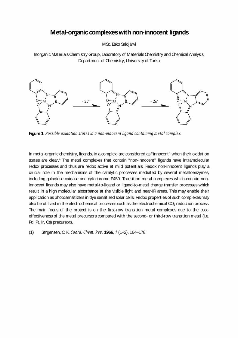
Figure 1. Possible oxidation states in a non-innocent ligand containing metal complex.
In metal-organic chemistry, ligands, in a complex, are considered as “innocent” when their oxidationstates are clear.1 The metal complexes that contain “non-innocent” ligands have intramolecularredox processes and thus are redox active at mild potentials. Redox non-innocent ligands play acrucial role in the mechanisms of the catalytic processes mediated by several metalloenzymes,including galactose oxidase and cytochrome P450. Transition metal complexes which contain non-innocent ligands may also have metal-to-ligand or ligand-to-metal charge transfer processes whichresult in a high molecular absorbance at the visible light and near-IR areas. This may enable theirapplication as photosensitizers in dye sensitized solar cells. Redox properties of such complexes mayalso be utilized in the electrochemical processes such as the electrochemical CO2 reduction process.The main focus of the project is on the first-row transition metal complexes due to the cost-effectiveness of the metal precursors compared with the second- or third-row transition metal (i.e.Pd, Pt, Ir, Os) precursors.
(1) Jørgensen, C. K. Coord. Chem. Rev. 1966, 1 (1–2), 164–178.
Inorganic nanomaterials for therapeutics and bioimaging
Jessica M. Rosenholm – Pharmaceutical Sciences Laboratory – Åbo Akademi University
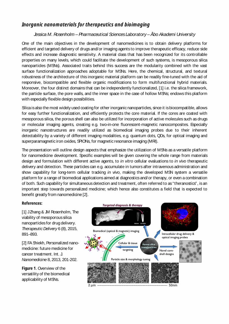
One of the main objectives in the development of nanomedicines is to obtain delivery platforms forefficient and targeted delivery of drugs and/or imaging agents to improve therapeutic efficacy, reduce sideeffects and increase diagnostic sensitivity. A material class that has been recognized for its controllableproperties on many levels, which could facilitate the development of such systems, is mesoporous silicananoparticles (MSNs). Associated traits behind this success are the modularity combined with the vastsurface functionalization approaches adoptable for MSNs. Here, the chemical, structural, and texturalrobustness of the architecture of this inorganic material platform can be readily fine-tuned with the aid ofresponsive, biocompatible and flexible organic modifications to form multifunctional hybrid materials.Moreover, the four distinct domains that can be independently functionalized, [1] i.e. the silica framework,the particle surface, the pore walls, and the inner space in the case of hollow MSNs; endows this platformwith especially flexible design possibilities.
Silica is also the most widely used coating for other inorganic nanoparticles, since it is biocompatible, allowsfor easy further functionalization, and efficiently protects the core material. If the cores are coated withmesoporous silica, the porous shell can also be utilized for incorporation of active molecules such as drugsor molecular imaging agents, creating e.g. two-in-one fluorescent-magnetic nanocomposites. Especiallyinorganic nanostructures are readily utilized as biomedical imaging probes due to their inherentdetectability by a variety of different imaging modalities, e.g. quantum dots, QDs, for optical imaging andsuperparamagnetic iron oxides, SPIONs, for magnetic resonance imaging (MRI).
The presentation will outline design aspects that emphasize the utilization of MSNs as a versatile platformfor nanomedicine development. Specific examples will be given covering the whole range from materialsdesign and formulation with different active agents, to in vitro cellular evaluations to in vivo therapeuticdelivery and detection. These particles can e.g. accumulate in tumors after intravenous administration andshow capability for long-term cellular tracking in vivo, making the developed MSN system a versatileplatform for a range of biomedical applications aimed at diagnostics and/or therapy, or even a combinationof both. Such capability for simultaneous detection and treatment, often referred to as “theranostics”, is animportant step towards personalized medicine; which hence also constitutes a field that is expected tobenefit greatly from nanomedicine [2].
References:
[1] J Zhang & JM Rosenholm, Theviability of mesoporous silicananoparticles for drug delivery.Therapeutic Delivery 6 (8), 2015,891–893.
[2] FA Shiekh, Personalized nano-medicine: future medicine forcancer treatment. Int. J.Nanomedicine 8, 2013, 201-202.
Figure 1. Overview of theversatility of the biomedicalapplicability of MSNs.

POSTERS (in alphabetical order)
01 Ilari AngervoSurface topography and electrical properties in Sr2FeMoO6 films studied atcryogenic temperatures.
02 Sergio E. DomínguezStudies on cationic conjugated polyelectrolytes
03 Mukarram Zaman Khan Growth temperature and thickness optimizations of superconducting YBCO thin films deposited on buffered hastelloy C276 tapes
04 Niko MoritzBioactive glass surface for fibre reinforced composite implants via surface etching by excimer laser: in vitro and in vivo results
05 Ermei MäkiläHydrolytic stabilization and chemical modification of porous silicon nanoparticles for biomedical applications
06 Isabella NorrboWhite emitting Li-hackmanite – a material with many uses
07 Tarek A. Omran Effect of interface surface texture on fracture behavior of bi-layered composites
08 Emilia Palo Surface modification of upconverting nanoparticles by layer-by-layer method
09 Janne PeltonenInfluence of relative humidity on the electrostatic charging of lactose powder mixed with salbutamol sulphate
10 Pasi SalonenBiomimetic oxoalkoxovanadium(v) complexes derived from proline-based aminophenol ligands
11 Khalil Shahramian Thrombogenicity of sol-gel derived TiO2 coated zirconia
12 Milla SuominenElectroactive composite materials for sustainable energy storage
13 Rahul YewaleVapor phase polymerized highly conducting PEDOT films as electrode andhigh capacitance material
Surface topography and electrical properties in Sr2FeMoO6
films studied at cryogenic temperatures.
I. Angervo1. 2, M. Saloaro1, J. Mäkelä2.3, J. -P. Lehtiö3, H. Huhtinen1 and P. Paturi
1 Wihuri Physical Laboratory, Department of Physics and Astronomy, FI-20014 University of Turku,Finland
2 University of Turku Graduate School (UTUGS), University of Turku, FI-20014 Turku, Finland3 Materials Research Laboratory, Department of Physics and Astronomy, FI-20014 University of
Turku, Finland
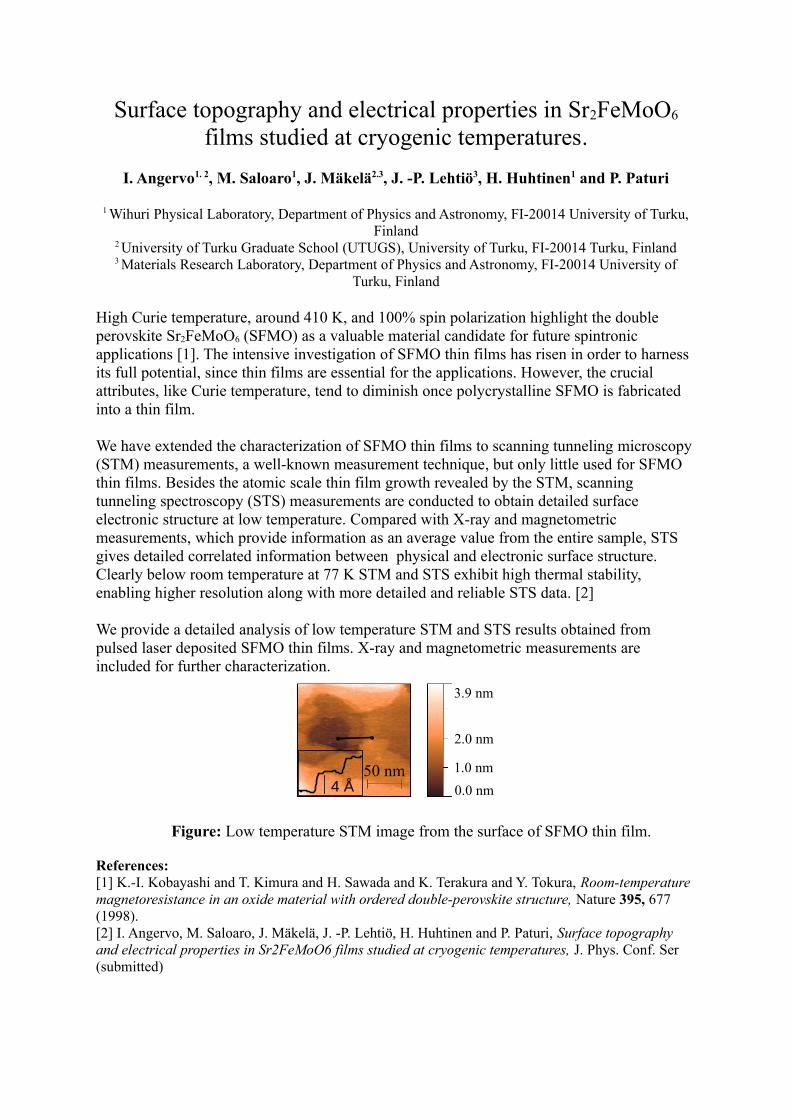
High Curie temperature, around 410 K, and 100% spin polarization highlight the double perovskite Sr2FeMoO6 (SFMO) as a valuable material candidate for future spintronic applications [1]. The intensive investigation of SFMO thin films has risen in order to harness its full potential, since thin films are essential for the applications. However, the crucial attributes, like Curie temperature, tend to diminish once polycrystalline SFMO is fabricated into a thin film.
We have extended the characterization of SFMO thin films to scanning tunneling microscopy (STM) measurements, a well-known measurement technique, but only little used for SFMO thin films. Besides the atomic scale thin film growth revealed by the STM, scanning tunneling spectroscopy (STS) measurements are conducted to obtain detailed surface electronic structure at low temperature. Compared with X-ray and magnetometric measurements, which provide information as an average value from the entire sample, STS gives detailed correlated information between physical and electronic surface structure. Clearly below room temperature at 77 K STM and STS exhibit high thermal stability, enabling higher resolution along with more detailed and reliable STS data. [2]
We provide a detailed analysis of low temperature STM and STS results obtained from pulsed laser deposited SFMO thin films. X-ray and magnetometric measurements are included for further characterization.
Figure: Low temperature STM image from the surface of SFMO thin film.
References:[1] K.-I. Kobayashi and T. Kimura and H. Sawada and K. Terakura and Y. Tokura, Room-temperature magnetoresistance in an oxide material with ordered double-perovskite structure, Nature 395, 677 (1998).[2] I. Angervo, M. Saloaro, J. Mäkelä, J. -P. Lehtiö, H. Huhtinen and P. Paturi, Surface topography and electrical properties in Sr2FeMoO6 films studied at cryogenic temperatures, J. Phys. Conf. Ser (submitted)
50 nm
3.9 nm
2.0 nm
1.0 nm
0.0 nm4 Å
Studies on cationic conjugated polyelectrolytesSergio E. Domínguez, Pia Damlin and Carita Kvarnström
Turku University Centre of Materials and Surfaces (MatSurf), Laboratory of MaterialsChemistry and Chemical Analysis, University of Turku, Vatselankatu 2, 20014 Turku, Finland
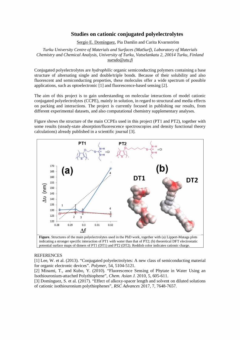
Conjugated polyelectrolytes are hydrophilic organic semiconducting polymers containing a basestructure of alternating single and double/triple bonds. Because of their solubility and alsofluorescent and semiconducting properties, these molecules offer a wide spectrum of possibleapplications, such as optoelectronic [1] and fluorescence-based sensing [2].
The aim of this project is to gain understanding on molecular interactions of model cationicconjugated polyelectrolytes (CCPE), mainly in solution, in regard to structural and media effectson packing and interactions. The project is currently focused in publishing our results, fromdifferent experimental datasets, and also computational chemistry supplementary analyses.
Figure shows the structure of the main CCPEs used in this project (PT1 and PT2), together withsome results (steady-state absorption/fluorescence spectroscopies and density functional theorycalculations) already published in a scientific journal [3].
Figure. Structures of the main polyelectrolytes used in the PhD work, together with (a) Lippert-Mataga plotsindicating a stronger specific interaction of PT1 with water than that of PT2; (b) theoretical DFT electrostaticpotential surface maps of dimers of PT1 (DT1) and PT2 (DT2). Reddish color indicates cationic charge.
REFERENCES[1] Lee, W. et al. (2013). “Conjugated polyelectrolytes: A new class of semiconducting materialfor organic electronic devices”. Polymer, 54, 5104-5121.[2] Minami, T., and Kubo, Y. (2010). “Fluorescence Sensing of Phytate in Water Using anIsothiouronium-attached Polythiophene”, Chem. Asian J. 2010, 5, 605-611.[3] Domínguez, S. et al. (2017). “Effect of alkoxy-spacer length and solvent on diluted solutionsof cationic isothiouronium polythiophenes”, RSC Advances 2017, 7, 7648-7657.
Poster Abstract
Growth temperature and thickness optimizations of superconductingYBCO thin films deposited on buffered hastelloy C276 tapes
M. Z. Khan ([email protected])a, Y. Zhaob, X. Wuc, M. Malmivirtaa,d, H. Huhtinena, P. Paturia
a Wihuri Physical Laboratory, Department of Physics and Astronomy, University of Turku, FI-20014 Turku, Finlandb Department of Electrical Engineering, Shanghai Jiao Tong University, 200240 Shanghai, People's Republic of China
c Shanghai Superconductor Technology Co. Ltd., 200240 Shanghai, People's Republic of Chinad University of Turku Graduate School (UTUGS), University of Turku, FI-20014 Turku, Finland
The growth mechanism of small-scale YBa2Cu3O6+x (YBCO) thin films laser deposited on a polycrystalline Hastelloy with new-fashioned and multilayered buffer layer architecture is studied from the flux pinning point of view. Compared to YBCOfilms grown on single crystal substrates, the most critical deposition issues that affect the suitable structural defect formation and thus the optimal vortex pinning landscapehave been observed to be the growth temperature and the film thickness evolution. In this work, we find that the best critical current density in a wide applied magnetic field range is realized in films grown at relatively low temperature and with intermediate thickness. This is explained to arise from the improved interface growth,film thickness related crystalline relaxation and from the formation of low-angle grain boundaries that form edge dislocations through the entire film thickness. Hence,the optimized buffer layer structure shown to be particularly suitable for new coated conductor solutions.
Acknowledgements
Jenny and Antti Wihuri Foundation is acknowledged for financial support. MM also wishes to thank the Finnish Cultural Foundation and the University of Turku Graduate School.
BIOACTIVE GLASS SURFACE FOR FIBER REINFORCED COMPOSITE IMPLANTS VIA SURFACE ETCHING BY EXCIMER LASER: IN VITRO AND IN VIVO RESULTS
Kulkova J, Moritz* N, Huhtinen H, Mattila R, Donati I, Marsich E, Vallittu PK
*Department of Biomaterials Science and Turku Clinical Biomaterials Centre – TCBCInstitute of Dentistry, University of Turku and others
In skeletal reconstructions, composites, such as bisphenol-A-glycidyldimethacrylate resin reinforced with glass fibers, are potentially useful alternatives to metallic implants. Recently, we reported a novel method to prepare bioactive surfaces for these composites. Surface etching by Excimer laser was used to expose bioactive glass granules embedded in the resin. The purpose of this study was to analyze two types of bioactive surfaces created by this technique. The surfaces contained bioactive glass and hydroxyapatite granules.
The selected processing parameters were adequate for the creation of the surfaces. However, the useof porous hydroxyapatite prevented the complete exposure the granules. In cell culture, for bioactive glass coatings, the pattern of proliferation of MG63 cells was comparable to that in the positive control group (Ti6Al4V) while inferior cell proliferation was observed on the surfaces containing hydroxyapatite granules. Scanning electron microscopy revealed osteointegration of implants with both types of surfaces.
The technique is suitable for the exposure of solid bioactive glass granules. However, the long-term performance of the surfaces needs further assessment.
Hydrolytic stabilization and chemical modification of porous silicon nanoparticles for
biomedical applications
Ermei Mäkilä1, Chang-Fang Wang2, Outi Keinänen2, Ulrika Jakobsson2, Martti Kaasalainen1, Sébastien
Lecommandoux3 Kerttuli Helariutta2, Anu Airaksinen2, Hélder A. Santos2, Jarno Salonen1
1University of Turku, Finland 2University of Helsinki, Finland 3Universite de Bordeaux, France
Introduction The silicon hydride based initial surface termination of porous silicon (PSi) enables a wide
range of chemical modifications for tailoring the properties of PSi suitable for various biomedical
applications. The surface chemistry of PSi can be altered to enable both high hydrolytic stability against
gradual erosion under physiological conditions and to provide different routes for specific chemical
functionalizations.
Methods PSi nanoparticles were fabricated from monocrystalline Si wafers by electrochemical anodization,
and passivated against dissolution using acetylene based thermal hydrocarbonization (THCPSi). Further
specific functionalizations were conducted through thermal addition of either an α,ω –diyne or alkenoic acid
onto the THCPSi surface. These modifications enabled both CuAAC and SPAAC based click reactions for
binding specific molecules onto the THCPSi nanoparticles (NPs). The chemical modifications were studied
with FTIR and XPS spectroscopies. The erosion of the NPs was monitored with ICP-OES.
Results Compared to oxidized PSi, the dissolution rate of both THCPSi and COOH-terminated THCPSi can
slowed down considerably. Under sink conditions in pH 7.4 at 37°C, oxidized PSi NPs disappeared within
12h, while the plain and modified THCPSi showed only 5 weight-% erosion after one week.
As the THCPSi surface allowed further modifications into alkyne and –COOH terminated NPs, the
applicability of the particles was studied using CuAAC reaction for binding azide-modified peptides on the
alkyne-terminated NPs. The COOH-terminated particles were further modified into azide-terminated NPs
and using a SPAAC reaction, a 18F radiotracer drug was bound to the NPs successfully.
Discussion We have shown here that the dissolution rate of PSi NPs in physiological conditions can be
reduced while retaining the ability to use the surface of the NPs for specific chemical functionalization. This
was demonstrated by modifying the particles to enable two common click chemistry routes, CuAAC and
SPAAC.
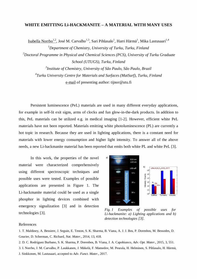
Fig. 1 Examples of possible uses forLi-hackmanite: a) Lighting applications and b)detection technologies [3].
WHITE EMITTING Li-HACKMANITE – A MATERIAL WITH MANY USES
Isabella Norrbo1,2, José M. Carvalho1,3, Sari Pihlasalo1, Harri Härmä1, Mika Lastusaari1,4
1Department of Chemistry, University of Turku, Turku, Finland2Doctoral Programme in Physical and Chemical Sciences (PCS), University of Turku Graduate
School (UTUGS), Turku, Finland3Institute of Chemistry, University of São Paulo, São Paulo, Brazil
4Turku University Centre for Materials and Surfaces (MatSurf), Turku, Finland
e-mail of presenting author: [email protected]
Persistent luminescence (PeL) materials are used in many different everyday applications,
for example in self-lit exit signs, arms of clocks and fun glow-in-the-dark products. In addition to
this, PeL materials can be utilized e.g. in medical imaging [1-2]. However, efficient white PeL
materials have not been reported. Materials emitting white photoluminescence (PL) are currently a
hot topic in research. Because they are used in lighting applications, there is a constant need for
materials with lower energy consumption and higher light intensity. To answer all of the above
needs, a new Li-hackmanite material has been reported that emits both white PL and white PeL [3].
In this work, the properties of the novel
material were characterized comprehensively
using different spectroscopic techniques and
possible uses were tested. Examples of possible
applications are presented in Figure 1. The
Li-hackmanite material could be used as a single
phosphor in lighting devices combined with
emergency signalization [3] and in detection
technologies [3].
References1. T. Maldiney, A. Bessiere, J. Seguin, E. Teston, S. K. Sharma, B. Viana, A. J. J. Bos, P. Dorenbos, M. Bessodes, D.
Gourier, D. Scherman, C. Richard, Nat. Mater., 2014, 13, 418.
2. D. C. Rodríguez Burbano, S. K. Sharma, P. Dorenbos, B. Viana, J. A. Capobianco, Adv. Opt. Mater., 2015, 3, 551.
3. I. Norrbo, J. M. Carvalho, P. Laukkanen, J. Mäkelä, F. Mamedov, M. Peurala, H. Helminen, S. Pihlasalo, H. Härmä,
J. Sinkkonen, M. Lastusaari, accepted to Adv. Funct. Mater., 2017.
Effect of interface surface texture on fracture behavior of bi-layered composites
Tarek A. Omran, Sufyan Garoushi, Lippo V. Lassila, Pekka K. Vallittu
Objectives: Bi-layered composite structures [BLS] are growing rapidly in restorative dentistry.Materials such as bulk-fill and fiber-reinforced composites require dentists to place a final occlusal-composite layer made of a different material. This provides structural-integrity and longevity to thewhole restoration. However, such a bi-layered system raises concerns about the interfacial segmentbetween the two materials. Therefore, this study aimed to evaluate the effect of different interfacial-surface textures on the load-bearing capacity of such BLS.
Methods: Cylindrical (πx3.52x5) specimens of BLS were prepared. Each specimen was made of a3mm base composite (bottom layer) and 2mm occlusal composite (top layer) (n=60). Two differentcomposite base materials where evaluated in this study; bulk-fill type SDR and fiber-reinforcedcomposite everX Posterior [EVERX]. While hybrid-type composite Gænial Posterior was used astop layer in all the specimen fabricated. Three different interfacial-textures were used in this study;(i) Pyramidal, (ii) longitudinal parallel grooves and (iii) Perpendicular grooves. 3D-printed moldswere fabricated to standardize the interfacial-texture between the top occlusal-composite and thebottom base composite materials. The base material was light-cured through the 3D-printed moldbefore application of the top-layer composite. The specimen where then statically-loaded with asteel ball (Ø 4mm) until fracture using a universal testing machine. Load values were evaluatedusing two-way ANOVA. Fracture types were classified into favorable or unfavorable bulk fracture.
Results:
Load-bearing capacities showed variations with different surface-textures. SDR showed lower meanload bearing capacity when compared to EVERX in all the surface-textures. Two-way ANOVArevealed that material had a significant effect on the load bearing capacity (p=0.008), whereassurface texture did not have a significant effect (p>0.05). Fracture analysis has shown that EVERXdemonstrated 80% favorable bulk-fractures whereas SDR demonstrated exclusively unfavorablebulk-fractures.
Conclusion: The presence of different interfacial-texture enhanced the fracture behavior of fiber-reinforced bi-layered composite structure.
Keywords: Bi-layered dental composite, interface surface texture, fiber composite

SURFACE MODIFICATION OF UPCONVERTING NANOPARTICLESBY LAYER-BY-LAYER METHOD
Emilia Palo1,2, Mikko Salomäki1,3, Mika Lastusaari1,3
1 University of Turku, Department of Chemistry, FI-20014 Turku, Finland2 University of Turku Graduate School (UTUGS), Doctoral Programme in Physical and Chemical
Sciences, Turku, Finland3 Turku University Centre for Materials and Surfaces (MatSurf), Turku, Finland
The use of upconverting nanoparticles in biomedical assays [1] and solar energy conversion [2] islimited by its sensitivity to surroundings. One of the possibilities for surface modifications andprotecting the delicate upconversion luminescence of the β-NaYF4:Yb3+,Er3+ nanoparticles is thelayer-by-layer method [3]. In addition to shielding the nanoparticles, the surface modificationimproves their further use and linking to biomolecules reducing the needed preparation steps inassays. In our study, we combined negative polyelectrolyte layers with positive lanthanide ions toobserve the bilayer formation and its effect on the upconversion luminescence intensity of thecore materials.
The layer-by-layer method was successfully used in coating the upconversion nanomaterials withselected deposition parameters. Presence of desired components on the particle surface wasconfirmed by FT-IR spectroscopy and thermal analysis for polyelectrolytes and reflectancespectroscopy for lanthanides. Furthermore, the bilayer formation was confirmed by zetapotential measurements. It was observed that small amount of bilayers shield the particlesincreasing the intensity of upconversion luminescence even when optically active lanthanide ions(e.g. Nd3+) were used as positively charged components.
Fig. Reflectance spectra of the layered NaYF4:Yb3+,Er3+@PP/Nd3+ nanomaterials prepared with deposition concentrations of 10 mM PP in 0.1 M NaCl(aq) and 10 mM NdCl3(aq).
References:1. R. Arppe, L. Mattson, K. Korpi, S. Blom, Q. Wang, T. Riuttamäki, T. Soukka, Anal Chem., 2015, 87,1782-1788.2. J. Roh, H. Yu, J. Jang, ACS Appl. Mater. Interfaces, 2016, 8, 19847–19852.3. J. Borges, J.F. Mano, Chem. Rev., 2014, 114, 8883-8942.
Influence of relative humidity on the electrostatic charging oflactose powder mixed with salbutamol sulphate
Janne Peltonen, Outi Alanen, Ermei Mäkilä, Matti Murtomaa, Jarno Salonen
University of Turku, Finland
Electrostatic charging, or triboelectrification, of powders has a significant effect on
pharmaceutical industry. The charging arises from powder−powder and
powder−surface contacts. The packing behaviour, flowability, and manufacturing of
the powders, among other things, may be hampered as a result. In dry powder
inhalers (DPIs), the drug is mixed with an additive powder, for instance with lactose.
Frictional contacts with these two unlike powders cause bipolar charging which can
lead to different problems. The oppositely charged drug particles may adhere on
lactose particles and end up in the throat instead of lungs. Moreover, if electrostatic
separation occurs during the transportation, mixing, and handling, the concentrations
of the drug−additive mixtures may considerably differ from the planned
concentrations. The charging may be reduced, for example, by increasing humidity.
However, the powders used in pharmaceutical industry are often sensitive to
moisture.
In this study, electrostatic charging of lactose and its mixtures with salbutamol
sulphate (SS) were studied as a function of relative humidity (RH). Powder adhesion
on a steel pipe surface was also investigated. The powders were charged by sliding
them in a steel pipe. Increase in RH decreased the charging of lactose and mixtures,
but the effect on SS was not evident. Furthermore, the charge of the mixtures was
reversed from negative to positive as RH was increased and remained positive as
the samples were again dried. Humidification also changed the adhesion behavior of
the mixtures onto the pipe surface.
Biomimetic oxoalkoxovanadium(v) complexes derived from proline-based aminophenol ligands
Pasi Salonena, Anssi Peuronenb and Ari Lehtonena*aLaboratory of Materials Chemistry and Chemical Analysis, Department ofChemistry, University of Turku, FinlandbLaboratory of Inorganic Chemistry, Department of Chemistry, University ofJyväskylä, Finland
The coordination chemistry of vanadium is generally studied due to thebiological relevance and catalytic properties of different vanadium-basedsystems.1-3 Especially, the role of vanadium in certain haloperoxidase andnitrogenase enzymes as well as the insulin-like effect of vanadiumcompounds have motivated synthetic chemists to prepare model compoundsfor structural studies along with for reactivity tests. For example, the activesites of haloperoxidase enzymes have been modelled by vanadiumphenoxides and catecholates. Moreover, artificial oxovanadium(V) complexescan be used as catalysts for a number of organic oxidation reactions.4
A common idea in coordination chemistry is to control the environment of themetal centre by using diverse ligands. Particularly, the electronic and stericproperties of ligands are used to tune the reactivity of metal species. In thepresent work, we have used phenol derivatives of L-proline as ligands tointroduce a chiral environment around the oxovanadium(V) centre.Consequently, four new complexes were prepared, characterized and studiesas catalyst precursors for epoxidation of cyclo-octene and sulfoxidation ofthioanisole by tBuOOH as well for the aerial oxidation of 3,5-di-tBu-catechol tothe corresponding o-quinone.
Figure 1. Active site of the haloperoxidase enzyme (left), the studiedcompounds (middle) with a crystal structure of 1 (right).
1D. C. Crans, J. J. Smee, E. Gaidamauskas, L. Yang, Chem. Rev. 2004, 104, 849.2C. Bolm, Coord. Chem. Rev., 2003, 237, 245.3M. Kirihara, Coord. Chem. Rev., 2011, 255, 2281.4 B. Floris, F. Sabuzi, A. Coletti, V. Conte, Catalysis Today, in press.
Thrombogenicity of sol-gel derived TiO2 coated zirconia
Khalil Shahramian,1,2 Aous Abdulmajeed,1,2,3 Timo Närhi,1,2,4 Lippo Lassila,1,2
1 Department of Prosthetic Dentistry and Stomatognathic Physiology, Institute of Dentistry,University of Turku, Lemminkäisenkatu 2, FI-20520 Turku, Finland2 Turku Clinical Biomaterials Centre–TCBC, University of Turku, Itäinen Pitkäkatu 4, 20520 Turku,Finland3 Department of General Practice, School of Dentistry, Virginia Commonwealth University,Richmond, Virginia4 Clinic of Oral Diseases, Turku University Central Hospital, Lemminkäisenkatu 2, 20520 Turku,Turku, Finland
Objective: This in vitro study was designed to evaluate the effect of sol-gel derived TiO2 coating on
zirconia to blood coagulation.
Methods: Square-shaped zirconia (10x10x2 mm3) was cut, ground, sintered and finally cleansed
ultrasonically in each of acetone and ethanol for 5 minutes. Two experimental groups (n=24) were
fabricated: (a) zirconia coated with sol-gel derived TiO2, (b) uncoated zirconia as control. The
coatings were prepared from tetraisopropyl orthotitanate solution by dip-coating. The
thrombogenicity of the specimens was evaluated using a whole blood kinetic clotting time method
where a 100 µl volume fresh human whole blood was placed on the coated and uncoated zirconia
surfaces (n=4) and incubated at room temperature for 10, 20, 30, 40, 50, and 60 min. 3 ml of
ultrapure water was then added at the end of each time point for lysing the non trapped red blood
cells and releasing hemoglobin into the water. After 5 min, each well was sampled in triplicate (200
µl each) and transferred to a 96-well plate. The concentration of hemoglobin in solution was
assessed by measuring the absorbance at 570 nm using ELISA plate reader. The optical density
reading is inversely related to clotting profile.
Results: Optical densities decreased gradually on both surfaces until 60 minutes. The total clotting
time was almost 40 minutes and 50 minutes for the coated and uncoated specimens, respectively.
Significant differences in the extent of clotting is observed at the 20, 30, 40, 50 minute time points
where the coated specimens demonstrate the lowest absorbance values reflecting the highest
amount of blood clotting.
Conclusions: TiO2 coating enhance blood coagulation on zirconia surface. Zirconia surface with
sol-gel derived TiO2 coatings exhibit better thrombogenicity in contact with blood.
Electroactive composite materials for sustainable energy storage
Milla Suominen, Pia Damlin, and Carita Kvarnström
Laboratory of Materials Chemistry and Chemical Analysis, University of Turku, Vatselankatu 2, FI-20014 Turku
In this work, composite materials of some electronically conducting polymers (ECPs) andelectrochemically reduced graphene oxide (ErGO) have been fabricated in an attempt toimprove the capacitance and cycle life of supercapacitors. Supercapacitors are promisingsustainable energy storage devices characterized by long cycle lives and high powerdensities, but so far their energy density is only 10 % of the energy density of a lithium ionbattery. ECPs have high intrinsic capacitances as well as simple and cheap manufacturingmethods, but swelling and shrinking during charging/discharging decreases their cycle life.On the other hand, graphene supercapacitors show remarkable cycling stabilities but exhibitlow energy densities.
We have combined poly(3,4-ethylenedioxythiophene) (PEDOT) and polyazulene (PAz) withErGO using ionic liquids (ILs) as electrolyte media to overcome each materials disabilities[1,2]. Graphene oxide (GO) produces stable dispersions in hydrophilic ILs [1]. It can beincorporated into ECP films electrochemically as a counter ion and it can be laterelectrochemically reduced inside the film [1]. This increases the capacitance of thecomposite due to change from electronically insulating material to conducting material. ILswere used as electrolyte since they improve the electrochemical activity of ECPs [3] andpossess better electrochemical stabilities than most conventional electrolytes allowingcomplete reduction of GO [4]. PEDOT has been extensively studied and shows quite goodproperties on its own, but we were able to increase its capacitance and stability byincorporating ErGO into the film in two different ILs [1,2]. PAz has received less interestthough it has been proven to contain high energy densities in organic solvents [5]. We haveshown that PAz exhibits higher capacitance than PEDOT in an asymmetric flexiblesupercapacitor using ILs as electrolyte [6], and by combining it with ErGO furtherimprovements on capacitance and cycling stability can be obtained [7].References[1] P. Damlin, M. Suominen, M. Heinonen, and C. Kvarnström, Ionic liquids for non-covalent modification ofgraphene sheets in conducting polymer composite materials, Carbon, 2015, 93, 533-543[2] S. Lehtimäki, M. Suominen, P. Damlin, S. Tuukkanen, C. Kvarnström, and D. Lupo, Preparation ofsupercapacitors on flexible substrates with electrodeposited PEDOT/graphene composites, ACS Appl. Mater.Int., 2015, 7, 22137-22147[3] A. Österholm, C. Kvarnström, and A. Ivaska, Ionic liquids in electrosynthesis and characterization of apolyazulene-fullerene composite, Electrochim. Acta, 2011, 56, 1490-1497[4] A. Viinikanoja, J. Kauppila, P. Damlin, M. Suominen, and C. Kvarnström, In situ FTIR and Ramanspectroelectrochemical characterization of graphene oxide upon electrochemical reduction in organic solvents,Phys. Chem. Chem. Phys., 2015, 17, 12115-12123[5] E. Grodzka, K. Winkler, B. M. Esteban, and C. Kvarnström, Capacitance properties of electrochemicallydeposited polyazulene films, Electrochim. Acta, 2010, 55, 970-978[6] M. Suominen, S. Lehtimäki, R.Yewale, P. Damlin, S. Tuukkanen, and C. Kvarnström, Electropolymerizedpolyazulene as active material in flexible supercapacitors, J. Power Sources, 2017, 356, 181-190[7] M. Suominen, P. Damlin, S. Granroth, and C. Kvarnström, Improved long term cycling ofpolyazulene/reduced graphene oxide composites fabricated in an ionic liquid, manuscript submitted to Carbon
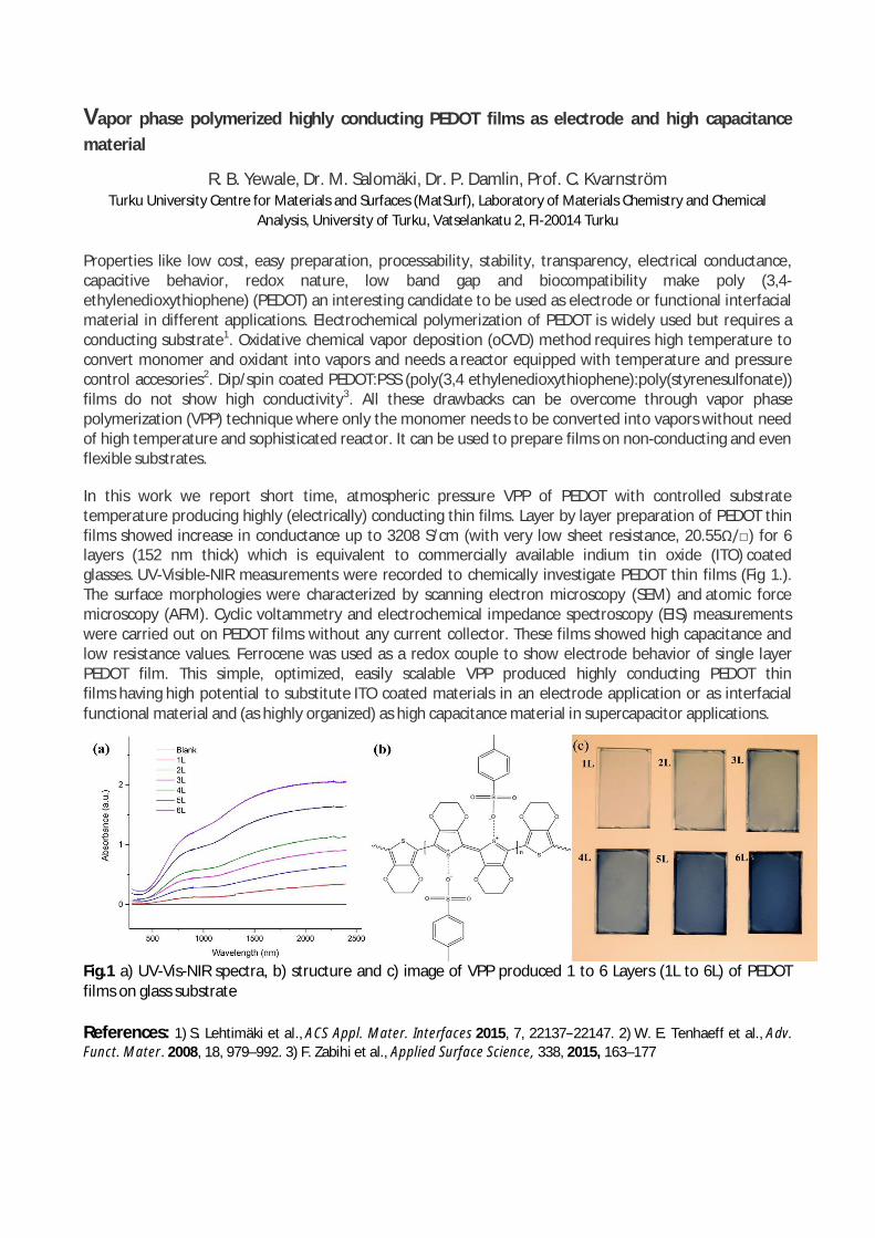
Vapor phase polymerized highly conducting PEDOT films as electrode and high capacitancematerial
R. B. Yewale, Dr. M. Salomäki, Dr. P. Damlin, Prof. C. KvarnströmTurku University Centre for Materials and Surfaces (MatSurf), Laboratory of Materials Chemistry and Chemical
Analysis, University of Turku, Vatselankatu 2, FI-20014 Turku
Properties like low cost, easy preparation, processability, stability, transparency, electrical conductance,capacitive behavior, redox nature, low band gap and biocompatibility make poly (3,4-ethylenedioxythiophene) (PEDOT) an interesting candidate to be used as electrode or functional interfacialmaterial in different applications. Electrochemical polymerization of PEDOT is widely used but requires aconducting substrate1. Oxidative chemical vapor deposition (oCVD) method requires high temperature toconvert monomer and oxidant into vapors and needs a reactor equipped with temperature and pressurecontrol accesories2. Dip/spin coated PEDOT:PSS (poly(3,4 ethylenedioxythiophene):poly(styrenesulfonate))films do not show high conductivity3. All these drawbacks can be overcome through vapor phasepolymerization (VPP) technique where only the monomer needs to be converted into vapors without needof high temperature and sophisticated reactor. It can be used to prepare films on non-conducting and evenflexible substrates.
In this work we report short time, atmospheric pressure VPP of PEDOT with controlled substratetemperature producing highly (electrically) conducting thin films. Layer by layer preparation of PEDOT thinfilms showed increase in conductance up to 3208 S/cm (with very low sheet resistance, 20.55Ω/) for 6layers (152 nm thick) which is equivalent to commercially available indium tin oxide (ITO) coatedglasses. UV-Visible-NIR measurements were recorded to chemically investigate PEDOT thin films (Fig 1.).The surface morphologies were characterized by scanning electron microscopy (SEM) and atomic forcemicroscopy (AFM). Cyclic voltammetry and electrochemical impedance spectroscopy (EIS) measurementswere carried out on PEDOT films without any current collector. These films showed high capacitance andlow resistance values. Ferrocene was used as a redox couple to show electrode behavior of single layerPEDOT film. This simple, optimized, easily scalable VPP produced highly conducting PEDOT thinfilms having high potential to substitute ITO coated materials in an electrode application or as interfacialfunctional material and (as highly organized) as high capacitance material in supercapacitor applications.
Fig.1 a) UV-Vis-NIR spectra, b) structure and c) image of VPP produced 1 to 6 Layers (1L to 6L) of PEDOTfilms on glass substrate
References: 1) S. Lehtimäki et al., ACS Appl. Mater. Interfaces 2015, 7, 22137−22147. 2) W. E. Tenhaeff et al., Adv.Funct. Mater. 2008, 18, 979–992. 3) F. Zabihi et al., Applied Surface Science, 338, 2015, 163–177

NOTES

ABSTRACT # 1
Turku University Centre for Materials and Surfaces(www.matsurf.utu.fi)
Participating laboratories:
Laboratory of Materials Research (Prof. Kalevi Kokko, Prof. Edwin Kukk)Laboratory of Industrial Physics (Doc. Jarno Salonen)Wihuri Physical Laboratory (Prof. Kurt Gloos, Prof. Petriina Paturi)Laboratory of Materials Chemistry and Chemical Analysis
(Prof. Carita Kvarnström, Dr. Mika Lastusaari, Prof. Jukka Lukkari)Turku Clinical Biomaterials Centre – TCBC (DDS Lippo Lassila,
Prof. Pekka Vallittu)Laboratory of Electron Microscopy (Dr. Markus Peurla,
Prof. emeritus Lauri Pelliniemi)
Sponsors of this year’s MatSurf Seminar:
Department of Physics and AstronomyTop Analytica Oy Ab, TurkuNordForsk