Embed Size (px)
Citation preview

Chapter 1
Introduction
Chapter 1
Introduction
1.1 Fundamentals of Medical Instrumentation
The field of computers in medicine is quite broad. During the last
quarter of the century, there has been a tremendous increase in the use of
the electrical and electronic equipment in the medical field for clinical and
research purposes. In a medical instrumentation system (MSI), the main
function is to measure and determine the presence of some physical
quantity that may be useful for diagnostic purposes. Therefore, many types
of instrumentation systems are used in hospitals and physicians clinics.
Knowledge of the structure of the living body and its function is essential
for understanding the functioning of most of the medical instruments. The
science of structure of the body is known as “ANATOMY” and that of its
function is known as, “PHYSIOLOGY”. Figure 1.1 shows the three basics
types of data that must be acquired, manipulated, and archived in the
hospital. Alphanumeric data include the patient’s name and address,
identification number, results of lab tests and physician’s notes. [89]

1.2 Overview of Medical Instruments
There are many different types of medical instruments. Our work is related
and concentrated on monitoring and analyzing physiological signals from a
patient using ECG.
Figure 1.2 shows block diagram that characterizes such instruments.
Sensors measure the patient’s physiological signals and produce electrical
signals that are analogs of the actual signals. A set of electrodes may be used
to sense a potential difference on the body surface commonly used in ECG.
The electrical signals produced by the sensors are interfaced to the processor
which is responsible for processing and analysis of the signals. The processor
block typically includes a microprocessor for performing the necessary tasks.
Many instruments have the ability to display, record, or distribute through a
network either the raw signals captured by the processor or the results of its
analysis. In some instruments, processor performs a control function [90].
Based on the results of the signal analysis, the processor performs a control
Fig. 1.1 Types of Medical Data
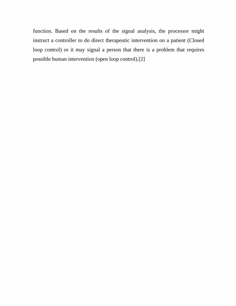
function. Based on the results of the signal analysis, the processor might
instruct a controller to do direct therapeutic intervention on a patient (Closed
loop control) or it may signal a person that there is a problem that requires
possible human intervention (open loop control).[2]

1.3 Physiological System Of The Body
Human body is a complex engineering marvel, which contains
various types of systems such as electrical, mechanical, hydraulic,
pneumatic, chemical and thermal etc. These systems communicate
internally with each other and also with an externally environment
[49][53]. By means of a multi-level control system and communications
network, the individual systems enable the human body to perform useful
tasks, sustain life and reproduce itself.
1.3.1 The Cardiovascular System
Patient Sensors Processor
Controller
Physiological signals Electrical Analogs (Voltages)
Display Recorder Network
Open/Closed Loop Control
Fig. 1.2 Basic Element of a Medical Instrumentation System

The cardiovascular system is a complex closed hydraulic system,
which performs the essential service of transportation of oxygen, carbon
dioxide, numerous chemical compounds and the blood cells. Structurally,
the heart is divided into right and left parts [53]. Each part has two
chambers called atrium and ventricle. The heart has four valves (seen in
figure 1.3): [90]
Tricuspid valve or right atrio-ventricular valve – between right
atrium and ventricle. It consists of three flaps or cusps. It prevents
backward flow of blood from right ventricle to right atrium.
Bicuspid Mitral or left atrio-ventricular valve – between left atrium
and left ventricle. The valve has two flaps or cusps. It prevents
backward flow of blood from right ventricle to atrium.
Pulmonary valve – at the right ventricle. It consists of three half
moon shaped cusps. This does not allow blood to come back to the
right ventricle.
Aortic valve- between left ventricle and aorta. Its construction is
like pulmonary valve. This valve prevents the return of blood back
to the ventricle from aorta.
The heart wall consists of three layers:
i) The Pericardium, which is the outer layer of the heart. It keeps the
outer layer moist and avoids friction as the heart beats.
ii) The Myocardium is the middle layer of the heart. It is main muscle
off the heart, which is made up of short cylindrical fibres. This muscle is
automatic in action, contracting and relaxing rhythmically throughout life.

iii) The Endocardium is the inner layer of the heart which provides
smooth lining for blood to flow.
The blood is carried to the various parts of the body through blood vessels,
which are hollow tubes. There are three types of blood vessels;
i) Arteries are thick walled and they carry the oxygenated blood away
from the heart.
ii) Veins are thin walled and carry de-oxygenated blood towards the
heart.
iii) Capillaries are the smallest and last level of blood vessels. They are
so small that the blood cells, which make blood, actually flow one at a
time through them. There are estimated to be over 800,000 km of
capillaries in human being, which include all the arteries and veins, which
carry blood.
Figure 1.3: Structure of the Heart

1.4 Overview of Cardiovascular diseases
Cardiovascular disease (CVD) including heart diseases and strokes,
accounts a major cause of morbidity and mortality in today’s world and
will become the leading cause of death and disability worldwide by 2020.
It is the leading cause of death in the USA with almost 2000 Americans
dying each day, i.e. one death in every 43 seconds. Among the non
communicable diseases, CVD are the leading cause of death responsible
for 30% of all deaths (17.5 million people in 2005 worldwide). According
the recent estimate cases of CVD may increase from 2.9 crores in 2004 to
as many as 6.4 crores in 2015. Prevalence rate of CVD in rural population
will continue to increase reaching around 13.5% in the age group of 60-69
years by 2015. The diseases constituting its range of fatal expression
include heart attack, myocardial infarction, acute coronary syndrome,
congestive failure, strokes, kidney disease and peripheral vascular disease
occur when the blood flow is blocked owing to the presence of blood clot
while strokes are the results of blocked or burst blood vessels in the brain.
Congenital heart defects and a range of other condition which occur due to
improper pumping of blood cause long term problems, and even deaths for
sufferers.
1.5 Introduction to Electrocardiography
One of the main techniques for diagnosing heart disease is based on
the electrocardiogram (ECG). The electrocardiograph or ECG machines
permits deduction of many electrical and mechanical defects of the heart
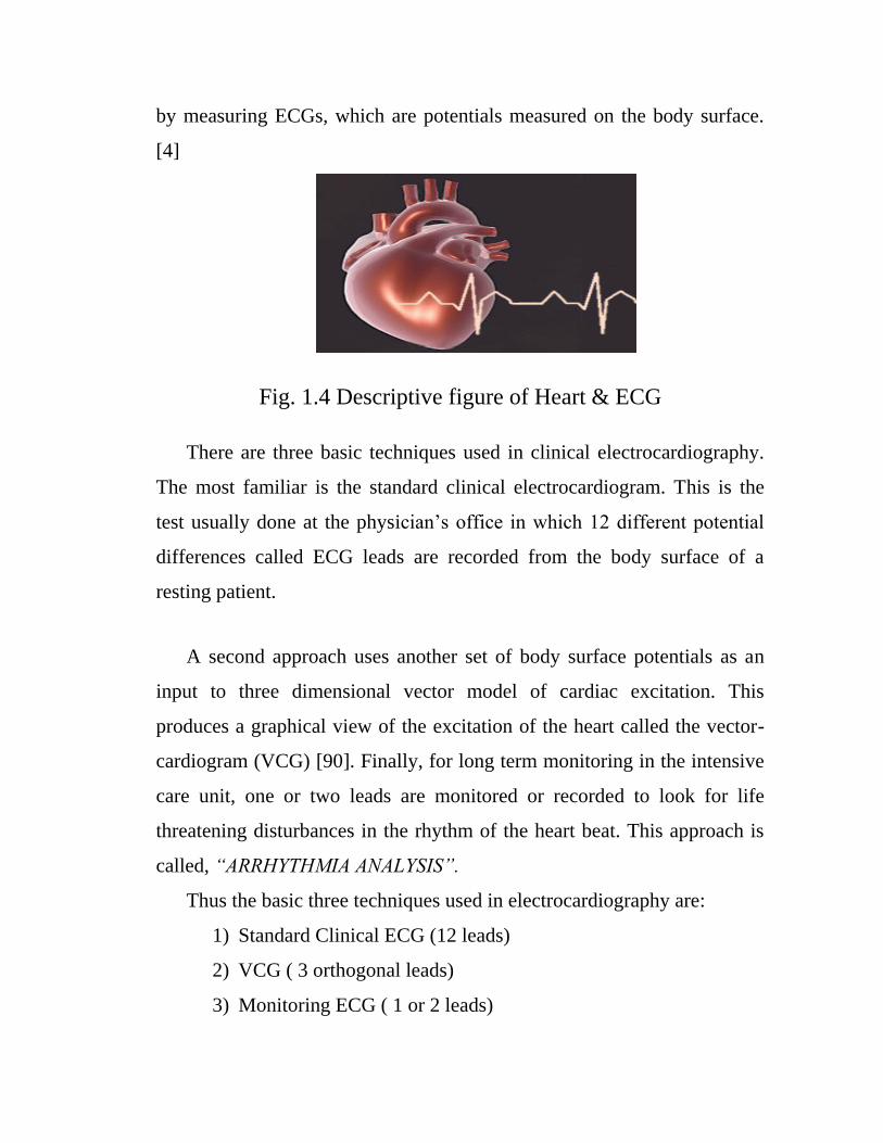
by measuring ECGs, which are potentials measured on the body surface.
[4]
Fig. 1.4 Descriptive figure of Heart & ECG
There are three basic techniques used in clinical electrocardiography.
The most familiar is the standard clinical electrocardiogram. This is the
test usually done at the physician’s office in which 12 different potential
differences called ECG leads are recorded from the body surface of a
resting patient.
A second approach uses another set of body surface potentials as an
input to three dimensional vector model of cardiac excitation. This
produces a graphical view of the excitation of the heart called the vector-
cardiogram (VCG) [90]. Finally, for long term monitoring in the intensive
care unit, one or two leads are monitored or recorded to look for life
threatening disturbances in the rhythm of the heart beat. This approach is
called, “ARRHYTHMIA ANALYSIS”.
Thus the basic three techniques used in electrocardiography are:
1) Standard Clinical ECG (12 leads)
2) VCG ( 3 orthogonal leads)
3) Monitoring ECG ( 1 or 2 leads)

Figure 1.5: Objective of the electrocardiography is to deduce the
electrical & mechanical condition of the heart by making non-
invasive body surface potential measurements.
Figure 1.5 shows the basic objective of electrocardiography. By looking at
the electrical signals recorded only on the body surface, a completely non-
invasive procedure, cardiologists attempt to determine the functional state
of the heart [89]. Although ECG is an electrical signal, changes in the
mechanical state of the heart lead to changes in how the electrical
excitation spreads over the surface of the heart, thereby changing the body
surface ECG. The study of cardiology is based on the recording of the
ECGs of thousands of patients over many years and observing the
relationships between various waveforms in the signal and different
abnormalities. Thus clinical electrocardiography is largely empirical,
based mostly on experiential knowledge.
1.6 Definition of ECG
An electrocardiogram (ECG or EKG, abbreviated from the German
Electrocardiogram) is a graphical representation of potential difference on
body surface, which records the electrical voltage in the heart in the form
of a continuous strip graph. Working of the cardiac system can be judged
from this ECG, representing the muscular contractions that the heart does
Condition of
the heart
Body Surface
Potentials
Electrocardiogram
Empirical

in-order to pump blood to the vascular system. In general, the
electrocardiogram is a technique of recording bioelectric currents
generated by the heart [7]. Clinicians can evaluate the conditions of a
patient’s heart from the ECG and perform further diagnosis. ECG records
are obtained by sampling the bioelectric currents sensed by several
electrodes, known as leads.
1.6.1 Characteristics of ECG
Figure 1.6 Characteristics of ECG w.r.t. the anatomy of Heart
The normal electrocardiogram (ECG) is comprised of characteristic
deflections referred to as P, Q, R, S, and T waves. The P wave is caused by
the current generated just before the contraction of the atria. The complex
QRS wave is the result of the currents generated in the ventricles during

depolarization just prior to ventricular contraction, the R wave being the
dominant component [5].
Characteristic waveform of ECG is as follows:
Figure 1.7 Components of ECG Waveform
From figure above, we conclude that; [5]
a) P wave - Marks the beginning of ECG wave
b) PR segment - Measured from beginning of P wave to beginning of
QRS complex
c) QRS Complex - Complex formed out of Q, R & S wave with
duration less than 0.12 sec
d) Q-T interval - Measured from beginning of Q wave to end of T
wave
e) S-T interval - Interval between S wave & T wave
f) T wave - Marks the end of ECG wave
The above shown deflections or nodes have specific amplitude and
duration.

Amplitude Duration
P wave 0.25mv P-R Interval 0.12 to
0.20 sec
Q wave 25% of R
wave
Q-T Interval 0.35 to
0.44 sec
R wave 1.60mv S-T 0.05 to 0.1 sec
T wave 0.1 to
0.5mv
P wave Interval 0.1
sec
QRS Interal0.09 sec
Note: These measurements are as per the standard ECG Signal on an ECG
sheet.
If these deflections and the interval between the points vary from standard
values then we can comment that there is some disturbance in the patient’s
rhythmic contractions. These disturbances are termed Cardiac arrhythmias.
Cardiac arrhythmia is a group of conditions in which the muscle
contraction of the heart is irregular or is faster or slower than normal.
Some arrhythmias are life-threatening medical emergencies that can cause
cardiac arrest and sudden death. Others cause aggravating symptoms, such
as an awareness of a different heart beat, or palpitation, which can be
annoying. Some are quite benign and normal [7].
1.6.2 Electrical Activity of Heart
The heart contains special group of cells called the ‘pacemaker
cells’ which have the ability to generate electrical activities on their own.
Electricity is produced when these cells change their electric charge from
positive to negative and back. The first electric wave is initiated at the top

of the heart in a heart beat and due to the inherent property of the heart
muscle cells to propagate electric charge to adjacent muscle cells; the
initial electric wave is enough to trigger a chain reaction. Specialized
fibers in the heart conduct the electrical impulse from the pacemaker to the
rest of the heart.
The three important parts of the heart’s electrical activity are (refer figure
1.6 Characteristics of ECG w.r.t. the anatomy of Heart) [53]
1. The SA node (Sinoatrial node) – It is the heart’s natural pacemaker
which initiates each heart beat
2. The AV node (Atrioventricular Node) - it acts as a bridge between
Atria and the ventricles, allowing electrical signals from the atria
into the ventricles.
3. His-Purkinje system- It carries the electrical signals throughout the
ventricles and consist of the following essential parts:
His-bundle
Right Bundle branch
Left bundle branch
Purkinje fibers
1.6.3 Blood Flow
The heart is filled with blood when then these heart muscles relax in
between heart beats and this process of relaxation is called diastole. Thus,
it can be summarized that the right side of the heart collects the
deoxygenated blood from the body and pumps it to the lungs for
oxygenation and release of waste products like CO2. The left side of the

heart collects oxygenated blood from the lungs and pumps it to the body
such that all the cells in the body receive the oxygen supply they require
for proper functioning. [47]
1.6.4 Uses of ECG
The ECG has a wide array of uses such as:
Determining the heart condition (normal or abnormal)
It may indicate acute or previous damage to heart muscles or
ischemia of heart muscle (angina)
Detection of potassium, calcium, magnesium, and other electrolytes
disturbances
Detection of conduction abnormalities
Used as screening tool for ischemic heart diseases during an
exercise tolerance test
Can provide information on physical conditions of the heart
Can suggest non cardiac diseases
1.7 Cardiac Arrhythmia
Cardiac arrhythmia is a term that denotes disturbances of the heart
rhythm. Cardiac arrhythmias can range in severity from entirely benign to
immediately life threatening. If arrhythmia is suspected, a cardiologist
should be consulted for confirmation. In addition, the use of natural
substances for arrhythmia should always be supervised by a doctor. A
cardiac Arrhythmia is also called as Dysrhythmia; disturbance in the
regular rhythm of the heartbeat. [20]

Normally the heart contracts 60 to 100 times per minute with each
contraction representing a heartbeat. Arrhythmias can occur when;
The natural pacemaker develops an abnormal rate
The normal flow of conduction is blocked
Another part of the heart acts as the pacemaker
It could also be caused by stress, caffeine, tobacco, alcohol and cough and
cold medicines. Some arrhythmias are life threatening medical emergency
that can cause cardiac arrest and sudden death others cause aggravating
symptoms, such as an awareness of a different heart beat, palpitation,
which can be annoying. Some are quite benign and normal. Sinus
arrhythmias are the mild acceleration followed by slowing of the normal
rhythm that occurs while breathing. In adults the normal heart rate ranges
from 60 to 100 bpm. The normal heart beat is controlled by a small area in
the upper chamber of the heart called the sinoatrial node or sinus node.
The sinus node contains specialized that have spontaneous electrical
activity that starts each heart beat.
1.7.1 Types of Cardiac Arrhythmia
Few of the commonly seen arrhythmias are listed below; [72]
1) Supraventricular Tachycardia – Supraventricular tachycardia
(SVT) is a series of very rapid heartbeats that begin in the heart’s upper
chambers. Generally supraventricular tachycardia is not a life threatening
problem although it can be quite bothersome and frightening. SVT may
occur when an extra pathway exists in the atria, the AV node or between
the atria and ventricles. An electrophysiology study can be used to identify
the origination site of SVT.

2) Atrial Fibrillation – In patients with atrial fibrillation,
multiple sites in the atria send electrical impulses in an uncoordinated
fashion. As a result, the atria beat very quickly and ineffectively. The AV
node, which acts as a relay station, allows only some of these impulses to
travel down the electrical system and simulate the ventricles. As a result,
the heart rhythm is irregular, erratic and usually rapid.
Figure 1.8: ECG Waveform – Atrial Fibrillation
3) Bradycardia: With a greater increase in the parasympathetic
tone and a decrease in sympathetic stimulation, the normal sinus rhythm
slows. Bradycardia is defined as slow heart rhythm. It is generally divided
into 2 categories, sick sinus syndrome and heart block. These slow heart
rhythms are usually treated by implantation of a permanent pacemaker,
which takes over the work of the hearts normal pacemaker.
Figure 1.9: ECG waveform – Bradycardia
4) Wolff-Parkinson-White Syndrome (WPW) – An abnormal
bridge of tissue connects the atria and ventricles. This extra pathway called

an accessory pathway makes it possible for electrical impulses to travel
from the atria to the ventricles without going through the AV node. Patient
with WPW syndrome experience arrhythmias when an impulse travels
down the AV node to the ventricles, and then up through the necessary
pathway to the atria. If the pulse continues to travel in the circular pattern,
it may cause the heart to contract with each cycle. This could result in a
very rapid heartbeat, which could allow extremely rapid and potentially
serious rhythms to occur.
5) Tachycardia - A reduction of parasympathetic tone or an
increase in sympathetic stimulation will raise the frequency of heartbeats.
When conduction is normal and the rate is greater than 120bpm, then the
rhythm is called sinus tachycardia.
Figure 1.10: ECG waveform - Tachycardia
6) Sinus Arrhythmia - This is characterized by variations in
heart rate from beat to beat that are greater than would be expected from
normal respiratory variation. It is irregular and caused by fluctuations of
autonomic tone that result in physic changes of the discharge rate.

Figure 1.11: ECG waveform - Sinus Arrhythmia
7) Bigeminy - (Latin: Bi-Two Gemini-twins) is a descriptor for a
heart arrhythmia in which abnormal heart beats occur every other
concurrent beat. A typical example is with bigeminal premature
ventricular beats, also known as premature ventricular
contractions/complexes (PVC). Following the PVC there is a pause and
then the normal beat returns - only to be followed by PVC.
Figure 1.12: ECG Waveform – Bigeminy
8) Ventricular Tachycardia - comes from the Greek words
tachys (rapid or accelerated) and kardia (of the heart). Tachycardia
typically refers to a heart rate that exceeds the normal range for a resting
heart rate (heart rate in an inactive or sleeping individual). In humans, the
upper threshold of a normal heart rate is usually based upon age,
sometimes it can be very dangerous depending on how hard the heart is
working and the activity.

Figure 1.13: ECG Waveform – Ventricular Tachycardia
9) Trigeminy - An arrhythmia in which 2 normal QRS
complexes are followed by a PVC; this rhythm usually does not progress
to dangerous forms of fast ventricular rhythms; in this rhythm, two VPCs
never occur one right after the other 2.
Figure 1.14: ECG Waveform – Trigeminy
1.7.2 Causes & Symptoms of Cardiac Arrhythmia
Cardiac arrhythmias occur when the heart beats improperly, as a
result of incorrect impulse generation, or impulse conduction. The heart
organ broadly differentiates into two types of cell, pacemaker cells, and
non-pacemaker cells. Pacemaker cells beat with their own independent
rhythm, in synchrony with their neighbors. The non pacemaker cells
require stimulus to beat or contract, this stimulus comes from the
pacemaker cells [72].

The symptoms of cardiac arrhythmia are not specially life
threatening, unless left untreated, cardiac arrhythmia can lead to more fatal
forms of rhythm disturbance, example: premature ventricular
depolarization may lead to ventricular fibrillation resulting in heart attack.
The signs and symptoms of cardiac arrhythmias range from completely
asymptomatic to loss of consciousness or sudden death. In general, more
severe symptoms are more likely to occur in the presence of structural
heart disease. For example, sustained monomorphic VT, particularly in a
normal heart, may be hemodynamically tolerated and causes marked
symptoms in patients with severe LV dysfunction. Complaints such as
lightheadedness, dizziness, quivering, shortness of breath, chest
discomfort, hart fluttering or pounding and forcefully or painful extra beats
are commonly reported with a variety of arrhythmias. Frequently patients
notice their arrhythmia only after checking peripheral pulses.

1.8 Basics of Patient Monitoring System
The objective of the patient monitoring is to have a quantitative
assessment of the important physiological variables of the patients during
critical periods of their biological functions. For diagnostic and research
purposes, it is necessary to know their actual value or trend of change.
Patient monitoring systems are used for measuring continuously or at
regular intervals, automatically, the values of the patient’s important
physiological parameters. There are several categories of patients who
may need continuous monitoring or intensive care. The long term
objective of patient monitoring is generally to reduce mortality and
morbidity by:
i) Organizing and displaying information in a form meaningful for
improved patient care,
ii) Correlating multiple parameters for clear demonstration of clinical
problems,
iii) Processing the data to set alarms on the development of abnormal
conditions,
iv) Providing information, based on automated data, regarding therapy,
v) Ensuring better care with fewer staff members.
The choice of proper parameters, which have high information
content, is an important issue in patient monitoring. It is, however,
generally agreed that monitoring of the following biological function is
often needed. Electrocardiogram (ECG), Heart Rate (Instantaneous or
Average), pulse rate, blood pressure, body temperature and respiratory rate
[37]. But the general requirements for patient monitoring equipments have
not changed much over the last past few decades. However, today’s

equipment monitors more parameters and processes more information.
Trends in monitoring include software control, arrhythmia monitoring,
Haemodynamics monitoring, monitoring during transportation of the
patient, and increased user friendly. With more than 10 parameters to
measure and scores of calculations to be made, the requirement for easy to
use user interface has assumed great significance.
1.8.1 Cardiac Monitoring
The most important physiological parameters monitored in the
intensive care units are the heart rate and the morphology or shape of the
electrical waveform produced by the heart. This is done to observe the
presence of arrhythmias or to detect changes in the heart rate that might be
indicative of a serious condition. Thus, a cardiac monitor is a specifically
useful for monitoring patients with cardiac problems and the special areas
in the hospitals where they are generally used are known as cardiac care
units or coronary care unit (CCU). These instruments are also called
‘Cardio-scopes’ and comprise of; [37]
i) Disposable type pre-gelled electrodes to pick up the ECG signal
ii) Amplifiers and a cathode ray tube for amplification and display of
ECG which enable direct observation of the ECG waveform.
iii) A heart rate meter indicates average heart rate with audible beep or
flashing light or both with each beat.
iv) An alarms system to produce signal in the event of abnormalities
occurring in the heart rate.
Modern ECG monitors not only include the non fade display facility but
also display heart rate along with the ECG trace.

1.8.2 Frequency response of Cardio-Scope
Some monitors have two selectable frequency response modes,
namely Monitor and Diagnostic [29]. In the ‘Monitor’ mode or ‘Filter-in’
mode, both the low frequency and high frequency components of an ECG
are attenuated. It is used to reduce baseline wander and high frequency
noise. The monitor mode bandwidth is generally 0.4 to 50 Hz (3dB
points). In the ‘Diagnostic’ mode, the instruments offers expanded
bandwidth capability of 0.05 to 100Hz.Some instruments include a 50 Hz
notch filter to improve the common mode rejection ratio and this factor
should be kept in mind while checking the frequency response of the
instrument. The patient circuit is isolated by using a transformer and by
modulating the 102 kHz carrier signal with amplified and filtered ECG.
The modulated carrier is demodulated in the cardio scope, amplified and
applied to an analog-to-digital converter. The converter samples the
waveform at a rate of 250samples/second, converts each sample to an 8-bit
parallel word, and enters the word into the re-circulating memory where it
replaces the oldest word stored. [12]

1.8.3 Proposed Patient Monitoring System
The proposed patient monitoring system performs following listed basic
tasks:
i) Detect ECG waveform using Embedded Controllers.
ii) Determine and display various deflections and nodes of the
waveform after accurate processing.
iii) Determine heart rate and display it.
iv) Determine various intervals (RR interval, PR segment, QRS
Complex interval)
v) Determine possible arrhythmia (cardiac disorder).
vi) Transmission of information to expert doctor available.
The whole system works under the control of the processor LPC2103
which works at 32MHz. The processor board for following listed
parameters is described in the subsequent sections;
1) Microprocessor board including analog signal multiplexer, A-D
converter and real time clock (Motherboard + ECG card + Power supply
card)
2) Video control board to convert the CPU commands into video
signals (HMI)
3) Motherboard including signal buses and analog input signal buffers
4) Keyboard – Using standard keyboard as the system is connected to
the computer

5) Software for calculation of heart rate, noise eradication, signal
processing, arrhythmia detection, transmission of signal, gain adjustment,
mode adjustment, and displaying of signal.