Embed Size (px)
Citation preview

76JAYPEE
Ajlana Mulic-LutvicaREVIEW ARTICLE
Postpartum UltrasoundAjlana Mulic-Lutvica
10.5005/jp-journals-10009-1228
ABSTRACT
This article describes uterine and uterine cavity changesthroughout the normal and pathological puerperium, as revealedby various ultrasound modalities. A gray scale ultrasound, colorand pulsed Doppler ultrasound and 3D ultrasound were used.It is based on results of several prospective longitudinal studies,which were designed so that every woman was examined atsix occasions during the puerperium, namely on postpartumdays 1, 3, 7, 14, 28 and 56. The first four examinations wereperformed transabdominally and the last two transvaginally. Thesame design was used in all studies. The involution process ofthe uterus was assessed by measuring the anteroposteriordiameter of the uterus and uterine cavity. Morphological findingswere recorded. The influence on the involution process of parity,breastfeeding, maternal smoking and infant's birth weight werealso evaluated. Besides conventional ultrasound, Dopplertechnology was used to study hemodynamic events occurringduring the normal puerperium. The pulsatility (PI) and resistance(RI) indices in the uterine arteries were measured, and thepresence or absence of early diastolic notches was recorded.A hypervascular area in myometrium was assessed by colorand power Doppler. Normal involution process was alsodescribed by means of 3D ultrasound. The volume of the uterusand uterine cavity after normal vaginal delivery were measuredusing virtual organ computer-aided analysis (VOCAL) using sixadjacent planes and a 30° rotation method. Reference valuesfrom uncomplicated puerperium were used when pathologicalconditions were studied.
Results from these studies, which shed light on normal andpathological changes during the puerperium, are summarizedin this chapter.
Keywords: Normal puerperium, Ultrasound, Uterus, Uterinecavity, Doppler, Uterine artery, 3D postpartum, VOCAL,Pathological puerperium.
How to cite this article: Mulic-Lutvica A. PostpartumUltrasound. Donald School J Ultrasound Obstet Gynecol 2012;6(1):76-92.
Source of support: Nil
Conflict of interest: None declared
INTRODUCTION
Postpartum period usually includes six subsequent weeksduring which normal pregnancy involution occurs and theuterus returns to the nonpregnant state. Our knowledge aboutpostpartum changes in the uterus has mainly been based onclinical examinations as well as from histological studiesfrom the end of the 19th century and the early part of the20th century when maternal mortality was high.1 Theinvolution of the uterus, as a main characteristic of thepuerperium was previously assessed by palpation of thefundal height.
Since, the introduction of ultrasound (USG) in clinicalpractice by Ian Donald et al2 in 1958 the uterus becameone of the first organs to be examined.3-7 However, fewstudies have focused on USG investigations during thepuerperium and results of published studies are notunambiguous.1-16 In published studies concerning theinvolution process, the length,4,6-9,11,12,14 width,8,9,12
anteroposterior diameter,3-7,11-13,16 area,9 thickness of theuterine wall10 and volume of the uterus and the uterinecavity,15 have been used as a measure of uterine involution.Majority of the studies described pathological conditionswithout knowledge about normal findings,4,5,8 they wererestricted to the early puerperium and designs were cross-sectional.3-7,12 A few studies concerning uterine cavityduring normal puerperium have been published.13-16
Postpartum complications involving the uterus occur inabout 8 to 10% of cases. Immediate and late postpartumhemorrhage, puerperal sepsis and septic pelvic thrombo-embolism are still potentially life-threatening conditions.Abnormal placentation (placenta accreta, increta or percreta)is a rare cause of postpartum hemorrhage that may continueafter delivery. Several studies investigated antenatalultrasound diagnosis of this condition17-23 but a few papershave focused on postpartum ultrasound monitoring ofretained placenta accreta.24 Ultrasound can help to diagnosevascular lesions, congenital or acquired,25-31 placental sitetumor32 and choriocarcinoma, which can also cause severepostpartum hemorrhage.
Thus, whenever puerperal complication occurs, theobstetricians should not hesitate to switch on ultrasoundmachine.
NORMAL PUERPERIUM
A description of normal ultrasound changes of the uterusand uterine cavity during puerperium is a prerequisite forultrasound diagnosis of pathological conditions. We canfollow the physiological involution of the uterus weighingmore than 1 kg soon after delivery to an organ weighing about80 gm at the end of the puerperium by means of ultrasound.The involution changes concerning the size, shape, positionand texture of the uterus have been relatively well-examinedby ultrasound.3-16 The influence on the involution process ofparity,7,9,11,13,15,16 route of delivery,11 oxytocin administrationduring labor7 breastfeeding6,7,9,11-13,15,16 or the infant’sweight11-13 have been studied. Previously published studies

Donald School Journal of Ultrasound in Obstetrics and Gynecology, January-March 2012;6(1):76-92 77
DSJUOG
Postpartum Ultrasound
involving sonographic examination of uterine cavity are notunambiguous.6,11,13-16
In the early and middle puerperium (in the first 2 weeks)the transabdominal approach is to be recommended. Arelatively short focal length of the vaginal probe limits itsuse during the early postpartum period, when the uterus istoo large and lies near the abdominal wall. In contrast, duringthe late postpartum period (> 2 weeks) a high frequencytransvaginal probe, which better distinguishes minor details,should be used. At that time, the uterus is considerablydecreased in size and it lies in the true pelvis. The postpartumuterus should be examined in three standard sections:sagittal, transverse and coronal (Figs 1 and 2). Urinarybladder should be moderately filled. Gentle compressionwith the probe should be used in order to avoid uterinedistortion.
We can differentiate three typical ultrasound imagesduring normal puerperium: In the early, middle and latepuerperium (Figs 3 and 4). The involution of the uterus is adynamic process that has no parallel process in normal adultlife.1 There are two physiological lifesaving processesoccurring soon after placenta delivery:1. Myotamponade (compression of the vessels by
myometrial contraction).2. Thrombotamponade (enhanced blood clotting activity).
The appearance of ultrasound finding in the earlypostpartum period reflects these physiological changes. Theuterus has an angulated form (Fig. 4A). It lies in a slightlyretroflexed position and arches over the sacral promontory.Wachsberg et al12 pointed out the impact of uterineangulation on the measurement of uterine length andrecommended segmental measurement. This angulated formof the early puerperal uterus is typical only in earlypuerperium and it is artificial. An extremely great degreeof uterine deformability is caused by a heavy uterine corpus,a hypotonic lower uterine segment and supine position ofthe examined woman. Lifesaving uterine contractionapproaches anterior and posterior uterine walls and just avirtual cavity appears. The uterine cavity is empty anddecidua appears as a thin white line from the fundus to thelevel of the internal cervical os (Fig. 4A). Sometimes, this
Figs 1A to C: Three standard ultrasound sections of thepuerperal uterus: (A) Longitudinal; (B) Coronal; (C) Transverse
Figs 2A to C: Transabdominal ultrasound scans of a normalpuerperal uterus on day 1: (A) Longitudinal scan; (B) Coronal scan;(C) Transverse scan
Figs 3A to C: The normal ultrasound appearance of the uterusand uterine cavity during the puerperium: (A) Transabdominalapproach during the early puerperium; (B) During the middle partof the puerperium; (C) Transvaginal approach during the latepuerperium
line can be irregular and thicker, which probably dependson the amount of retained decidua (Fig. 5A). The separationof the placenta and membranes generally occurs in thespongy layer; however the level varies. In 1931, Williamswrote concerning the line of separation of the placenta andmembranes: ‘While separation generally occurs in thespongy layer, the line is very irregular so that in places athick layer of decidua is retained, in others only a few layers
78JAYPEE
Ajlana Mulic-Lutvica
Figs 4A to C: Three typical USG images during normal puerperium: (A) In the early puerperium: uterus is retroverted. The cavity is seenas a thin white line; (B) In the middle puerperium: uterus is anteverted. An abundant fluid or mixed echo pattern with echogenic and echo-free area is seen in the whole cavity; (C) In the late puerperium: uterus is considerably decreased in size; the cavity is empty and appearsas a thin white line
Figs 5A to D: Transabdominal, longitudinal scans of the uterus from an uncomplicated puerperium: (A) On day 1;(B) On day 7; (C) On day 14; (D) On day 28
of cells remain, while in still others the muscularis ispractically bare”.33 The variation in sonographic appearanceof the cavity could be seen as a demonstration of thesephysiological variations in retained decidua. The white thinline seen on ultrasound might possibly represent cases inwhich only the basal decidual layer is retained or if themuscularis is practically bare (Fig. 4A). Whereas the thickerand more irregular lines might represent cases with retentionof more amount of spongy decidual layer and perhapsfragments of membranes (Fig. 5A).
Fluid or echogenic mass is not common finding in thecavity in the early postpartum period.13 Small echogenic orecholucent dots in the cavity are harmless physiological
findings.13,34 A heterogeneous mass with fluid and solidcomponents can be seen in the cervical area.13,14,34,35 Thisfinding has no clinical significance and the mass is usuallyexpelled spontaneously. It probably reflects a collection ofblood, blood clots and parts of membranes. On the posteriorwall of the uterus the prominent uterine vascular channelsare regularly seen.11 They usually disappear during the 2ndand 3rd postpartum weeks as a result of involution process,which decreases both the size and the amount of uterinevessels. Gas in the cavity is not common finding in the earlypostpartum period although it can be occasionally seen.13
Wachsberg detected gas in 19% of normal population duringthe early postpartum period.36
Donald School Journal of Ultrasound in Obstetrics and Gynecology, January-March 2012;6(1):76-92 79
DSJUOG
Postpartum Ultrasound
In the middle part of the puerperium (1-2 weekspostpartum) the uterus is diminished, the shape of the uterusis oval. It rotates along its internal cervical os toward ananteflexed position probably due to forming a firmisthmus.13 The vascular channels are not so prominent.Either pure fluid or mixed echo with fluid and solidcomponents can be seen in the whole cavity not only in thecervical area (Figs 4B, 5B and C). This finding reflects anormal healing process of the placental site inside uterinecavity, necrotic changes of retained decidua and an abundantshedding of lochia. Echogenic mass or gas is not commonfinding during middle part of the puerperium. In contrastEdwards et al15 found an echogenic mass in a greatproportion of normal puerperal women.
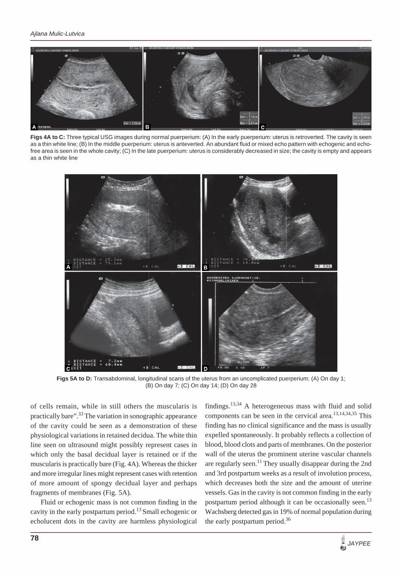
During late puerperium (>2 weeks postpartum), theuterus is considerably diminished (Figs 4C and 5D). It liesin an anteflexed position in 88% of cases.13 In 12 % of casesthe uterus has a retroflexed position corresponding well tonormal prevalence of retroversion of the uterus in generalpopulation (Fig. 6A). The uterine cavity is again empty.Decidua and necrotic vessel ends are exfoliated, theplacental site is recovered and a new endometrium isregenerated from the basal layer of the decidua adjacent tothe myometrium. Ultrasonically the cavity in the latepuerperium appears as a thin white line (Figs 4C and 5D).This corresponds to an inactive endometrium and reflectsthe hypoestrogenic state of the puerperium (‘the physiologicmenopause’). Sometimes, a small amount of fluid orechogenic dots can be seen (arrow) (Fig. 6B).
In 1953, Sharman performed endometrium biopsies andidentified fully restored endometrium from the 16thpostpartum day.37 In contrast, a study published in 1986 byOppenheimer38 showed that duration of puerperal lochiamay be up to 60 days in 13% of women. Similarly in arecently published study,39 on the duration of postpartumbleeding among 477 breastfeeding women, it was reported
that the median duration of lochia was 27 days with a rangefrom 5 to 90 days. Only 15% of the women reported thattheir lochia had stopped within two weeks postpartum. Theyalso pointed to the fact that bleeding associated with thepostpartum healing process commonly stops and startsagain. So, the normal physiological time span for theplacental site to recover is probably 4 to 6 weeks and nottwo weeks as previously considered.
Doppler Ultrasound During Normal Puerperium
Besides conventional ultrasound, Doppler technology isused to study hemodynamic events occurring during thepuerperium. Normal pregnancy requires the growth of manynew vessels. Consequently, during puerperium dramaticallyregressive changes must occur. The physiological involutionof the uterus involves not only muscle cells and deciduabut also the arteries. From histological studies, we knowthat normal involuted placental bed is characterized by adisappearance of trophoblasts and completely thrombosedspiral arteries.40-42 High diastolic flow velocities incombination with a disappearance of the early diastolicnotch are the main characteristics of the uterine arteryDoppler flow pattern from gestational week 20 to 26 andthey reflect the physiological conversion from high(nonpregnant) to low (pregnant) resistance state.43,44 Howfast these physiological changes return to the nonpregnantstate is a controversial issue.45-48 Tekay and Jouppila14
assessed the peripheral vascular resistance of the uterinearteries in 42 postpartum women and found that thepulsatility index (PI) increased significantly in earlypuerperium compared to pregnancy, remained unchangedduring the next 6 weeks and then gradually started toincrease again. However, nonpregnant values were notreached even three months after delivery. Jaffa et al,46 onthe other hand, described that PI decreased in the 2nd andremained relatively low until the 4th postpartum week.
Figs 6A and B: (A) Transvaginal ultrasound image of the uterus on day 28 postpartum shows a retroverted uterus on day 28 postpartum;(B) Transvaginal ultrasound image of the uterus on day 28 postpartum shows a small amount of fluid with echogenic foci in the cavity(white arrow)

80JAYPEE
Ajlana Mulic-Lutvica
Similar differences regarding the reappearance of the earlydiastolic notch have been reported. Tekay and Jouppila14
noted a reappearance of the early diastolic notch already inearly puerperium in 40 of 42 women, while Jaffa et al46
found that the early diastolic notch had reappeared in onlyone of 60 women five weeks postpartum.
According to our findings,48 in early puerperium themeans of Doppler flow resistance indices are higher thanthose reported in late pregnancy. Thereafter, they do notchange markedly until day 28 postpartum. On day 56postpartum, they are still lower compared to the valuesreported for nonpregnant women, which speaks for longerduration of physiological vascular return from a pregnantto a nonpregnant state. We observed a diastolic notch in13% of women on day one and in 90.6% of women on day56 postpartum (Figs 7A and B).
Color and power Doppler ultrasound may detect alocalized area of increased vascularity within themyometrium. It may be a common transient ultrasoundfinding if asymptomatic and it does not require treatment.47
THREE-DIMENSIONAL ULTRASOUNDPOSTPARTUM
Although the volume of the uterus and uterine cavity werepreviously measured using 2D ultrasound,15 the volumes
assessed by 3D (three-dimensional) ultrasound may providemore accurate measurements than does the conventionalultrasound. 3D ultrasound using VOCAL program (VirtualOrgan Computer-aided Analysis) has recently been used tomeasure the volumes of the uterus and the uterine cavityafter normal delivery.49 It is shown in Figures 8A and B.
3D power Doppler angiography is a new unexploredmethod for quantifying noninvasively the vascular networkof the uterus (Fig. 8C).
RETAINED PLACENTAL TISSUE
Both ultrasound diagnosis of RPT (retained placental tissue)and appropriate management for SPH (secondary postpartumhemorrhage) is still a controversial issue. SPH is defined asany abnormal bleeding from the uterus occurring between24 hours and 12 weeks postpartum49 and occurs in 1 to 2%of deliveries.50,51 In developed countries, half of postpartumwomen who are admitted to hospital with this conditionundergo uterine surgical evacuation.49-53 In developingcountries, it is a major contributor to maternal death.49 Themost common causes of SPH are abnormal involution of theplacental site in the uterine cavity that may be idiopathic42
or it can be caused by RPT54 or by endometritis.52
Subinvolution of the placental bed in the absence of RPT or
Figs 7A and B: (A) Normal flow velocity waveforms of the uterine artery on day 1 (Transabdominal approach) and; (B) 56(Transvaginal approach) postpartum
Figs 8A to C: (A) Three-dimensional USG of the volume of the uterus on day 28 and; (B) Uterine cavity on day 7 after normaldelivery; (C) With 3D power Doppler a localized area of increased vascularity within the myometrium is seen

Donald School Journal of Ultrasound in Obstetrics and Gynecology, January-March 2012;6(1):76-92 81
DSJUOG
Postpartum Ultrasound
endometritis is a distinctive entity, characterized by widelydistended spiral arteries, only partly occluded by thrombi ofvarious ages and invested with extravillous trophoblasts.40,42
The diagnosis, however, requires histological examinationand clinically it is a diagnosis of exclusion. Moreover,placental vascular subinvolution is often under-recognizedby general surgical pathologists.42 Carlan et al55 performedmanual exploration of the cavity on 131 asymptomaticwomen, 5 minutes after placental delivery and within2 minutes after an ultrasound examination. They found that24 of 131(18.8%) women had documented evidence of RPT.This is a surprisingly higher figure compared to Jones et al56
who performed manual intrauterine explorations routinelyafter 1000 births and removed placental fragments or bits ofmembranes in only 2 to 4% of cases. Defective decidua, whichcan be scanty or completely absent in some patients, is apredisposing factor for abnormal attachment of the placentaand for partially RPT.40 Vascular abnormalities of the uterushave recently been described as possibly more commoncauses of severe SPH than previously thought.25-31
In a Cochrane Review, Alexander et al50 identified 45papers about the management of SPH and concluded thatlittle information is available from randomized trials to guideclinicians in the management of this condition. Since, thecauses of SPH may be numerous, the best treatment optionsshould be chosen according to the underlying cause ofbleeding. However, an essential problem is that theunderlying cause of SPH often is unknown and that clinicalor ultrasound diagnosis of RPT, which is the indication forsurgical treatment, is still a controversial issue.58-68 Thedecision whether to perform uterine evacuation for RPTdepends on both, clinical finding and the ability to visualizeretained placenta by ultrasound.58-69 Although promptcurettage seems to be necessary, in many cases it usuallydoes not remove identifiable placental tissue. Moreover, itis more likely to traumatize the implantation site and incitemore bleeding. Consequently, the complications rate is high.Hoveyda et al reported in his review regarding secondarypostpartum hemorrhage that the frequency of perforationof the uterus was 3% and hysterectomy about 1%.54 Similarresults are reported from an audit of 200 cases concerningpuerperal curettage.70 They showed that 8.5% of patientsexperienced major morbidity and 7% required a repeatprocedure with further morbidity. In addition to immediatecomplications, late sequelae related to surgical treatmentfor SPH may influence the reproductive health of women.If curettage damages the endometrium 1 to 4 weekspostpartum, the endometrium may fail to regenerate, leadingto Asherman’s syndrome Jensen and Stromme.71
Westendorp et al72 prospectively examined 50 women
undergoing either a repeat removal of placental remnantsafter delivery or a repeat curettage for incomplete abortion.At a later hysteroscopy, 20 out of 50 (40%) women hadintrauterine adhesions. The prevalence of Asherman’ssyndrome is 2% after manual evacuation of the placentabut,37 5% after postpartum curettage.72 Recently, an updateon intrauterine adhesions has been published and theimportance of prevention has been emphasized.73
First studies concerning RPT performed with oldultrasound equipment showed high rate of false-positivediagnosis.4,5,8 Similar results have been obtained by modernultrasound equipment.58-68 Published studies havedemonstrated a variable sensitivity (42-94%) and specificity(62-92%) for ultrasound diagnosis of RPT.58-68 On the otherside, ultrasound appears as a valuable tool to confirm anempty cavity. Lee and Mandrazzo8 found empty cavity in20 of 27 patients with late puerperal bleeding. In only onecase, RPT was confirmed. The same authors reported thathistological confirmation was obtained in eight of ninepatients with ultrasound suspected RPT. Althoughultrasound technology improved considerably, the diagnosisof RPT is still difficult. Ultrasound finding of RPT mayvary depending on many different factors. We cannot expectthe same ultrasound image during early (Figs 4A and 5A)and late period of the puerperium (Figs 4C and 5D). Thepresence of blood, blood clots, necrotic decidua, membranesor gas can give various ultrasound images and a properdiagnosis is sometimes difficult. Nevertheless, the mostcommon ultrasound finding associated with RPT is anechogenic mass8,34-35,55,57-68 (Figs 9A to C, 10A and B, 11Aand B, 12A, 13A to C and 14A). In contrast, Edwards et al15
found in his study an echogenic mass on day 7 in 51% ofnormal cases, in 21% on day 14 and in 6% on day 21. Hequestioned ultrasound finding of an echogenic mass inuterine cavity as a sign of RPT. However, the definition ofan echogenic mass was not specified and we mayhypothesize that others investigators would probablyclassify many of their ‘echogenic mass’ as ‘heterogeneouspatterns’. A heterogeneous pattern is a common andinsignificant finding of the involuting uterus13 (Figs 4B and5C to D). It is located in the cervical area in the earlypuerperium, in the whole uterine cavity in the middle partof the puerperium and it is not common during latepostpartum period.13 Sokol et al16 used the sameclassification and found ‘echogenic material’ in 40% ofwomen 48 hours after a normal delivery. However, 14 ofthe 16 cases demonstrated echogenic material in the loweruterine segment, while only two had such findings in thefundus. It is unclear if ‘echogenic material’ is the same as

82JAYPEE
Ajlana Mulic-Lutvica
Figs 9A to D: Puerperal abnormalities revealed by ultrasound: (A) Retained placental tissue 2 days postpartum; (B) Blood flow in relationto retained placental tissue; (C) Retained placental tissue 6 weeks postpartum; (D) After curettage a thin, echogenic endometrium
Figs 10A and B: Puerperal abnormalities revealed by ultrasound: (A) Transabdominal transverse scan, 9 days postpartum, showsretained placental tissue seen as an echogenic mass; (B) A low resistance blood flow is seen on one side of the echogenic mass
Figs 11A and B: (A) Transvaginal longitudinal scan shows retained placental tissue 6 weeks postpartum; (B) By color Doppler,feeding vessels are seen inside the echogenic mass

Donald School Journal of Ultrasound in Obstetrics and Gynecology, January-March 2012;6(1):76-92 83
DSJUOG
Postpartum Ultrasound
Figs 12A and B: (A) Transabdominal GD: longitudinal scan, 11 days postpartum, shows retained placental tissue seen as anechogenic mass; (B) A thin echogenic endometrium is visible soon after curettage
Figs 13A to C: (A) Transabdominal longitudinal scan of the uterus on day 17 postpartum. Suspected retained placental tissue seen asan echogenic mass in the uterine cavity (red arrow); (B) 3D USG shows the volume of the suspected retained placental tissue; (C) PowerDoppler angiography, glass body mode shows vessels in the placental tissue (red arrow)
Figs 14A to D: Ultrasound image of placenta praevia perccreta left in situ: (A) Color Doppler and; (B) Power Doppler show the interfacebetween the uterus and urinary bladder 7 days after cesarean section; (C) Retained placenta occupies the most part of the uterine cavity(arrow); (D) Power Doppler shows increased myometrial vascularity behind the retained placenta (arrow)

84JAYPEE
Ajlana Mulic-Lutvica
an ‘echogenic mass’ or if it might be a mixed echo pattern.If dysfunctional postpartum bleeding persists for a long time,RPT is highly suspected. Hertzberg et al34 described so-called ‘stippled pattern’ of scattered hyperechogenic focithat later on became increasingly generalized echogenic,reflected secondary regressive changes in RPT (Figs 9Cand 11A).
Two studies61,62 compared the diagnostic accuracy ofclinical assessment with transabdominal USG in themanagement of SPH and concluded that both methods wereof limited value. In contrast, recently published studies thatassessed diagnostic accuracy of combined clinical andsonographic protocol, concluded that the combinedapproach was accurate and highly sensitive tool for thediagnosis of retained placental tissue.66-69
There are many reasons for discrepancies in thepublished reports. Factors that might explain the lowsensitivity and high false-positive rate include a vaguedefinition of the USG diagnosis of RPT,58-62 retrospectivestudy design34,60,64,65 and mixed study populations includingwomen with bleeding after an abortion and women withpostpartum hemorrhage.8,60,64-66 Three studies often citedin the published literature evaluated asymptomaticwomen.55,58 The accuracy of postpartum USG for detectionof RPT was calculated either from a small proportion ofwomen who underwent curettage, assuming that womenwho had an uneventful puerperal course after conservativetreatments had no RPT,34,60-62 or from histological findingsamong asymptomatic women.55,58 Finally, the patients andclinicians have not been blinded to the sonographic resultsin any of the published studies. If ultrasound finding showsan empty cavity with thin white decidua/endometriumduring early (Figs 2A and 4A) or late puerperium (Figs 4C,5D, 6A and 9D), pure fluid/heterogeneous content in thecavity during the middle part of the puerperium (Figs 4Band 5B and C), or only small echolucent or hyperechogenicdots throughout whole postpartum period, a clinicallysignificant amount of retained placental tissue isunlikely.13,34 Transvaginal ultrasound with high frequencyprobe as well as transvaginal sonohysterography may betterdifferentiate intrauterine puerperal pathology.74-77
Doppler Ultrasound During PathologicalPuerperium
A few studies investigated pulsed and color Doppler duringpuerperium in order to improve diagnostic accuracy ofultrasound regarding RPT.60,74,75 Some investigatorsobserved low resistance blood flow around intracavitarycontents74-78 (Figs 9A and 10A). Ashiron et al74 measuredresistance index (RI) in relation to RPT and found thatdiagnosis is highly suspected if RI is below 0.35 (Fig. 9B).
These patients are suitable for invasive treatment. RI above0.45 should exclude diagnosis. Values between 0.35 and0.45 form a ‘gray zone’ (Fig. 10B). Conservative treatmentand repeated ultrasound examinations should be performed.
Power Doppler seems to be a new unexplored modalitythat could improve our abilities to diagnose clinicallysignificant RPT. Retained placental tissue in the uterinecavity might cause a delay in the normal involution of uterinevessels.40,41 By color Doppler ultrasound, a localized areaof increased vascularity within the myometrium may bedetected.47,78-83 The presence of a hypervascular area in themyometrium, within or close to the echogenic mass, haspreviously been interpreted alternatively as a commonphysiological finding,47 as a finding associated with thepresence of RPT60,74,75,83,84 or with arteriovenous (AV)malformations.25,78,79 Pulsed Doppler usually demonstratesa low resistance turbulent flow with high systolic velocity,resembling AV malformations. It has recently beensuggested that curettage should not be performed on patientswho present with SPH and a color Doppler image of ahypervascular area within the myometrium.78,79 Van denBosch80 examined 385 consecutive postpartum women andreported that a hypervascular area in the uterus was relativelycommon (8.3%) and disappeared either spontaneously orafter removal of placental remnants. Mungen81 has drawnattention to a tendency to overdiagnose true AV malfor-mations. He pointed out that a majority of hypervascularareas in the myometrium probably represented normal ‘peri-villous flow’ in the spiral arteries. The regression periodmay be prolonged in the presence of RPT. Only in veryrare instances do they represent true arteriovenous malfor-mations. In our recent work on angiographic embolizationfor treatment of major postpartum hemorrhage, no true AVmalformation was diagnosed among 20 patients but fourcases had pseudoaneurysm85 (Figs 15A and B).
Our knowledge on uterine artery flow in women withRPT is sparse. It could be that RPT prevents the physio-logical changes in uterine blood flow during the puerperium.The results of our small study83 showed the resistance flowindices in uterine artery below the 10th percentile for 8 of20 (40.0%) women of which seven had histologicalconfirmation of RPT and one did not. There was, however,considerable overlap. No patient had resistance indicesabove the 90th percentile. In 12 of 20 (60.0%) patients, anearly diastolic notch was absent. Early diastolic notchesappeared relatively late compared to the findings in normalpopulation. Only one woman had a notch before postpartumday 28. Color Doppler showed a hypervascular area closeto the echogenic mass in 12 of 20 (60%) patients, all withhistologically confirmed RPT. This figure is slightly higher

Donald School Journal of Ultrasound in Obstetrics and Gynecology, January-March 2012;6(1):76-92 85
DSJUOG
Postpartum Ultrasound
than that reported by Durfee et al60 (55%) and by Zalel etal77 (46%). A hypervascular area was absent in eight patients(40%) of which six had an echogenic mass that washistologically confirmed RPT. Our findings that the absenceof blood flow does not exclude RPT are in concordancewith previously reported results.60,77
POSTPARTUM ENDOMETRITIS
Postpartum endometritis is a fairly common clinicalcondition, affecting 2 to 5% women following delivery.86,87
Cesarean section (CS) is the leading predisposing factor.88
It has been considered that the typical ultrasound finding incases of endometritis is the presence of gas in the uterinecavity.10 Madrazo found gas in uterine cavity in 15% ofpatients with puerperal endometritis.10 Nowadays, infectionscaused by gas-forming organisms C perfringens are veryrare and large gas-bubbles are almost never seen. Moreover,Wachsberg and Kurtz36 detected gas in about 19% of normalcases, which is in accordance with results of a computedtomographic study performed within 24 hours ofuncomplicated vaginal delivery (21%). Ultrasoundappearance of gas is seen as an intensively hyperechogenicfocus equivalent in echogenicity to bowel gas with cleanand dirty shadowing or a reverberation artefact.89 Accordingto our experience, gas is mostly observed followingintrauterine manipulations90 (Figs 12B and 16A) althoughit is occasionally observed after normal vaginal delivery.13
The detection of gas within the uterine cavity may be anormal finding during the puerperium and does notnecessarily indicate the presence of endometritis or RPT.13,36
After CS or intrauterine manipulations highly echogenicfoci can obscure an existing mass in the uterine cavity or bemistaken for retained placental tissue.34,90 Thus, whenever
highly echogenic foci are present in the uterine cavity, thephysician who interprets ultrasound finding must be awareof recent uterine manipulations. Gas usually disappearswithin 1 to 2 weeks after instrumentation90 (Fig. 16B).
Figs 15A and B: (A) Transabdominal scan of the uterus on day 8 postpartum shows a huge defect in the uterus forming apseudoaneurysm (arrow). Color Doppler reveals a feeding damaged uterine artery; (B) Angiography confirmed the ultrasound finding
Figs 16A and B: (A) Gas in uterine cavity one-daypost-curettage; (B) Six days postcurettage

86JAYPEE
Ajlana Mulic-Lutvica
Furthermore, it has been claimed that ultrasound image ofRPT and endometritis overlap.8,52 Results from publishedstudies on this issue are inconsistent.8,41,52-53,62,70,90 Pelageet al91 described 14 cases with uncontrollable SPH under-going selective angiographic embolization. Six of 14patients had clinical and ultrasound signs of endometritiswith RPT. In four cases, histological confirmation wasobtained. Two patients had pure endometritis. Conversely,Kong et al41 pointed out that endometritis appeared to be anoverstated cause of SPH. He found that less than 5% ofcases could be ascribed to endometritis. Ben-Ami et al62
found that a majority of the patients presenting with fever>38°C postpartum were falsely diagnosed by ultrasound ashaving suspected RPT.
The presence of RPT may result in intrauterine infectionand in these cases ultrasound examination can help us toselect patients suitable for invasive treatment. In the vastmajority of cases of isolated endometritis, ultrasoundfindings are normal and have no pathognomonic ultrasoundimage.35,90 Kirkinen et al45 found that blood flow to theinfected uterus could be different from normal. Deutchmanand Hartman described postpartum pyometra as a lucentarea within the uterus.92 They also advocated the usage ofUltrasound to assist in guiding a drainage procedure. Septicpelvic thrombophlebitis, well known as an ‘enigmaticpuerperal fever’ is another uncommon complication of thepuerperium. It most commonly presents in early postpartumperiod and antibiotic treatment is usually unsuccessful.Rudoff et al93 suggests ultrasound examination in case ofclinical suspicion of pelvic thrombophlebitis. Althoughultrasound diagnosis of ovarian vein thrombophlebitis iswell described,94-96 the diagnosis is still difficult and anultrasound expertise is needed. Asymmetric dilatation ofthe ovarian or other pelvic vein may sometimes beobserved.95 Furthermore a complex or hypoechoic mass nearthe lower pole of the kidney particularly in clinical settingof an ‘enigmatic puerperal fever’ should suggest thrombo-
phlebitis. An echogenic intracaval mass is considereddiagnostic and anticoagulation treatment should be added.96
Cesarean Section
Nowadays, when CS rates are continuously rising, higherincidence of all puerperal complications can be expected.88
The ultrasound image of the uterus following CS usuallyshows three distinctive patterns:1. Gas in the cavity.2. A small rounded area at the incision site that reflects
tissue reaction due to localized edema.3. Several echogenic dots at the incision site, which is
related to the type of closure and the suture materialused82,90,97 (Figs 17A and B). All these characteristicsare normal findings and no correlation with pathologicalconditions is found. The involution rate of the uterusfollowing CS is not markedly different from theinvolution rate after vaginal delivery.90,98-100 Weobserved a few morphological differences between thewomen delivered by CS and the women who had avaginal delivery, such as the less common; antevertedposition of the uterus and the empty uterine cavity inearly puerperium, which might reflect slightly delayeduterine involution process.90 The significant infectiousmorbidity is associated with CS.88 Ultrasound may beuseful in postpartum women with clinical suspicion ofa postoperative complication like phlegmona,101 abscess,pyometra, hematometra (Fig. 17C), wound infection,subfascial hematoma or intra-abdominal postoperativehemorrhage. Baker et al described bladder flaphematoma after a low uterine transverse CS.102 A solidor complex mass between the posterior bladder wall andthe anterior uterine wall may be observed by ultrasound.An abscess appears as a cystic structure with internaldebris surrounded by thicker irregular walls. An infectedhematoma initially has similar ultrasound appearance.During the resolution process, it may change and appearsmore solid. However, the physician must be aware that
Figs 17A to C: (A) The uterus after cesarean section—longitudinal section; (B) Coronal section—hyperechogenic scar in loweruterine segment; (C) Hematometra

Donald School Journal of Ultrasound in Obstetrics and Gynecology, January-March 2012;6(1):76-92 87
DSJUOG
Postpartum Ultrasound
ultrasound diagnosis is just a complement and clinicalcondition of the patient should guide the therapeuticapproach.
UNCOMMON BUT POTENTIALLYLIFE-THREATENING CAUSES OF POSTPARTUMBLEEDING
There are a few uncommon but potentially life-threateningcauses of postpartum bleeding which contribute significantlyto maternal mortality and morbidity. These are placentaaccreta/increta/percreta,17-23 vessel’s lesions (true AVmalformations,25 pseudoaneurysm26-31) placental sitetumor32 and choriocarcinoma.
Placenta Accreta/Increta/Percreta
The incidence of abnormal placentation, placenta accreta/increta/percreta, has increased in recent years, particularlydue to the increasing rates of cesarean section. It is the mostcommon factor for uncontrolled postpartum hemorrhageleading to emergency postpartum hysterectomy, which isassociated with significant maternal morbidity andmortality.17 Antenatal diagnosis of severe invasive placenta-tion is feasible with ultrasound. Both, conventional 2D and3D ultrasound have been used and several reports have beenrecently published.18-23 Optimal management strategies forplacenta accreta is highly dependent on accurate antenataldiagnosis and it is also associated with decreased maternalmorbidity.103,104 Placenta accreta may be left in situ, as thewhole or just partly103 and ultrasound may be of help tomonitor spontaneous resorption of the retained tissue.However, the reports about ultrasound findings in thesecases are sparse24 (Figs 14A to D).
Uterine Arteriovenous Lesions (True AVMalformations, Acquired AV Malformations/Pseudoaneurysm)
True arteriovenous malformations (AV) are a rare errors ofmorphogenesis, which do not regress spontaneously andthey are extremely rare causes of SPH.25,81 In contrast,acquired AV abnormalities of the uterus are associated withtrauma after previous intrauterine procedures, RPT,infection or malignancy and are more common.26-31 Thedifference between normal perivillous blood flow increasedmyometrial flow related to RPT and AV malformationsseems to be difficult.25,47,81 Three typical ultrasound signsof pseudoaneurysm include: A pulsating hypoechoic areaconnected to feeding artery by a narrow neck on Gray-scaleultrasound, a turbulent flow inside the pseudoaneurysm oncolor Doppler and a reversed flow on the neck of thepseudoaneurysm on pulse Doppler. During systole, blood
enters the pseudoaneurysm and during diastole it reversesback into the uterine artery because of the pressure gradientbetween the pseudoaneurysm and the feeding artery(Figs 15A and B). Curettage should not be performed onpatients who present with severe postpartum bleeding anda pseudoaneurysm is suspected on color Doppler scan.Further investigation with pelvic angiography or MRIshould be performed.
Placental Site Tumor and Choriocarcinoma
These are rare forms of gestational trophoblastic diseaseand ultrasound image is difficult to distinguish them fromeach other as well as from retained placenta accreta andfrom acquired arteriovenous lesions. MRI findings are moresensitive but not specific. Thus, the appropriate diagnosismay be delayed.
Placental site tumor32 and choriocarcinoma may appearas an irregular mass with both cystic and hyperechoiccomponents in the cavity often involving myometrium(Figs 18A and B). With color Doppler, low-resistance flowmay be seen.
CONGENITAL UTERINE MALFORMATIONS
The prevalence of the congenital uterine malformations ingeneral population is largely unknown. Failed fusion of thetwo Mullerian ducts to form the genital organs may causereproductive, fetal and maternal hazards (infertility,premature labor, abnormal fetal presentations, retainedplacental tissue and postpartum hemorrhage). It is wellknown that uterine anomalies may remain undiscoveredexcept when they are associated with reproductive orobstetric problems. Already in 1976, Bennett suggestedpuerperal ultrasonic hysterography as a screening procedureprior to radiological examination in women whosereproductive performance suggests a diagnosis of congenitalmalformation of the uterus.105 Since then, a few studiesconcerning the issue were published. Szoke and Kiss106 in1977, manually examined patients, revealing a uterusdiffering in shape from normal, the patient had a breachpresentation in her previous or present pregnancy and theinvolution of the uterus was slow. The ultrasound echotechnique was applied and uterine anomalies were found infive cases postpartum.106 In 1984, Land et al performedultrasonic hysterography in 104 patients between the 2ndand 5th postpartum day. An unexpectedly high number ofwomen (16%) showed an abnormal uterine configuration.107
The coronal section seems to be the most appropriatesection in order to reveal uterine cavity anatomy (Figs 19Ato C).13,82 It is difficult to obtain the coronal section by

88JAYPEE
Ajlana Mulic-Lutvica
abdominal examination in nonpregnant patients. However,the puerperium when the uterus is extremely large makesan exception. The ultrasound examination should performin the early puerperium because a large uterus lies in nearproximity to the ultrasound probe and highly echogenicdecidua outlines well the shape of the cavity. Puerperalultrasound might detect uterine developmental abnormality,providing an explanation for complications in labor and thepuerperium (Fig. 19C).
Postpartum Urinary Retention
Postpartum urinary retention is a relatively commoncondition and incidence ranges between 1 and 18%.108
According to the International Continence Society, 100 mlis considered as the upper limit of residual urine. Ultrasoundis the method of choice when assessing urinary bladder andresidual urine postpartum. Invasive catheterization with thediscomfort and the risk of infection can be avoided.Conventional bladder scanner is not to be recommendedduring the puerperium. Large uterus may have content offluid and thus a misinterpretation may be done. Manydifferent techniques for bladder volume measurement areused and the accuracy of the method varies widely. We
prefer a method where the longest distance of the maternalbladder (d1) is measured in a longitudinal section, and thentwo perpendicular diameters (d2 and d3) are measured inthe transverse section (Fig. 20). The estimated amount ofresidual urine can be calculated using the formula forapproximation of the ellipsoid:
Volume (ml) = (d1 × d2 × d3)/2 (Figs 13A to C).
Puerperal Mastitis and Breast Abscess
Puerperal mastitis is a common complication in lactatingwomen, particularly in primiparous women. Reportedincidence varies from 1 to 24%.109 If treatment with
Figs 18A and B: (A) Transvaginal, longitudinal scan shows an irregular heterogeneous mass in the cavity involving the myometrium(arrow); (B) By color Doppler moderately increased myometrial vascularity is seen. Histological finding was choriocarcinoma
Figs 19A to C: (A) A coronal section shows a subseptate uterus one day after manual evacuation of the placenta; (B) The uterus of thesame patient 8 days later; (C) A coronal section on day 3 postpartum shows uterus arcuatus in a woman who had twice breach positionand preterm delivery
Fig. 20: Residual urine volume measurement

Donald School Journal of Ultrasound in Obstetrics and Gynecology, January-March 2012;6(1):76-92 89
DSJUOG
Postpartum Ultrasound
antibiotics is delayed or inadequate it can progress to moreserious complication, breast abscess. Breast tendernesslimits clinical assessment and clinical diagnosis may bedifficult, particularly if the abscess lies deeply in the breastor if it is too small. Ultrasound with a 7.5 to 12 MHz probesis being used to differentiate abscess from puerperalmastitis.109 Ultrasound findings of mastitis are increasedparenchyma/fat echogenicity, skin thickening and increasedvascularity by color Doppler. Ultrasound diagnosis of breastabscess, in contrast, is made when a round/oval or irregularhypoechogenic lesion is detected (Fig. 21A). Color Dopplerdetects any vessels. The traditional treatment of breastabscess by surgical incision and drainage requires generalanesthesia, major duct may be damaged and it usually makesa poor cosmetic result due to scar formation. More recentlyUSG guided needle aspiration of breast abscess or catheterplacement was used instead of surgical treatment. Ulitzschet al109 reported ultrasound treatment of 56 breast abscessesamong 43 breastfeeding women and the treatment wassuccessful in all but one woman. In 52% of cases repeatneedle aspirations were required. The authors recommendeda 21 gauge needle aspiration alone if the abscess diameter<3 cm. In contrast an abscess >3 cm should treat withplacement of pigtail catheter (Fig. 21B) which should beremoved when ultrasound shows no residual fluid and whenonly minimal saline can be irrigated into the residual cavity.Breast-feeding is not contraindicated. However, in cases ofrecurrence, lactation stop with dopamine agonist is to berequired.
CONCLUSION
Present ultrasound technology with high image resolutionhas made ultrasound a valuable diagnostic tool for assessing
numerous postpartum clinical conditions. Suspicion ofretained placental tissue, unknown cause of the puerperalsepsis, surgical complications or acute abdominal pain aresome of the possible reasons to switch on ultrasoundmachine. Not only the involution changes of the uterus orpathological changes in uterine cavity but also the otherorgans like kidneys, urinary bladder, gallbladder, ovariesand abdominal cavity can be easily examined by ultrasoundduring postpartum period.
Sonohysterography may better differentiate intrauterinepathology by injecting saline under sonographic control andso improve the accuracy of the diagnosis of puerperalpathology.
Color, pulsed and power Doppler have improved ourability to study for the first time the vascular changes of theuterine involution noninvasively. With three-dimensionalultrasound, the possibility to measure volumes of the uterusand the cavity postpartum has been introduced. Uterinevascular network can also be investigated by 3D PDangiography more extensively.
More studies are required in this important area and allthese new modalities need further evaluation. Moreover,the knowledge obtained through ultrasound examinationscan help us to better understand both the physiology andpatophysiology of the puerperium. The usefulness ofultrasound examinations during puerperium is notquestioned any more, but ultrasound has become the firstimaging modality used whenever, puerperal complicationsare suspected.
REFERENCES
1. Hytten F. The clinical physiology of the puerperium. London,UK: Farrand Press; 1996.
2. Donald I, MacVicar J, Brown TG. Investigation of abdominalmasses by pulsed ultrasound. Lancet 1958;1(7032):1188-95.
3. Robinson HP. Sonar in the puerperium. A means of diagnosingretained products of conception. Scott Med J 1972;17(11):364-66.
4. Szoke B, Kiss D. The use of the ultrasonic echo technique inexamining the normal and pathological involution in thepuerperium. Int J Gynaecol Obstet 1976;14(6):513-16.
5. Malvern J, Campbell S. Ultrasonic scanning of the puerperaluterus following postpartum haemorrhage. J Obstet GynaecolBr Commonw 1973;80(4):320-24.
6. Rodeck CH, Newton JR. Study of the uterine cavity byultrasound in the early puerperium. Br J Obstet Gynaecol 1976;83(10):795-801.
7. Defoort P, Benijts G, Thiery M, et al. Ultrasound assessment ofpuerperal uterine involution. Eur J Obstet Gynaecol 1978;8(2):95-97.
8. Lee CY, Madrazo B, Drukker BH. Ultrasonic evaluation of thepostpartum uterus in the management of postpartum bleeding.Obstet Gynaecol 1981;58(2):227-32.
Figs 21A and B: (A) Ultrasound image of breast abscess beforedrainage with pigtail catheter; (B) After drainage with pigtail catheter(arrow)

90JAYPEE
Ajlana Mulic-Lutvica
9. VanRees D, Bernstine RL, Crawford W. Involution of thepostpartum uterus: An ultrasonic study. J Clin Ultrasound1981;9(2):55-57.
10. Madrazo BL. Postpartum sonography. The principle and practiceof ultrasonography in obstetrics and gynecology (3rd ed). EastNorwalk: Appleton-Century-Crofts 1985;449-56.
11. Lavery JP, Shaw LA. Sonography of the postpartum uterus. JUltrasound Med 1989;8:481-86.
12. Wachsberg RH, Kurtz AB, Levine CD, et al. Real-timeultrasonographic analysis of the normal postpartum uterus:technique, variability and measurements. J Ultrasound Med1994;13:215-21.
13. Mulic-Lutvica A, Bekuretzion M, Axelsson O, et al. Ultrasonicevaluation of the uterus and uterine cavity after normal, vaginaldelivery. Ultrasound Obstet Gynecol 2001;18:491-98.
14. Tekay A, Jouppila P. A longitudinal Doppler ultrasonographicassessment of the alterations in peripheral vascular resistanceof uterine arteries and ultrasonographic findings of the involutinguterus during the puerperium. Am J Obstet Gynecol 1993;168(1Pt 1):190-98.
15. Edwards A, Ellwood DA. Ultrasonographic evaluation of thepostpartum uterus. Ultrasound Obstet Gynecol 2000;16(7):640-43.
16. Sokol ER, Casele H, Haney EI. Ultrasound examination of thepostpartum uterus: What is normal? J Maternal Fetal NeonatMed 2004;15(2):95-99.
17. Rossi AC, Lee RH, Chmait RH. Emergency postpartumhysterectomy for uncontrolled postpartum bleeding: Asystematic review. Obstet Gynecol 2010;115(3):637-44.
18. Chou MM, Ho ESC, Lee YH. Prenatal diagnosis of placentaprevia accreta by transabdominal color Doppler ultrasound.Ultrasound Obstet Gynecol 2000;15(1):28-35.
19. Yang JI, Lim YK, Kim HS, et al. Sonographic findings ofplacental lacunae and the prediction of adherent placenta inwomen with placenta previa totalis and prior Cesarean section.Ultrasound Obstet Gynecol 2006;28(2):178-82.
20. Japaraj RP, Mimin TS, Mukudan K. Antenatal diagnosis ofplacenta previa acreta in patients with previous cesarean scar. JObstet Gynaecol Res 2007,33(4),431-37.
21. Bauer ST, Bonanno C. Abnormal placentation. Semin Perinatol2009;33:88-96.
22. Shin JC, Jaraquemada JMP, Su YN, et al. Ultrasound ObstetGynecol 2009;33:193-203.
23. Chou M-M, Chen W-C, Tseng J-J, et al. S-C Prenatal detectionof bladder wall involvement in invasive placentation withsequential two-dimensional and adjunctive three-dimensionalultrasonography. Taiwan J Obstet Gynecol 2009;48(1):38-45.
24. Shapiro JL, Sherer DM, Hurley JT, et al. Postpartumultrasonographic findings associated with placenta accreta. AmJ Obstet Gynecol 1992;167(3):601-02.
25. Kelly SM, Belli AM, Campbell S. Arteriovenous malformationof the uterus associated with secondary postpartum hemorrhage.Ultrasound Obstet Gynecol 2003;21(6):602-05.
26. Henrich W, Fuchs I, Luttkus A, et al. Pseudoaneurysm of theuterine artery after cesarean delivery: Sonographic diagnosisand treatment. J Ultrasound Med 2002;21(12):1431-34.
27. Cooper BC, Hocking-Brown M, Sorosky JI, et al. Pseudo-aneurysm of the uterine artery requiring bilateral uterine arteryembolization. J Perinat 2004;24(9):560-62.
28. Eason DE, Tank RA. Avoidable morbidity in a patient withpseudoaneurysm of the uterine artery after cesarean section. JClin Ultrasound 2006;34(8):407-11.
29. Mammen T, Shanthakumari H, Gopi K, et al. Iatrogenicsecondary postpartum haemorrhage: Apropos of two uncommoncases. Australas Radiol 2006;50(4):392-94.
30. McGonegle SJ, Scott Dziedzic T, Thomas J, et al. Pseudo-aneurysm of the uterine artery after an uncomplicated sponta-neous vaginal delivery. J Ultrasound Med 2006;25(12):1593-97.
31. Marnela K, Saarelainen S, Palomäki O, et al. Sonographicdiagnosis of postpartum pseudoaneurysms of the uterine artery:A report of 2 cases. J Clin Ultrasound 2010;38(4):205-08.
32. Vaswani K, Vitellas KM, Bennet WF, et al. Sonography caseof the day. Am J Roentgenol 2000;175:895-901.
33. Williams JW. Regeneration of the uterine mucosa after deliverywith special reference to the placental site. Am J Obstet Gynecol1931;22:640, 664.
34. Hertzberg BS, Bowie JD. Ultrasound of the postpartum uterus,prediction of retained placental tissue. J Ultrasound Med1991;10:451-56.
35. Sakki A, Kirkinen P. Ultrasonography of the uterus at earlypuerperium. Eur J Ultrasound 1996;4:99-105.
36. Wachsberg RH, Kurtz AB. Gas within the endometrial cavityat postpartum US. A normal finding after spontaneous vaginaldelivery. Radiology 1992;183(2):431-33.
37. Sharman A. Reproductive physiology of the postpartum period.Livingston: Edinburgh E and S; 1966.
38. Oppenheimer LW, Sherriff EA, Goodman JDS, et al. Theduration of lochia. Br J Obstet Gynaecol 1986;93:754-57.
39. Visness CM, Kennedy KI, Ramos R. The duration and characterof postpartum bleeding among breast-feeding women. ObstetGynecol 1997;89(2):159-63.
40. Andrew AC, Bulmer JN, Wells M, et al. Subinvolution of theuteroplacental arteries in the human placental bed.Histopathology 1989;15(4):395-405.
41. Khong TY, Khong TK. Delayed postpartum hemorrhage: Amorphologic study of causes and their relation to other pregnancydisorders. Obstet Gynecol 1993;82(1):17-22.
42. Weydert JA, Benda JA. Subinvolution of the placental site asan anatomic cause of postpartum uterine bleeding. A Review.Arch Pathol Lab Med 2006;130:1538-42.
43. Campbell S, Diaz-Recasen J, Griffin D, et al. New Dopplertechnique for assessing uteroplacental blood flow. Lancet1983;1(8326 Pt 1):675-77.
44. Bernstein IM, Ziegler WF, Leavitt T, et al. Uterine arteryhemodynamic adaptations through the menstrual cycle into earlypregnancy. Obstet Gynecol 2002;99(4):620-24.
45. Kirkinen P, Dudenhausen J, Baumann H, et al. Postpartum bloodflow velocity waveforms of the uterine arteries. J Reprod Med1988;33(9):745-48.
46. Jaffa AJ, Wolman I, Har-Toov J, et al. Changes in uterine arteryresistance to blood flow during puerperium: A longitudinal study.J Matern-Fetal Invest 1996;6:27-30.
47. Van Schoubroeck D, Van den Bosch T, Scharpe K, et al.Prospective evaluation of blood flow in the myometrium anduterine arteries in the puerperium. Ultrasound Obstet Gynecol2004;23(4):378-81.
48. Mulic-Lutvica A, Eurenius K, Axelsson O. Longitudinal studyof Doppler flow resistance indices of uterine artery after normalvaginal delivery. Acta Obstet Gynecol Scand 2007;86(10):1207-14.
49. Belachew J, Mulic-Lutvica A, Eurenius K. Three-dimensionalultrasound of the uterus postpartum. Abstract, Presented as anoral poster at 20th World Congress on ultrasound in Obst andGynecol in Prag 10-14 October, 2010.

Donald School Journal of Ultrasound in Obstetrics and Gynecology, January-March 2012;6(1):76-92 91
DSJUOG
Postpartum Ultrasound
50. Alexander J, Thomas P, Sanhghera J. Treatments for secondarypostpartum haemorrhage. The Cochrane Library, Issue 4:2002.
51. Dewhurst C. Secondary postpartum hemorrhage. J ObstetGynaecol Br Commonwealth 1966;73:53-58.
52. Rome RM. Secondary postpartum haemorrhage. Br J ObstetGynaecol 1975;82(4):289-92.
53. King PA, Duthie SJ, Dong ZG, et al. Secondary postpartumhaemorrhage. Aust NZ Obstet Gynaecol 1989;29(4):394-98.
54. Hoveyda F, MacKenzie IZ. Secondary postpartum haemorrhage:Incidence, morbidity and current management. Br J ObstetGynaecol 2001;108(9):927-30.
55. Carlan SJ, Scott WT, Pollack R, et al. Appearance of the uterusby ultrasound immediately after placental delivery withpathologic correlation. J Clin Ultrasound 1997;25(6):301-08.
56. Jones RF 3rd, Warren BL Jr, Thorton WN Jr. Planned postpartumexploration of uterus cervix and vagina. Obstet Gynecol1996;27(5):699-702.
57. Shalev J, Royburt M, Fite G, et al. Sonographic evaluation ofthe puerperal uterus: Correlation with manual examination.Gynecol Obstet Invest 2002;53(1):38-41.
58. Shen O, Rabinowitz R, Eisenberg VH, et al. Transabdominalsonography before uterine exploration as a predictor of retainedplacental fragments. J Ultrasound Med 2003;22(6):561-64.
59. Sadan O, Golan A, Girtler O, et al. Role of sonography in thediagnosis of retained products of conception. J Ultrasound Med2004;23(3):371-74.
60. Durfee SM, Frates MC, Luong A, et al. The sonographic andcolor Doppler features of retained products of conception. JUltrasound Med 2005;24(9):1181-86.
61. Neill AMC, Nixon RM, Thornton S. A comparison of clinicalassessment with ultrasound in the management of secondarypostpartum haemorrhage. Eur J Obstet Gynecol Reprod Biol2002;104(2):113-15.
62. Ben-Ami I, Schneider D, Maymon R, et al. Sonographic versusclinical evaluation as predictors of residual trophoblastic tissue.Hum Reprod 2005;20(4):1107-11.
63. Mulic-Lutvica A, Axelsson O. Ultrasound finding of anechogenic mass in women with secondary postpartumhemorrhage is associated with retained placental tissue.Ultrasound Obstet Gynecol 2006;28(3):312-19.
64. Rufener SL, Adusumilli S, Weadock WJ, et al. Sonography ofuterine abnormalities in postpartum and postabortion patients.A potential pitfall of interpretation. J Ultrasound Med2008;27(3):343-48.
65. Kamaya A, Petrovitch I, Chen B, et al. Retained products ofConception: Spectrum of color Doppler Findings. J UltrasoundMed 2009;28(8):1031-41.
66. Van den Bosch T, Daemen A, Van Schoubroeck D, et al.Occurrence and outcome of residual trophoblastic tissue. JUltrasound Med 2008;27(3):357-61.
67. Matijevic R, Knezevic M, Grgic O, et al. Diagnostic accuracyof sonographic and clinical parameters in the prediction ofretained products of conception. J Ultrasound Med2009;28(3):295-99.
68. Wolman I, Altman E, Faith G, et al. Combined clinical andultrasonographic work-up for the diagnosis of retained productsof conception. Fertil Steril 2009;92:1162-64.
69. Wolman I, Altman E, Faith G, et al. Evaluating retained productsof conception in the setting of an ultrasound unit. Fertil Steril2009;91(4):1586-88.
70. Pather S, Ford M, Reid R, et al. Postpartum curettage: An auditof 200 cases. Aust N Z J Obstet Gynaecol 2005;45(5):368-71.
71. Jensen PA, Stromme WB. Amenorrhhea secondary to puerperalcurettage (Asherman’s syndrom). Am J Obstet Gynecol1972;113:150-57.
72. Westendorp IC, Ankum WM, Mol BW, et al. Prevalence ofAsherman’s syndrome after secondary removal of placentalremnants or a repeat curettage for incomplete abortion. HumReprod 1998;13(12):3347-50.
73. Al-Inany H. Intrauterine adhesions. An update. Acta ObstetGynecol Scand 2001;80:986-93.
74. Achiron R, Goldenberg M, Lipitz S, et al. Transvaginal duplexDoppler ultrasonography in bleeding patients suspected ofhaving residual trophoblastic tissue. Obstet Gynecol1993;81(4):507-11.
75. Alcazar JL, Lopez-Garcia G, Zornoza A. A role of color velocityimaging and pulsed Doppler sonography to detect retainedtrophoblastic tissue. Ultrasound Obstet Gynecol 1996;8(Suppl 1):41.
76. Wolman I, Hartoov J, Amster R, et al. Transvaginal sonohystero-graphy for the early detection of residual trophoblastic tissue.Ultrasound Obstet Gynecol 1986;8:37.
77. Zalel Y, Gamzu R, Lidor A, et al. Color Doppler imaging in thesonohysterographic diagnosis of residual trophoblastic tissue. JClin Ultrasound 2002;30(4):222-25.
78. Timmerman D, Van Den Bosch T, Peeraer K, et al. Vascularmalformations in the uterus: Ultrasonographic diagnosis andconservative management. Eur J Obstet Gynecol Reprod Biol2000;92(1):171-78.
79. Timmerman D, Wauters J, Van Calenbergh S, et al. ColorDoppler imaging is a valuable tool for the diagnosis andmanagement of uterine vascular malformations. UltrasoundObstet Gynecol 2003;21(6):570-77.
80. Van den Bosch T, Van Schoubroeck D, De Brabanter J, et al.Color Doppler and gray- scale ultrasound evaluation of thepostpartum uterus. Ultrasound Obstet Gynecol 2002;20(6):586-91.
81. Müngen E. Vascular abnormalities of the uterus: Have werecently over-diagnosed them? Opinion. Ultrasound ObstetGynecol 2003;21:529-31.
82. Mulic-Lutvica A, Axelsson O. The chapter labor and puerperiumin the section ultrasound in perinatal medicine in the Textbookof Perinatal Medicine, July 2001.
83. Mulic-Lutvica Ajlana, Eurenius K, Axelsson O. Uterine arteryDoppler ultrasound in postpartum women with retained placentaltissue. Acta Obstet Gynecol Scand 2009;88(6):724-28.
84. Kido A, Togashi K, Koyama Y, et al. Retained products ofconception masquerading as acquired arteriovenousmalformation. J Comput Assist Tomogr 2003;27(1):88-92.
85. Eriksson LG, Mulic-Lutvica A, Jangland L, et al. Massivepostpartum hemorrhage treated with transcatheter arterialembolization: Long-term effects, implication on fertility andtechnical considerations. Acta Radiologica 2007;48:635-42.
86. Stovall TG, Ambrose SE, Ling FW, et al. Short-term courseantibiotic therapy for the treatment of chorioamnionitis andpostpartum endomyometritis. Am J Obstet Gynecol.1998;159(2):404-07.
87. Calhoun BC, Brost B. Emergency management of suddenpuerperal fever. Obstet Gynecol Clin North Am 1995;22(2):357-67.
88. Zelop C Heffner LJ. The downside of caesarean delivery; short-and long-term complications. Clin Obstet Gynecol 2004;47:386-93.

92JAYPEE
Ajlana Mulic-Lutvica
89. Carson PL. Clean and dirty shadowing at US: A reappraisal.Radiology 1991;181:231-36.
90. Mulic-Lutvica A, Axelsson O. Postpartum ultrasound in womenwith postpartum endometritis, after cesarean section and aftermanual evacuation of the placenta. Acta Obstet Gynecol Scand2007;86:210-17.
91. Pelage JP, Soyer P, Repiquet D, et al. Secondary postpartumhemorrhage: Treatment with selective arterial embolization.Radiology 1999;212:385-89.
92. Deutchman ME, Hartmann KJ. Postpartum pyometra: A casereport. J Fam Pract 1993;36:449-52.
93. Rudoff JM, Astranskas LJ, Rudoff JC, et al. Ultrasonographicdiagnosis of septic pelvic thrombophlebitis. J Ultrasound Med1988;7:287-91.
94. Warhit JM, Fagelman D, Goldman MA, et al. Ovarian veinthrombophlebitis: Diagnosis by ultrasound and CT. J ClinUltrasound 1984;12:301.
95. Wilson PC, Lerner RM. Diagnosis of ovarian veinthrombophlebitis by ultrasonography. J Ultrasound Med1983;2:187.
96. Sherer DM, Fern S, Mester J, et al. Postpartum ultrasonographicdiagnosis of inferior vena cava thrombus associated with ovarianvein thrombosis. Am J Obstet Gynecol 1997;177(2):474-75.
97. Burger NF, Dararas B, Boes EGM. An echogenic evaluationduring the early puerperium of the uterine wound after caesareansection. J Ultrasound Med 1983;2:18.
98. Meyenburg M, Schulze-Hagen K, Schaller G. Involution of theuterus following vaginal or abdominal delivery. Z GeburtshilfePerinatol 1983;187(4):200-02.
99. Negishi H, Kishida T, Yamada H, et al. Changes in uterine sizeafter vaginal delivery and caesarean section determined byvaginal sonography in the puerperium. Arch Gynecol Obstet1999;263(1-2):13-16.
100. Koskas M, Nizard J, Salomon LJ, et al. Abdominal and pelvicultrasound findings within 24 hours following uneventfulcesarean section. Ultrasound Obstet Gynecol 2008;32(4):520-26.
101. Lavery JP, Howell RS, Shaw L. Ultrasonic demonstration of aphlegmona following caesarean section: Case report. J ClinUltrasound 1985;13:134-36.
102. Baker ME, Bowie JD, Killan AP. Sonography of postcaesarean-section bladder-flap hematoma. Am J Roentgenol 1984;144:757-59.
103. Sentilhes L, Ambroselli C, Kayem G, et al. Maternal outcomeafter conservative treatment of placenta accreta. Obstet Gynecol2010;115(3):526-34.
104. Warshak CR, Ramos GA, Eskander R, et al. Effect of predeliverydiagnosis in 99 consecutive cases of placenta accreta. ObstetGynecol 2010;115(1):65-69.
105. Bennett MJ. Puerperal ultrasonic hysterography in the diagnosisof congenital uterine malformations. Br J Obstet Gynaecol1976;83(5):389-92.
106. Szoke B, Kiss D. The use of ultrasonic echo technique in thediagnosis of developmental anomalies of the uterus. Ann ChirGynaecol 1977;66(1):59-61.
107. Land JA, Stoot JE, Evers JL. Puerperal ultrasonic hysterography.Gynecol Obstet Invest 1984;18(3):165-68.
108. Weissman A, Grisarn D, Shenhav M, et al. Postpartumsurveillance of urinary retention by ultrasonography: The effectof epidural analgesia. Ultrasound Obstet Gynecol 1995;6:130-34.
109. Ulitzsch D, Nyman MKG, Carlson RA. Breast abscess inlactating women: US guided treatment. Radiology 2004;232(3):904-09.
ABOUT THE AUTHOR
Ajlana Mulic-Lutvica
Senior Consultant, Department of Obstetrics and Gynecology, Fetal-Maternal Medicine Unit, Uppsala University Hospital, Uppsala,Sweden, Phone: 46(0) 18/6110000, Fax: +46(0) 18/559775e-mail: [email protected]