Embed Size (px)
Citation preview
1
THROMBOPHILIATHROMBOPHILIA
2
ThrombophiliaThrombophilia
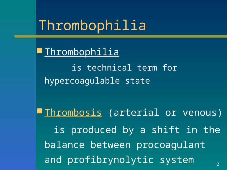
Thrombophilia
is technical term for hypercoagulable state
Thrombosis (arterial or venous)
is produced by a shift in the balance
between procoagulant and
profibrynolytic system
3
ThrombophiliaThrombophilia
inherited
acquired
3
4
Epidemiology of VTEEpidemiology of VTE
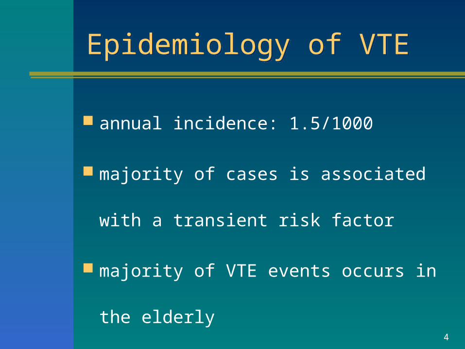
annual incidence: 1.5/1000
majority of cases is associated with a
transient risk factor
majority of VTE events occurs in the elderly
5
Hereditary thrombophiliaHereditary thrombophilia
is a genetically determined
increased risk of thrombosis
6
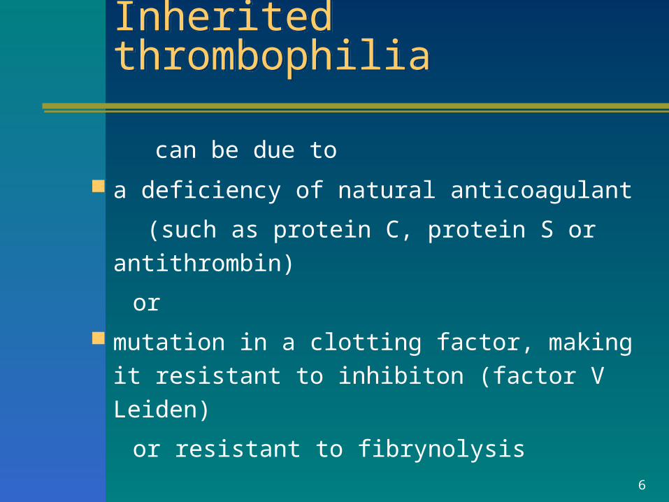
Inherited thrombophiliaInherited thrombophilia
can be due to a deficiency of natural anticoagulant
(such as protein C, protein S or antithrombin)
or
mutation in a clotting factor, making it
resistant to inhibiton (factor V Leiden)
or resistant to fibrynolysis
7
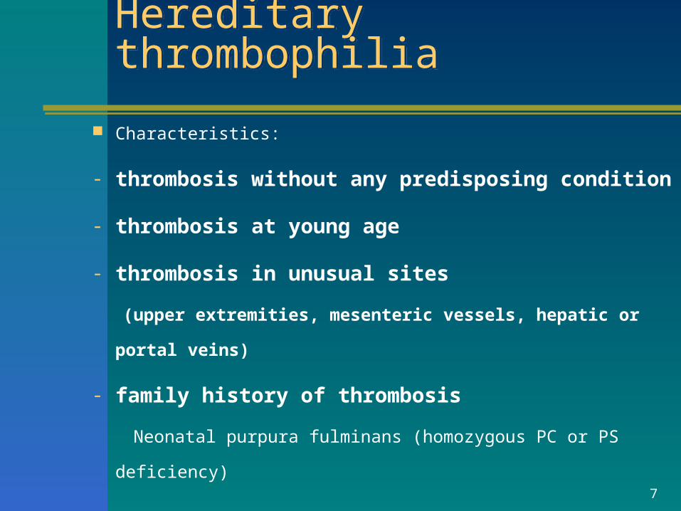
Hereditary thrombophiliaHereditary thrombophilia
Characteristics:
- thrombosis without any predisposing condition
- thrombosis at young age
- thrombosis in unusual sites
(upper extremities, mesenteric vessels, hepatic or portal
veins)
- family history of thrombosis
Neonatal purpura fulminans (homozygous PC or PS deficiency)
8
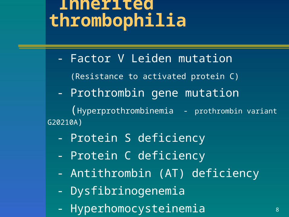
Inherited thrombophilia Inherited thrombophilia
- Factor V Leiden mutation
(Resistance to activated protein C)
- Prothrombin gene mutation
(Hyperprothrombinemia - prothrombin variant G20210A)
- Protein S deficiency
- Protein C deficiency
- Antithrombin (AT) deficiency
- Dysfibrinogenemia
- Hyperhomocysteinemia
9
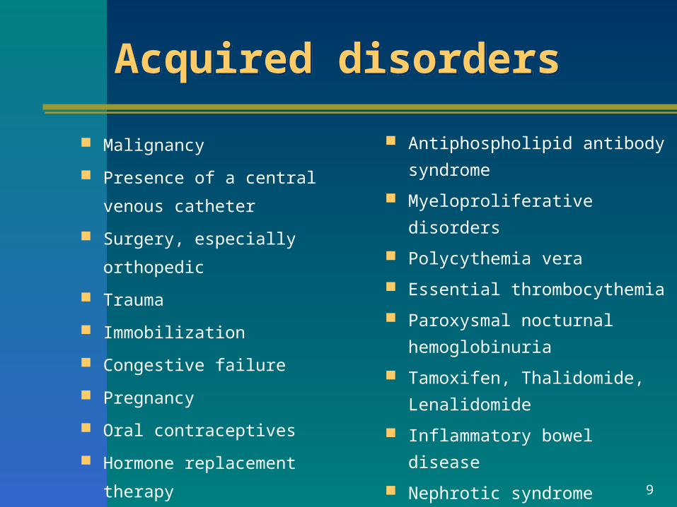
Acquired disorders Acquired disorders
Malignancy
Presence of a central venous
catheter
Surgery, especially orthopedic
Trauma
Immobilization
Congestive failure
Pregnancy
Oral contraceptives
Hormone replacement therapy
Antiphospholipid antibody
syndrome
Myeloproliferative disorders
Polycythemia vera
Essential thrombocythemia
Paroxysmal nocturnal
hemoglobinuria
Tamoxifen, Thalidomide,
Lenalidomide
Inflammatory bowel disease
Nephrotic syndrome
10
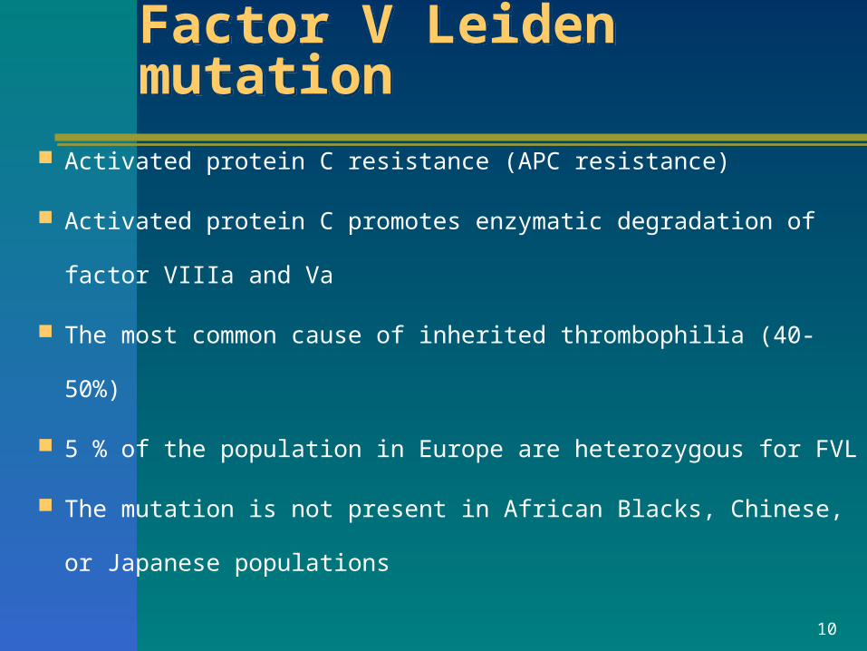
Factor V Leiden mutationFactor V Leiden mutation
Activated protein C resistance (APC resistance)
Activated protein C promotes enzymatic degradation of factor
VIIIa and Va
The most common cause of inherited thrombophilia (40-50%)
5 % of the population in Europe are heterozygous for FVL
The mutation is not present in African Blacks, Chinese, or
Japanese populations
11
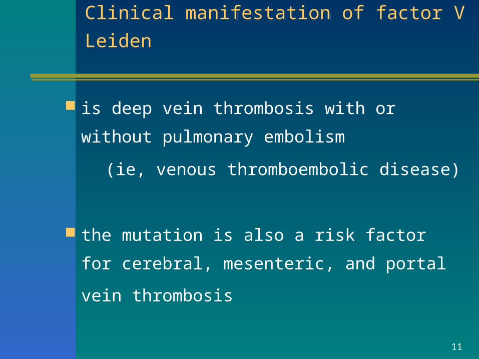
Clinical manifestation of factor V Leiden Clinical manifestation of factor V Leiden
is deep vein thrombosis with or without
pulmonary embolism
(ie, venous thromboembolic disease)
the mutation is also a risk factor for cerebral,
mesenteric, and portal vein thrombosis
12
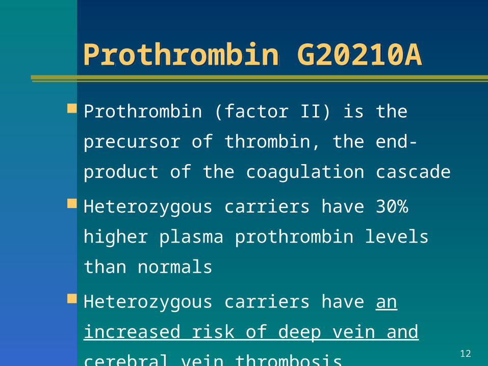
Prothrombin G20210A Prothrombin G20210A
Prothrombin (factor II) is the precursor of
thrombin, the end-product of the coagulation
cascade
Heterozygous carriers have 30% higher
plasma prothrombin levels than normals
Heterozygous carriers have an increased risk
of deep vein and cerebral vein thrombosis
13
Protein C (PC) deficiencyProtein C (PC) deficiency
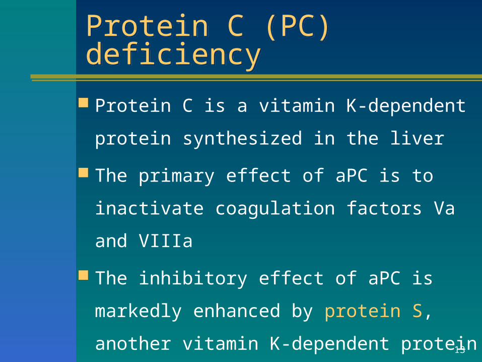
Protein C is a vitamin K-dependent protein
synthesized in the liver
The primary effect of aPC is to inactivate
coagulation factors Va and VIIIa
The inhibitory effect of aPC is markedly
enhanced by protein S, another vitamin K-
dependent protein
14
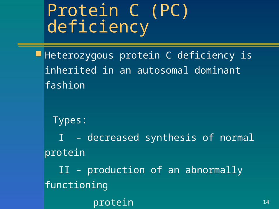
Protein C (PC) deficiencyProtein C (PC) deficiency
Heterozygous protein C deficiency is inherited
in an autosomal dominant fashion
Types:
I – decreased synthesis of normal protein
II – production of an abnormally functioning
protein
15
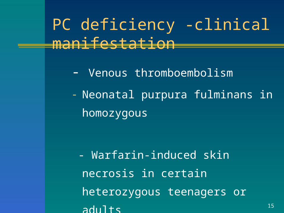
- Venous thromboembolism
- Neonatal purpura fulminans in
homozygous
- Warfarin-induced skin necrosis in certain
heterozygous teenagers or adults
PC deficiency -clinical manifestation
16
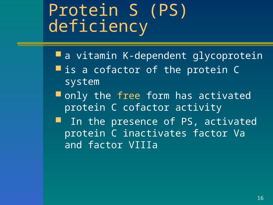
Protein S (PS) deficiencyProtein S (PS) deficiency
a vitamin K-dependent glycoprotein is a cofactor of the protein C system only the free form has activated protein C
cofactor activity In the presence of PS, activated protein
C inactivates factor Va and factor VIIIa
17
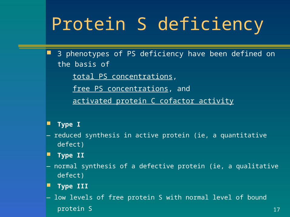
Protein S deficiencyProtein S deficiency 3 phenotypes of PS deficiency have been defined on the basis of
total PS concentrations,
free PS concentrations, and
activated protein C cofactor activity
Type I
— reduced synthesis in active protein (ie, a quantitative defect)
Type II
— normal synthesis of a defective protein (ie, a qualitative defect)
Type III
— low levels of free protein S with normal level of bound protein S
18
CLINICAL MANIFESTATIONS OF PS DEFICIENCY CLINICAL MANIFESTATIONS OF PS DEFICIENCY
Autosomal dominant trait
Similar to those of PC deficiency
19
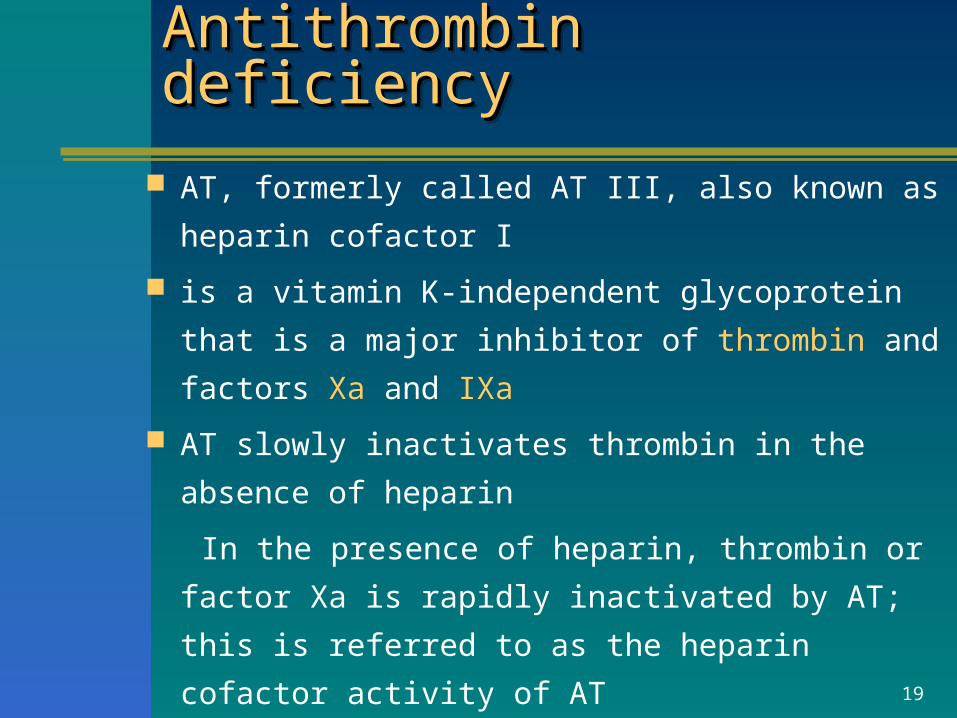
Antithrombin deficiencyAntithrombin deficiencyAntithrombin deficiencyAntithrombin deficiency
AT, formerly called AT III, also known as
heparin cofactor I
is a vitamin K-independent glycoprotein that is a major
inhibitor of thrombin and factors Xa and IXa
AT slowly inactivates thrombin in the absence of
heparin
In the presence of heparin, thrombin or factor Xa is
rapidly inactivated by AT; this is referred to as the
heparin cofactor activity of AT
20
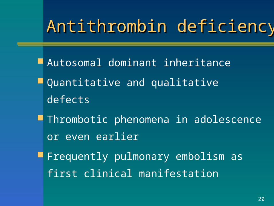
Antithrombin deficiencyAntithrombin deficiencyAntithrombin deficiencyAntithrombin deficiency
Autosomal dominant inheritance
Quantitative and qualitative defects
Thrombotic phenomena in adolescence or
even earlier
Frequently pulmonary embolism as first
clinical manifestation
21
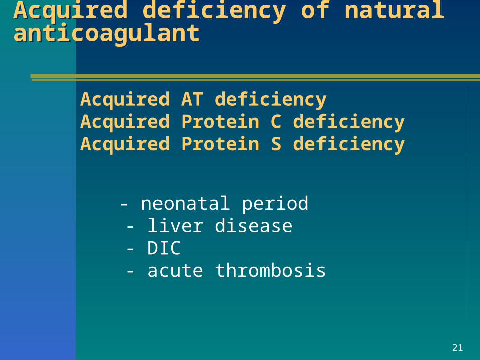
Acquired deficiency of natural anticoagulantAcquired deficiency of natural anticoagulant
Acquired AT deficiencyAcquired Protein C deficiencyAcquired Protein S deficiency
- neonatal period - liver disease - DIC - acute thrombosis
22
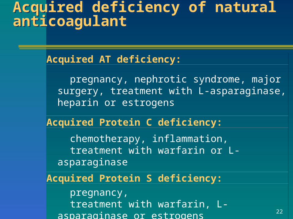
Acquired deficiency of natural anticoagulantAcquired deficiency of natural anticoagulant
Acquired AT deficiency:
pregnancy, nephrotic syndrome, major surgery, treatment with L-asparaginase, heparin or estrogens
Acquired Protein C deficiency:
chemotherapy, inflammation, treatment with warfarin or L-asparaginase
Acquired Protein S deficiency:
pregnancy, treatment with warfarin, L-asparaginase or estrogens
23
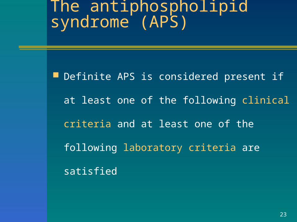
The antiphospholipid syndrome (APS) The antiphospholipid syndrome (APS)
Definite APS is considered present if at least one of
the following clinical criteria and at least one of the
following laboratory criteria are satisfied
24
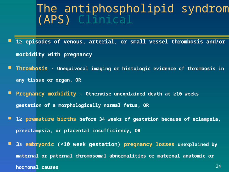
The antiphospholipid syndrome (APS) ClinicalThe antiphospholipid syndrome (APS) Clinical
1≥ episodes of venous, arterial, or small vessel thrombosis and/or morbidity
with pregnancy
Thrombosis - Unequivocal imaging or histologic evidence of thrombosis in any
tissue or organ, OR
Pregnancy morbidity - Otherwise unexplained death at ≥10 weeks gestation of a
morphologically normal fetus, OR
1≥ premature births before 34 weeks of gestation because of eclampsia,
preeclampsia, or placental insufficiency, OR
3≥ embryonic (<10 week gestation) pregnancy losses unexplained by maternal
or paternal chromosomal abnormalities or maternal anatomic or hormonal causes
25
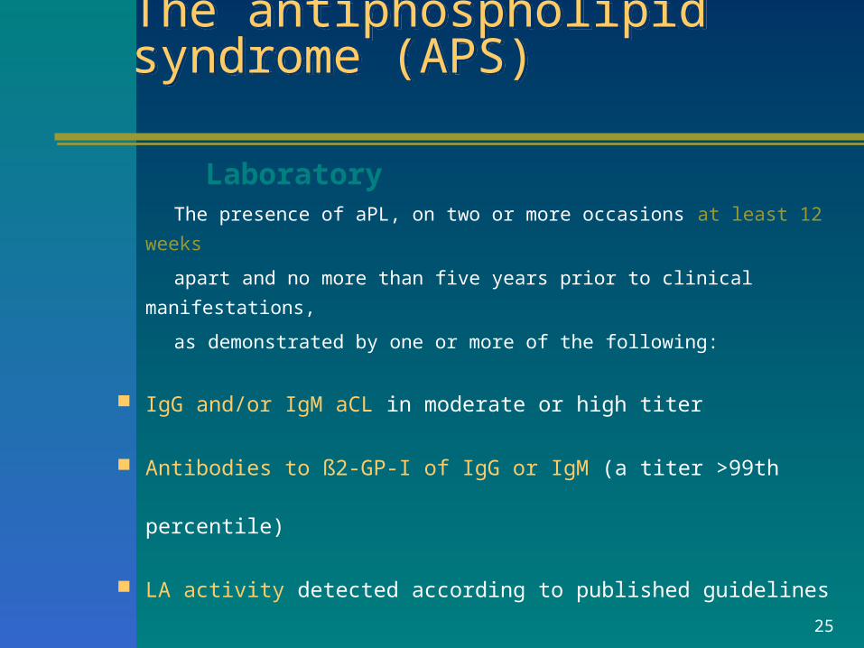
The antiphospholipid syndrome (APS)The antiphospholipid syndrome (APS)
Laboratory The presence of aPL, on two or more occasions at least 12 weeks
apart and no more than five years prior to clinical manifestations,
as demonstrated by one or more of the following:
IgG and/or IgM aCL in moderate or high titer
Antibodies to ß2-GP-I of IgG or IgM (a titer >99th percentile)
LA activity detected according to published guidelines
26
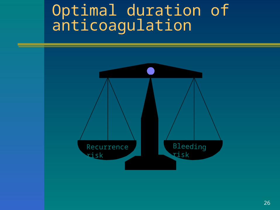
Optimal duration of anticoagulationOptimal duration of anticoagulation
Recurrence risk Bleeding risk
27
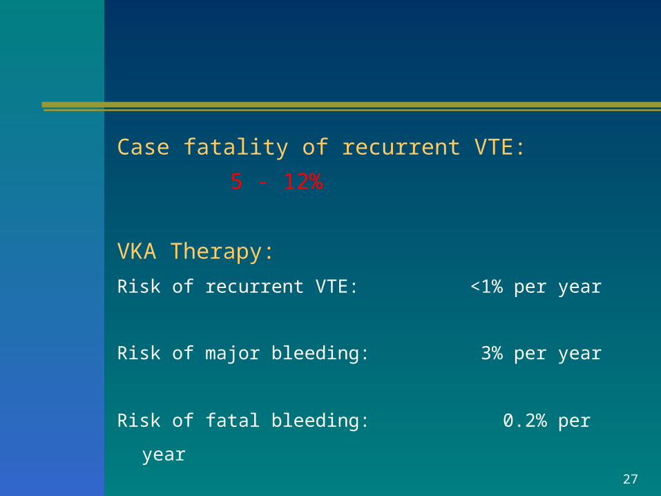
Case fatality of recurrent VTE: 5 - 12%
VKA Therapy:
Risk of recurrent VTE: <1% per year
Risk of major bleeding: 3% per year
Risk of fatal bleeding: 0.2% per year
28
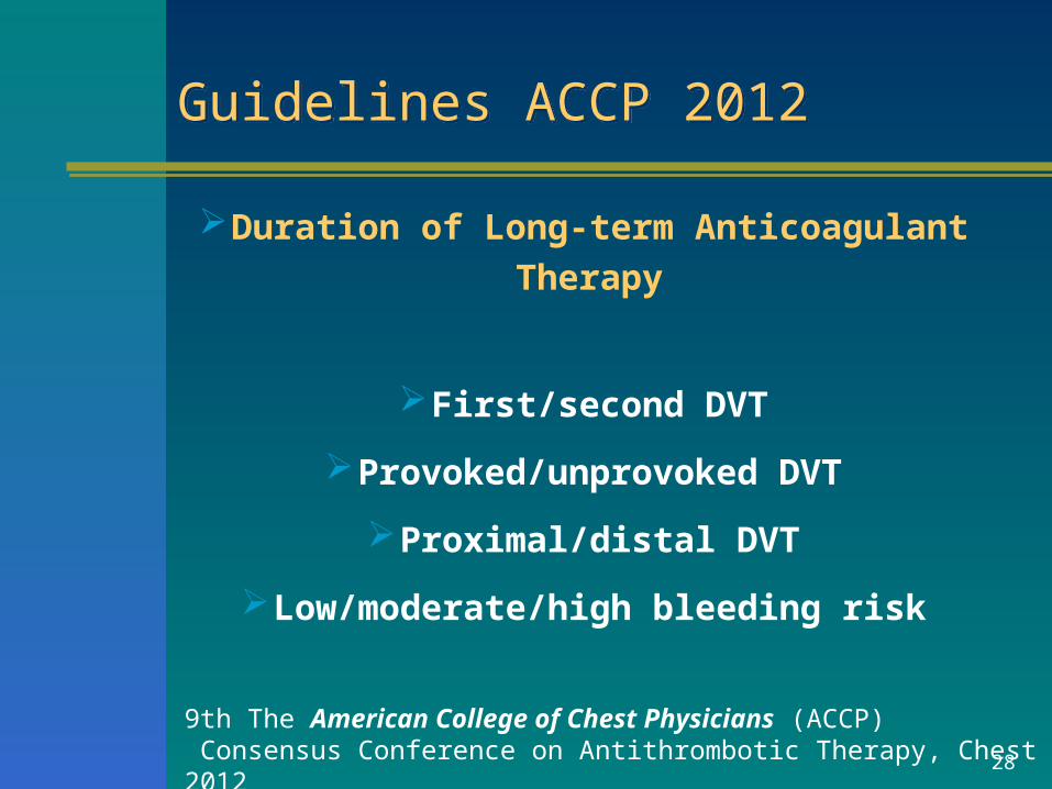
Guidelines ACCP 2012Guidelines ACCP 2012
Duration of Long-term Anticoagulant Therapy
First/second DVT
Provoked/unprovoked DVT
Proximal/distal DVT
Low/moderate/high bleeding risk
9th The American College of Chest Physicians (ACCP) Consensus Conference on Antithrombotic Therapy, Chest 2012
Guidelines ACCP 2012Guidelines ACCP 2012
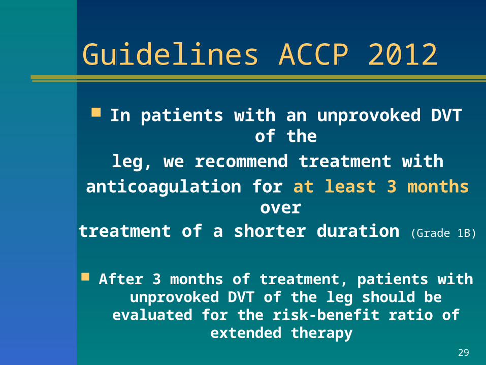
In patients with an unprovoked DVT of the
leg, we recommend treatment with
anticoagulation for at least 3 months over treatment of a shorter duration (Grade 1B)
After 3 months of treatment, patients with unprovoked DVT of the leg should be evaluated
for the risk-benefit ratio of extended therapy
29
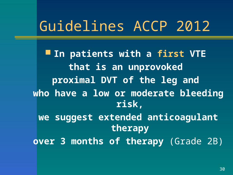
Guidelines ACCP 2012Guidelines ACCP 2012
In patients with a first VTE
that is an unprovoked
proximal DVT of the leg and
who have a low or moderate bleeding risk,
we suggest extended anticoagulant therapy
over 3 months of therapy (Grade 2B)
30
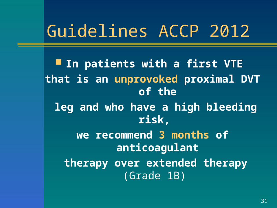
Guidelines ACCP 2012Guidelines ACCP 2012
In patients with a first VTE
that is an unprovoked proximal DVT of the
leg and who have a high bleeding risk,
we recommend 3 months of anticoagulant
therapy over extended therapy (Grade 1B)
31
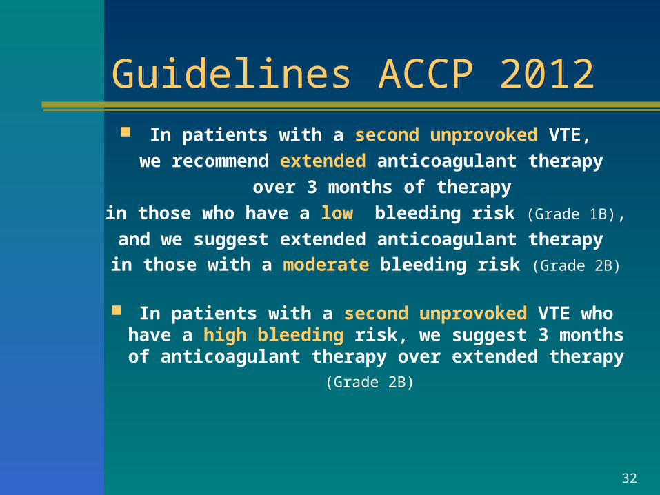
Guidelines ACCP 2012Guidelines ACCP 2012 In patients with a second unprovoked VTE,
we recommend extended anticoagulant therapy
over 3 months of therapy
in those who have a low bleeding risk (Grade 1B),
and we suggest extended anticoagulant therapy
in those with a moderate bleeding risk (Grade 2B)
In patients with a second unprovoked VTE who have a high bleeding risk, we suggest 3 months of anticoagulant therapy over extended therapy (Grade
2B)
32
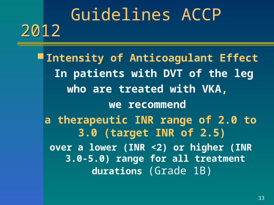
Guidelines ACCP 2012 Guidelines ACCP 2012 Intensity of Anticoagulant Effect
In patients with DVT of the leg
who are treated with VKA,
we recommend
a therapeutic INR range of 2.0 to 3.0 (target INR of 2.5)
over a lower (INR <2) or higher (INR 3.0-5.0) range for all treatment durations (Grade 1B)
33
34
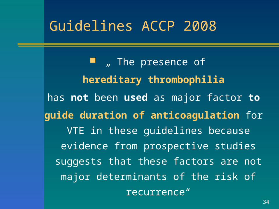
Guidelines ACCP 2008Guidelines ACCP 2008
„ The presence of
hereditary thrombophilia
has not been used as major factor to
guide duration of anticoagulation for
VTE in these guidelines because evidence
from prospective studies suggests that
these factors are not major determinants
of the risk of recurrence“


![Thrombosis%20and%20Thrombophilia[1].pptscscls.les3z.org/.../thrombosis-and-thrombophilia...CVA Venous Thromboembolism • Third most common cardiovascular disease • Significant morbidity](https://img.pdfslide.us/doc/110x75/5e432760b2114b1eb170ce76/thrombosis20and20thrombophilia1-cva-venous-thromboembolism-a-third-most.jpg)