Embed Size (px)
Citation preview

This is an Open Access document downloaded from ORCA, Cardiff University's institutional
repository: http://orca.cf.ac.uk/120326/
This is the author’s version of a work that was submitted to / accepted for publication.
Citation for final published version:
Al-Sabah, Ayesha, Burnell, Stephanie E.A., Simoes, Irina N., Jessop, Zita, Badiei, Nafiseh, Blain,
Emma and Whitaker, Iain S. 2019. Structural and mechanical characterization of crosslinked and
sterilised nanocellulose-based hydrogels for cartilage tissue engineering. Carbohydrate Polymers
212 , pp. 242-251. 10.1016/j.carbpol.2019.02.057 file
Publishers page: http://dx.doi.org/10.1016/j.carbpol.2019.02.057
<http://dx.doi.org/10.1016/j.carbpol.2019.02.057>
Please note:
Changes made as a result of publishing processes such as copy-editing, formatting and page
numbers may not be reflected in this version. For the definitive version of this publication, please
refer to the published source. You are advised to consult the publisher’s version if you wish to cite
this paper.
This version is being made available in accordance with publisher policies. See
http://orca.cf.ac.uk/policies.html for usage policies. Copyright and moral rights for publications
made available in ORCA are retained by the copyright holders.

1
Structural and Mechanical Characterization of Crosslinked and Sterilised Nanocellulose-Based 1
Hydrogels for Cartilage Tissue Engineering 2
3
ǂAyesha Al-Sabaha, ǂStephanie EA Burnella, ǂIrina N Simoesa, Zita Jessopa,b, Nafiseh Badieic, Emma 4
Blaind and *Iain S Whitakera,b 5
aReconstructive Surgery & Regenerative Medicine Group (ReconRegen), Institute of Life Sciences, 6
Swansea University Medical School, SA2 8PP, Swansea, Wales, UK, bThe Welsh Centre for Burns 7
and Plastic Surgery, Morriston Hospital, SA6 6NL, Swansea, Wales, UK, cNanoHealth Centre, 8
Institute of Life Sciences, Swansea University Medical School, SA2 8QA, Swansea, Wales, UK, 9
dArthritis Research UK Biomechanics and Bioengineering Centre, School of Biosciences, Cardiff 10
University, CF10, 3AX, Cardiff, Wales, UK 11
Ayesha Al-Sabah: [email protected], Stephanie EA Burnell: [email protected], Irina 12
N Simoes: [email protected], Zita Jessop: [email protected], Nafiseh 13
Badiei: [email protected], Emma Blain: [email protected] and Iain S Whitaker: 14
16
ǂEquivalent contributors 17
18
Corresponding author: 19
Professor Iain S. Whitaker 20
Reconstructive Surgery & Regenerative Medicine Group (ReconRegen) 21
Institute of Life Sciences 2, Swansea University Medical School 22
Swansea, SA2 8PP, Wales, United Kingdom 23
Telephone: +44 1792 606311 24
Fax: +44 1792 703875 25
E-mail: [email protected] 26

2
Abstract 27
Nanocellulose is a natural biopolymer derived from cellulose. Combined with sodium alginate, it is 28
used to 3D print hydrogels for articular and nasal cartilage engineering and shows good integration, 29
promising cartilage regeneration and mechanical stability over 60 days of implantation in mice. Yet, 30
little is known about their structural and mechanical properties, particularly the impact of crosslinking 31
and sterilisation methods. This study investigates the impact of different calcium chloride crosslinker 32
concentrations and sterilization methods on the structural and mechanical properties of nanocellulose-33
based hydrogels containing plant-derived cellulose nanofibrils, cellulose nanocrystals or a blend of 34
the two. Crosslinking significantly alters the overall network distribution, surface morphology, pore 35
size and porosity of the hydrogels. Sterilisation has a striking effect on pore size and affects swelling 36
depending on the sterilisation method. The effect of crosslinker and sterilisation on the overall 37
properties of the hydrogels was reliant on the form of nanocellulose that comprised them. 38
39
Keywords: Nanocellulose, Crosslinking, Sterilisation, Hydrogels, Cartilage Tissue Engineering 40
41
1. Introduction 42
Tissue engineering can provide advanced alternatives to the current standard surgical procedures used 43
in the field of cartilage repair and reconstruction. For many years, the cartilage tissue engineering 44
field has explored the use of biomaterials, more specifically hydrogels, to create tissue substitutes 45
(Park & Lee, 2014; Tibbitt & Anseth, 2009; Xiao, Friis, Gehrke, & Detamore, 2013). Hydrogels 46
mimic the native extracellular matrix (ECM) and can be tailored to resemble the native structure and 47
mechanics of the tissues, enhance mass transport and support cell adhesion and protein sequestration 48
(S. Lin, Sangaj, Razafiarison, Zhang, & Varghese, 2011; Tibbitt & Anseth, 2009). Importantly, 49
through techniques such as three-dimensional (3D) bioprinting, these hydrogels can be used as 50
bioinks to create high-resolution 3D structures, with any shape or size, to support cell growth and 51
tissue formation (Mouser et al., 2017). As the use of synthetic materials often leads to infection, 52

3
extrusion and foreign body reaction, more natural biomaterials are increasingly explored (Anderson, 53
Rodriguez, & Chang, 2008; Baker, Walsh, Schwartz, & Boyan, 2012). 54
Alginate is a natural and abundant polysaccharide that occurs in marine brown algae and other sources 55
(Hecht & Srebnik, 2016). Water-soluble sodium alginate ((NaC6H7O6)n), the sodium salt of alginic 56
acid, is commonly used as a component of hydrogels for cartilage engineering due to its recognized 57
chondrogenicity and ability to enhance the structural properties of hydrogels (Ansari et al., 2017; 58
Chou, Akintoye, & Nicoll, 2009; Markstedt et al., 2015; Miao et al., 2017). 59
Within the past decade, nanocellulose (NC) was appointed as an exciting novel biomaterial for 60
biomedical applications due to its attractive physicochemical properties, abundance, sustainability, 61
non-cytotoxicity and biodegradability (Dumanli, 2016; N. Lin & Dufresne, 2014). NC is a biopolymer 62
derived from cellulose, a polysaccharide composed by D-glucopyranose linked by β-1,4 glycosidic 63
bonds (Endes et al., 2016) that is the most abundant, renewable and natural resource available 64
(Dumanli, 2016; N. Lin & Dufresne, 2014). Cellulose contains three hydroxyl groups (-OH) at C-2, 65
C-3 and C-6 positions which determine its physical properties. NC can be found in plants and marine 66
animals and is naturally available in two forms: nanofibrils and nanocrystals (N. Lin & Dufresne, 67
2014). Additionally, NC is biotechnologically produced in bacteria (N. Lin & Dufresne, 2014). 68
Although the cellulose molecular backbone is common to all forms of NC, the surface morphology, 69
size, chemical and physical properties can vary depending on the material source and extraction 70
methods (Mao et al., 2017). Cellulose nanofibrils and nanocrystals are produced through several 71
chemical, mechanical and/or enzymatic methods that introduce functional groups in the surface of 72
the NC (Kim & Song, 2015). Yet, NC produced through the American Value Added Pulping 73
(AVAP®) technology chemically pre-treats wood-pulp derived biomass and produces NC that is free 74
from any additional functional groups, apart from the -OH groups (Kyle et al., 2018). Importantly, 75
the lack of post-hydrolysis modifications allows facile surface functionalization of the hydroxyl 76
groups resulting in promising potential for novel, advanced and multifunctional biomaterials with 77
improved biocompatibility and tissue generation (Bodin et al., 2007). Bacterial NC can be produced 78

4
with high purity and has shown promise for tissue engineering applications (water-holding capacity, 79
mechanical strength and morphological similarities with collagen) and 3D bioprinting (good 80
rheological properties) (Ahrem et al., 2014; Markstedt et al., 2015; Paakko et al., 2007). Yet, the use 81
of bacterial NC for large scale commercialization is limited by the high cost of substrates, low 82
productivity of strains and expensive culture media (Paakko et al., 2007; Revin, Liyaskina, 83
Nazarkina, Bogatyreva, & Shchankin, 2018). Despite the efforts to increase productivity and decrease 84
costs using various waste-products, the production of bacterial NC is still far from large-scale 85
commercialisation and needs further development (Revin et al., 2018). Additionally, there are still 86
concerns regarding residual bacterial toxins/epitopes in bacterial NC (Paakko et al., 2007). 87
The combination of crosslinked sodium alginate and NC has been recently explored for cartilage 88
tissue engineering, for articular and nasal reconstruction (Ahrem et al., 2014; Martínez Ávila et al., 89
2015; Möller et al., 2017; Müller, Öztürk, Arlov, Gatenholm, & Zenobi-Wong, 2016; Nguyen et al., 90
2017). The chondrogenic potential and biocompatibility of these composite hydrogels was reported 91
in both in vitro and in vivo studies using bacterial NC (Ahrem et al., 2014; Martínez Ávila et al., 92
2015; Möller et al., 2017; Müller et al., 2016; Nguyen et al., 2017; Svensson et al., 2005). Recently, 93
Müller and colleagues used 3D bioprinted alginate-NC hydrogels and articular bovine chondrocytes 94
to demonstrate high cell viability, proliferation and high collagen type II deposition after 28 days in 95
culture (Müller et al., 2016). Similarly, Nguyen et al. reported, using the same composite hydrogels 96
as a scaffold for the differentiation of human induced pluripotent stem (iPS) cells, significant increase 97
in RNA expression of chondrogenic markers and matrix deposition. This increase was confirmed by 98
histology staining and immunohistochemistry upon 5 weeks of differentiation (Nguyen et al., 2017). 99
In 2015, Martínez-Avila and his team reported in vivo neocartilage formation using co-cultures of 100
human nasoseptal chondrocytes and bone marrow mononuclear cells in bilayer alginate-NC 101
hydrogels (Martínez Ávila et al., 2015). Briefly, the constructs were implanted subcutaneously in 102
nude mice showing non-pathological foreign body reaction, deposition of proteoglycans and collagen 103

5
type II and increased instantaneous modulus upon 8 weeks of implantation (Martínez Ávila et al., 104
2015). 105
In these composite hydrogels, sodium alginate provides structural integrity through chemical 106
crosslinking promoting the transition of the hydrogel into a solid material (Caliari & Burdick, 2016). 107
Sodium alginate can be ionically crosslinked by adding calcium ions (crosslinker) which substitute 108
the sodium ions in the alginate, creating strong bonds between alginate chains and ultimately creating 109
a mesh (Hecht & Srebnik, 2016). The concentration of crosslinker can regulate the characteristics of 110
the solid material by tailoring its structural and mechanical properties (S. Lin et al., 2011). Such 111
changes affect the network mesh distribution and pore size that ultimately impact cellular phenotype, 112
proliferation and ECM production (Bryant, Chowdhury, Lee, Bader, & Anseth, 2004; Hwang et al., 113
2007; Lien, Ko, & Huang, 2009; Villanueva, Klement, von Deutsch, & Bryant, 2009). In cartilage 114
engineering, this has a particularly significant impact on mass transport and the spatial distribution of 115
the ECM – increased hydrogel mesh size leads to higher collagen content, for example (Bryant & 116
Anseth, 2002; Buxton et al., 2007; Chung, Mesa, Randolph, Yaremchuk, & Burdick, 2006; S. Lin et 117
al., 2011). Apart from the extensive body of literature reporting on the chondrogenicity of hydrogels 118
combining sodium alginate and NC, the detailed microenvironment and mechanical properties of 119
these hydrogels remains fairly unknown (Leppiniemi et al., 2017; Markstedt et al., 2015; Martínez 120
Ávila et al., 2015; Müller et al., 2016; Nguyen et al., 2017). Moreover, prior to any application, these 121
hydrogels require sterilisation to limit or prevent the risk of contamination, infection and rejection 122
(Matthews, Gibson, & Samuel, 1994; Veerachamy, Yarlagadda, Manivasagam, & Yarlagadda, 2014). 123
Despite the importance of this topic, less than 1% of the scientific publications in the past decade 124
have focused on the sterilisation methods of hydrogel-based biomedical systems (Galante, Pinto, 125
Colaco, & Serro, 2017). Along with this trend, the effect of sterilisation on the intrinsic properties of 126
NC-based hydrogels also remains elusive. Few studies have performed side-by-side comparison of 127
the architecture, structure and mechanics of the different forms of NC-based hydrogels. Plant-derived 128

6
cellulose nanofibrils, cellulose nanocrystals and blend, produced using AVAP® technology have been 129
thoroughly characterised without additives or crosslinking by Kyle et al. in 2018 (Kyle et al., 2018). 130
The first aim was to investigate the effect of crosslinking – using calcium chloride (CaCl2) – on the 131
structural and mechanical properties of AVAP® produced plant-derived cellulose nanofibrils, 132
cellulose nanocrystals and blend (combination of nanofibrils and nanocrystals) NC-based hydrogels 133
combined with sodium alginate. Inspired by the hydrogels described in the literature, NC-based 134
hydrogels were crosslinked using increasing concentrations of crosslinker to understand its impact 135
on the overall architecture and characteristic properties (Ahrem et al., 2014; Martínez Ávila et al., 136
2015; Möller et al., 2017; Müller et al., 2016; Nguyen et al., 2017; Svensson et al., 2005). Secondly, 137
the same type of characterisation was performed upon exposure of the NC-based hydrogels to 138
different sterilisation methods: exposure to ultraviolet (UV) light, autoclaving and ethanol immersion. 139
Finally, the characteristics of the microenvironment of NC-based hydrogels used herein was 140
compared with the reported “ideal” conventional environment for cartilage engineering (Nava, 141
Draghi, Giordano, & Pietrabissa, 2016; Oh, Kim, Im, & Lee, 2010; Pan et al., 2015). 142
143
2. Hypothesis 144
The concentration of crosslinker and the sterilisation methods affect the structural and mechanical 145
properties (i.e. pore size, overall network organisation, swelling, porosity and elastic modulus) of 146
NC-based hydrogels. 147
148
3. Material and Methods 149
All reagents were purchased from Sigma-Aldrich® (Dorset, UK) unless stated otherwise. All reagents 150
were of analytical grade or above. 151
3.1 Preparation of nanocellulose-based hydrogels 152
Plant-derived nanocellulose (hydrophilic Bioplus® cellulose nanofibrils gel, hydrophilic Bioplus® 153
cellulose nanocrystals gel and hydrophilic Bioplus® blend gel – a blend of fibrils and crystal) was 154

7
provided by American Process, Inc. (Georgia, USA) (Kyle et al., 2018). All nanocellulose forms are 155
produced via the AVAP® technology (Kyle et al., 2018) which fractionates biomass into cellulose, 156
hemicelluloses and lignin using ethanol and sulfur dioxide (Kyle et al., 2018). The final nanocellulose 157
product morphology – fibrils (3 wt.% solids), crystals (6 wt.% solids) and blend (3 wt.% solids) was 158
controlled by the time and temperature of the pre-treatment step (Kyle et al., 2018). The blend 159
nanocellulose is produced in situ during production and is not an actual blend of fibrils and crystal, 160
yet for simplicity it will be referred to as blend. Hydrogels were prepared by mixing nanocellulose 161
with 2.5% (w/v) sodium alginate (alginic acid sodium salt, from brown algae, 80,000-120,000 Da, 162
medium viscosity (2% at 25oC), 1.56 mannuronate/guluronate ratio) solution in ultrapure water. 163
Briefly, nanocellulose was centrifuged at 1500 g for 5 min, excess water was removed, and 2.5% 164
sodium alginate solution was added in a 1:4 proportion (Markstedt et al., 2015). NC-based hydrogels 165
were named as follows: NC-blend and 2.5% sodium alginate (NCB), NC-fibrils and 2.5% sodium 166
alginate (CNF) and NC-crystals and 2.5% sodium alginate (CNC). All NC-based hydrogels contained 167
75% of NC, where CNF and NCB had a final concentration of 2.25 wt.% solids and CNC contained 168
4.5 wt.% solids. 169
3.2 Crosslinking of nanocellulose-based hydrogels 170
Nanocellulose-based hydrogels were shaped into: (a) 1.5 ml discs (Ø=~14mm) for mechanical testing 171
and (b) 100 µl pellets for all other assays (Figure 1A). The discs were produced using 24 well plates 172
(Cellstar®) and the pellets were produced using 1ml syringes (BD Biosciences©, Oxford, UK) and 173
the indentations of a 96 well plate lid (Cellstar®) as a mould. Crosslinking was performed at room 174
temperature using 0.1 M, 0.5 M or 1.0 M calcium chloride (CaCl2) solutions prepared in ultrapure 175
water. Hydrogels of 2.5% sodium alginate were also prepared as mentioned above. The experimental 176
layout is depicted in Figure 1B. 177

8
178
Figure 1. Graphical experimental layout. (A) Overall aspect of NC-based hydrogels (pellets and discs). Photos are from 179
NCB-based crosslinked hydrogels. (B) Experimental layout: different NC-based hydrogels were crosslinked with varying 180
concentrations of CaCl2 – structural and mechanical properties were assessed post-crosslinking; NC-based hydrogels 181
crosslinked with the least concentrated CaCl2 solution were subjected to different sterilisation methods – structural and 182
mechanical properties were assessed post-sterilisation. Sodium alginate hydrogels were used as controls in all 183
experiments. NCB – nanocellulose blend of fibrils and crystal; CNC – nanocellulose crystal; CNF – nanocellulose fibrils; 184
CaCl2 – calcium chloride. Image partially created with BioRender©. 185
3.3 Sterilization of nanocellulose-based hydrogels 186
Nanocellulose-based hydrogels were sterilised using (a) autoclave, (b) UV light (UV-C germicidal 187
light) or (c) ethanol (70% absolute ethanol in ultrapure water). Autoclave sterilisation was performed 188
for 20 min at 126°C using a Classic bench-size autoclave (Prestige Medical, Blackburn, UK). UV 189
sterilisation was completed in petri dishes inside a laminar flow hood using UV-C 254 nm for 1 hour. 190
After sterilisation, hydrogels were crosslinked using 0.1 M CaCl2. Ethanol sterilisation was carried 191
out by immersion of nanocellulose-based hydrogels in ethanol for 20 min – the crosslinking was 192
performed in tandem (i.e. the CaCl2 was dissolved in 70% ethanol). Sodium alginate hydrogels were 193
also processed as mentioned above. The experimental layout is depicted in Figure 1B. 194
3.4 Scanning electron microscopy and average pore size calculation 195
Hydrogels were washed with 50 mM sodium cacodylate-HCl buffer solution (pH 7.2-7.4, SPI 196
Supplies®, West Chester, PA, USA) for 10-20 min, fixed overnight in 2% glutaraldehyde and 197

9
dehydrated using a series of graded ethanol concentrations (30%-100%). These were subsequently 198
rinsed with 50% hexamethyldisilazane solution (HMDS) in 100% ethanol for 10 min, then in 100% 199
HMDS and left overnight to dry. The specimens were coated with a thin layer of gold (~15 nm) using 200
sputter coating and examined using scanning electron microscopy (SEM, Hitachi 4800, Hitachi, 201
Schamumburg, IL, USA). Pore size was determined using ImageJ 1.51 software from the National 202
Institutes of Health, USA. 203
3.5 Swelling and porosity assays 204
Pellets were immersed in 1x PBS (Gibco®, ThermoFisher Scientific, Loughborough, UK) and 205
incubated at 37°C for 24 hours. After blotting the excess PBS on the surface, each pellet was weighed 206
individually (Mw). After drying for 48h at room temperature (using desiccant inside a Styrofoam box), 207
the pellets were again weighed individually (Md). Swelling and porosity percentages (%) were given 208
by the Equation 1 and 2 (Caliari & Burdick, 2016; Gupta & Shivakumar, 2012; K. Pal, 2009). PBS 209
density was considered as 1.06 g cm-3. 210 � � � % = �− Equation 1 211
� � % = �− ���� � � �� Equation 2 212
3.6 Mechanical testing 213
Mechanical tests were performed on wet discs at room temperature using a Bose Electroforce® 3200 214
(Bose Corp., TA Instruments, MN, USA) equipped with a compression plate. Compressive loading 215
was applied using a 1 Hz frequency at 5 N for 20 cycles. Young’s modulus was given by Equation 3, 216
4, and 5. 217
� �′ � �� = � � −2� �� % Equation 3 218
Where, 219 � = � �� � �� 2 Equation 4 220
� �� = � � ℎ −� � − � � ℎ −� � � � ℎ −� � Equation 5 221

10
The length and surface area were determined pre- and post-loading using a digital calliper. 222
3.7 Bacterial persistence 223
Bacterial persistence post-sterilisation was determined through optical density (OD) at 600 nm using 224
a spectrophotometer. Samples were horizontally and vertically cut into four equal pieces with similar 225
exposed surface area and added to a tube containing 10 ml of lysogeny broth (LB). After 24h and 48h 226
at 37°C under constant stirring, 1ml samples were taken out and used to measure OD. 227
3.8 Cell viability 228
Human naso-septal chondrocytes were isolated from healthy donors, after informed consent from 229
patients (IRAS ID 99202) at ABM University Health Board, Swansea, United Kingdom. Samples 230
were collected during routine septorhinoplasty procedures where the cartilage would have otherwise 231
been discarded (institutional review committee approved the study, ethics approval: REC 232
12/WA/0029), following an adjusted protocol (Dowthwaite et al., 2004; Fickert, Fiedler, & Brenner, 233
2004). Cells were extracted overnight using 2.0 mg ml-1 pronase and 2.4 mg ml-1 collagenase I and 234
cultured in DMEM with 10% fetal bovine serum (FBS), 1% penicillin-streptomycin solution, 1mM 235
D-glucose solution and 0.1% minimum essential medium (MEM) non-essential amino acids (all from 236
Gibco®) in a humidified 37°C incubator with 5% CO2. After 2.5 weeks, chondrocytes were mixed 237
with the sterilised hydrogels (3 x 10⁵ cells per pellet) to prepare 100 µl pellets, as mentioned 238
previously, and crosslinked using 0.1 M CaCl2. Pellets were cultured for up to 7 days – cell viability 239
was determined at 24h and 7 days using Live/Dead assay kit (ThermoFisher Scientific) according to 240
manufacturer’s instructions. The pellets were imaged using confocal microscopy (Zeiss 710 confocal 241
microscope, Zeiss, Cambridge, UK) and ZEN software (Zeiss). 242
3.9 Statistical analysis 243
Data are expressed as mean ± standard error of the mean (mean ± SEM). All data were checked for 244
normality (Anderson-Darling Test) and equal variance (Levene’s Test) to meet the assumptions of 245
ANOVA. An ANOVA followed by a Tukey test for post-hoc pairwise comparisons were used. 246

11
Alternatively, Mann-Whitney U tests were used for data with unequal variance. Statistical analysis 247
was performed using Minitab® 18 (Minitab Inc.). A p-value < 0.05 was considered significant. 248
249
4. Results and Discussion 250
In recent years, NC-based hydrogels have been used for cartilage engineering purposes providing 251
promising in vitro and in vivo outcomes (Martínez Ávila et al., 2015; Nguyen et al., 2017). However, 252
little is known about the effects of crosslinking concentrations and sterilisation methods on the 253
hydrogel structure, microarchitecture, and mechanical properties. If these hydrogels are to be 254
translated to clinic, it is essential to understand how such processing methods affect their properties. 255
In this study, we initially looked at the effect of increasing crosslinker concentrations and later 256
investigated the impact of different sterilisation methods. NC-based hydrogels possess excellent 257
rheological properties for applications such as 3D bioprinting, however these must be supplemented 258
with a biomaterial that enables crosslinking to ensure post-printing shape fidelity (Kyle et al., 2018). 259
To that end, we used plant-derived cellulose nanofibrils, cellulose nanocrystals and a blend, produced 260
via the AVAP® technology which do not crosslink on their own when exposed to various 261
concentrations of CaCl2 (data not shown). Sodium alginate was used to provide structural integrity 262
via ionic crosslinking using CaCl2 (Hecht & Srebnik, 2016). NC-based hydrogels were formulated 263
by mixing sodium alginate and different NC forms: crystalline (CNC), fibrillar (CNF) and blend 264
(NCB) (Figure 1 and Supplementary Material, Figure S1). The surface charge of the different NC 265
forms were previously evaluated by means of the zeta potential (Kyle et al., 2018). All NC forms 266
showed negative zeta potential in neutral water. As such, this feature was dismissed for the discussion 267
as it would not explain structural and mechanical differences between the composite hydrogels. 268
4.1 Characterization of crosslinked NC-based hydrogels 269
The surface morphology and network distribution of the different NC-based hydrogels were observed 270
through SEM images (Figure 2A-P). CNC and CNF showed different surface morphologies – CNF 271
contains a fibrillar-like network with varying thickness (Figure 2I) while CNC holds a leaf-like net 272

12
architecture (Figure 2M). The overall surface morphology of NCB, a blend of CNC and CNF, is 273
apparently more porous and interconnected than CNF and CNC individually (Figure 2E). Upon 274
addition of the crosslinker, the overall structural morphology and network distribution change 275
noticeably. Increasing concentrations of CaCl2 developed a denser and more organised network in the 276
sodium alginate hydrogels, creating an apparent flatter external surface (Figure 2A-D). In the NC-277
based hydrogels the same trend was observed, although visible differences were more obvious when 278
comparing the highest CaCl2 concentration (Figure 2H, 2L and 2P) with the other two concentrations 279
(Figure 2F-G, 2J-K and 2N-O). As sodium alginate is the structural crosslinked component of the 280
NC-based hydrogels, the NC is in the interstitial framework of sodium alginate and thus seems 281
relatively disorganised, making these changes only noticeable at higher crosslinker concentration. 282
With increasing CaCl2, the gelation rate increases as it is directly proportional to the concentration of 283
calcium ions (Lee & Rogers, 2012). The resulting hydrogel has increased interactions between 284
sodium alginate chains, as additional binding sites on alginate become occupied by calcium ions 285
(Fang et al., 2007). The network of NC-based hydrogels is moderately different between NCB, CNC 286
and CNF, yet noticeably different from the sodium alginate hydrogels: alginate has a more organised 287
and uniform pore distribution whereas NC-based hydrogels are more irregular with varying pore 288
distribution and pore interconnectivity. These findings are related to the structural organisation of 289
sodium alginate as linear unbranched chains – the differences observed are more prominent due to a 290
higher level of organisation of alginate when compared to NC-based hydrogels (Vold, Kristiansen, & 291
Christensen, 2006). Average pore size was confirmed through ImageJ measurements (Figure 2Q), 292
showing significant differences (p<0.05) in all hydrogels when exposed to the crosslinker. The impact 293
of different CaCl2 concentrations on pore size was particularly accentuated in the sodium alginate 294
hydrogels (p<0.0001), confirming the tendency observed through SEM (Figure 2A-D). Interestingly, 295
CNC exposed to the lowest CaCl2 concentration had smaller pores than the ones subjected to the 296
highest concentration (0.68 ± 0.05 µm versus 0.84 ± 0.08 µm, respectively, Figure 2Q). The lower 297
porosity along higher crosslinking concentrations is due to the enhanced association of sodium 298
13
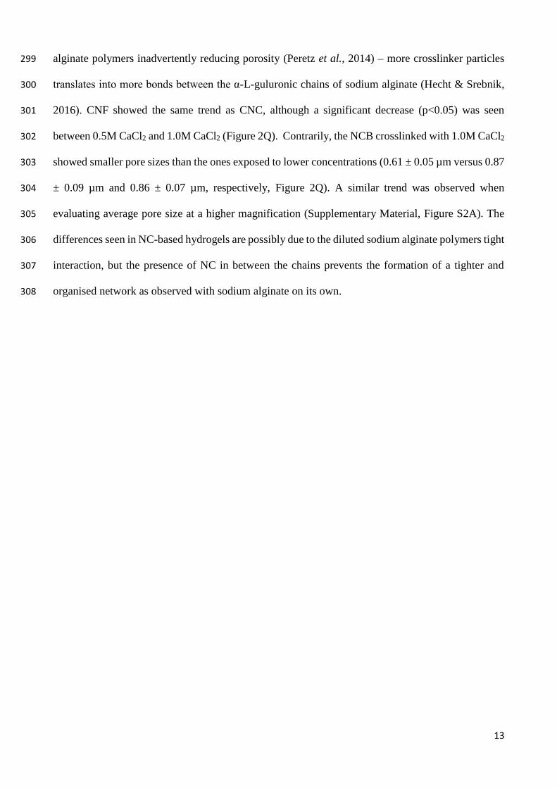
alginate polymers inadvertently reducing porosity (Peretz et al., 2014) – more crosslinker particles 299
translates into more bonds between the α-L-guluronic chains of sodium alginate (Hecht & Srebnik, 300
2016). CNF showed the same trend as CNC, although a significant decrease (p<0.05) was seen 301
between 0.5M CaCl2 and 1.0M CaCl2 (Figure 2Q). Contrarily, the NCB crosslinked with 1.0M CaCl2 302
showed smaller pore sizes than the ones exposed to lower concentrations (0.61 ± 0.05 µm versus 0.87 303
± 0.09 µm and 0.86 ± 0.07 µm, respectively, Figure 2Q). A similar trend was observed when 304
evaluating average pore size at a higher magnification (Supplementary Material, Figure S2A). The 305
differences seen in NC-based hydrogels are possibly due to the diluted sodium alginate polymers tight 306
interaction, but the presence of NC in between the chains prevents the formation of a tighter and 307
organised network as observed with sodium alginate on its own. 308

14
309
Figure 2. Structure and mechanics of NC-based hydrogels post-crosslinking with different CaCl2 concentrations. (A-P) 310
Overall network architecture and pore distribution. Images taken at 9k and 20k magnifications. Scale bar = 2 µm. (Q) 311
Average pore size (µm) post-crosslinking based on 9k magnification SEM images. Mean ± SEM, n=80 measurements. 312

15
(R) Porosity and (S) swelling percentages post-crosslinking. Mean ± SEM, n=6. (T) Young’s modulus (kPa) based on 313
compression post-crosslinking. Mean ± SEM, n=6. NCB – nanocellulose blend; CNC – nanocellulose crystal; CNF – 314
nanocellulose fibrils; CaCl2 – calcium chloride; 0.1M CaCl2 – black; 0.5M CaCl2 – light grey; 1.0M CaCl2 – grey. Mann 315
Whitney (Q and T) and ANOVA (R and S) statistical tests: *, p ≤ 0.05; **, p ≤ 0.01; ****, p ≤ 0.0001. 316
317
The overall porosity of NCB was not affected by the concentration of the crosslinker (p>0.05, Figure 318
2R). In all other hydrogels, the porosity decrease was more accentuated between the lowest and the 319
highest CaCl2 concentrations: sodium alginate (56.7 ± 0.8% versus 37.2 ± 1.9%, p<0.0001), CNF 320
(45.6 ± 0.3% versus 34.2 ± 1.0%, p<0.0001) and CNC (43.7 ± 1.4% versus 34.6 ± 1.60%, p<0.01) 321
(Figure 2R). The swelling capacity of NCB and CNF was not affected by the crosslinker 322
concentration (Figure 2S). Conversely, the swelling of sodium alginate and CNC was affected by 323
CaCl2 concentration – swelling decreased to at least half when exposed to 1.0M CaCl2 (p<0.0001, 324
Figure 2S). Overall, in NC-based hydrogels, the use of 0.5M CaCl2 showed milder effects for both 325
porosity and swelling percentages (Figures 2R and 2S). Finally, the stiffness of the crosslinked 326
hydrogels was measured based on Young’s Modulus (Figure 2T). NCB and CNF retained similar 327
stiffness independent of the crosslinker concentration (p>0.05, Figure 2T). However, CNC and 328
sodium alginate yielded stiffer hydrogels when exposed to higher CaCl2 concentrations (Figure 2T). 329
These similarities may be related to the ordered structural organisation of both sodium alginate and 330
CNC (Ma et al., 2017). The sodium alginate crosslinked with 1.0M CaCl2 produced the stiffest 331
hydrogel tested (361 ± 40 kPa, Figure 2T). 332
Overall, the effect of crosslinker is more striking in sodium alginate hydrogels than NC-based 333
hydrogels. Among the different NC forms, CNC seems to be the most affected by varying crosslinker 334
concentrations while NCB retains most of its characteristics independent of crosslinker 335
concentrations. 336
4.2 Characterization of sterilised NC-based hydrogels 337
The use of low crosslinker concentrations has been widely demonstrated as the optimal crosslinking 338
method as it promotes a slower gelation rate, uniform structure, and enhanced mechanical integrity 339

16
(Kuo & Ma, 2001; Skjåk-Bræk, Grasdalen, & Smidsrød, 1989). As a result, 0.1M CaCl2 was used to 340
assess the effects of the various sterilisation methods on sodium alginate and NC-based hydrogels. 341
Due to the temperature sensitive nature of sodium alginate, autoclave sterilisation was not pursued as 342
the high temperatures promote depolymerisation of alginate (Leo, Mcloughlin, & Malone, 1990). 343
Changes in the surface morphology and network distribution in the different NC-based hydrogels 344
upon sterilisation was confirmed through SEM (Figure 3A-P). All sterilisation methods showed an 345
apparent impact on the overall network distribution. The most striking differences were observed in 346
sodium alginate hydrogels when exposed to any sterilisation method (Figure 3A-D). Both UV and 347
ethanol sterilisations transformed the network of the NC-based hydrogels into a more leaf-like 348
architecture while autoclave seemed to accentuate the fibrillar features of the network (Figure 3E-P). 349
No visible network differences were observed between different NC-based hydrogels exposed to the 350
same sterilisation method. Sterilisation significantly decreased the average pore size of all hydrogels 351
by 17% – 86 % (p<0.0001, Figure 3Q), which is similarly seen in sterilisation of silk-fibroin hydrogels 352
in other studies (Hofmann, Stok, Kohler, Meinel, & Müller, 2014). Autoclave sterilisation resulted in 353
hydrogels with the largest pore size – a trend that was observed in all tested hydrogels (Figure 3Q). 354
Heat sterilisation using the autoclave process replaces the air in the container, creating pressure and 355
leading to the formation of larger pores. Similar trends were observed when evaluating average pore 356
size at a higher magnification (Supplementary Material, Figure S2B). UV and ethanol sterilisations 357
have shown roughly similar pore sizes in all NC-based hydrogels however, the porosity was not 358
affected. This might be related to the rearrangement and fragmentation of the pores during 359
sterilisation, resulting in smaller pores but no changes in overall porosity. This is evident when 360
examining the swelling percentage of NC-based hydrogels. Apart from the alterations in average pore 361
size post-sterilisation, the overall porosity was maintained in all hydrogels except for sodium alginate, 362
where ethanol significantly decreased overall porosity by ~6% (p<0.05, Figure 3R). The swelling 363
capacity of sodium alginate hydrogels decreased significantly post-UV (p<0.05) and post-ethanol 364
sterilisation (p<0.0001, Figure 3S). However, with regards to NC-based hydrogels there was an 365

17
overall increase in swelling capacity post-sterilisation, with UV sterilisation yielding hydrogels with 366
the highest swelling percentage (p<0.001, Figure 3S). UV irradiation has sufficient energy to disrupt 367
covalent bonds and result in the formation of free radicals which propagate degradation 368
(Wasikiewicz, Yoshii, Nagasawa, Wach, & Mitomo, 2005). The results suggest that UV treatment 369
potentiated the formation of smaller pores which enhanced the swelling potential of all NC-based 370
hydrogels. This is corroborated by the pore size measurements of UV treated NC-based hydrogels. 371
Measurements of stiffness post-sterilisation showed that overall ethanol creates hydrogels with a 372
higher Young’s modulus (Figure 3T). This trend was significantly higher in sodium alginate (306 ± 373
32.8 kPa, p<0.001), CNF (508 ± 94.5 kPa, p<0.05), and CNC (420 ± 62.7 kPa, p<value 0.001) 374
hydrogels. Ethanol is known for its dehydration properties resulting in the compaction of hydrogels 375
– which explains the higher mechanical strength post-sterilisation as the resultant gels are stiffer 376
(Eltoum, Fredenburgh, Myers, & Grizzle, 2001). The two other sterilisation methods showed variable 377
effects on hydrogel stiffness (Figure 3T): autoclave sterilisation significantly reduced the Young’s 378
modulus of CNF hydrogels (148 ± 12 kPa, p<0.05), whereas it had the opposite effect on CNC 379
hydrogels (173 ± 12.7 kPa, p<0.05). Although it has not been reported in the literature, we theorize 380
that UV and autoclave treatments cause the breakage of clusters of CNC within the hydrogel, resulting 381
in an increase in homogeneity which can be observed in the SEM images post-sterilisation. CNF was 382
not degraded by the thermal energy generated from the autoclave, yet the SEM images show that the 383
fibrils have undergone structural alterations, such as fibril realignment, thus resulting in larger pore 384
size post-sterilisation out of all NC-based hydrogels, which translated into weaker mechanical 385
properties (Kyle et al., 2018; Yang, Yan, Chen, Lee, & Zheng, 2007). All sterilisation methods did 386
not significantly affect the stiffness of NCB hydrogels (Figure 3T). In contrast, all the sterilisation 387
methods used affected the stiffness of CNC (UV, 177 ± 6.6 kPa, p<0.05; autoclave, 173 ± 12.7 kPa, 388
p<0.05; ethanol, 420 ± 62.7 kPa, p<0.001, Figure 3T). 389

18
390
Figure 3. Structure and mechanics of NC-based hydrogels post-sterilisation with different methods. (A-P) Overall 391
network architecture and pore distribution. Images taken at 9k and 20k magnification. Scale bar = 2 µm. (Q) Average 392
pore size (µm) post-sterilisation based on 9k magnification SEM images. Mean ± SEM, n=80 measurements. (R) Porosity 393
and (S) swelling percentages post-sterilisation. Mean ± SEM, n=5-6. (T) Young’s modulus (kPa) based on compression 394

19
post-sterilisation. Mean ± SEM, n=6. NCB – nanocellulose blend; CNC – nanocellulose crystal; CNF – nanocellulose 395
fibrils; Non-sterile – black; UV – grey; Autoclave – dark grey; Ethanol – light grey. ●, absent graph bar: autoclaved 396
sodium alginate hydrogels were not tested. Mann Whitney (Q and T) and ANOVA (R and S) statistical tests: *, p ≤ 0.05; 397
**, p ≤ 0.01; ***, p ≤ 0.001; ****, p ≤ 0.0001. 398
4.3 Bacterial persistence in sterilised NC-based hydrogels 399
The efficiency of the sterilisation processes was examined through bacterial persistence (Figure 4). 400
Sterilisation efficiency was evaluated using the respective non-sterile material as a control and all 401
sterilisation methods were confirmed as effective in removing bacterial content. All sterilisation 402
methods showed significant reduction of OD in the hydrogels post-sterilisation (p<0.05, Figure 4). 403
Overall, UV sterilisation was the most inefficient method for the sterilisation of NC-based hydrogels 404
(Figure 4). UV sterilisation was very efficient in sodium alginate hydrogels, indicating it is optimal 405
for materials that are transparent – due to its limited penetrability – but not ideal for NC-based 406
hydrogels (Lerouge, 2012). Conversely, the autoclave method was the most efficient for all hydrogels 407
(p<0.0001, Figure 4). Although it resulted in structural alterations, it is the optimal method to ensure 408
the elimination of potential contaminants including fungal and bacterial spores (Rogers, 2012). In 409
practice, the use of ethanol is unfeasible as this would result in cell death, as observed in the cell 410
viability tests. 411
412
Figure 4. Bacterial persistence at 24h and 48h post-sterilisation. Mean ± SEM, n=4-5. NCB – nanocellulose blend; CNC 413
– nanocellulose crystal; CNF – nanocellulose fibrils. Non-sterile – black; UV – grey; Autoclave – dark grey; Ethanol – 414

20
light grey. ●, absent graph bar: autoclaved sodium alginate hydrogels were not tested. Mann Whitney statistical test: *, p 415
≤ 0.05; **, p ≤ 0.01; ***, p ≤ 0.001; ****, p ≤ 0.0001. 416
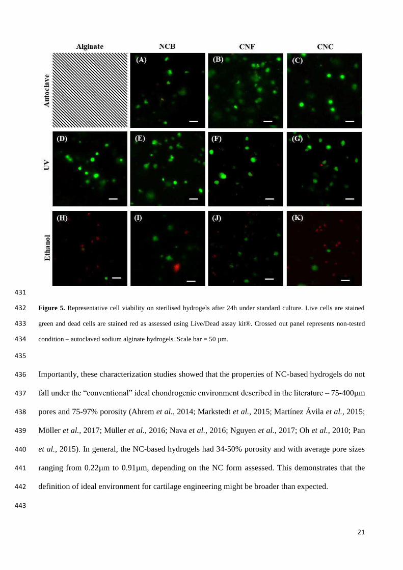
4.4 Cell viability assessment using sterilised NC-based hydrogels 417
Cell viability in the hydrogels was assessed at 1- and 7-days post-sterilisation using human naso-418
septal chondrocytes and Live/Dead® assay kit (Figure 5 and Supplementary Material, Figure S3). 419
Upon 24h in culture in sterilised hydrogels, cell viability was minimally affected, apart from the cells 420
in ethanol-sterilised hydrogels where most of the cells were dead (Figure 5). After 7 days, the number 421
of dead cells increased in all hydrogels although it was visibly lower than the number of live cells 422
which is indicative of cellular turnover (Supplementary Material, Figure S3). Yet, the low cell 423
viability outcomes in ethanol sterilisation could be related to a limitation of this study – ethanol 424
sterilisation and crosslinking were performed in tandem, meaning that the cells were exposed to 70% 425
ethanol for 20 min, which resulted in higher cell death when compared to other methods. Technically, 426
for the ethanol sterilisation, it was not possible to sterilise the non-crosslinked NC-based hydrogels 427
with ethanol because the removal of ethanol by centrifugation would result in decreased water content 428
in the hydrogels. 429
430
21
431
Figure 5. Representative cell viability on sterilised hydrogels after 24h under standard culture. Live cells are stained 432
green and dead cells are stained red as assessed using Live/Dead assay kit®. Crossed out panel represents non-tested 433
condition – autoclaved sodium alginate hydrogels. Scale bar = 50 µm. 434
435
Importantly, these characterization studies showed that the properties of NC-based hydrogels do not 436
fall under the “conventional” ideal chondrogenic environment described in the literature – 75-400µm 437
pores and 75-97% porosity (Ahrem et al., 2014; Markstedt et al., 2015; Martínez Ávila et al., 2015; 438
Möller et al., 2017; Müller et al., 2016; Nava et al., 2016; Nguyen et al., 2017; Oh et al., 2010; Pan 439
et al., 2015). In general, the NC-based hydrogels had 34-50% porosity and with average pore sizes 440
ranging from 0.22µm to 0.91µm, depending on the NC form assessed. This demonstrates that the 441
definition of ideal environment for cartilage engineering might be broader than expected. 442
443

22
5. Conclusion 444
Previous studies have shown that NC contains favourable properties for diverse biological and 445
medical applications. NC-based hydrogels have been extensively explored for cartilage engineering 446
purposes, mainly using 3D bioprinting technologies. 447
In this study, composite hydrogels containing sodium alginate and different forms of plant-derived 448
NC (nanocellulose fibrils, nanocellulose crystals or a blend), ionically crosslinked with CaCl2, were 449
shown to have alterations in their structural and mechanical properties upon standard processing 450
methods such as crosslinking and sterilisation. Increasing concentrations of the crosslinker CaCl2 451
yielded visible changes in overall architecture, pore size (as demonstrated through SEM) and porosity. 452
As sodium alginate crosslinks faster with increasing concentrations of CaCl2, the resulting mesh 453
network will have a different distribution and size, with the different NC forms entrapped in the 454
interstitial areas of the mesh – providing characteristic architectures according their structure (i.e. 455
fibrils or crystals). The swelling capacity and the mechanical properties (as assessed by the Young’s 456
Modulus) of the NC-based hydrogels were also affected with increasing crosslinker concentrations, 457
yet not all NC forms were significantly affected. 458
When exposed to different sterilisation methods (physical, thermal and chemical), the crosslinked 459
NC-based hydrogels showed striking significant decreases in average pore size, while porosity was 460
maintained. From all the properties tested, pore size was the most affected by the sterilisation method, 461
possibly due to the re-arrangement of particles inside the hydrogels. The mechanical properties of the 462
hydrogels were mildly affected by the sterilisation method, apart from the chemical sterilisation using 463
ethanol that yielded significantly stronger hydrogels, possibly due to the dehydration of the hydrogels. 464
Importantly, differential effects were observed based on the NC form contained in the composite 465
hydrogels. Among the NC forms, CNC was more affected by the crosslinker concentrations, CNF 466
and CNC were affected by all sterilization methods with different methods affecting properties 467
differently while NCB was more resilient to changes when exposed to different sterilisation methods 468
and crosslinker concentrations. This indicates that the crosslinking reactions and the sterilisation 469

23
method used to process these hydrogels need to be chosen and tailored to the final aim (e.g. tissue 470
type) as these will significantly alter the final environment to which cells will be exposed. 471
The study of structural and mechanical alterations upon different processing methods is important as 472
it impacts the characteristics of the final product. These will directly affect, for example, its 473
microstructure and microenvironment, ultimately impacting cell phenotype and behaviour when 474
targeting biomedical applications. 475
476
Acknowledgments 477
This work was supported by Abertawe Bro Morgannwg University (ABMU) Health Board, Health 478
and Care Research Wales, Oakgrove Medical Charitable Trust, the Royal College of Surgeons of 479
England and the Medical Research Council (grant no. MR/N002431/1). We acknowledge the 480
Arthritis Research UK Biomechanics and Bioengineering Centre (Cardiff University) for access to 481
the ElectroForce™ 3200 loading equipment and Mrs. Kavitha Saw for collecting the human naso-482
septal samples at Singleton Hospital, Swansea, UK. Dr. Ayesha Al-Sabah, Dr. Stephanie EA Burnell, 483
and Dr. Irina N Simoes have equally contributed to this work. 484
485
References 486
Ahrem, H., Pretzel, D., Endres, M., Conrad, D., Courseau, J., Müller, H., . . . Kinne, R. W. (2014). Laser-structured 487
bacterial nanocellulose hydrogels support ingrowth and differentiation of chondrocytes and show potential as cartilage 488
implants. Acta Biomaterialia, 10(3), 1341-1353. 489
Anderson, J. M., Rodriguez, A., & Chang, D. T. (2008). Foreign body reaction to biomaterials. Semin Immunol, 20(2), 490
86-100. 491
Ansari, S., Diniz, I. M., Chen, C., Aghaloo, T., Wu, B. M., Shi, S., & Moshaverinia, A. (2017). Alginate/hyaluronic 492
acid hydrogel delivery system characteristics regulate the differentiation of periodontal ligament stem cells toward 493
chondrogenic lineage. J Mater Sci Mater Med, 28(10), 162. 494
Baker, M. I., Walsh, S. P., Schwartz, Z., & Boyan, B. D. (2012). A review of polyvinyl alcohol and its uses in cartilage 495
and orthopedic applications. J Biomed Mater Res B Appl Biomater, 100(5), 1451-1457. 496
Bodin, A., Ahrenstedt, L., Fink, H., Brumer, H., Risberg, B., & Gatenholm, P. (2007). Modification of nanocellulose 497
with a xyloglucan-RGD conjugate enhances adhesion and proliferation of endothelial cells: implications for tissue 498
engineering. Biomacromolecules, 8(12), 3697-3704. 499

24
Bryant, S. J., & Anseth, K. S. (2002). Hydrogel properties influence ECM production by chondrocytes 500
photoencapsulated in poly(ethylene glycol) hydrogels. J Biomed Mater Res, 59(1), 63-72. 501
Bryant, S. J., Chowdhury, T. T., Lee, D. A., Bader, D. L., & Anseth, K. S. (2004). Crosslinking density influences 502
chondrocyte metabolism in dynamically loaded photocrosslinked poly(ethylene glycol) hydrogels. Ann Biomed Eng, 503
32(3), 407-417. 504
Buxton, A. N., Zhu, J., Marchant, R., West, J. L., Yoo, J. U., & Johnstone, B. (2007). Design and characterization of 505
poly(ethylene glycol) photopolymerizable semi-interpenetrating networks for chondrogenesis of human mesenchymal 506
stem cells. Tissue Eng, 13(10), 2549-2560. 507
Caliari, S. R., & Burdick, J. A. (2016). A practical guide to hydrogels for cell culture. Nat Methods, 13(5), 405-414. 508
Chou, A. I., Akintoye, S. O., & Nicoll, S. B. (2009). Photo-crosslinked Alginate Hydrogels Support Enhanced Matrix 509
Accumulation by Nucleus Pulposus Cells In Vivo. Osteoarthritis and cartilage / OARS, Osteoarthritis Research 510
Society, 17(10), 1377-1384. 511
Chung, C., Mesa, J., Randolph, M. A., Yaremchuk, M., & Burdick, J. A. (2006). Influence of gel properties on 512
neocartilage formation by auricular chondrocytes photoencapsulated in hyaluronic acid networks. J Biomed Mater Res 513
A, 77(3), 518-525. 514
Dowthwaite, G. P., Bishop, J. C., Redman, S. N., Khan, I. M., Rooney, P., Evans, D. J., . . . Archer, C. W. (2004). The 515
surface of articular cartilage contains a progenitor cell population. J Cell Sci, 117(Pt 6), 889-897. 516
Dumanli, A. G. (2016). Nanocellulose and its Composites for Biomedical Applications. Curr Med Chem. 517
Eltoum, I., Fredenburgh, J., Myers, R. B., & Grizzle, W. E. (2001). Introduction to the Theory and Practice of Fixation 518
of Tissues. Journal of Histotechnology, 24(3), 173-190. 519
Endes, C., Camarero-Espinosa, S., Mueller, S., Foster, E. J., Petri-Fink, A., Rothen-Rutishauser, B., . . . Clift, M. J. D. 520
(2016). A critical review of the current knowledge regarding the biological impact of nanocellulose. Journal of 521
Nanobiotechnology, 14. 522
Fickert, S., Fiedler, J., & Brenner, R. E. (2004). Identification of subpopulations with characteristics of mesenchymal 523
progenitor cells from human osteoarthritic cartilage using triple staining for cell surface markers. Arthritis Res Ther, 524
6(5), R422-432. 525
Galante, R., Pinto, T. J. A., Colaco, R., & Serro, A. P. (2017). Sterilization of hydrogels for biomedical applications: A 526
review. J Biomed Mater Res B Appl Biomater. 527
Gupta, N. V., & Shivakumar, H. G. (2012). Investigation of Swelling Behavior and Mechanical Properties of a pH-528
Sensitive Superporous Hydrogel Composite. Iran J Pharm Res, 11(2), 481-493. 529
Hecht, H., & Srebnik, S. (2016). Structural Characterization of Sodium Alginate and Calcium Alginate. 530
Biomacromolecules, 17(6), 2160-2167. 531

25
Hofmann, S., Stok, K. S., Kohler, T., Meinel, A. J., & Müller, R. (2014). Effect of sterilization on structural and 532
material properties of 3-D silk fibroin scaffolds. Acta Biomaterialia, 10(1), 308-317. 533
Hwang, N. S., Varghese, S., Lee, H. J., Theprungsirikul, P., Canver, A., Sharma, B., & Elisseeff, J. (2007). Response of 534
zonal chondrocytes to extracellular matrix-hydrogels. FEBS Lett, 581(22), 4172-4178. 535
K. Pal, A. K. B., D. K. Majumdar. (2009). Polymeric Hydrogels: Characterization and Biomedical Applications - A 536
mini review. Designed Monomers and Polymers, 12, 34. 537
Kim, D. H., & Song, Y. S. (2015). Rheological behavior of cellulose nanowhisker suspension under magnetic field. 538
Carbohydr Polym, 126, 240-247. 539
Kuo, C. K., & Ma, P. X. (2001). Ionically crosslinked alginate hydrogels as scaffolds for tissue engineering: Part 1. 540
Structure, gelation rate and mechanical properties. Biomaterials, 22(6), 511-521. 541
Kyle, S., Jessop, Z. M., Al-Sabah, A., Hawkins, K., Lewis, A., Maffeis, T., . . . Whitaker, I. S. (2018). Characterization 542
of pulp derived nanocellulose hydrogels using AVAP(R) technology. Carbohydr Polym, 198, 270-280. 543
Leo, W. J., Mcloughlin, A. J., & Malone, D. M. (1990). Effects of Sterilization Treatments on Some Properties of 544
Alginate Solutions and Gels. Biotechnology Progress, 6(1), 51-53. 545
Leppiniemi, J., Lahtinen, P., Paajanen, A., Mahlberg, R., Metsa-Kortelainen, S., Pinomaa, T., . . . Hytonen, V. P. 546
(2017). 3D-Printable Bioactivated Nanocellulose-Alginate Hydrogels. ACS Appl Mater Interfaces, 9(26), 21959-21970. 547
Lerouge, S. (2012). 5 - Non-traditional sterilization techniques for biomaterials and medical devices. In S. Lerouge & 548
A. Simmons (Eds.), Sterilisation of Biomaterials and Medical Devices (pp. 97-116): Woodhead Publishing 549
Lien, S. M., Ko, L. Y., & Huang, T. J. (2009). Effect of pore size on ECM secretion and cell growth in gelatin scaffold 550
for articular cartilage tissue engineering. Acta Biomater, 5(2), 670-679. 551
Lin, N., & Dufresne, A. (2014). Nanocellulose in biomedicine: Current status and future prospect. European Polymer 552
Journal, 59, 24. 553
Lin, S., Sangaj, N., Razafiarison, T., Zhang, C., & Varghese, S. (2011). Influence of physical properties of biomaterials 554
on cellular behavior. Pharm Res, 28(6), 1422-1430. 555
Ma, X., Li, R., Zhao, X., Ji, Q., Xing, Y., Sunarso, J., & Xia, Y. (2017). Biopolymer composite fibres composed of 556
calcium alginate reinforced with nanocrystalline cellulose. Composites Part A: Applied Science and Manufacturing, 96, 557
155-163. 558
Mao, Y., Liu, K., Zhan, C., Geng, L., Chu, B., & Hsiao, B. S. (2017). Characterization of Nanocellulose Using Small-559
Angle Neutron, X-ray, and Dynamic Light Scattering Techniques. J Phys Chem B, 121(6), 1340-1351. 560
Markstedt, K., Mantas, A., Tournier, I., Martínez Ávila, H., Hägg, D., & Gatenholm, P. (2015). 3D Bioprinting Human 561
Chondrocytes with Nanocellulose–Alginate Bioink for Cartilage Tissue Engineering Applications. Biomacromolecules, 562
16(5), 1489-1496. 563
26
Martínez Ávila, H., Feldmann, E.-M., Pleumeekers, M. M., Nimeskern, L., Kuo, W., de Jong, W. C., . . . Gatenholm, P. 564
(2015). Novel bilayer bacterial nanocellulose scaffold supports neocartilage formation in vitro and in vivo. 565
Biomaterials, 44, 122-133. 566
Matthews, I. P., Gibson, C., & Samuel, A. H. (1994). Sterilisation of implantable devices. Clin Mater, 15(3), 191-215. 567
Miao, Z., Lu, Z., Wu, H., Liu, H., Li, M., Lei, D., . . . Zhao, J. (2017). Collagen, agarose, alginate and Matrigel 568
hydrogels as cell substrates for culture of chondrocytes in vitro: A comparative study. J Cell Biochem. 569
Möller, T., Amoroso, M., Hägg, D., Brantsing, C., Rotter, N., Apelgren, P., . . . Gatenholm, P. (2017). In Vivo 570
Chondrogenesis in 3D Bioprinted Human Cell-laden Hydrogel Constructs. Plastic and Reconstructive Surgery Global 571
Open, 5(2), e1227. 572
Mouser, V. H. M., Levato, R., Bonassar, L. J., D'Lima, D. D., Grande, D. A., Klein, T. J., . . . Malda, J. (2017). Three-573
Dimensional Bioprinting and Its Potential in the Field of Articular Cartilage Regeneration. Cartilage, 8(4), 327-340. 574
Müller, M., Öztürk, E., Arlov, Ø., Gatenholm, P., & Zenobi-Wong, M. (2016). Alginate Sulfate–Nanocellulose Bioinks 575
for Cartilage Bioprinting Applications. Annals of Biomedical Engineering, 1-14. 576
Nava, M. M., Draghi, L., Giordano, C., & Pietrabissa, R. (2016). The effect of scaffold pore size in cartilage tissue 577
engineering. J Appl Biomater Funct Mater, 14(3), e223-229. 578
Nguyen, D., Hagg, D. A., Forsman, A., Ekholm, J., Nimkingratana, P., Brantsing, C., . . . Simonsson, S. (2017). 579
Cartilage Tissue Engineering by the 3D Bioprinting of iPS Cells in a Nanocellulose/Alginate Bioink. Sci Rep, 7(1), 658. 580
Oh, S. H., Kim, T. H., Im, G. I., & Lee, J. H. (2010). Investigation of pore size effect on chondrogenic differentiation of 581
adipose stem cells using a pore size gradient scaffold. Biomacromolecules, 11(8), 1948-1955. 582
Paakko, M., Ankerfors, M., Kosonen, H., Nykanen, A., Ahola, S., Osterberg, M., . . . Lindstrom, T. (2007). Enzymatic 583
hydrolysis combined with mechanical shearing and high-pressure homogenization for nanoscale cellulose fibrils and 584
strong gels. Biomacromolecules, 8(6), 1934-1941. 585
Pan, Z., Duan, P., Liu, X., Wang, H., Cao, L., He, Y., . . . Ding, J. (2015). Effect of porosities of bilayered porous 586
scaffolds on spontaneous osteochondral repair in cartilage tissue engineering. Regen Biomater, 2(1), 9-19. 587
Park, H., & Lee, K. Y. (2014). Cartilage regeneration using biodegradable oxidized alginate/hyaluronate hydrogels. J 588
Biomed Mater Res A, 102(12), 4519-4525. 589
Peretz, S., Florea-Spirou, M., Anghel, D.-F., Bala, D., Stoian, C., & Zgherea, G. (2014). Preparation of Porous 590
Calcium Alginate Beads and Their Use for Adsorption of O-Nitrophenol from Aqueous Solutions. 591
Revin, V., Liyaskina, E., Nazarkina, M., Bogatyreva, A., & Shchankin, M. (2018). Cost-effective production of 592
bacterial cellulose using acidic food industry by-products. Braz J Microbiol, 49 Suppl 1, 151-159. 593
Rogers, W. J. (2012). 2 - Steam and dry heat sterilization of biomaterials and medical devices. In S. Lerouge & A. 594
Simmons (Eds.), Sterilisation of Biomaterials and Medical Devices (pp. 20-55): Woodhead Publishing 595

27
Skjåk-Bræk, G., Grasdalen, H., & Smidsrød, O. (1989). Inhomogeneous polysaccharide ionic gels. Carbohydr Polym, 596
10(1), 31-54. 597
Svensson, A., Nicklasson, E., Harrah, T., Panilaitis, B., Kaplan, D. L., Brittberg, M., & Gatenholm, P. (2005). Bacterial 598
cellulose as a potential scaffold for tissue engineering of cartilage. Biomaterials, 26(4), 419-431. 599
Tibbitt, M. W., & Anseth, K. S. (2009). Hydrogels as extracellular matrix mimics for 3D cell culture. Biotechnol 600
Bioeng, 103(4), 655-663. 601
Veerachamy, S., Yarlagadda, T., Manivasagam, G., & Yarlagadda, P. K. (2014). Bacterial adherence and biofilm 602
formation on medical implants: a review. Proc Inst Mech Eng H, 228(10), 1083-1099. 603
Villanueva, I., Klement, B. J., von Deutsch, D., & Bryant, S. J. (2009). Cross-linking density alters early metabolic 604
activities in chondrocytes encapsulated in poly(ethylene glycol) hydrogels and cultured in the rotating wall vessel. 605
Biotechnol Bioeng, 102(4), 1242-1250. 606
Wasikiewicz, J. M., Yoshii, F., Nagasawa, N., Wach, R. A., & Mitomo, H. (2005). Degradation of chitosan and sodium 607
alginate by gamma radiation, sonochemical and ultraviolet methods. Radiation Physics and Chemistry, 73(5), 287-295. 608
Xiao, Y., Friis, E. A., Gehrke, S. H., & Detamore, M. S. (2013). Mechanical testing of hydrogels in cartilage tissue 609
engineering: beyond the compressive modulus. Tissue Eng Part B Rev, 19(5), 403-412. 610
Yang, H., Yan, R., Chen, H., Lee, D. H., & Zheng, C. (2007). Characteristics of hemicellulose, cellulose and lignin 611
pyrolysis. Fuel, 86(12), 1781-1788. 612
613