Embed Size (px)
Citation preview
1
Saquiba Yesmine, Saquiba Yesmine, PhDPhD
Fall 2014

2
Brain function is the single most important aspect of physiology that defines the difference between humans and other species.
Disorders of brain function, whether primary or secondary to malfunction of other systems, are a major concern of human society,
It is a field in which pharmacological intervention plays a key role.

3
Brain function is the single most important aspect of physiology that defines the difference between humans and other species.
Disorders of brain function, whether primary or secondary to malfunction of other systems, are a major concern of human society,
It is a field in which pharmacological intervention plays a key role.

4
"Men ought to know that from nothing else but the brain come joys, delights, laughter and sports, and sorrows and griefs,”-Hippocrates
Through much of history, the mind was thought to be separate from the brain.

Neuropharmacology can be defined simply as the
study of drugs that affect nervous tissue.
Neuropharmacology is the study of drugs
that interact with neurons in the brain to
affect mood, sensation, behavior, and
thinking.
What is Neuropharmacology? What is Neuropharmacology?

But….
this is not an accomplished definition since a great many
drugs whose therapeutic value is extraneural can affect the
nervous system.
For example,
the cardiotonic drug digitalis commonly produce central
nervous system (CNS) effects ranging from blurred vision to
disorientation.
What is Neuropharmacology? What is Neuropharmacology?
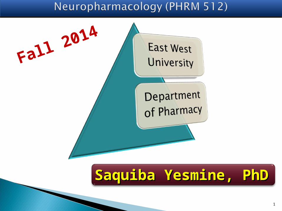
Using drugs non-medicinally to alter states of consciousness is not a new concept.
Alcohol, caffeine, nicotine, heroin, cocaine, cannabis all transitioned from medicinal use to the recreational improvement of mood and performance
What is Neuropharmacology? What is Neuropharmacology?
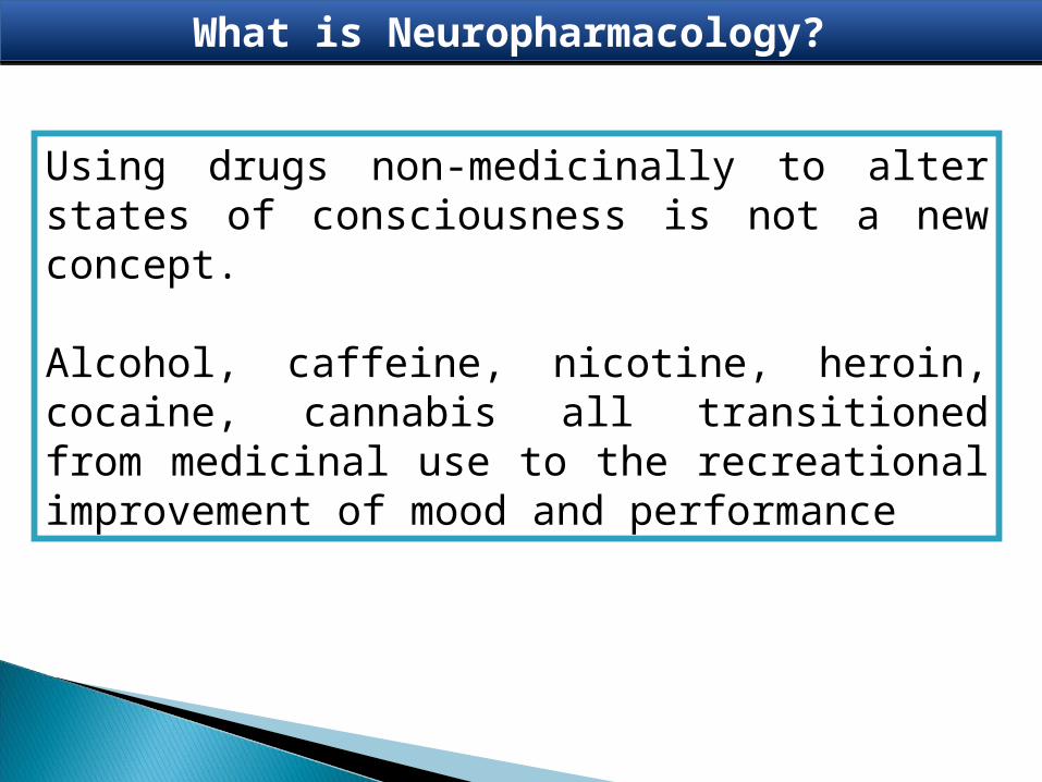
The scope of neuropharmacology generally limits to those
drugs which specifically employed to affect the
nervous system.
Scope of Neuropharmacology Scope of Neuropharmacology
Psychotropic drugs that affect mood and behavior, Anesthetics drugs, Sedatives drugs, Hypnotics drugs, Narcotics drugs, Anticonvulsants drugs, Analgesics drugs, and Drugs that affect the Autonomic Nervous System.
The domain of neuropharmacology include -

9
Therapeutic
Non-therapeutic(e.g. alcohol, tea and coffee, cannabis, nicotine, opioids, amphetamines etc.).
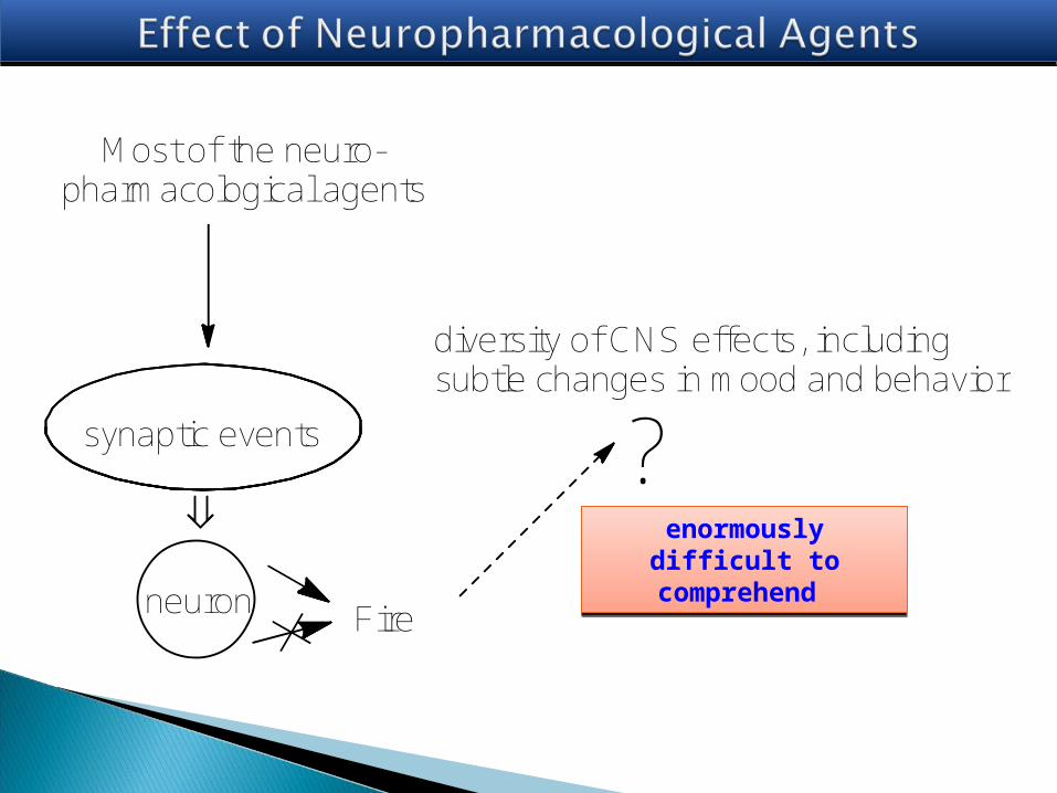
Most of the neuro-pharmacological agents
synaptic events
Fireneuron
diversity of CNS effects, including subtle changes in mood and behavior
?enormously difficult
to comprehend enormously difficult
to comprehend

at the molecular level, an explanation of the action
of a drug is often possible;
at the cellular level, an explanation is sometimes
possible; but –
at the behavioral level, our ignorance is abysmal.

NeuronsNeurons
Neurons are highly polarized cells (it has distinct subcellular domains that subserve different functions).
Morphologically, in a typical neuron, three major regions can be defined:
1) the cell body, or perikaryon, which contains the nucleus and the major cytoplasmic organelles;
2) a variable number of dendrites, which emanate from the perikaryon and which differ in size and shape and
3) a single axon, which extends in most cases much farther from the cell body than does the dendritic arbor.
many axons are surrounded by an insulating myelin sheath, which facilitates rapid impulse conduction.

Sensory (afferent) neurons
-- carry signals to the central
nervous system (CNS)
Interneurons
- contained entirely within the
CNS and carry signals from
one neuron to another
Motor (efferent) neurons
- carry signals from the CNS
to muscles and glands.
Functional Classes of NeuronsFunctional Classes of Neurons

The term neuroglia, or “nerve glue,” was coined in 1859 by Rudolph Virchow, who conceived of the neuroglia as an inactive “connective tissue” holding neurons together in the central nervous system.
Supporting cells in the central nervous system (CNS)

Supportive Cells (Neuroglia)Supportive Cells (Neuroglia)
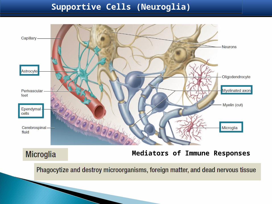
Supportive Cells (Neuroglia)Supportive Cells (Neuroglia)
Mediators of Immune Responses

Supportive Cells (Neuroglia)Supportive Cells (Neuroglia)
Form myelin in brain and spinal cord
Oligodendrocytes
branched and starlike shape
Supportive Cells (Neuroglia)Supportive Cells (Neuroglia)
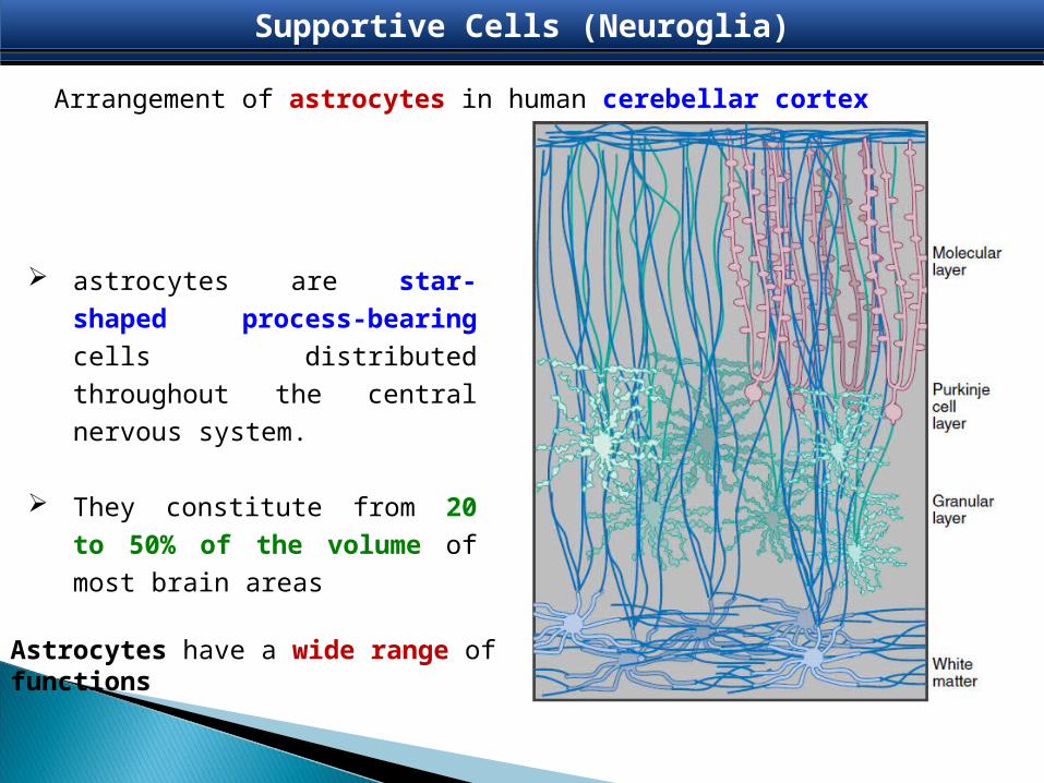
Arrangement of astrocytes in human cerebellar cortex
astrocytes are star-shaped
process-bearing cells distributed
throughout the central nervous
system.
They constitute from 20 to 50% of
the volume of most brain areas
Astrocytes have a wide range of functions

bulbous body with as many as 15 arm like processesAstrocytes
Supportive Cells (Neuroglia)Supportive Cells (Neuroglia)
1. Cover brain surface and nonsynaptic regions of neurons; 2. form supportive framework in CNS;
induce formation of blood-brain barrier;3. nourish neurons;
4. produce growth factors that stimulate neurons; 5. communicate electrically with neurons and influence
synaptic signalling;6. remove neurotransmitters and K from ECF of brain and
spinal cord;7. help to regulate composition of ECF;8. form scar tissue to replace damaged nervous tissue

Glutamate–Glutamine cycle Glutamate–Glutamine cycle
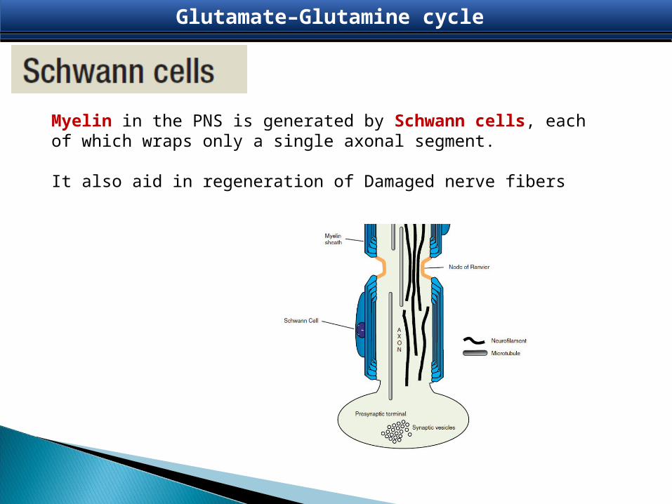
Myelin in the PNS is generated by Schwann cells, each of which wraps only a single axonal segment.
It also aid in regeneration of Damaged nerve fibers
Glutamate–Glutamine cycle Glutamate–Glutamine cycle

Overviews of Nervous systemOverviews of Nervous system

NeuropharmacologyNeuropharmacologyThe nervous system can be separated into parts based on structure and on function.
structurally, it can be divided into the central nervous system (CNS) and the peripheral nervous system (PNS)
functionally, it can be divided into –•somatic and •visceral parts.
The CNS -composed of the brain and spinal cord The PNS - composed of all nervous structures outside the CNS that connect the CNS to the body.
PNS consists of the spinal nerves cranial nerves,visceral nerves and plexusesenteric system.

NeuropharmacologyNeuropharmacology
Brain
1.cerebral hemispheres,
2.cerebellum, and
3.brainstem.
1. The cerebral hemispheres consist of - an outer portion or the gray matter containing cell
bodies, an inner portion or the white matter made up of
axons forming tracts or pathways, and the ventricles, which are spaces filled with
cerebrospinal fluid (CSF).
2. The cerebellum has two lateral lobes and a midline portion.
3. The components of the brainstem are diencephalon, midbrain, pons, and medulla.
Central nervous system – Brain and Spinal cord

The spinal cord is the part of the CNS in the superior two-thirds of the vertebral canal. It is roughly cylindrical in shape, and is circular to oval in cross-section with a central canal.
Spinal cord has a small central canal surrounded by gray and white matter. the gray matter is rich in nerve cell bodies, which form longitudinal columns along the cord and in cross section, these columns form a characteristic H-shaped appearance in the central regions of the cord.
the white matter surrounds the gray matter and is rich in nerve cell processes, which form large bundles or tracts that ascend and descend in the cord to other spinal cord levels or carry information to and from the brain.
Spinal Cord Spinal Cord

MeningesMeninges
The meninges are three connective tissue coverings that surround, protect, and suspend the brain and spinal cord within the cranial cavity and vertebral canal.
1. Dura mater is the thickest and most external of the coverings;
2. Arachnoid mater is against the internal surface of the dura mater;
3. Pia mater is adherent to the brain and spinal cord.

Spinal nerves
Each spinal nerve is connected to the spinal cord by posterior and anterior roots. contains the processes of sensory neurons carrying information to the CNS - the cell bodies of the sensory neurons are clustered in a spinal ganglion at the distal end of the posterior root.
Peripheral Nervous System Peripheral Nervous System
anterior root contains motor nerve fibers, which carry signals away from the CNS – the cell bodies of the primary motor neurons are in anterior regions of the spinal cord.
Remember: all sensory information passes into the posterior aspect of the spinal cord, and all motor fibers leave anteriorly
Functionally, the nervous system can be divided into
somatic parts
visceral parts
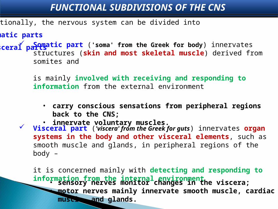
FUNCTIONAL SUBDIVISIONS OF THE CNS FUNCTIONAL SUBDIVISIONS OF THE CNS
Somatic part ('soma' from the Greek for body) innervates structures (skin and most skeletal muscle) derived from somites and
is mainly involved with receiving and responding to information from the external environment
Visceral part ('viscera' from the Greek for guts) innervates organ systems in the body and other visceral elements, such as smooth muscle and glands, in peripheral regions of the body –
it is concerned mainly with detecting and responding to information from the internal environment
• sensory nerves monitor changes in the viscera; • motor nerves mainly innervate smooth muscle, cardiac muscle, and glands.
• carry conscious sensations from peripheral regions back to the CNS; • innervate voluntary muscles.

Visceral Motor Neurons Visceral Motor Neurons
Visceral motor neurons arise from cells in lateral regions of spinal
cord and send processes out anteriorly.
These processes synapse with other cells, usually other visceral
motor neurons
The visceral motor neurons located in the spinal cord are referred to
as preganglionic motor neurons and their axons are called
preganglionic fibers;
the visceral motor neurons located outside the CNS are referred to as
postganglionic motor neurons and their axons are called
postganglionic fibers.

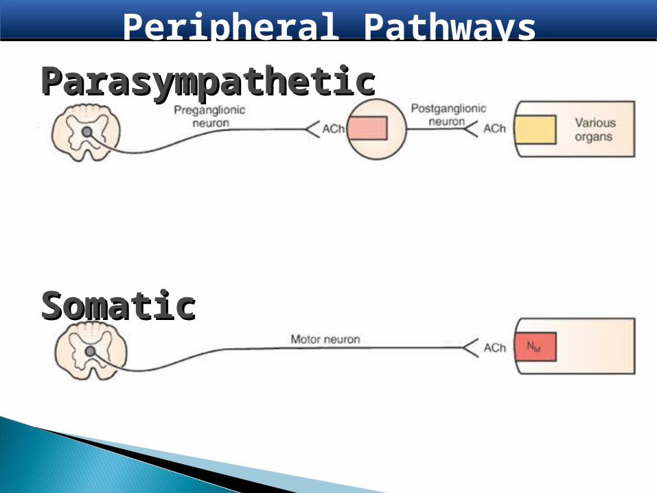
Peripheral Pathways

Somatic◦ Muscle movement◦ No ganglia◦ Transmitter: Acetylcholine ◦ Receptor: Nicotinic-M (“M” for muscle)
Peripheral Pathways
Peripheral Pathways
ParasympatheticParasympathetic
SomaticSomatic
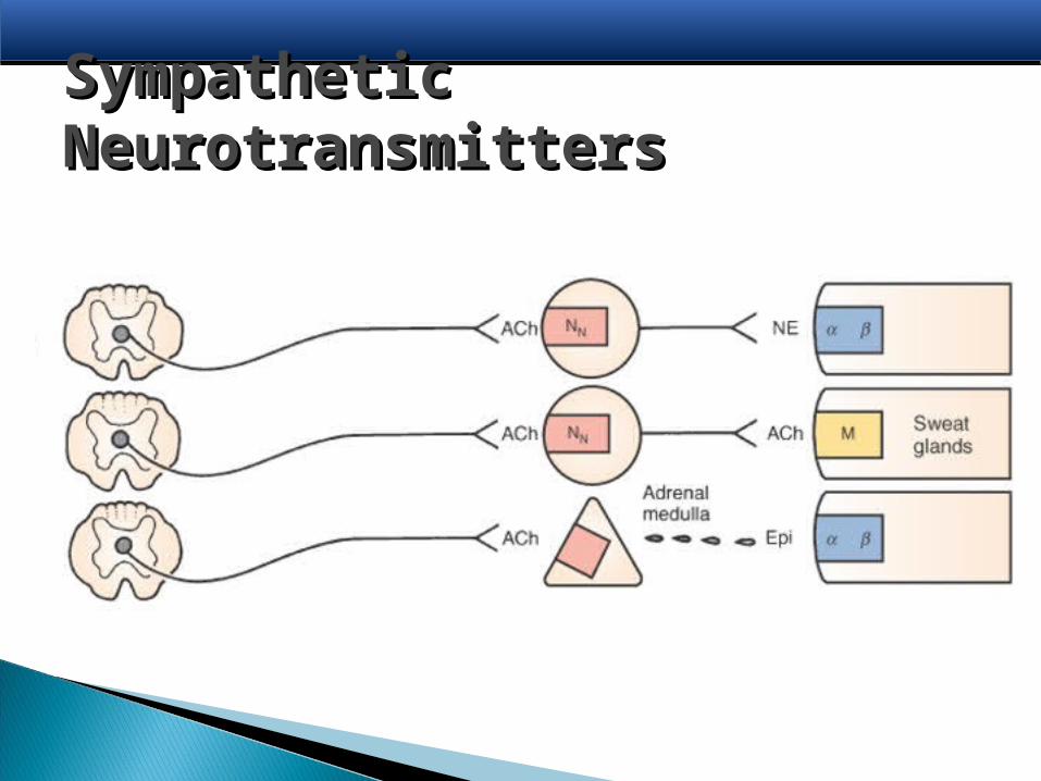
Sympathetic Sympathetic NeurotransmittersNeurotransmitters
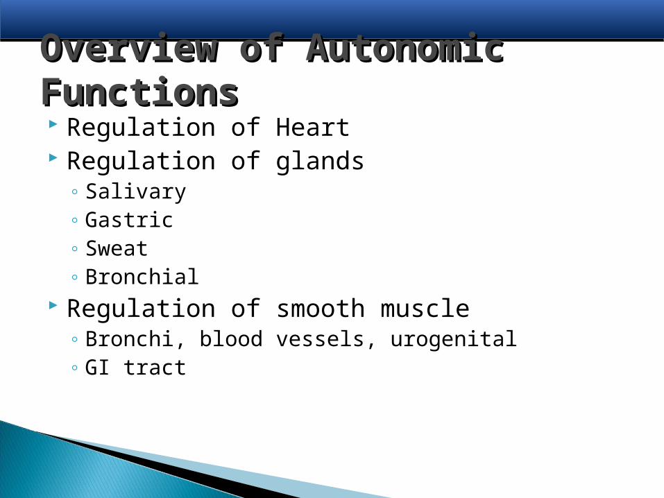
Overview of Autonomic Overview of Autonomic FunctionsFunctions Regulation of Heart Regulation of glands
◦ Salivary◦ Gastric◦ Sweat◦ Bronchial
Regulation of smooth muscle◦ Bronchi, blood vessels, urogenital◦ GI tract

Parasympathetic FunctionsParasympathetic Functions
Slow heart Increase gastric secretion and motility Emptying Bowel Focusing eye for near vision Constriction of pupil Contraction of bronchial smooth muscle
Most cholinergic drugs affect: GI, bladder, eye
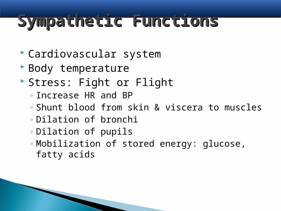
Sympathetic FunctionsSympathetic Functions
Cardiovascular system Body temperature Stress: Fight or Flight
◦ Increase HR and BP◦ Shunt blood from skin & viscera to muscles◦ Dilation of bronchi◦ Dilation of pupils◦ Mobilization of stored energy: glucose, fatty acids
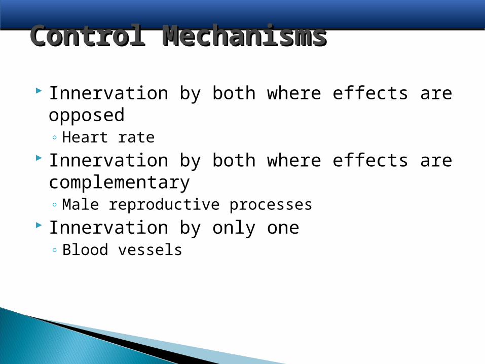
Control MechanismsControl Mechanisms
Innervation by both where effects are opposed◦ Heart rate
Innervation by both where effects are complementary◦ Male reproductive processes
Innervation by only one◦ Blood vessels

Autonomic ToneAutonomic Tone
Steady day-to-day influence exerted by the autonomic system◦ Usually only one division provide tone◦ Parasympathetic system usually provides the
basal tone

![Pancreatic cancer: What defines resectabilityWhat …1].1 Evans.Pancreatic Cancer.pdfPancreatic cancer: What defines resectabilityWhat defines ... SMA margin uninvolved with distance](https://img.pdfslide.us/doc/110x75/5ad222427f8b9a72118cc0ad/pancreatic-cancer-what-defines-resectabilitywhat-11-evanspancreatic-cancerpdfpancreatic.jpg)

![Pancreatic cancer: What defines resectabilityWhat defines …gicancers.org/syllabus/1[1].1 Evans.Pancreatic Cancer.pdf · 2010-11-01 · Pancreatic cancer: What defines resectabilityWhat](https://img.pdfslide.us/doc/110x75/5f57a92ea7197928bc5ea35f/pancreatic-cancer-what-defines-resectabilitywhat-defines-11-evanspancreatic.jpg)