Embed Size (px)
Citation preview

1
Perception
• Chris Rorden• Lecture 8: Vision and perception
• Low level visual deficits:• Visual field defects
• Blindsight• Achromatopsia :: cortical colorblindness (V4)• Akinetopsia :: motion perception (MT/V5)• Agnosias :: apperceptive, associative, prosop- (FFA)
• ‘How’ versus ‘what’
www.mricro.com

2
Vision
Human vision: Lots of real-estate
3
Visual Pathway
Each eye sees both left and right visual field.
Ipsilateral information crosses over at optic chiasm.
Some connections to superior colliculi.– Reflexive eye movments
Others go to thalamus (lateral geniculate nuclei) and then cortex.
4
Visual Defects
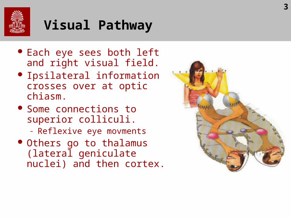
Field defects reveal anatomical injury
A. Monocular blindnessB. C. Bitemporal hemianopiaD. Homonymous
hemianopiaE. Upper quadrantanopiaF. Lower quadrantanopiaG. Homonymous
hemianopia
5
V1
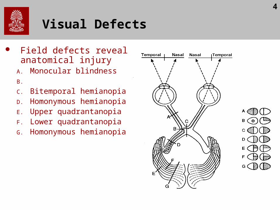
Primary visual cortex (V1) lies in calcarine fissure.
Complete damage leads to Homonymous hemianopia.
Partial damage leads to scotomas
6
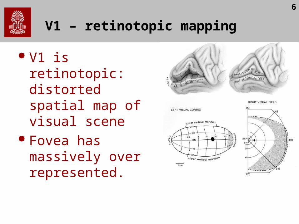
V1 – retinotopic mapping
V1 is retinotopic: distorted spatial map of visual scene
Fovea has massively over represented.
7
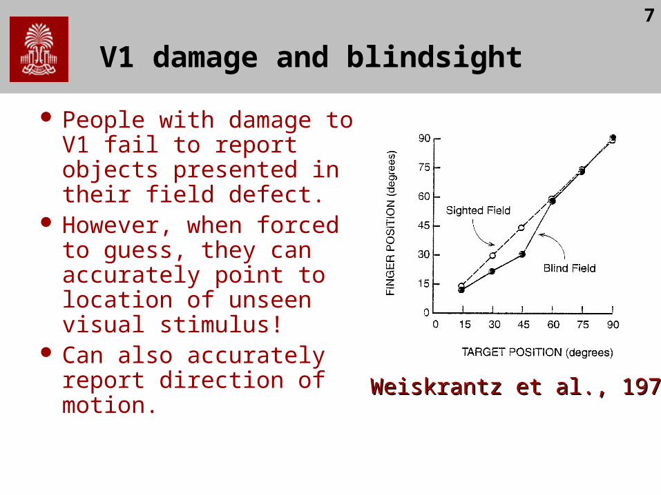
V1 damage and blindsight
People with damage to V1 fail to report objects presented in their field defect.
However, when forced to guess, they can accurately point to location of unseen visual stimulus!
Can also accurately report direction of motion. Weiskrantz et al., 1974Weiskrantz et al., 1974
8
Implications of Blindsight
V1 is crucial for conscious awareness. What explains blindsight? Why do only 20% of
V1 patients show blindsight?– Incomplete damage to V1? Islands of spared tissue
(Gazzaniga, 1994).– Typically seen in people who had injury while young
– neural plasticity?– Small number of indirect connections to later cortical
visual centers?– Visual connections to colliculi?
9
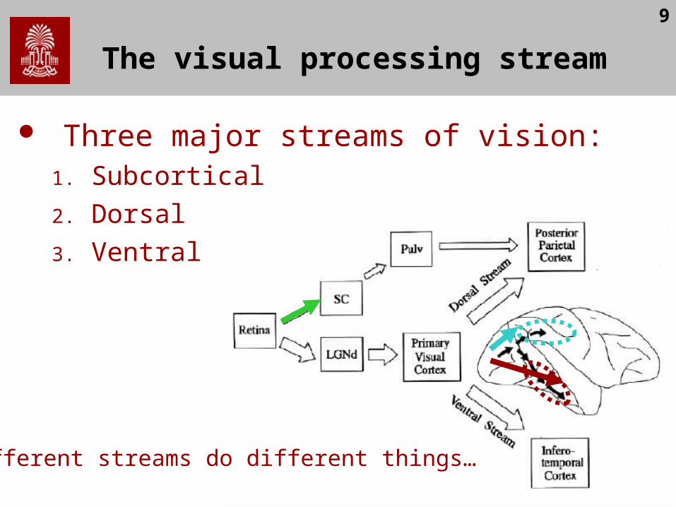
The visual processing stream
Three major streams of vision:1. Subcortical
2. Dorsal
3. Ventral
Different streams do different things…
10
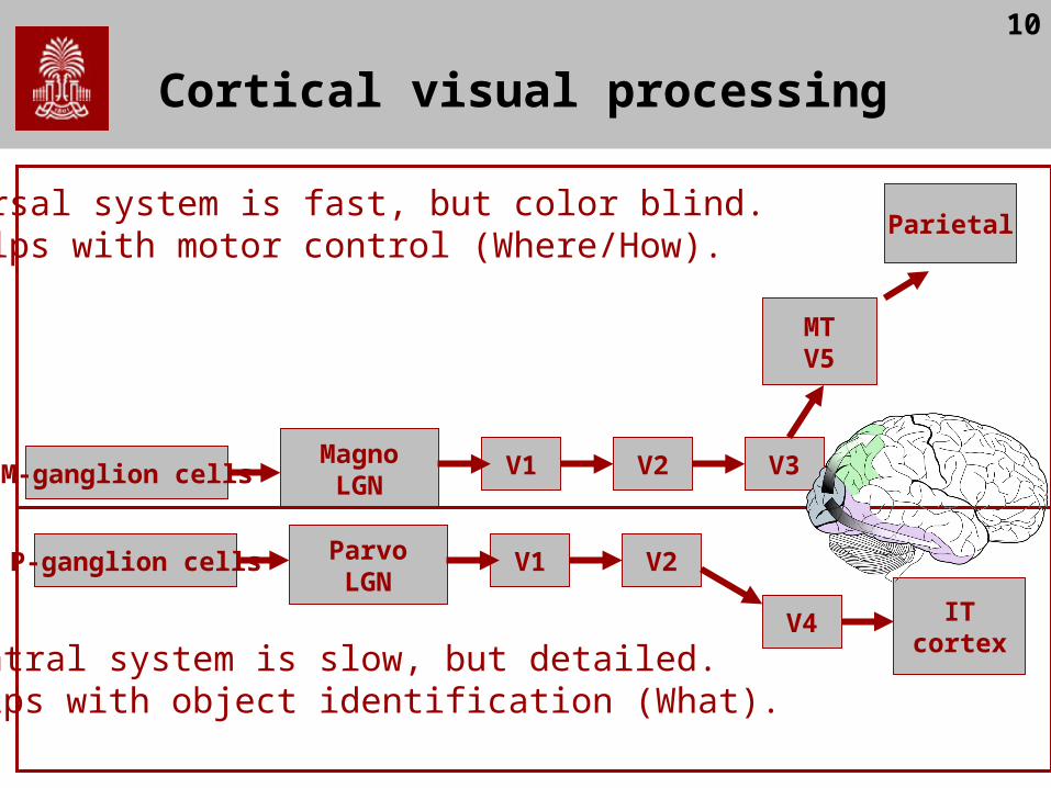
Cortical visual processing
M-ganglion cells
P-ganglion cells
MagnoLGN
ParvoLGN
V1
V1
V2
V2
V3
V4
MTV5
ITcortex
ParietalDorsal system is fast, but color blind.Helps with motor control (Where/How).
Ventral system is slow, but detailed.Helps with object identification (What).

11
Achromatopsia :: V4
Achromatopsia is usually caused by bilateral damage to V4 - lingual and fusiform gyri (occipitotemporal junction) and is characterized by an inability to identify or discriminate colour
Still able to perceive form and motion

12
Akinetopsia (Motion Blindness)
Zilles reported first case of akinetopsia. Pure cases are rare, as requires bilateral injury.
– Case LM - akinetopsia 43 yr old. Sinus vein thrombosis V5 damaged bilaterally - V1 spared Could not see movement of objects but could see still objects. People
would suddenly appear Diagnosed as agoraphobic Can see movements/reach for/catch very slow moving objects (< 10°/s)
13
V5 timecourse
Beckers & Zeki (1995) examined brief V5 disruption using TMS.
Motion perception disrupted most with V5 stimulation up to 30ms after visual stimulation onset
V1 stimulation also partially disrupts motion perception, but later (60-70ms after VS onset).
Takes 30-50ms for signals to go from V1 to V5– Direct route to V5?– Reafference to V1?– May explain motion performance in blindsight?

14
Agnosias
Three reasons why people might fail to recognize objects:– Perceptual Deficit: e.g. acuity, field cut, loss
of color vision– Apperceptive agnosia: unable to perceive
full shape of object despite intact low level processing.
– Associative agnosia: ability to perceive shape, but unable to recognize it.
15
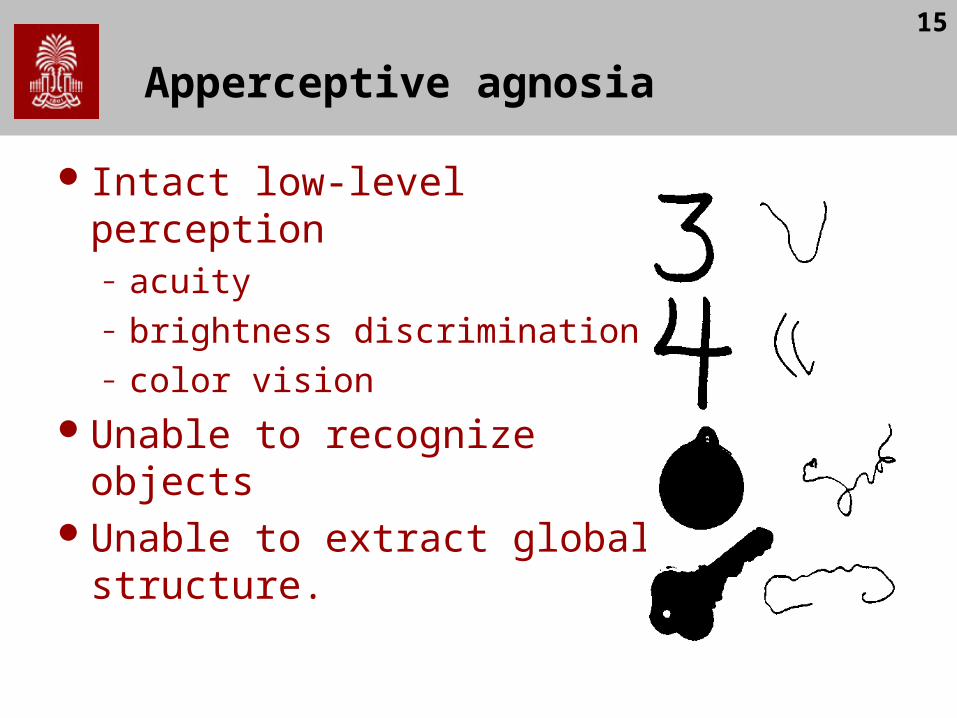
Apperceptive agnosia
Intact low-level perception– acuity– brightness discrimination– color vision
Unable to recognize objectsUnable to extract global
structure.

16
Associative agnosia
Able to see whole form of shapes
No problem copying figures
However, unable to recognize the objects

17
Associative agnosia
Theoretical explanations:
– Disconnection between visual representation and language?
– Damage to visual memory representation?
– Slightly impaired perception?
18
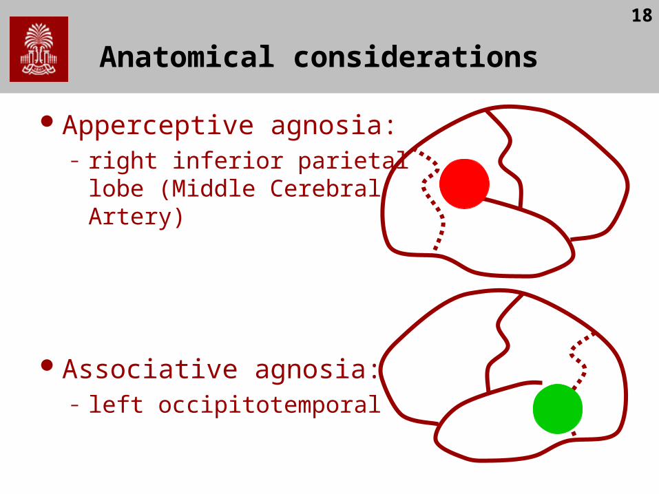
Anatomical considerations
Apperceptive agnosia:– right inferior parietal lobe
(Middle Cerebral Artery)
Associative agnosia:– left occipitotemporal

19
Prosopagnosia
Wigan (1844), Quaglino & Borelli (1867), Hughlings Jackson (1872), Charcot & Bernard (1883), Wilbrand (1892)
Inability to visually recognize facesEven a spouse’s face does not seem
familiar
20
Prosopagnosia - specificity
Seems specific to faces. Patients can still recognize others by:– Silhouette– Voice– Clothing
Note: not like amnesia
21
Prosopagnosia
Is face processing special?Or, are faces simply the most difficult
objects we discriminate?Most people withprosopagnosia have
difficulty recognizing differences within categories:– types of car– porcelain fixtures– breed of dog
Also, often suffer achromatopsia

22
Faces are difficult
Most objects are identified by unique components
However, faces have the same basic components:nose, eyes,ears, hair

23
Are faces special?
Farah tried to find objects as difficult as faces:– Spectacle frames– Undergrads recognized 87% of faces, 67%
of eyeglass frames (faces easier)– LH recognized only 64%
of faces, and 63% of eyeglass frames

24
Are faces special?

25
Are faces special?
26
Are faces special?
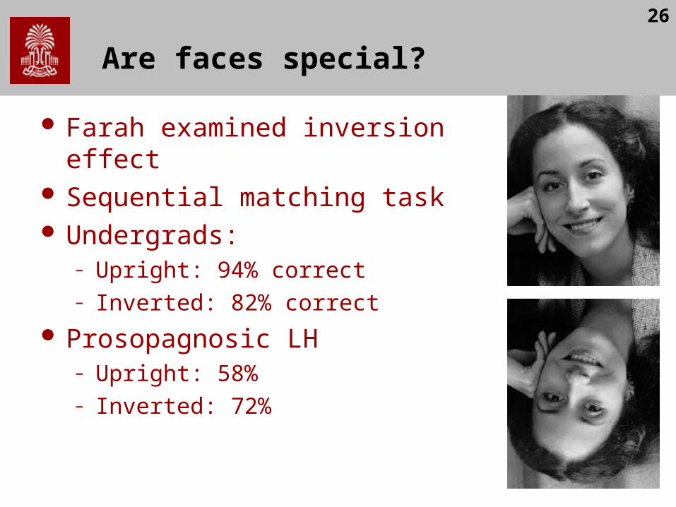
Farah examined inversion effect Sequential matching task Undergrads:
– Upright: 94% correct– Inverted: 82% correct
Prosopagnosic LH– Upright: 58%– Inverted: 72%
27
Double dissociations
Assal, Faure & Anderes (1984) report zooagnosic farmer MX– Lost ability to recognise cows– Still recognises faces
Bruyer et al (1983) report reverse– Fails to recognise faces– Intact perception of cows

28
Double Dissociation
If faces are simply difficult, we should not find patients with spared face recognition who are impaired on other tasks.M
X
Faces Cows
Acc
ura
cy
MX
RB
RB
29
Prosopagnosia
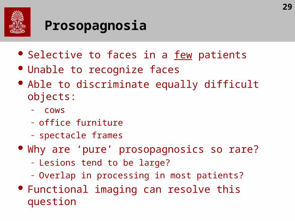
Selective to faces in a few patients Unable to recognize faces Able to discriminate equally difficult objects:
– cows– office furniture– spectacle frames
Why are ‘pure’ prosopagnosics so rare?– Lesions tend to be large?– Overlap in processing in most patients?
Functional imaging can resolve this question
30
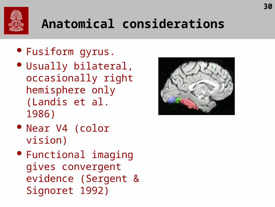
Anatomical considerations
Fusiform gyrus. Usually bilateral,
occasionally right hemisphere only (Landis et al. 1986)
Near V4 (color vision) Functional imaging
gives convergent evidence (Sergent & Signoret 1992)
31
Vision in split brain patients
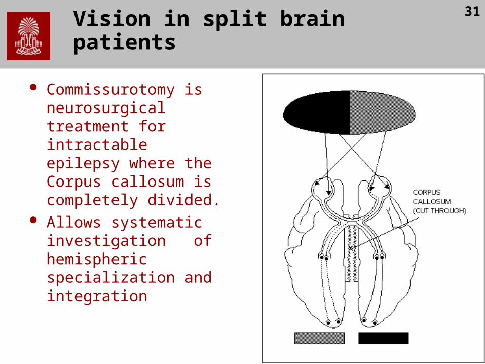
Commissurotomy is neurosurgical treatment for intractable epilepsy where the Corpus callosum is completely divided.
Allows systematic investigation of hemispheric specialization and integration
32
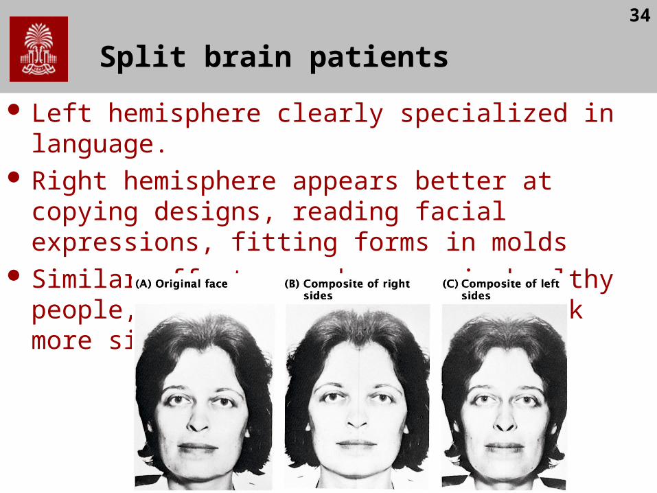
Split brain patients
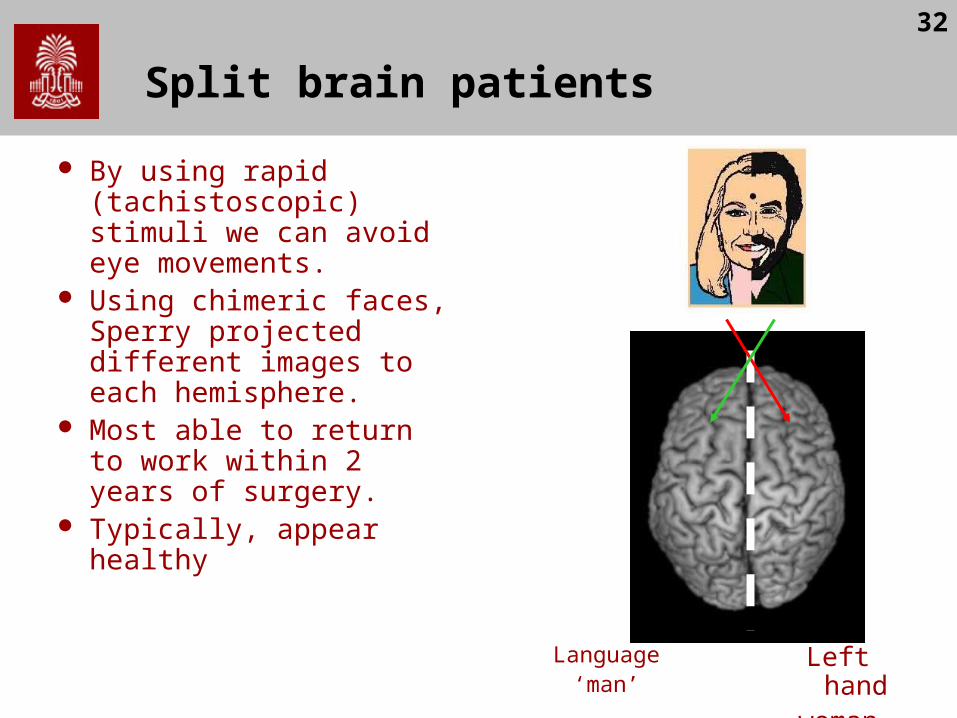
By using rapid (tachistoscopic) stimuli we can avoid eye movements.
Using chimeric faces, Sperry projected different images to each hemisphere.
Most able to return to work within 2 years of surgery.
Typically, appear healthy
Language‘man’
Left handwoman

33
Split brain patients
Picture presented in RVF (i.e. to LH)
– Patient could name or reach for the object correctly with right hand.
Picture presented in LVF (i.e. to RH)
– Patients could not name/describe the object
– Subjects could reach for the correct
– object with their left hand Likewise, unable to find a
object felt with one hand by using the other hand.
34
Split brain patients
Left hemisphere clearly specialized in language. Right hemisphere appears better at copying designs,
reading facial expressions, fitting forms in molds Similar effects can be seen in healthy people, e.g.
most think A and C look more similar than A and B

35
Cortical visual processing
M-ganglion cells
P-ganglion cells
MagnoLGN
ParvoLGN
V1
V1
V2
V2
V3
V4
MTV5
ITcortex
ParietalDorsal system is fast, but color blind.Helps with motor control (Where/How).
Ventral system is slow, but detailed.Helps with object identification (What).
36
Visual Form Agnosia
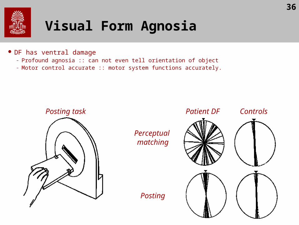
DF has ventral damage– Profound agnosia :: can not even tell orientation of object– Motor control accurate :: motor system functions accurately.
Patient DF ControlsPosting task
Perceptual matching
Posting
37
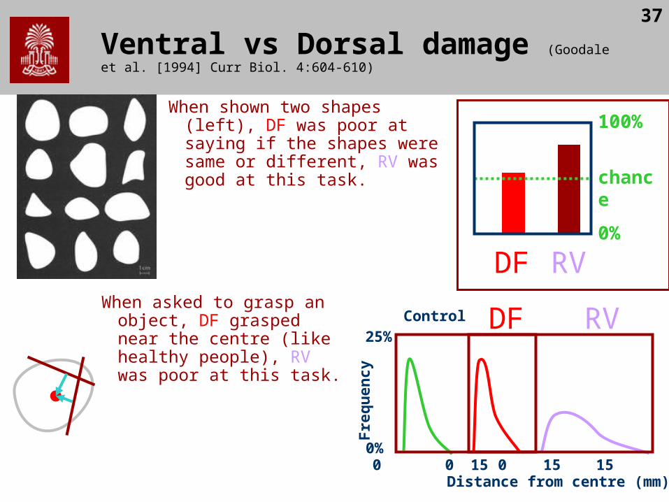
Ventral vs Dorsal damage (Goodale et al. [1994] Curr Biol. 4:604-610)
When shown two shapes (left), DF was poor at saying if the shapes were same or different, RV was good at this task.
DF RV
chance
100%
0%
DFControl RVF
req
uen
cy
25%
0%
Distance from centre (mm)0 15 0 15 0 15 30
When asked to grasp an object, DF grasped near the centre (like healthy people), RV was poor at this task.


![BlindSight: Eyes-Free Access to Mobile Phones · Luk demonstrates piezoelectric-driven feedback for mobile devices [17]. Mobile input BlindSight allows for one-handed input using](https://img.pdfslide.us/doc/110x75/5fcaf0d551b8492f4740006a/blindsight-eyes-free-access-to-mobile-luk-demonstrates-piezoelectric-driven-feedback.jpg)