Embed Size (px)
Citation preview

1
Methods in isolation and characterization of bovine monocytes and macrophages 1
2
Ceciliani F.a*, Ávila Morales G.a, De Matteis G.b, Grandoni F.b, Furioso Ferreira R.c,d, Roccabianca 3
P.a, Lecchi C.a 4
5
a Department of Veterinary Medicine, Università degli Studi di Milano, Milan, Italy 6
b Research Centre of Animal Production and Aquaculture, Council for Agricultural Research and 7
Economics (CREA), Monterotondo, Rome, Italy 8
c Faculty of Veterinary Medicine, University of Zagreb, Zagreb, Croatia 9
d Institute for Animal Sciences, Physiology and Hygiene Unit, University of Bonn, Bonn, Germany 10
* Corresponding author at Department of Veterinary Medicine, Università degli Studi di Milano, 11
Milan, Italy 12
Email address: [email protected] (F.Ceciliani) 13
14
Abstract 15
16
Monocytes and macrophages belong to the mononuclear phagocyte system and play important roles 17
in both physiological and pathological processes. The cells belonging to the monocyte/macrophage 18
system are structurally and functionally heterogeneous. Several subsets of monocytes have been 19
previously identified in mammalian blood, generating different subpopulations of macrophages in 20
tissues. Although their distribution and phenotype are similar to their human counterpart, bovine 21
monocytes and macrophages feature differences in both functions and purification procedures. The 22
specific roles that monocytes and macrophages fulfil in several important diseases of bovine species, 23
including among the others tuberculosis and paratuberculosis, brucellosis or the disease related to 24
peripartum, remain still partially elusive. The purpose of this review is to discuss the current 25
knowledge of bovine monocytes and macrophages. We will describe methods for their purification 26
and characterization of their major functions, including chemotaxis, phagocytosis and killing, 27
oxidative burst, apoptosis and necrosis. An overview of the flow cytometry and morphological 28
procedures, including cytology, histology and immunohistochemistry, that are currently utilized to 29
describe monocyte and macrophage main populations and functions is presented as well. 30
31
32
Keywords: 33
monocytes; macrophages; innate immunity; bovine 34

2
1. Introduction: monocytes/macrophages and their main functions. 35
36
Monocytes are myeloid cells produced by the bone marrow that play pivotal roles in the immune 37
response against infections and injuries. Monocytes and their corresponding tissue derivates, 38
including macrophages and subpopulations of dendritic cells, are involved in almost all phases of the 39
inflammation and bridge innate and specific immune responses [1]. In the bone marrow, 40
hematopoietic stem cells differentiate to precursor cells. Pre-monocytic cells produce peripheral 41
blood monocytes that transform into resident macrophages upon tissue entry [2]. Resident 42
macrophages are among the first cell populations to sense a tissue pathogen or injury and generate 43
the initial wave of cytokines and chemokines, alerting the immune system to incoming pathogens or 44
tissue damage, skewing immune defences and activating and recruiting additional immune cells at 45
the site of inflammation [2]. Functions of macrophages vary according to their location and type of 46
activation. Alveolar macrophages are located in the lungs, and provide cellular defence against 47
inhaled antigens, clearing the air spaces of infectious, toxic, or allergic particles that have evaded the 48
mechanical defences of the respiratory tract [3,4]. In the brain, macrophages are identified as resident 49
microglial cells and protect the Central Nervous System (CNS). Microglial macrophages play a 50
pivotal role in synaptic pruning during development [5]. In a healthy status, microglia facilitate 51
learning and memory and scavenge cellular or other debris. After CNS infections and injury, activated 52
microglia fulfil the functions of resident macrophages during inflammation, recognizing pathogens 53
or injured cells, activating phagocytic, antigen-presenting and cytokine/chemokine secretion 54
functions. Activated microglia also contribute to the resolution of inflammation via switching to anti-55
inflammatory cytokine patterns, and promote intercellular matrix synthesis and angiogenesis.[6]. In 56
the CNS, beside microglia, macrophages are also found in perivascular, meningeal and choroid plexus 57
spaces, participating to the blood-brain barrier by modulating the entrance and the phenotype of 58
immune cells during inflammation [7]. In lymph nodes, macrophages are classified as spinal or 59
subcapsular sinusoidal, their main role being that of capturing antigens and presenting them to B cells 60
[8]. The intestine provides one of the largest pool of macrophages in the body. Resident macrophages 61
act as sentinels for pathogen recognition and elimination, maintaining the intestinal homeostasis by 62
shaping host-microbiota symbiosis and regulating gut inflammation, promoting the crosstalking with 63
T cells [9,10]. The splenic red pulp is a large tissue compartment containing macrophage lines able 64
to perform several and diverse functions. The main role of splenic red pulp macrophages is to engulf 65
senescent and altered erythrocytes, break down haemoglobin, recovering iron to the bone marrow, 66
thus contributing to iron homeostasis [11] and to monitor, detect and phagocytize bloodborne 67
pathogens. Other equally important functions of splenic red pulp macrophages include presenting 68

3
antigens to T lymphocytes of the splenic periarteriolar sheats [12]. In the liver, finally, resident 69
macrophages are named Kupffer cells. In humans, Kupffer cells account for a large pool of tissue 70
macrophages, and their main role is to remove toxins and pathogens [13]. 71
Although the main role of macrophages is related to the protection against pathogens, they contribute 72
to the resolution of inflammation by at least three ways: a) removing the pathologic stimulus by 73
phagocytosis; b) producing anti-inflammatory cytokines, including for example TNF- soluble 74
receptor of IL1-R antagonist and c) removing leukocytes and necrotic debris in the inflammatory site. 75
Macrophages carry out also tissue-repairing function and actively participate in the development of 76
many autoimmune diseases development [1]. 77
Peripheral blood (circulating) monocytes are cells with a consistent morphology (Fig. 1), and for 78
many years they were regarded as a single functional population identified by the surface expression 79
of the lipopolysaccharide receptor (CD14). However, the development of flow cytometry and multi-80
colour immunofluorescence, have allowed more detailed differentiation of monocytes, based on the 81
additional expression of CD16 (FcIIIR), the low-affinity receptor for the Fc regions of IgG [14]. 82
According to CD14 and CD16 expression over the surface of the cells, at least two subpopulations of 83
human monocytes: (CD14++(bright)CD16−and CD14+(dim)CD16+) have been identified. Further 84
studies introduced, according with CD16 intensity of expression, other monocyte subpopulations, 85
namely CD14++CD16+ and CD14+CD16++ [15]. Given their CD14 and CD16 membrane surface 86
asset, circulating human blood monocytes can be divided into three different subpopulations [16]: 87
a) Classical monocytes (cM): “CD14++ CD16− “cells providing approximately 90% of human 88
blood monocytes. 89
b) Intermediate monocytes (intM): “CD14++ CD16+ cells” (approximately 10% of human blood 90
monocytes) 91
c) Non-classical monocytes (ncM): “CD14+ CD16++” cells (in humans, a population with 92
variable percentage). 93
Similar to humans, three monocyte subsets have been identified in bovine peripheral blood as well 94
[17,18]. The heterogeneity and the functional differences between the three populations of bovine 95
monocytes have been recently and extensively reviewed [19]. The authors compared bovine 96
monocytes with their analogue human population, focusing on sub-type distribution and their main 97
inflammatory functions, including adhesion and chemotaxis. Indeed, bovine monocyte specific 98
functions (Table 1) have been studied in a relatively limited number of papers [18–20] and only 99
partially resemble those of human counterparts. 100

4
Table 1 101 102
The inflammatory activity of bovine monocytes subsets
Sub-type of monocytes Relative
percentage
Phagocytic
capability 1
ROS
production2
Inflammasome
activation
Classical monocytes (cM)
CD14++CD16-
89% +++ ++ ++
Intermediate monocytes (intM)
CD14++CD16-
5-10% ++ +++ +++
Non-classical monocytes
(ncM) CD14+CD16++
5/10% + + +
103 1 Phagocytic capability is expressed as percentage of FITC-positive cells within viable monocytes 104
[18]: +++ = 80%, ++ = 56%, + = 40% 105 2 ROS production is expressed in Mean Fluorescence intensity (MFI). +++ = 30 x 103, ++ = 50 x 106
103, + = 5 x 10³. 107 3 IL1 production (pg/mL) after ATP and LPS stimulation. +++ 4000 pg/mL, ++1000 pg/mL, + 108
negligible 109
110
Differences between macrophage activation patterns 1q1q2ubetween cattle and other species also 111
exist. For example, stimulating bovine monocyte-derived macrophages with pro-inflammatory 112
signals do not produce the same reactions as compared to human and murine macrophages [19,21]. 113
In humans and rodents, several other macrophage categories have been reviewed [22], the most 114
important of which are listed in Table 2. The presence of most of these sub-groups in bovine 115
peripheral blood has yet to be demonstrated. 116
Table 2 117
Principal macrophage subcategories and phenotypes in human and rodents 118
Macrophages
sub-group
Type of response Activity Reference
M1 Pro-inflammatory, Facilitate immunity to remove foreign
pathogens and tumour cells, mediate tissue
damage induced by ROS, impair wound
healing and tissue regeneration
[22,23]
M2 Anti-inflammatory [24]
M2a Tissue repair and wound healing [25,26]
M2b Stress oxidative, efferocytosis [27]
M2c Pro-fibrotic activity phagocytosis of
apoptotic cells
M2d Tumour-associated macrophages,
angiogenesis and cancer metastasis
[28]
M3 Pro-inflammatory Switch between M2 to M1 response,
anti-tumour effect
[29,30]
M4 Pro-inflammatory Predominant in atherosclerotic lesions [31]

5
low phagocytosis capability, high tissue
repair capability
M17 Pro-inflammatory Increased phagocytosis in the presence of
oxidized LDL
[21,32]
Mreg Anti-inflammatory Produce high levels of nitric oxide, CD86 and
MHC Class II,
Intense activation and proliferation, of IL-10
producing T cells
[33]
Mox No clear pro- or
anti-inflammatory
activity
Weak phagocytosis, proatherogenic
Development in response to oxidative stress
damage
[34,35]
119
The transcriptomic data also revealed differential gene transcription between the three major 120
monocyte subclasses of genes involved in immune defences, providing the evidence that monocytes 121
subsets express different transcriptomes [36]. Indeed, the functions related to CD16+ have to be 122
revised and probably attributed between intermediate and non-classical subsets [37,38]. A sequential 123
development between the two subsets was suggested [39], based on the concept that macrophages 124
adopt in vivo several functional phenotypes, depending on continuous changes of the tissues' 125
microenvironments. Given this background, M1/M2 and other subgroups are therefore not definite 126
subsets but may represent the extremes of a continuum of different functional states [40]. 127
128
2. Purification of bovine monocytes from blood: sorting or adherence 129
130
Normal blood values for monocytes in cows range from 25 to 840 cells/L (average 400 cells/L) 131
and changes with age [41]. Purification of bovine monocytes starts from the collection of peripheral 132
blood from the jugular or coccygeal veins, using a sterile Vacutainer™ blood collection vial 133
containing EDTA (Becton Dickinson, USA). When a large amount of blood is needed (e.g. more than 134
200 L) the use of blood bags containing citrate phosphate dextrose (Terumo, Japan) is preferred as 135
a less stressful procedure for the animal. Blood collection must be as fast as possible for welfare 136
issues and because the production of glucocorticoids associated with prolonged containment can 137
profoundly affect in vitro monocyte activities. Although published several years ago, the paper by 138
Godderies and co-workers [42] still provides a very good background for monocyte purification from 139
bovine blood, specifically for the techniques related to adherence to glass or plastic wares. In general, 140
methods to purify monocytes can be divided into two groups: those related to the adhesive capability 141
of monocytes and those related to monocyte sorting by magnetic-activated cell sorting (MACS, 142
Miltenyi Biotec, Germany). 143
The purification procedure based on the adhesiveness to tissues has been previously described by the 144
team of Godderies and coworkers [42], whereas the procedure for the monocyte sorting by MACS 145

6
technique has been first described by Ceciliani et al. [43]. Figure 2 presents the morphology of 146
cultured monocytes following MACS purification technique after 1, 2 and 4 days of culture. The first 147
steps of purification are similar for both methods. Blood is collected into tubes (or bags) containing 148
anticoagulant and processed within 1 hour of collection. Blood is centrifuged at 1260 × g for 30 min 149
at RT to obtain the buffy coat. Mononuclear cells are recovered after dilution of the buffy coat 1:2.5 150
(v/v) in PBS/2mM EDTA, and 30 ml of this solution is overlaid on 15 ml Ficoll 1077 (Amersham 151
Biosciences) and centrifuged for 30 min at 1500 × g at 4 °C. Mononuclear cells (i.e. monocytes and 152
lymphocytes) are collected from the interface, also called mononuclear ring. The cells are 153
resuspended in 50 ml PBS/2 mM EDTA and centrifuged for 7 min at 400 × g at 4 °C to remove 154
platelets. To avoid non-specific activation of monocytes, it is pivotal to remove as many platelets as 155
possible from the mononuclear cell fraction by performing second centrifugation in a 4% to 20% 156
sucrose gradient layered over Ficoll-Paque PLUS (Sigma-Aldrich, USA). This procedure will 157
effectively remove any platelet contamination [45,46]. Alternatively, consecutive centrifugations at 158
low speed (400 x g for bovine monocytes), considerably reduces the number of platelets, that remains 159
in the supernatant [43]. Table 3 summarizes the two protocols. 160
161
Table 3. 162
Purification methods for bovine monocytes * 163
Methods Protocol
Adherence
A. Polystyrene surfaces 1. Isolated PBMCs are cultured in 75 cm2 polystyrene culture flasks and
incubated for 2 h at 38°C.
2. No-adherent cells are removed by pipetting out the supernatant and rinsing the
flask with warm medium (38°C).
3. Adherent cells (monocytes) are detached by incubating them with Hank’s
Balanced Salt Solution (HBSS) without Ca2+ and Mg2+, containing a mixture
of 0.5 mg/mL Trypsin + 0.6 mM EDTA for 10 min at 23°C.
4. Cells are pelleted and washed (10 min 180 x g, 4°C) with unsupplemented
RPMI 1640 medium, and finally resuspended in the culture medium.
5. After detaching monocytes from polystyrene flasks, to avoid monocytes loss
by the binding to polystyrene, all procedures should be carried out at 4°C in
polycarbonate conical tubes.
B. Plasma coated gelatin surfaces
1. PBMCs are cultured in 75 cm2 plasma coated gelatin (2%) culture flasks and
incubated for 1 h at 38°C.
2. No-adherent cells are aspirated, and the flask rinsed with culture medium
(38°C).
3. Adherent cells are detached by incubating the cells with HBSS without Ca2+
and Mg2+, containing 10 mM EDTA for 5 min at 23°C.
4. Flasks are gently shaken, and cells recover with pipette and pelleted.
5. Cells are washed and resuspended in culture medium at 4°C.

7
Sorting
1. Monocytes are isolated from PBMCs with magnetic-activated cellular sorting
technique, using a monocyte-specific monoclonal antibody (CD14+).
2. PBMCs are incubated with anti-human CD14 labelled superparamagnetic
beads (Miltenyi-Biotech) for 15 min at 4°C.
3. Cells are separated through magnetic separation using Macs column
(Miltenyi-Biotech), by first eluting the cells not bound to the column (CD14-)
and then CD14+ cells by removing the column from the magnet.
4. The purity of sorted cells is evaluated by flow cytometry.
164
The aforementioned techniques have both advantages and disadvantages that must be considered 165
when choosing the adequate isolation method according to each experimental requirement. MACS 166
technique is based on the immunomagnetic separation of the cells, which allows the specific isolation 167
of a subset of monocytes of interest (e.g., CD14+ fraction) with higher purity (> 98%) [43] than with 168
the adherence based method [44]. Such specific marker separation is not possible when working on 169
the isolation of monocytes by adherence from PMBC, where the authors often report the cells only 170
as adherent (monocytes) and non-adherent (lymphocytes) [42]. Despite its advantages, this immune-171
based method also presents some disadvantages such as higher costs and the unavailability of 172
antibodies used for the separation of monocytes of other species than humans. This issue can be 173
addressed by purifying cells with the adherence-based method. Finally, the latter method is also 174
cheaper and simpler, as no complex equipment is needed. The main disadvantage of the adherence 175
method is the potential presence of other cellular population, such as eosinophils. When blood is 176
collected from animals with a high concentration of eosinophils, the first step of purification, e.g. 177
centrifugation over Ficoll layer, cannot entirely remove eosinophils, which in turn can contaminate 178
monocyte population. 179
Once purified, population purity is assessed through flow-cytometric techniques. 180
Briefly, a total of 100 μL of cell suspensions are labelled with FITC-anti-CD14 antibody (clone 181
TÜK4) and incubated in the dark for 15 minutes. Cells are washed twice and resuspended in PBS for 182
flow cytometry. Fig. 3 shows representative flow cytometry dot plots of CD14+ cells before and after 183
MACS purification. The recovery of cells from either method yields an amount of homogenous 184
monocyte population ranging from 8 to 10 millions of cells, starting from an initial blood volume of 185
200 mL. 186
Flow cytometric analysis shows that after MACS sorting, an enrichment of CD14+ monocytes 187
population > of 95% is obtained. 188
189
3. Monocyte apoptosis, pyroptosis and necrosis (necroptosis) 190
191
8
In multicellular organisms, a balance between cell proliferation and cell death is maintained to assure 192
homeostasis [47]. Cells that are useless or superfluous, aged, damaged, and/or potentially harmful are 193
selectively eliminated using genetically encoded mechanisms [48]. Up to now several types of cell 194
death have been described: apoptosis, necrosis, both accidental and programmed (necroptosis), 195
pyroptosis, cell death associated with autophagy, ferroptosis, netosis, among others [49]. However, 196
apoptosis and necrosis are the best-characterized cell death ways so far. A comprehensive list of 197
different cell death mechanisms has been recently published by the Nomenclature Committee on Cell 198
Death [48]. 199
Apoptosis is an ATP-dependent type of programmed cell death, generally characterized by distinctive 200
morphological and biochemical changes. The morphological features of apoptosis include reduction 201
of cellular volume, condensation of the chromatin, nucleus fragmentation, plasma membrane 202
blebbing, maintenance of plasma membrane integrity until late stages and formation of apoptotic 203
bodies followed by dead cells phagocytosis [50]. Cells undergoing apoptosis also show biochemical 204
changes such as externalization of phosphatidylserine on the outer plasma membrane [51], the release 205
of mitochondrial intermembrane space proteins (Cytochrome C, Smac/DIABLO) [52], DNA 206
fragmentation and caspase-dependent activation [53]. Several of these features were exploited to 207
measure apoptosis rate. Apoptosis is generally not associated with inflammation, due to the absence 208
of cells’ contents release, resulting in cell clearance from the body with minimal damage to the 209
surrounding tissues [54]. 210
Understanding the monocyte cell death mechanism(s) is important, as the way of cell death (necrosis 211
vs. apoptosis vs. pyroptosis) can influence the pro- and anti-inflammatory responses [55] and 212
therefore interfere with the proper resolution of inflammation, or the clearance of the inflammatory 213
focus from the pathogens, or cancer development [56]. Moreover, apoptosis rate is critical for normal 214
cell turnover and proper development and functioning of the immune system. Indeed, apoptosis 215
provides a way to control the activity of blood monocytes, by increasing or reducing their abundance 216
in the inflammatory environment. 217
In contrast, necrosis is generally described as an “accidental or uncontrolled” way of cell death caused 218
by extreme physical or chemical stress [57]. The rapid cytoplasmic granulation/vacuolization, cellular 219
and organelles’ swelling in necrotic cells, results in plasma membrane rupture, organelle breakdown, 220
intracellular content spillage, inflammation and surrounding tissue damage [47,54]. Nevertheless, it 221
is now known that necrosis can also occur as a caspase-independent form of regulated cell death, also 222
known as necroptosis, following many defined steps and signalling events, just like apoptosis [57]. 223
Necrosis is the term currently used for nonapoptotic death and has been characterized as passive, 224
accidental cell death resulting from environmental perturbations with the uncontrolled release of 225

9
inflammatory cellular contents. The presence of necrosis provides evidence that a cell has died, but 226
not necessarily how the death occurred. The necroptosis is defined as the caspase-independent, 227
regulated cell death (RCD). In the recent years, research studies concerning caspase-independent, 228
non-apoptotic forms of programmed cell death (PCD) have enabled the identification of backup 229
mechanisms to allow the cells to undergo suicide or RCD under conditions where the caspase 230
machinery is defective or inhibited [58]. 231
Another type of regulated necrosis so far described and measured in monocytes and macrophages is 232
pyroptosis. Pyroptosis occurs in response to microbial products or other intracellular perturbations, 233
which cause the production of proinflammatory cytokines like IL-1, leading to cell swelling and 234
death [57]. Being initially thought to occur only in macrophages and leukocytes during inflammatory 235
conditions, pyroptosis has now been observed in several other cell types, including fibroblasts and 236
endothelial cells [59]. Pyroptosis is generally characterized morphologically by membrane pore 237
formation, cell swelling, membrane disruption and DNA damage, whereas biochemically by an 238
inflammatory caspase activation (non-apoptotic caspase-1), that mediates cytokine maturation 239
[57,60]. The identification of distinctive and specific markers in all of these three types of cell death 240
has long been used for their in vitro and in vivo discrimination in many cell types including monocytes 241
and macrophages. 242
243
3.1. In vitro assays measuring monocytes/macrophages apoptosis 244
245
In vitro apoptosis can be determined through several methods: by characterizing cell morphology 246
through microscopy or by measuring biochemical cell death features, like caspase activation, that is 247
activated uniquely during the apoptosis [47]. Methods relying on flow cytometry have also been 248
widely applied in human and murine research to distinguish apoptosis from necrosis [61]. 249
Specifically, the measurement of the activity of the two main effectors enzymes in apoptosis, namely 250
caspase-3 and -7, has been already reported and validated in bovine monocytes (CD14+) [20,32]. In 251
these studies, the activation of the two enzymes was measured through fluorometry, using a detection 252
assay that contains aa pro-fluorescent substrate the rhodamine 110, bis-(N-CBZ-L-aspartyl-L-gluta-253
myl-L-valyl-L-aspartic acid amide - Z-DEVD-R110) and a cell lysis buffer that supports optimal 254
caspase activity. The assay is adapted to small well formats, such as 96 or 384-well plates. Depending 255
on the selected format, between 5x10⁴ cells for 384-well up to 1x10⁵ cells for 96-well plates are seeded 256
in final volumes of 25 𝜇L to 50 𝜇L, respectively [43,63]. In a second step, an equal volume of Apo-257
ONETM Homogeneous Caspase 3/7 Reagent (Promega, USA), containing the pro-fluorescent 258
substrate and previously diluted 1:100 in the reaction buffer, is added to each well. Plates are then 259

10
incubated in a plate shaker at 300 rpm for 1 h at room temperature. According to the manufacturer’s 260
instructions, incubation times should be optimized for each model as minimal apoptotic induction 261
and low cell numbers may require extended incubation periods. Finally, upon cleavage of the DEVD 262
peptides by caspase-3 and -7 activities, the fluorescent compound rhodamine 110 is released and 263
fluorescence intensity can be measured using a fluorescence plate reader (excitation at 485 nm, 264
emission at 530 nm). 265
Caspase activity can also be measured through colourimetric assays. For example, a caspase 266
colourimetric protease assay (Aportaget; Invitrogen, Canada) kit has been used to determine the 267
activity of caspases-3, -6, and -9 in monocytes infected with Mycobacterium bovis [64]. This assay 268
can be carried out by lysing a pellet of infected monocytes containing 5 × 10⁶ cells with 50 μL of 269
chilled cell lysis buffer provided by the kit and incubated for 10 min, before centrifuging them at 270
10,000 × g. The supernatant containing the cytosolic fraction is transferred to a fresh tube and the 271
protein concentration is quantified. After having adjusted the cytosolic fraction concentration (200 272
μg/mL) in the cell lysis buffer provided in the kit, 50 μL of it are transferred to 96-well round-plates 273
and 50 μL of 2×kit reaction buffer, containing 10 mM of dithiothreitol (DTT) and 200 μM the 274
respective substrate for the caspases, is added in a volume of 5 μL. Finally, samples are incubated at 275
37°C for 2 h in the dark and then read at 405 nm with a 490-nm reference filter in a microplate reader. 276
Bovine monocyte-derived macrophages apoptosis has also been detected trough fluorometric analysis 277
of caspase-3 activity [63] and relative quantification of histone-complexed DNA fragments, that are 278
often out of the cytoplasm of cells undergoing apoptosis [65], using Cell Death Detection 279
ELISAPLUS kit (Roche, Switzerland) [66]. Because histone-complexed DNA release is not an 280
exclusive phenomenon of apoptosis but also can occur in necrotic cells, using complementary assays 281
is preferred [67]. 282
283
3.2. In vitro assays measuring monocytes necrosis 284
285
The permeabilization of cells’ plasma membranes and its consequent release of cellular content are 286
some of the hallmarks of necrosis. The release of lactate dehydrogenase (LDH), an ubiquitous 287
cytosolic enzyme, into the supernatant after membrane integrity loss has long been measured to detect 288
necrosis in multiple cell types by spectrophotometry, including bovine monocyte-derived 289
macrophages [63,66,68]. Specifically, in the study of Denis and coworkers [66], a commercially 290
available kit (CytoTox 96®, Promega, USA) was used to measure the LDH released to the 291
supernatants by a 30-minute coupled enzymatic assay that leads to the conversion of a tetrazolium 292
salt (iodonitro-tetrazolium violet; INT) into a red formazan product, which is proportional to the 293

11
amount of LDH present and thus to the number of damaged cells. Besides INT, the kit reagents also 294
contain the substrates lactate, NAD+ and diaphorase to complete the reaction, as the first two are 295
needed to produced reduced nicotinamide adenine dinucleotide (NADH) that is later used to convert 296
INT into the coloured product in the presence of diaphorase [68]. According to the manufacturer 297
instructions, cells are first seeded in 96- or 384-well plates and treated with the desired experimental 298
design. Afterwards, the supernatants are transferred to a fresh plate (50 μL for a 96-well and 12.5 μL 299
for a 384-well plate) and an equal volume of CytoTox 96® Reagent is added. Plates are protected 300
from light and incubated for 30 min at room temperature. Finally, 50 or 12.5 μL of Stop Solution are 301
added and absorbance measured 490nm or 492nm [66]. Other techniques based on differential uptake 302
of DNA binding dyes, like propidium iodide (PI), that do not enter living cells, have also been used 303
for determination of bovine necrotic neutrophils [69], blood monocytes and macrophages (CD14+) 304
isolated from the mammary gland through flow cytometry [70]. 305
306
3.3.In vitro assays measuring monocytes pyroptosis 307
308
The discrimination between necrosis and pyroptosis can be a challenging task as many of the 309
hallmarks for regulated necrosis such as plasma membrane rupture, cytoplasmic 310
granulation/vacuolization and cellular/organelle swelling are also shared by pyroptosis [71]. The 311
measurement of LDH release has also been implemented for pyroptosis determination of human and 312
murine bone marrow-derived macrophages, as it is a simple and cost-effective technique [62,72]. 313
DiPeso and coworkers monitored the cells’ LDH secretion, as previously described [59,73], after 314
reacting with a pyroptosis inducer called RodTox that activates the NAIP2/NLRC4 inflammasome, 315
leading to caspase-1 activation and pyroptosis. Additionally, a more specific way to measure 316
pyroptotic death of individual cells in vitro can be carried out through the measurement of caspase-1 317
activation and inflammasome formation (NLRP3) by both fluorescence or confocal microscopy [74]. 318
Cells are labelled with the caspase-1 activity probe FAM-YVAD-FMK that specifically binds to the 319
caspase-1 family proteases, and with antibodies against inflammasome components such as the 320
adaptor protein ASC that recruits pro-caspase-1. For that, murine marrow-derived macrophages are 321
first seeded in 96-well plates and stimulated with 100 ng/mL of LPS for 3 h at 37°C to prime Toll-322
Like Receptor 4 (TLR4), the first step needed for NLRP3 activation. Afterwards, the medium is 323
removed, and cells are stimulated with fresh DMEM medium-containing 5 μM nigericin to induce 324
the second step for the activation of the NLRP3 inflammasome, and 5 mM glycine to reduce the cell 325
lysis caused during caspase-1 activation. Cells are then incubated for 60 min at 37°C and 5% CO2, 326
and during the last 45 min incubated as well with 30x of the FAM-YVAD-FMK probe, which is 327
12
prepared according to the manufacturer's instructions. After incubation, the medium is removed and 328
the cells are washed three times for 5 min with cold PBS. Finally, the cells are fixed and stained with 329
a fluorescent nucleic acid stain to label the nuclei and mounted for a correct visualization by confocal 330
microscopy, with an excitation/emission of 492/520nm for the FAM-YVAD-FMK probe. For the 331
antibody staining of the inflammasome component (ASC), after seeding, priming and exposing the 332
cells to nigericin, the cells are washed with cold PBS, fixed and permeabilized on ice and in dark for 333
30 min. Macrophages are washed with the washing buffer and the primary antibody anti-ASC (1:500) 334
is added and cells are incubated for 1 h on ice. The cells are again washed and the secondary antibody 335
(fluorescent dye conjugated goat anti-mouse; 1:500) is added for another 1 h on ice. During the last 336
5 min of this incubation, the dye to stain the cells’ nuclei is also added. Finally, cells are washed and 337
prepared for confocal analysis with an excitation/emission for the secondary antibody of 555/580 nm. 338
According to Hartigh and Fink, to successfully determine pyroptotic cells the nuclear morphology 339
should be considered, as cells with active caspase-1 possess a rounded and condensed nuclei without 340
visible nucleoli, often present in cells without caspase-1 activation [74]. 341
Even though the previously described techniques have been carried out mostly in human and murine 342
macrophage models, the same ones can be applied to bovine monocytes/macrophages’ pyroptosis 343
determination, although they have been not tested so far. 344
345
3.4. Flow cytometric assays to evaluate apoptosis, necrosis (necroptosis) and pyroptosis 346
347
Determining the number of dead cells during a flow cytometry study is a prerequisite to obtain reliable 348
data, given that dead cells can bind in a non-specific way to many reagents. Therefore, removing dead 349
cells from flow cytometry data is a prerequisite and a critical step to ensure accurate results and 350
analysis [55]. A widely used flow cytometric assay to define cell viability is based on the reaction of 351
a fluorescent dye, such for example propidium iodide (PI), with cellular amines. This die cannot 352
penetrate live cell membranes: consequently, only cell surface proteins are available to react with the 353
dye, resulting in dim staining. The reactive dye can permeate the damaged membranes of dead cells 354
and stain both the interior and exteriors amines, resulting in more intense staining [72]. In normal 355
living cells, the phosphatidylserine (PS) is located at the side of the cytoplasmic surface of the cell 356
membrane. However, during early apoptosis, PS is translocated from the cytosolic side of the intact 357
plasma membrane to the extracellular surface. Early apoptotic cells cannot, therefore, be reliably 358
identified using approaches that are based on membrane permeability. Annexin V is a protein with 359
high affinity, specificity, and sensitivity for PS. Thus, the binding of annexin V to cells can be used 360

13
as a marker of early apoptosis [75] and provides the most commonly used approach for determining 361
apoptosis [55]. 362
A typical experimental protocol to detect apoptosis is the following: 100 L of cells suspension 363
(1x10⁶ cells/mL) in 1X annexin-binding buffer are incubated with 5 L of fluorescein isothiocyanate 364
(FITC) annexin V and 1 L of PC5.5-PI (100g/mL) in the dark at room temperature for 15 min. 365
Finally, 400 L of the annexin V binding buffer is added and the cells are analyzed by flow cytometry. 366
The binding of annexin V to PS is reversible, and so samples must be analyzed as soon as possible 367
and no later than 1 h after labelling. Fig. 4 illustrates the typical aspect of a dot plot flow cytometry 368
analysis using annexin V/PI double staining to determine in the percentages of live, apoptotic and 369
necrotic bovine peripheral blood lymphocytes and monocytes. Beside annexin V staining, the 370
measurement of caspases activity is also widely used to determine the apoptosis rate. Caspases are a 371
family of cysteine proteases that plays a key role in apoptosis and inflammation. These enzymes are 372
present in healthy cells as inactive zymogens, but when stimulated they undergo autolytic cleavage 373
to become fully active. Consequently, many of the cleaved fragments remain intact during apoptosis 374
and can be detected using specific antibodies. These antibodies can be used to specifically label cells 375
in which caspase cleavage has occurred, allowing to quantify these cells by flow cytometry. Several 376
antibodies that specifically recognize caspase-cleaved fragments have been generated, including 377
antibodies that recognize the cleaved form of caspase-3, a key protease activated during the early 378
stage of apoptosis [76]. 379
For flow cytometric detection of active caspase3, 1x10⁶ cells are washed with PBS and then 380
resuspended with 100 L of fixation/permeabilization buffer (Cytofix/CytopermTM, BD Biosciences, 381
USA)) for 20 min at room temperature. In the following step, cells are washed twice with 600 L of 382
erm/WashTM (BD Biosciences, USA) buffer and stained with the PE- rabbit anti-active caspase-3 383
antibody (clone C92-605) for 40 min at 4°C. Cells are then washed and resuspended in Perm/WashTM 384
buffer for flow cytometric analysis. 385
Both pyroptosis and apoptosis are programmed cell death mechanisms but are dependent on different 386
caspases. Pyroptosis is triggered by caspase-1 after its activation by various inflammasomes and 387
results in lysis of the affected cell. Pyroptotic cell death could be assessed in PBMC and monocytes 388
by flow cytometry by measuring the double-positive staining of activated caspase-1 and PI [77]. 389
Furthermore, Hussen and coworkers [20], analyzed the inflammasome activation in bovine 390
monocytes through the evaluation of caspase-1 activation. Authors used the FLICACaspase-1 391
detection KIT (Thermo-Fisher, USA), a fluorescent inhibitor of active caspase-1 (FAM-YVAD-392
FMK), which binds activated caspase-1. First, bovine PBMC or purified monocytes (1 x10⁶) are 393
stimulated for 2 h with LPS (100 ng/mL). Then ATP (2 mmol/L) is added to the cell culture for a 394

14
further 30 min. After stimulation, cells are detached from the plates by Accutase™ (Sigma-Aldrich, 395
USA) treatment (incubation for 15 min at 37°C and 5% CO2). Cells are then suspended in 300 L 396
RPMI medium and 10 L of the 30X FLICA solution is added. After the incubation (1 h at 37°C and 397
5% CO2) with FLICA, cells were washed two times with the FLICA buffer and suspended in 200 L 398
of the same buffer. For flow cytometric analysis PI (2 g/mL final concentration) is added to exclude 399
dead cells. 400
401
4. Monocyte and macrophage chemotaxis 402
403
Chemotaxis is the ability of cells to migrate in response to a chemical signal or chemoattractant 404
gradient concentration. It is a crucial function of leukocytes, including monocytes, as they fulfil their 405
defensive functions by migrating from blood to affected tissues, where they produce pro-406
inflammatory cytokines and reactive oxygen species (ROS) and eventually phagocyte and destroy the 407
invading pathogens [78]. 408
Monocyte chemotaxis is determined by using the Boyden chamber assay, composed of an upper and 409
a lower compartment separated by a porous membrane (Fig. 5a). This assay consists mainly of 410
monitoring the migration of the cells through a microporous membrane. In general, the cells are 411
seeded in the medium-filled upper compartment and are allowed to migrate through the pores of the 412
membrane, towards the medium-filled lower compartment where chemoattractants are present [79]. 413
To assure the correct migration of the cells through the membrane, this one must be selected with the 414
appropriate pore size for the cells of interest. Human monocyte chemotaxis is usually triggered by N-415
Formylmethionine-leucyl-phenylalanine (N-fLMP), a formylated peptide derived from bacteria [80], 416
which specifically reacts with Formyl Peptide Receptors [81]. Bovine cells do not have N-fLMP 417
receptors [82,83]. In this species, therefore, chemotaxis should be triggered with other 418
chemoattractants, including, such as, for example, zymosan activated serum (ZAS) [84]. The in vitro 419
chemotactic capacity of bovine monocytes has been determined using this principle with a modified 420
Boyden chamber [84]. In this study, bovine monocyte migration was measured using a 48-well 421
microchamber, where the chemoattractant ZAS was added in the lower part of the chamber. A PVP-422
free polycarbonate filter (5 μm pore size) was layered onto the lower wells and covered with a silicone 423
gasket and finally with the top plate above. In the upper wells, 1.5 x 10⁶ cells/mL were seeded (50 μL 424
final volume) and the chamber was incubated at 37°C in 5% CO2 for 105 min. The microchamber 425
was disassembled and the filters were removed carefully, the non-migrated cells on the upper part of 426
the filter were removed by scraping and the migrated cells found in the lower part of it were stained 427

15
with Romanowsky type stains including May‐Grünwald‐Giemsa and Wright's stains. Finally, cells 428
were counted in different high-power oil-immersion fields. 429
Modified Boyden chamber devices, allowing a more convenient and user-friendly methodology to 430
carry out the cell migration studies, are commercially available. Such is the case of the transwell 431
migration plates (Fig. 5b), that contain easy-to-use permeable cell culture membrane inserts of 432
different pore sizes that can be removed from the plates when needed to stain and count the cells that 433
have migrated through the membrane. The transwell migration assay has been described to assess the 434
chemotactic activity of human blood monocytes and the human monocyte cell line THP-1 [85] and 435
can be also used for bovine studies. In this study, 1x10⁵ of THP-1 and blood monocytes were seeded 436
in the upper chamber of a 24-well transwell migration plate in a final volume of 125 μL of serum-437
free media. The monocyte chemoattractant protein-1 (MCP-1) and “platelet-releasate”, which is the 438
content released by secretory granules of activated platelets, were added as chemoattractants to the 439
lower chamber in a final volume of 1 mL and plates incubated for 2 h at 37°C. After incubation, the 440
upper chambers were removed and placed into 2.5% glyceraldehyde for 15 min to fix the migrated 441
cells found on the underside of the membrane. The membranes were washed with PBS, and the upper 442
chambers were stained with 0.1% of crystal-violet for 45 min. Finally, cells were washed twice with 443
PBS and cells in ten 40x fields were counted using light microscopy. 444
Chemotaxis of monocyte-derived macrophages has been measured as well [86], using the same 445
technique, e.g. the migration through Boyden chamber, previously described. 446
The Boyden chamber technique has been modified over the years to allow the quantification of total 447
migrating leukocytes concurrently with the determination of their phenotype to associate specific 448
inflammatory responses with distinct leukocyte subsets. Gomez-Lopez and coworkers [87] validated 449
in humans a combined Boyden-flow cytometry chemotaxis assay (CBFCA), that allows the accurate 450
quantification and recovery of the migrating leukocytes for subsequent phenotyping and 451
characterization in a single assay, facilitating the understanding of leukocyte trafficking during the 452
inflammatory response. The combined chemotaxis assay is performed under the same conditions 453
described for the classic method, but only the polycarbonate membrane was used. Following 454
incubation, the leukocyte suspension in the lower well is recovered and cells counted by flow 455
cytometry. To our knowledge, the CBFCA assay has not yet been applied to study chemotaxis of 456
bovine monocytes. 457
458
5. Monocyte/macrophages phagocytosis and killing 459
460
16
Phagocytosis and respiratory burst are two of the most important functions for 461
monocytes/macrophages and polymorphonuclear cells and are essential for both the elimination of 462
invading microorganisms and the presentation of antigens to T-cells. Phagocytosis is a process in 463
which target particles are engulfed and taken up by other cells, crucial to the innate immune response 464
and consequently regulating the adaptive immune response [88]. Phagocytic affinity is related to 465
particular marker expression patterns, directed by a complex range of cell surface receptors, and may 466
also change in different populations of monocytes [88]. Bovine monocytes mediate the inflammatory 467
response activated in phagocytosis and killing capability, which leads to different abilities to 468
phagocytose bacteria between the different monocyte’s subsets – cM are the ones with the highest 469
phagocytosis, followed by intM and ncM with the lowest ability [18]. The phagocytosis ability of 470
monocytes can also relate to other factors such as age, breed, nutrition and the clinical status of the 471
animal [89–91]. Lymphocytes in neonatal calves have poorer phagocytosis ability but counterbalance 472
with higher absolute numbers, in contrast to monocytes of neonatal calves that showed a higher 473
bacterial uptake than 3-9 week old calves, suggesting that the monocytes contribute to the 474
compensation of impaired earlier innate immune defence in infancy [92]. A similar mechanism seems 475
to take place in the mammary gland, where a greater number of viable macrophages may partially 476
compensate for the functional deficit in polymorphonuclear cells in cows with high somatic cell 477
counts [93]. 478
The in vitro phagocytosis assay of bovine monocytes can be performed by co-culturing the cells with 479
opsonised fluorescein-labelled bacteria and then incubating them at 37oC in a humidified atmosphere 480
of 5% CO2 for 1 h. The cells are washed twice with sterile HBSS and then incubated with trypan blue 481
to quench non-internalised fluorescent bacteria, and after two subsequential washes with HBSS, the 482
intensity of the fluorescence, which can be correlated with the phagocytic ability, can be measured 483
[44]. Improved results have been achieved in vitro in the study of monocyte-derived macrophages 484
phagocytosis when opsonizing bacteria with naive-autologous sera than bacteria opsonized with fetal 485
bovine serum [94]. 486
Phagocytosis assays of monocyte-derived macrophages can also be performed by co-incubating the 487
cells with opsonised bacteria in a ratio of one bacterium to one macrophage, in a water bath at 37o C 488
under continuous rotation. The suspension is removed and added to ice-cold gelatine-Hanks’ salt 489
solution to stop phagocytosis, and the macrophages are centrifuged (110 x g for 4 min) to remove the 490
non-ingested bacteria, that remain in suspension in the supernatant fluid. The supernatant is then 491
seeded on tryptic soy agar plates, in dilutions between 10 and 100 colony counts, incubated in 8% 492
CO2 at 37oC for 72 hours. The results are expressed as the percentage of survival of non-493
phagocytized/non-cell-associated bacteria based on the formula [94]: 494

17
495
𝑃𝑒𝑟𝑐𝑒𝑛𝑡 𝑠𝑢𝑟𝑣𝑖𝑣𝑎𝑙 =𝐶𝐹𝑈 𝑜𝑓 𝑏𝑎𝑐𝑡𝑒𝑟𝑖𝑎 𝑎𝑓𝑡𝑒𝑟 𝑖𝑛𝑐𝑢𝑏𝑎𝑡𝑖𝑛𝑔 𝑓𝑜𝑟 120 𝑚𝑖𝑛
𝐶𝐹𝑈 𝑜𝑓 𝑏𝑎𝑐𝑡𝑒𝑟𝑖𝑎 𝑎𝑡 𝑡𝑖𝑚𝑒 0𝑥 100 496
497
The monocyte’s and macrophage's ability to bind, internalize and kill bacteria occur as independent 498
and sequential steps, and, as a result, a phagocyte may be able to ingest a pathogen and yet not manage 499
to kill it. Thus, the phagocytosing and killing capabilities of monocytes and macrophages should be 500
measured by separate assays [95]. The in vitro intracellular bacterial killing assay is performed by 501
co-incubating bovine monocytes (5 x 10⁵ cells) with opsonised bacteria (E. coli) for 60 min. The 502
unbound bacteria are removed, by centrifuging them at 110 x g for 5 min, and the cell pellet is 503
resuspended in HBSS with 100 µg/mL gentamicin, to kill any remaining extracellular bacteria, and 504
then incubated for 1 h at 37o C. Cells are washed with warm HBSS and then centrifuged again at 110 505
x g for 5 min to remove the gentamicin. The cells are then lysed by incubation on ice with 0.5% Triton 506
X-100 for 10 min and the surviving bacteria are seeded on MacConkey agar plates for CFU counting 507
[44]. 508
Flow cytometric assays have been developed to quantify phagocytosis and respiratory burst in whole 509
blood, as well as isolated cells from blood, tissue or secretions [96,97]. Hussen and coworkers [98] 510
reported a flow cytometric assay to evaluate phagocytosis capacity of bovine monocytes. FITC-511
conjugated and heat-killed S. aureus or E. coli were opsonized with heterologous bovine serum 512
(diluted 1:10 with PBS) for 45 minutes at 37°C. MACS-separated bovine monocyte subsets were 513
incubated for 24 h (37°C, 5% CO2) in RPMI medium. After this resting period, cells were incubated 514
with opsonized bacteria (50 bacteria/cell) for 40 minutes (37°C, 5% CO2). Control samples were 515
incubated without bacteria. After incubation, samples were analyzed using flow-cytometry after the 516
addition of propidium iodide (2 mg/mL final concentration) to exclude dead cells. Phagocytic activity 517
of different monocyte subsets is defined as the percentage of green fluorescing cells among viable 518
cells. 519
520
6. Monocyte/macrophages ROS production 521
522
Generation of reactive oxygen species (ROS) is a critical factor in the antimicrobial activities of 523
monocytes, though it may also lead to cellular cytotoxicity and/or tissue damage. The intracellular 524
presence of ROS can result in damage to the intracellular constituents and oxidative stress, which 525
may have consequential alterations in monocyte’s function [88]. As other functional abilities, ROS 526
production differ between different monocyte populations – bovine intM is reported to present the 527

18
strongest production between the subsets [18] – and virulent pathogens of cattle, such as N. caninum, 528
may also regulate ROS production of infected macrophages [88]. 529
The production of oxidative burst activity of bovine monocytes can be measured in vitro using a 530
cytochrome C reduction assay, as similarly used in bovine and caprine neutrophils and goat 531
monocytes [99,100]. The measurement of cytochrome C reduction is commonly used for the 532
measurement of extracellular superoxide anion [101] and demonstrated a dose-dependent specificity 533
in detecting extracellular superoxide production in bovine neutrophils [99]. The extracellular ROS 534
production is measured by mixing 2 x 10⁵ monocytes in RPMI-1640 medium (adjusted final volume 535
on wells of 200 L) and cytochrome C (1 mM). Parallel studies to assess the ROS production after 536
inducing a pro-inflammatory challenge to the cells, by adding phorbol 12-myristate-13-acetate 537
(PMA), can also be performed. PMA (2.5 g/mL final concentration) and cytochrome C (1 mM) are 538
added to the cells and the absorbance is measured immediately with a plate reader every 30 min for 539
2 h at a wavelength of 550 nm. Background values are measured from wells with cytochrome C 540
diluted in RPMI-1640 and subtracted from all values [102]. Analysis of ROS generation via 541
cytochrome C reduction assay has been used in bovine monocytes and alveolar macrophages (AM), 542
revealing that monocytes appear to be less responsive in ROS formation than AM, likely for being 543
more immature and thus possessing less cytoplasmic organelles enabling metabolic processes [103]. 544
Production of ROS reduces substrates such as dichloro‐dihydro‐fluorescein‐diacetate (DCHF) or 545
dihydro‐rhodamine 123 (DHR 123) that become fluorescent and can be quantified by flow cytometry. 546
Hussen and Coworkers [98] reported a typical flow cytometric analysis of ROS generation on bovine 547
monocyte. The analysis is performed after stimulation of cells with PMA (50 nmol/L) or heat-killed 548
and serum opsonized E. coli (50 bacteria/cell) for 15 min (37°C, 5% CO2) in the presence of the 549
fluorogenic substrate DHR 123 (750 ng/mL). ROS generation is calculated as Mean Fluorescence 550
Intensity (MFI) of gated monocyte region after subtracting the MFI of non-stimulated cells. 551
552
7. Monocyte/macrophages production of cytokines 553
554
Cytokines are important chemical messengers produced by both innate and adaptive immune cells, 555
that allow the communication between both types of immunity and within the adaptive immune 556
system [104]. Specifically, monocytes and macrophages play a critical role in immune regulation by 557
producing a wide range of pro and anti-inflammatory cytokines like TNF-𝛼, IL-1, IL-6, IL-8, IL-558
10, IL-12, and TGF-𝛽1 [105]. At the same time, these cytokines modulate their functions by 559
stimulating or inhibiting their microbial activities in an autocrine and paracrine manner [106] and are 560
also involved in cell migration, activation, proliferation and antibodies’ production. Moreover, 561

19
monocytes and macrophages can be further classified into different subsets according to the cytokine 562
profile they produce [107]. Therefore, the measurement of monocyte and macrophage cytokine 563
production is a highly useful approach for phenotyping and characterizing their functionality. 564
Bovine monocytes’ cytokine production can be measured mainly by two approaches: detection of 565
secreted cytokines at the protein level through enzyme-linked immunoabsorbent assays (ELISAs) 566
[64] or by measuring cytokines’ mRNA abundance by real-time quantitative PCR (q-PCR) 567
[16,21,62,108]. 568
Several commercially available ELISA kits have been used for the quantification of bovine cytokines, 569
including IFN-γ [109,110], TNF-α [111], IL-1β [112] and IL-4 [111]. However, the quantification of 570
secreted cytokines in monocytes/macrophages can be performed by other methods. Recently, type 1 571
IFN was measured in ovine alveolar macrophages’ supernatant by using an IFN bioassay [113]. An 572
IFN bioassay is a method to quantify IFN biological activity units, meaning it measures the actual 573
biological power of an IFN preparation in a specific cell line or biological system [114]. It is well 574
known that IFNs types’ cytokines induce a potent antiviral activity in vertebrate cells [115]. 575
Therefore, this method mainly relies on type 1 IFN antiviral properties and determines the amount of 576
IFN in a solution- usually secreted by virus-infected monocytes/macrophages- according to its ability 577
to neutralize viral replication and protect the infected cells in culture from cytopathic effects. One of 578
the most common IFN bioassays is based on the quantification of the fluorescence intensity of viral-579
encoded reporter proteins, like green fluorescent protein (GFP), during the virus replication in cell 580
culture [116]. The protective effects of IFN are then quantified by measuring the fluorescence 581
intensity and is determined by the ED50 that is the amount of IFN needed to reduce the fluorescence 582
by 50% [114]. Briefly in ovine, the bioactivity of IFN secreted by Sendai virus-infected ovine alveolar 583
macrophages was quantified by first serially diluting the IFN containing supernatant in DMEM 584
medium supplemented with FBS and antibiotics. This supernatant was then added to 2 × 10⁴ ovine 585
fibroblasts that were seeded the day before in 96 well plates, and cells were incubated at 37 ◦C for 24 586
h. After incubation, supernatants were removed and ovine fibroblasts were infected with 587
Recombinant Vesicular Stomatitis virus expressing GFP (VSV-GFP) at a multiplicity of infection of 588
0.01 and incubated at 37 ◦C. After 18 h post-infection, the green fluorescence of VSV-GFP infected 589
cells was detected by using a plate reader with an excitation wavelength of 480 nm and emission of 590
518 nm [113]. This method gives extra information when compared to ELISA, as by quantifying IFN 591
with a bioassay one can measure the antiviral activity on a specific experimental condition without 592
needing absolute numbers [114]. 593
To measure bovine monocytes’ cytokines mRNA abundance using qPCR [62], RNA from at least 1 594
x 106 cells/well is extracted by using guanidinium-acid-phenol extraction methods, such as TRizol™ 595

20
(Thermo-Fisher, USA) or Qiazol™ (Qiagen, Germany), or commercial kit [117]. After having 596
assessed the quality and concentration of RNA using a NanoDrop ND-1000 UV–vis 597
spectrophotometer, an Agilent Bioanalyzer or a Qubit fluorometer, genomic DNA is eliminated using 598
DNase I, RNase free kit. Reverse transcription is then carried out on RNA using commercially 599
available cDNA Synthesis kit. The cDNA is then used as a template to perform qPCR or microarray. 600
To carry out a reliable relative quantification, at least three adequate reference genes should be 601
identified and the geometric mean is used for normalization [118,119]. In the published MIQE 602
(Minimum Information for publication of Quantitative real-time PCR Experiments) guidelines is 603
indeed proposed the validation of RGs for all qPCR experiments [120]. Statistical softwares like 604
geNormTM, NormFinder© and BestKeeper can be used to assess the appropriateness and stability of 605
these genes [121–124]. The list of reference genes that have been used for quantitative real-time RT-606
PCR gene expression studies in monocytes and macrophages is reported in Table 4. 607
608
Table 4. 609
Validated reference genes for monocytes and macrophages used for qPCR. 610
Symbol Name Accession
number
Cell target Target
specie
Ref.
HMBS
B2M
Hydroxymethyl-bilane
synthase
NM_013551 Macrophages Murine [121]
-2-microglobulin
NM_009735
ACTB
RPL37A
-actin NM_001101.3 THP-1
monocyte
cell line
Human [122]
Ribosomal protein L37a NM_000998.4
SF3A1
ACTB
HMBS
Splicing factor 3 subunit 1 NM 001081510 Monocytes/
Other tissues
Bovine [123]
-actin BT030480.1
Hydroxymethyl-bilane
synthase
NM 001046207.1
H3F3A
SF3A1
YWHAZ
H3 histone, family 3A (H3-
3B)
NM_001014389.2 Monocytes Bovine [62]
Splicing factor 3 subunit 1 NM 001081510
Tyrosine 3-
monooxygenase/tryptophan
5-monooxygenase
activation
protein, zeta polypeptide
XM_025001429.1
611
Additional studies have also reported the adequateness of some of the previously mentioned reference 612
genes like YWHAZ and ACTB in different ovine tissues (brain, spleen, mesenteric lymph nodes) 613
[125]; and of YWHAZ in goats’ frozen whole blood [126], but not specifically in 614

21
monocytes/macrophages. Remarkably, the stability of all the genes tested in different ovine tissues 615
changed significantly within each tissue, suggesting that there is not a unique universal reference gene 616
for all tissues or cell types and that their stability must be evaluated prior the start of each study. Such 617
a statement can be further confirmed by the fact that in a previous study, ACTB was not an appropriate 618
reference gene for gene expression analyses in ovine interstitial cells[127]. 619
Intracellular cytokine staining (ICS) of stimulated whole blood or isolated PBMC followed by flow 620
cytometric analysis is a well-established method for detecting cytokines production at the single-cell 621
level. Recent studies developed a cytokine flow cytometric assay to detect intracellular IFN-γ in cattle 622
naturally infected with M. bovis [128]. The detection of intracellular IFN-γ may represent an 623
important alternative approach for an improved method of detection of cattle secreting IFN-γ below 624
levels of detection in the culture medium. 625
It is important to consider that the analysis of intracellular targets using flow cytometry presents 626
several technical challenges that are not generally encountered in the measurement of cell surface 627
epitopes. In general, cells must be first fixed to preserve and maintain both the structure and location 628
of target epitopes, then permeabilized to allow antibodies access to all cellular compartments, such 629
as the cytoplasm. In general, cell fixation is accomplished using either crosslinking fixatives, such as 630
formaldehyde, or low molecular weight alcohols (methanol), which generally act to coagulate 631
proteins. However, the fixation–permeabilization and methanol treatment need to be validated for the 632
complete set of antibody conjugates used for a new experiment [55]. 633
Short-term stimulation of PBMC (6-16 h) with antigen or peptide mixtures is required to induce 634
cellular activation and production of cytokines. Cells can also be stimulated in 15 mL conical 635
polypropylene tubes or plates. Secretion inhibitors such as Brefeldin A and/or Monensin are added at 636
the time of stimulation (for peptides) or after 2 h (for proteins) to allow secreted cytokines and other 637
proteins to be retained intracellularly. At the end of the stimulation period, cells can be held at 4–638
18°C until ready to process. They are then co-incubated with 0.2 mg/mL of EDTA at room 639
temperature to remove adherent cells, fixed, permeabilized and stained for intracellular determinants. 640
In some cases, surface marker staining is carried out jointly with intracellular staining. However, most 641
other cell-surface markers require staining before fixation, because the epitopes recognized by 642
staining antibodies are sensitive to fixation and/or permeabilization reaction [129]. 643
644
8. Phenotypic characterization of monocyte subsets: the flow cytometry approach 645
646
The flow cytofluorimetric approach allows the immunophenotypic characterization and absolute 647
count of the monocyte subsets. Hussen et al. [17] demonstrated for the first time that bovine 648

22
monocytes can be distinguished in three subsets on the differential expression of CD14 and CD16. 649
Similar to humans, bovine cM are CD14+CD16- and intM CD14+CD16+. On the contrary, ncM are 650
CD14-CD16+. Further evidence [19,130,131] suggested that CD14 is not a useful marker for the 651
immunophenotyping studies and cell sorting experiments of non-classical monocytes. 652
CD172a, a member of signal regulatory protein (SIRP) family, is a marker for myeloid cells and 653
neurons [132]. Two specific mAbs (Table 5), can be successfully used to identified bovine 654
monocytes, granulocytes, macrophages and dendritic cells [133], suggesting their potential use as 655
pan-monocyte markers. Interestingly, clone DH59B recognizes a conserved epitope across a wide 656
range of other species including small ruminants [134], dog [135], water buffalo [136] and dolphins 657
[137]. 658
Although CD163 is a monocyte/macrophage differentiation antigen [138], and its expression in ncM 659
is very low or absent [16,17], it is still regarded as an important functional marker. 660
A multicolour panel designed for a proper resolution of these subsets should, therefore, contain the 661
markers: CD14, CD16, CD163, CD172a and a fluorescent dye for dead cell exclusion. Besides, 662
CD335 (NKp46) should also be used to exclude NK cells. In Table 5 a list of monoclonal antibodies 663
that can be used with either direct or indirect labelling protocols using anti-mouse secondary 664
fluorescent antibodies is reported, although labelling of monocytes is still a hot topic within the flow 665
cytometric community [55]. 666
Table 5. 667
List of monoclonal antibodies reactive with bovine antigens 668
669
Specificity Clone Isotype Target specie Reference
CD14 CAM36A IgG1 Bovine [139]
CAM66A IgM Caprine [108]
CC-G33 IgG1 Bovine [140]
M5E2 IgG2a Human [141]
MM61A IgG1 Bovine [142]
TÜK4 IgG2a Human [143]
VPM65 IgG1 Ovine [144]
CD16 KD1 IgG2a Human [145]
CD163 2A10/11 IgG1 Pig [17]
EDHu-1 IgG1 Human [16]
LND5A IgG1 Bovine [146]
LND37A IgG1 Bovine [146]
LND37A IgG1 Bovine [146]
CD172a CC149 IgG2b Bovine [147]
DH59B IgG1 Bovine [148]
670
671
A typical stain/lyse/wash protocol is the following: 672

23
1) 100 µL of whole blood anticoagulated with Heparin [149] are labelled with saturating 673
concentrations of monoclonal antibodies (Table 5) in a final volume of 120 μL with PBS (pH 674
7.2), in a 5 mL polypropylene flow cytometric tube for 30 min at 4°C in the dark. 675
2) Erythrocytes are lysed with 1.0 mL of Tris-buffered ammonium chloride solution (0.87% w/v, 676
pH 7.3) for 10 min, then 2 mL of cold PBS are added 677
3) The sample is centrifuged at 300 g for 5 min. 678
4) Pellet of labelled cells is re-suspended with 200 μL of cold PBS and immediately acquired by 679
flow cytometry. 680
A simple indirect labelling protocol is done by introducing simple changes to the protocol previously 681
described. In brief, whole blood is incubated under the same conditions with 0.75 µg of purified mAbs 682
and then is processed in according to step 2 a 3. Pellet is re-suspended in 100 μL of fluorescent goat 683
anti-mouse isotype-specific secondary mAbs and incubated for 15 min at 4°C in the dark. After the 684
final wash, cells are re-suspended and acquired. 685
Appropriate controls are necessary to evaluate auto-fluorescence, background staining and proper 686
sample compensation, as well as to perform a careful antibody titration. 687
The gating strategy to be used to identify the three subsets of monocytes should include: 1) time 688
parameter vs FL or SSC to exclude event burst, 2) FSC-A vs FSC-H to exclude doublets, 3) exclusion 689
of dead cells in the fluorescent live/dead channel, 4) FSC vs SSC to exclude debris and granulocytes 690
from single live leukocytes, 5) CD172a vs CD16 dot plots to select CD172a positive mononuclear 691
cells (all monocytes), 6) CD16 vs CD14 to separate cM subsets from intM and ncM, 7) back gating 692
on dot plots 2, 3, and 4 to confirm the validity of the gating strategy. 693
To avoid false positives, the use of the fluorescence minus one (FMO) control for quadrant positions 694
of the dot plot CD16 vs CD14 is recommended. 695
Pomeroy and coworkers [131] found a significant association between postpartum diseases (mastitis 696
and metritis) and counts of monocyte subsets. Therefore, it is meaningful to evaluate the enumeration 697
of absolute count of these subsets. Two different approaches can be used: dual-platform (flow 698
cytometry and haematology) or single platform (flow cytometry). In the first method, the percentage 699
of subsets (e.g. cM, intM, ncM) is measured with a flow cytometer while the absolute monocyte count 700
is assessed by automated haematology analyzer. The absolute subset counts are therefore indirect. In 701
the single-platform method, the flow cytometer is therefore used for directly counting monocyte 702
subset in a given sample volume. 703
In humans, the precise and accurate leukocyte subset count (e.g. CD4+ T cells in HIV patients, CD34+ 704
for autotransplantation) is of primary importance, and the two methods have been extensively 705
described [150]. According to European Working Group on Clinical Cell Analysis (EWGCCA) 706

24
[135], the single-platform method should be regarded as the gold standard for absolute cell 707
assessments, and the use of stain/lyse/no-wash labelling protocols is recommended. 708
709
9. Characterization of tissue macrophages via immunohistochemistry. 710
Circulating monocytes migrate into various tissues and transform into macrophages. Macrophages 711
have several roles in tissues monitoring, inflammation and repair representing one of the dominant 712
cell types in chronic inflammatory reactions. 713
Under non-inflammatory conditions, circulating monocytes localize in tissues and organs becoming 714
resident macrophages to monitor healthy tissues. However, many resident macrophages are 715
established before birth and then self- maintained during adult life. Resident macrophages are found 716
in connective and adipose tissues, liver (Kupffer cells), splenic red pulp, bone marrow, lymph 717
nodes, lung, brain, serous fluids, skin [151]. 718
Under inflammatory conditions, most of the macrophages that accumulate at diseased sites typically 719
derive from circulating monocytes and take on several functions according to the inciting factors 720
and milieu including antigen presentation, phagocytosis, and immunomodulation through the 721
production of various cytokines and growth factors. Macrophages can be classified based on their 722
function and activation pathways. According to this grouping, there are classically-activated (M1) 723
macrophages, wound-healing macrophages (also known as alternatively-activated (M2) 724
macrophages), and regulatory macrophages (Mregs) [23,152,153]. 725
Macrophages can be activated directly by bacterial lipopolysaccharides, via TLR and extracellular 726
matrix proteins. Under TH1 influence, macrophages react to IFN-γ, and TNF- to become 727
classically activated M1 macrophages producing Nitric Oxide and Reactive Oxygen Species and 728
upregulating lysosomal enzymes to kill ingested organisms or digest foreign substances [23,152]. In 729
TH2 conditions macrophages act in response to IL-4, IL-5 and IL-13 by becoming wound healing 730
M2 macrophages that secrete growth factors promoting angiogenesis, activating fibroblasts, and 731
collagen synthesis [23,152]; under T regulatory secretion of IL-10 become regulatory/anti-732
inflammatory Mreg macrophages producing IL-10 to suppress inflammation [153]. 733
734
9.1 Morphological characterization of macrophages: cytology and histology 735
736
Following tissue entry and activation, monocytes in tissue undergo functional and morphological 737
transformation into reactive macrophages that are morphologically readily recognizable. 738
Thus, macrophages can be identified morphologically on cytological and histological specimens. 739

25
Cytological specimens are obtained by several techniques including imprints from tissues, scrapings 740
to sample firm lesions which are less likely to exfoliate cells, swabs or by the needle aspiration 741
technique. Sampled material by any of the aforementioned sampling methods can be smeared and 742
air-dried [154]. Fluid specimens such as bronchoalveolar lavages, cerebrospinal fluid, articular fluid 743
can be collected into an EDTA or a plain (if bacteriology is required) tube. Fluids can be 744
cytocentrifuged or directly smeared according to their cellularity and again air-dried. Routine 745
cytological stains are represented by alcohol‐based Romanowsky type stains including May‐746
Grünwald‐Giemsa and Wright's stains [155]. 747
On cytological specimens, macrophages are easy to identify: 748
a-Reactive macrophages (Fig. 6A ) are cells characterized by round to irregular shape, variably 749
large size (averaging18-30 m), low N/C ratio, abundant light blue foamy to highly vacuolated 750
cytoplasm with a paracentral to eccentric round to oval/reniform nucleus with granular chromatin 751
and often one visible nucleolus. Macrophages phagocytize and degrade foreign exogenous or 752
endogenous material and several etiological agents such as mycobacteria thus, they are recognized 753
also because of their cytoplasmic content (Fig. 6B). 754
b-Multinucleated macrophagic giant cells are cells developing in persistent granulomatous 755
inflammation by fusion of two or more activated macrophages in the attempt to engulf and degrade 756
large or undigestible material or organisms. Multinucleated giant macrophages can be extremely 757
large (40-80 m), have two to multiple nuclei that may be haphazardly arranged termed foreign 758
body type giant cells or have nuclei arranged at the periphery along the cell membrane in the so-759
called horseshoe fashion. These cells are named Langhans cells and develop commonly but are not 760
exclusive of tuberculosis in humans as in cows. 761
c-Epithelioid macrophages develop in response to persistent intracellular pathogens such as 762
mycobacteria. Epithelioid macrophages are characterized by large size, round to polygonal to 763
spindle shape, abundant homogeneous light blue homogeneous, smooth cytoplasm with little to no 764
vacuoles, an oval variably eccentric nucleus with finely granular chromatin. These cells are 765
specialized in the production of cytokines and chemokines and do not have or have minimal 766
phagocytic functions. 767
In tissue specimens, macrophages, their architectural organization and possibly the engulfed 768
causative agent can be viewed by histology. For histological evaluation, tissues are fixed in 10% 769
neutral phosphate-buffered formalin. Fixation in formalin provides a standardized method to 770
immortalize tissues providing with optimal tissue architecture and morphology. Formalin is a 771
solution of Formaldehyde fixing by adducts crosslink (methylene bridges) firming the tissue. 772
Fixation times vary according to with type and size of tissues with a penetrance of 1mm per hour of 773

26
a 1 mm3 tissue specimen with a volume of 1:10-1:20 with general fixation times of 24-48 h. 774
Following adequate fixations, tissues are routinely processed, dehydrated in graded alcohol scales to 775
remove free water, followed by tissue clearing (alcohol removal) in xylene and embedding in 776
paraffin. Tissue sections of 3-6 m are cut, punt onto glass slides, dried, deparaffinized and stained 777
routinely with hematoxylin and eosin. Macrophage morphology is similar to that described for 778
cytological specimens (see the previous section) with observation or foamy reactive macrophages 779
with pink to grey granular to vacuolated cytoplasm with variably distinct cell boundaries (Fig. 6C), 780
multinucleated giant cells (Fig.6C and Fig. 6D) and epithelioid cells. Histopathology provides with 781
reduced cellular detail compared with cytology, however, give relevant information regarding the 782
organization of the inflammation. Specifically, granulomatous inflammation is a chronic 783
inflammation in which monocyte-macrophages predominate. Granulomatous inflammation can be 784
diffuse (lepromatous) with macrophages randomly dispersed in sheets (Fig. 6E ) or nodular 785
(tuberculoid) with macrophages arranged in distinct masses termed granulomas (Fig. 6F). Diffuse 786
granulomatous inflammation is currently believed to derive from TH2 driven stimuli while nodular 787
granulomatous inflammation derives from the predominance of TH1 immunologic responses [156–788
159]. The prototypic diffuse granulomatous inflammation in bovines is seen in Johne’ diseases 789
where lamina propria of the intestine is expanded by sheets of macrophages (Fig. 6G associating to 790
a heavy cytoplasmic burden of M. avium subsp. paratuberculosis (Fig. 6H) and often with minimal 791
lymphoplasmacytic associated inflammation. In contrast, well-formed granulomas have a specific 792
stratigraphy. The prototype of nodular tuberculoid granuloma develops in bovines and humans 793
following M. bovis and M. tuberculosis infection. Granulomas in these conditions are generally 794
paucibacillary. Histologically, granulomas are recognized as caseating that is with a central core of 795
necrosis or non-caseating. Non-caseating granulomas contain a core of macrophages with variable 796
epithelioid and multinucleated giant cells and may be surrounded by mature lymphocytes, plasma 797
cells and rimmed by fibroblasts and collagen. Caseating granulomas have a similar arrangement but 798
with variably extensive central areas of necrosis and can be seen in bovine (Fig. 6I) and human 799
tuberculosis. In granulomas developing in the context of inflammatory reactions to persistent and 800
poorly degradable antigens (e.g. Nocardia spp., Actinobacillus spp., Actinomyces spp., S. aureus) 801
central accumulation of organisms rimmed and/or embedded in intensely eosinophilic material can 802
be seen. This material represents the accumulation of immunoglobulins and is termed splendore-803
Hoeppli phenomenon (Fig. 6J). Furthermore, granulomas can be also formed after vaccine or alum 804
administration as described in many animal species including cats, dogs, swine, cattle and sheep. 805
Histochemical stains for macrophage identification are occasionally applied and include lysozyme 806
and acid phosphatase 5 [160] that however, should not be considered specific for macrophages. 807

27
808
9.2 Immunohistochemistry and Immunofluorescent characterization of macrophages. 809
810
Immunohistochemistry and immunofluorescence are techniques that have been utilized to identify 811
macrophages and macrophage subpopulations by definition of their antigen surface expression. The 812
phenotype of macrophages has been evaluated on cytological specimens, [161] frozen tissues and 813
formalin-fixed, paraffin-embedded tissue specimens. Immunofluorescence is a technique that 814
provides the same information of immunohistochemistry (protein expression by a cell or a tissue) 815
but by activation of fluorescent dyes. Compared to immunohistochemistry, immunofluorescence 816
allows for single and double stains on tissues with an easier interpretation of co-expression of 817
antigens. However, immunofluorescence stained tissues need to be kept in the dark and must be 818
evaluated with a fluorescence microscope. Tissue sections stained with fluorescent dyes need to be 819
analysed promptly since fluorescence intensity decays rapidly (few days to few weeks). Also, 820
autofluorescence interferes with the detection of specific fluorescent signals, especially when the 821
signals of interest are very dim. Immunofluorescence is generally applied to frozen tissue sections 822
while is seldom applied to formalin-fixed and paraffin-embedded samples because of tissue 823
processing associates with increased autofluorescence that may interfere with positive 824
immunofluorescent signals especially if these are dim. Additionally, immunofluorescent expression 825
of markers on tissue macrophages should be interpreted with care since degradation components 826
such as lipofuscins accumulating in macrophage cytoplasm are autofluorescent. 827
Immunohistochemistry is the most frequently applied technique to assess bovine macrophage 828
phenotype in formalin-fixed tissue specimens, however, while formalin is the most common 829
substance used worldwide for tissue fixation, induces cross-links (methylene bridges) that can block 830
antibody access to antigenic epitopes or can damage surface antigens. To overcome antigen damage 831
or masking caused by formalin fixation, immunofluorescence and immunohistochemistry can be 832
applied to air-dried cytological specimens [162] or frozen tissue sections. Once the specimen is 833
adequately sectioned immunohistochemistry and immunofluorescence techniques are similar. 834
Immunohistochemistry on tissue sections has the advantage of visualizing protein expression in the 835
context of the tissue, tissue lesion and specific cell type. This is less so for frozen specimens that 836
bear inferior morphology and more so for immunofluorescence that reduces visualization of 837
morphology. However, immunofluorescence is still favoured in specific studies because allows with 838
easier double or multiple stains on tissues. 839
For immunohistochemistry, 3-6-m sections are cut and put onto glued or charged slides to avoid 840
detachment. Sections are deparaffinized and rehydrated using xylene and graded ethanol series. For 841

28
formalin-fixed tissue specimens in specific cases, antigen retrieval may be necessary. Antigen 842
retrieval methods using enzymatic or heat-based protocols vary according to each molecular target 843
and comprise: heat-induced (heating by pressure cookers or microwave in citrate buffer for 10 to 20 844
min), enzymatic (0.1% trypsin or pepsin in phosphate-buffered saline for 30 min at 37°C) or 845
proteinase K (10 μg/mL proteinase K in Tris buffer for 15 min) may be necessary [163–168]. 846
Endogenous peroxidase is quenched with 3% H2O2 in PBS or 3% H2O2 in methanol for 20-30 847
minutes to avoid false-positive results. Following washes in buffers, nonspecific antibody binding is 848
inhibited by incubating tissues with blocking agents that vary according to primary and secondary 849
antibody types and techniques. Primary antibody incubation temperatures (4-37°C) and times (1 850
hour to overnight) vary according to the reagents and techniques with longer incubation times 851
favoured for paraffin-embedded tissue sections [164]. Secondary biotinylated antibody incubation 852
and signal amplification with different systems such as avidin-biotin (ABC), streptavidin-853
peroxidase complexes are followed by visualization with chromogens such as 3, 3′-854
diaminobenzidine or 3-Amino-9-ethyl carbazole (AEC) carbazole and counterstained with 855
hematoxylin. 856
For immunofluorescence, tissue specimens should be snap-frozen in isopentane cooled at the freezing 857
temperature by immersion in liquid nitrogen. Frozen samples should be stored at -80˚C or cut on a 858
cryostat immediately after freezing. For immunofluorescence, sections are dried and autofluorescence 859
is quenched with 3-3 DAB. The following steps parallel those described for immunohistochemistry. 860
Primary antibodies can be directly labelled with the fluorescent dyes thus the secondary antibodies 861
and steps involving amplification of the signal and visualization with chromogens can be avoided. If 862
primary antibodies are unconjugated, visualization is achieved by incubating the samples with the 863
specific secondary antibody generally conjugated to rhodamine or FITC. Nuclei can be counterstained 864
with 4’,6- diamidino-2-phenylindole, dihydrochloride (DAPI) or Hoechst 33342. 865
Dual-colour immunofluorescence has been seldom applied to bovine tissues for macrophage 866
evaluation [169]. 867

29
Table. 5
List and details of molecular targets utilized for the immunohistochemical or immunofluorescent examination of bovine macrophages in formalin-
fixed or frozen tissue sections.
Primary
Ab Target
Main cellular expression Known or proposed functions IHC/IF Ref.
CD14 Monocytes, Macrophages Coreceptor for bacterial LPS, recruiting LPS to TLR,
clearance of apoptotic cells,
IF [169,170]
CD11b Monocytes, Macrophages,
natural killer cells, neutrophil
granulocytes,
Heterodimer binding to beta integrin CD18 (Mac-1
complex) mediating leukocyte adhesion and migration
IF [169]
CD68 Monocytes, Macrophages,
Osteoclasts
Lysosomal membrane activated phagocytosis IHC, IF [138, 141–143]
CD107b Placenta, lung and liver (wide
tissue distribution),
activated neutrophil
granulocytes, activated
platelets, activated T cells,
activated endothelium
Modulates chaperone-mediated autophagy. Decreases
presentation of endogenous antigens by MHCII
Maintenance of lysosomal integrity, pH and catabolism
IF [160]
CD163 Monocytes, Macrophages, M2
polarization
Acute phase-regulated receptor, clearance and
endocytosis of haemoglobin/haptoglobin complexes by
macrophages (protect tissues from free haemoglobin-
mediated oxidative damage). Innate immune sensor for
bacteria inducer of proinflammatory cytokine production
IF, IHC [136–138,144–
147]
CD172a Neutrophil granulocytes,
Monocytes, Macrophages M2
polarization
Receptor-type transmembrane glycoprotein negative
regulator of receptor tyrosine kinase-coupled signalling
IHC [175,176]
CD204 Myeloid cells Endocytosis of modified/oxidized low-density
lipoproteins
IHC [163]
CD206 Macrophages,
Immature Dendritic cells
Scavenger receptor that binding and internalization and
lysosomal transport of microbial high mannose
oligosaccharides
IF [173]
Iba-1 Activated macrophages, Actin and calcium-binding protein with a role in
Chemotaxis. Induced by cytokines and interferon promote
IHC [163,166,167,171]

30
Microglia, activated
endothelial cells
Neutrophils
macrophage activation and growth of vascular smooth
muscle cells and T-lymphocytes.
Mac387 Granulocytes, Monocytes,
Macrophages
calcium- and zinc-binding protein with proinflammatory,
antimicrobial, oxidant-scavenging and apoptosis-inducing
activities.
IHC [166]
Nramp-1 Monocytes, Macrophages Role in resistance to intracellular bacterial pathogens IHC [165]
CD14 ( Monocyte Differentiation Antigen CD14), CD11b (Integrin alpha), CD68 (Scavenger Receptor Class D, LAMP4, SCARD1), CD107b
(Mac3, LAMP2-lysosomal associated membrane protein 2), CD163 (AM-3K, Scavenger Receptor Cysteine-Rich Type 1 Protein M130), CD172a
(SIRPA, Signal Regulatory Protein Alpha), CD204 (MSR1, Macrophage scavenger receptor 1), CD206 (Mannose receptor MRC1), Iba-1 (Ionizing
binding adaptor molecule 1, Allograft inflammatory factor 1 - AIF-1), Mac387 (Calprotectin beta chain), Nramp-1 (Natural resistance-associated
macrophage protein 1)

31
Table 6. List of molecular targets used for the immunohistochemical or immunofluorescent
identification of bovine macrophage M1/M2 polarization in formalin-fixed or frozen tissue sections.
Cytokine
target
M1/M2
Polarization
Technique Disease Ref.
IFN M1 IF, IHC Paratuberculosis [166,173]
IL1Ra M2 IF Paratuberculosis [173]
IL1- M1
IF, IHC Paratuberculosis
Contagious
pleuropneumonia
[173,177]
IL-10 Mreg IF Paratuberculosis [165,173]
IL-12p40 M1 IF Paratuberculosis [173]
IL-17A M1 IHC Contagious
pleuropneumonia
[177]
IL-23 M1 IF Paratuberculosis [173]
iNOS M1 IHC Paratuberculosis [165]
TGF- M2 IF, IHC Paratuberculosis [165,173]
TLR4
(CD284)
M1 IF Paratuberculosis [173]
TNF- M1 IF, IHC Paratuberculosis
Contagious
pleuropneumonia
[165,173,177]
uNOS
(all NOS
isoforms)
M1 IF Paratuberculosis
[173]

32
Several molecular targets have been utilized for the identification of bovine macrophages in
formalin-fixed (Fig. 6K) or frozen tissue specimens. The molecular targets for macrophage
recognition in cattle, their function and corresponding references are listed in Table 5.
Immunohistochemistry and immunofluorescence have been utilized to identify, enumerate and
characterize tissue macrophages in formalin-fixed [160,163–167,175–177] or frozen [168–170,172–
174] sections of a variety of bovine tissue samples including adipose tissue [160,167,168,175], in
the intestinal tissue of cattle with spontaneous paratuberculosis at different stages of the disease
[165,166,172–174], the endometrium of pregnant and post parturient dairy cows [169,170,176],
fibrotic liver lesions in bovines infected with Fasciola hepatica [163], lung lesion in bovine
contagious pleuropneumonia [177] and early lesions during experimental M. bovis aerosol infection
[164].
Polarization of M1-host defence macrophages versus M2-resolution/tissue repair macrophages has
been investigated on tissue sections in bovine paratuberculosis and contagious bovine
pleuropneumonia [165,173,177]. The target molecules assessed to study bovine macrophage
polarization in tissues comprise several cytokines (Fig. 6L) and are listed in Table 6. Regarding
macrophage polarization, the evaluation of CD163 expression as a specific marker form M2
polarization has been questioned [174] because of its widespread expression in macrophages
independently of disease stage of bovine paratuberculosis. Thus, CD163 is now favoured as a
marker for all bovine macrophages and this reason has been listed in Table 5 and not in Table 6.
Studies on paratuberculosis have demonstrated significantly higher numbers of M2 macrophages in
the ileum of bovines with the clinical stage multibacillary lesions [165,173]. On the contrary,
significant polarization versus M1 phenotype has been observed in paucibacillary lesions [166]. In
lung lesions of contagious bovine pleuropneumonia, a general M1 polarization has been detected
[177].
10. Conclusions
The present work reviews the current methods to classify in subpopulations and study the main
functions of monocyte and macrophages in bovine species. The concept of monocyte/macrophage
polarization in subsets has been validated and accepted in bovine species, and monocyte distribution
in blood, and macrophage in tissues, mostly resembles that of human species. Indeed, new knowledge
on monocyte and macrophage function basic biology and functions is needed to understand their
impact on immune response in cows. Further studies are needed to demonstrate if monocytes can be
further classified in subsets and to determine what is the function of each sub-set in bovine species as
well. Among the various techniques applied so far, flow cytometry is the most promising and rapid
method to identify new monocyte populations in bovine species and study their functions. The

33
application of flow cytometry in veterinary medicine has lagged behind human medical uses: the
number of specific antibodies is limited, and the cross-reactivity of most of those available for human
and murine studies is not validated in cows. The lack of specific reagents is what holds major
challenges in advancing the knowledge in bovine monocytes. Lacking a labelled anti-Cd16 antibody
allowing purification of ncM and intM is what prevents a full understanding of the functions of these
important sub-classes of monocytes in the cow. Transcriptome analysis of resting and activated
monocytes and macrophages is also limited to few studies: the role of different monocyte subset
would be much more clear with a system biology approach by integrating transcriptome, metabolome,
proteome and microRNAome of each subset after inflammatory activation.
This information would provide important advancement in diagnosis and investigation of the
pathogenesis of important cattle diseases, such for example tuberculosis, brucellosis or Johne’s
disease that affect human health as well.
References
[1] G. Lauvau, L. Chorro, E. Spaulding, S.M. Soudja, Inflammatory monocyte effector mechanisms.,
Cell. Immunol. 291 (n.d.) 32–40. https://doi.org/10.1016/j.cellimm.2014.07.007.
[2] P.J. Murray, T.A. Wynn, Protective and pathogenic functions of macrophage subsets., Nat. Rev.
Immunol. 11 (2011) 723–37. https://doi.org/10.1038/nri3073.
[3] C.G. Chitko-McKown, F. Blecha, Pulmonary intravascular macrophages: a review of immune
properties and functions., Ann. Rech. Vet. 23 (1992) 201–14.
http://www.ncbi.nlm.nih.gov/pubmed/1416724.
[4] S. Arora, K. Dev, B. Agarwal, P. Das, M.A. Syed, Macrophages: Their role, activation and
polarization in pulmonary diseases., Immunobiology. 223 (n.d.) 383–396.
https://doi.org/10.1016/j.imbio.2017.11.001.
[5] J. Herz, A.J. Filiano, A. Smith, N. Yogev, J. Kipnis, Myeloid Cells in the Central Nervous System,
Immunity. 46 (2017) 943–956. https://doi.org/10.1016/j.immuni.2017.06.007.
[6] A. Fani Maleki, S. Rivest, Innate Immune Cells: Monocytes, Monocyte-Derived Macrophages and
Microglia as Therapeutic Targets for Alzheimer’s Disease and Multiple Sclerosis., Front. Cell.
Neurosci. 13 (2019) 355. https://doi.org/10.3389/fncel.2019.00355.
[7] P. Thériault, A. ElAli, S. Rivest, The dynamics of monocytes and microglia in Alzheimer’s disease.,
Alzheimers. Res. Ther. 7 (2015) 41. https://doi.org/10.1186/s13195-015-0125-2.
[8] T.A. Wynn, A. Chawla, J.W. Pollard, Macrophage biology in development, homeostasis and disease.,

34
Nature. 496 (2013) 445–55. https://doi.org/10.1038/nature12034.
[9] T. Joeris, K. Müller-Luda, W.W. Agace, A.M. Mowat, Diversity and functions of intestinal
mononuclear phagocytes., Mucosal Immunol. 10 (2017) 845–864.
https://doi.org/10.1038/mi.2017.22.
[10] S. Wang, Q. Ye, X. Zeng, S. Qiao, Functions of Macrophages in the Maintenance of Intestinal
Homeostasis., J. Immunol. Res. 2019 (2019) 1512969. https://doi.org/10.1155/2019/1512969.
[11] T. Ganz, Macrophages and systemic iron homeostasis., J. Innate Immun. 4 (2012) 446–53.
https://doi.org/10.1159/000336423.
[12] B.S. Steiniger, Human spleen microanatomy: why mice do not suffice., Immunology. 145 (2015)
334–46. https://doi.org/10.1111/imm.12469.
[13] C. Zannetti, G. Roblot, E. Charrier, M. Ainouze, I. Tout, F. Briat, N. Isorce, S. Faure-Dupuy, M.
Michelet, M. Marotel, S. Kati, T.F. Schulz, M. Rivoire, A. Traverse-Glehen, S. Luangsay, O. Alatiff,
T. Henry, T. Walzer, D. Durantel, U. Hasan, Characterization of the Inflammasome in Human
Kupffer Cells in Response to Synthetic Agonists and Pathogens., J. Immunol. 197 (2016) 356–67.
https://doi.org/10.4049/jimmunol.1502301.
[14] B. Passlick, D. Flieger, H.W. Ziegler-Heitbrock, Identification and characterization of a novel
monocyte subpopulation in human peripheral blood., Blood. 74 (1989) 2527–34.
http://www.ncbi.nlm.nih.gov/pubmed/2478233.
[15] P. Ancuta, R. Rao, A. Moses, A. Mehle, S.K. Shaw, F.W. Luscinskas, D. Gabuzda, Fractalkine
Preferentially Mediates Arrest and Migration of CD16+ Monocytes, J. Exp. Med. 197 (2003) 1701–
1707. https://doi.org/10.1084/jem.20022156.
[16] L. Ziegler-Heitbrock, P. Ancuta, S. Crowe, M. Dalod, V. Grau, D.N. Hart, P.J.M. Leenen, Y.-J. Liu,
G. MacPherson, G.J. Randolph, J. Scherberich, J. Schmitz, K. Shortman, S. Sozzani, H. Strobl, M.
Zembala, J.M. Austyn, M.B. Lutz, Nomenclature of monocytes and dendritic cells in blood, Blood.
116 (2010) e74–e80. https://doi.org/10.1182/blood-2010-02-258558.
[17] Y. Corripio-Miyar, J. Hope, C.J. McInnes, S.R. Wattegedera, K. Jensen, Y. Pang, G. Entrican, E.J.
Glass, Phenotypic and functional analysis of monocyte populations in cattle peripheral blood
identifies a subset with high endocytic and allogeneic T-cell stimulatory capacity, Vet. Res. 46 (2015)
112. https://doi.org/10.1186/s13567-015-0246-4.
[18] J. Hussen, A. Düvel, O. Sandra, D. Smith, I.M. Sheldon, P. Zieger, H.-J. Schuberth, Phenotypic and
Functional Heterogeneity of Bovine Blood Monocytes, PLoS One. 8 (2013) e71502.
https://doi.org/10.1371/journal.pone.0071502.
[19] J. Hussen, H.-J. Schuberth, Heterogeneity of Bovine Peripheral Blood Monocytes., Front. Immunol. 8
35
(2017) 1875. https://doi.org/10.3389/fimmu.2017.01875.
[20] J. Hussen, A. Düvel, M. Koy, H.-J. Schuberth, Inflammasome activation in bovine monocytes by
extracellular ATP does not require the purinergic receptor P2X7, Dev. Comp. Immunol. 38 (2012)
312–320. https://doi.org/10.1016/j.dci.2012.06.004.
[21] G. Zizzo, P.L. Cohen, IL-17 stimulates differentiation of human anti-inflammatory macrophages and
phagocytosis of apoptotic neutrophils in response to IL-10 and glucocorticoids., J. Immunol. 190
(2013) 5237–46. https://doi.org/10.4049/jimmunol.1203017.
[22] J.R. de Sousa, P.F. Da Costa Vasconcelos, J.A.S. Quaresma, Functional aspects, phenotypic
heterogeneity, and tissue immune response of macrophages in infectious diseases., Infect. Drug
Resist. 12 (2019) 2589–2611. https://doi.org/10.2147/IDR.S208576.
[23] P.J. Murray, J.E. Allen, S.K. Biswas, E.A. Fisher, D.W. Gilroy, S. Goerdt, S. Gordon, J.A. Hamilton,
L.B. Ivashkiv, T. Lawrence, M. Locati, A. Mantovani, F.O. Martinez, J.-L. Mege, D.M. Mosser, G.
Natoli, J.P. Saeij, J.L. Schultze, K.A. Shirey, A. Sica, J. Suttles, I. Udalova, J.A. van Ginderachter,
S.N. Vogel, T.A. Wynn, Macrophage activation and polarization: nomenclature and experimental
guidelines., Immunity. 41 (2014) 14–20. https://doi.org/10.1016/j.immuni.2014.06.008.
[24] A. Mantovani, A. Sica, S. Sozzani, P. Allavena, A. Vecchi, M. Locati, The chemokine system in
diverse forms of macrophage activation and polarization., Trends Immunol. 25 (2004) 677–86.
https://doi.org/10.1016/j.it.2004.09.015.
[25] S. Colin, G. Chinetti-Gbaguidi, B. Staels, Macrophage phenotypes in atherosclerosis, Immunol. Rev.
262 (2014) 153–166. https://doi.org/10.1111/imr.12218.
[26] M.H. Abdelaziz, S.F. Abdelwahab, J. Wan, W. Cai, W. Huixuan, C. Jianjun, K.D. Kumar, A.
Vasudevan, A. Sadek, Z. Su, S. Wang, H. Xu, Alternatively activated macrophages; a double-edged
sword in allergic asthma., J. Transl. Med. 18 (2020) 58. https://doi.org/10.1186/s12967-020-02251-w.
[27] L. Wang, S. Zhang, H. Wu, X. Rong, J. Guo, M2b macrophage polarization and its roles in diseases,
J. Leukoc. Biol. 106 (2019) 345–358. https://doi.org/10.1002/JLB.3RU1018-378RR.
[28] A. Shapouri-Moghaddam, S. Mohammadian, H. Vazini, M. Taghadosi, S.-A. Esmaeili, F. Mardani,
B. Seifi, A. Mohammadi, J.T. Afshari, A. Sahebkar, Macrophage plasticity, polarization, and function
in health and disease., J. Cell. Physiol. 233 (2018) 6425–6440. https://doi.org/10.1002/jcp.26429.
[29] I. Malyshev, Y. Malyshev, Current Concept and Update of the Macrophage Plasticity Concept:
Intracellular Mechanisms of Reprogramming and M3 Macrophage “Switch” Phenotype, Biomed Res.
Int. 2015 (2015) 1–22. https://doi.org/10.1155/2015/341308.
[30] C. Jackaman, T.L. Yeoh, M.L. Acuil, J.K. Gardner, D.J. Nelson, Murine mesothelioma induces
locally-proliferating IL-10(+)TNF-α(+)CD206(-)CX3CR1(+) M3 macrophages that can be selectively

36
depleted by chemotherapy or immunotherapy., Oncoimmunology. 5 (2016) e1173299.
https://doi.org/10.1080/2162402X.2016.1173299.
[31] H. Jinnouchi, L. Guo, A. Sakamoto, S. Torii, Y. Sato, A. Cornelissen, S. Kuntz, K.H. Paek, R.
Fernandez, D. Fuller, N. Gadhoke, D. Surve, M. Romero, F.D. Kolodgie, R. Virmani, A. V Finn,
Diversity of macrophage phenotypes and responses in atherosclerosis., Cell. Mol. Life Sci. (2019).
https://doi.org/10.1007/s00018-019-03371-3.
[32] M. de la Paz Sánchez-Martínez, F. Blanco-Favela, M.D. Mora-Ruiz, A.K. Chávez-Rueda, M.
Bernabe-García, L. Chávez-Sánchez, IL-17-differentiated macrophages secrete pro-inflammatory
cytokines in response to oxidized low-density lipoprotein., Lipids Health Dis. 16 (2017) 196.
https://doi.org/10.1186/s12944-017-0588-1.
[33] P. Riquelme, J. Haarer, A. Kammler, L. Walter, S. Tomiuk, N. Ahrens, A.K. Wege, I. Goecze, D.
Zecher, B. Banas, R. Spang, F. Fändrich, M.B. Lutz, B. Sawitzki, H.J. Schlitt, J. Ochando, E.K.
Geissler, J.A. Hutchinson, TIGIT+ iTregs elicited by human regulatory macrophages control T cell
immunity., Nat. Commun. 9 (2018) 2858. https://doi.org/10.1038/s41467-018-05167-8.
[34] A. Kadl, A.K. Meher, P.R. Sharma, M.Y. Lee, A.C. Doran, S.R. Johnstone, M.R. Elliott, F. Gruber, J.
Han, W. Chen, T. Kensler, K.S. Ravichandran, B.E. Isakson, B.R. Wamhoff, N. Leitinger,
Identification of a novel macrophage phenotype that develops in response to atherogenic
phospholipids via Nrf2., Circ. Res. 107 (2010) 737–46.
https://doi.org/10.1161/CIRCRESAHA.109.215715.
[35] D.A. Chistiakov, Y. V Bobryshev, A.N. Orekhov, Changes in transcriptome of macrophages in
atherosclerosis., J. Cell. Mol. Med. 19 (2015) 1163–73. https://doi.org/10.1111/jcmm.12591.
[36] S.C. Talker, A. Baumann, G.T. Barut, I. Keller, R. Bruggmann, A. Summerfield, Precise Delineation
and Transcriptional Characterization of Bovine Blood Dendritic-Cell and Monocyte Subsets., Front.
Immunol. 9 (2018) 2505. https://doi.org/10.3389/fimmu.2018.02505.
[37] K.L. Wong, J.J.-Y. Tai, W.-C. Wong, H. Han, X. Sem, W.-H. Yeap, P. Kourilsky, S.-C. Wong, Gene
expression profiling reveals the defining features of the classical, intermediate, and nonclassical
human monocyte subsets, Blood. 118 (2011) e16–e31. https://doi.org/10.1182/blood-2010-12-
326355.
[38] P. Italiani, D. Boraschi, From Monocytes to M1/M2 Macrophages: Phenotypical vs. Functional
Differentiation, Front. Immunol. 5 (2014). https://doi.org/10.3389/fimmu.2014.00514.
[39] L. Ziegler-Heitbrock, T.P.J. Hofer, Toward a Refined Definition of Monocyte Subsets, Front.
Immunol. 4 (2013). https://doi.org/10.3389/fimmu.2013.00023.
[40] A. MANTOVANI, S. SOZZANI, M. LOCATI, P. ALLAVENA, A. SICA, Macrophage polarization:

37
tumor-associated macrophages as a paradigm for polarized M2 mononuclear phagocytes, Trends
Immunol. 23 (2002) 549–555. https://doi.org/10.1016/S1471-4906(02)02302-5.
[41] J. Kramer, Normal Hematology of Cattle, Cheep, and Goats, in: B. Feldman, J. Zinkl, N. Jain (Eds.),
Vet. Hematol., 5th ed., Lippincott Williams & Wilkins, Baltimore, 2000: pp. 1075–1084.
[42] B.M. Goddeeris, C.L. Baldwin, O. Ole-MoiYoi, W.I. Morrison, Improved methods for purification
and depletion of monocytes from bovine peripheral blood mononuclear cells. Functional evaluation
of monocytes in responses to lectins., J. Immunol. Methods. 89 (1986) 165–73.
https://doi.org/10.1016/0022-1759(86)90354-6.
[43] F. Ceciliani, V. Pocacqua, A. Miranda-Ribera, V. Bronzo, C. Lecchi, P. Sartorelli, alpha(1)-Acid
glycoprotein modulates apoptosis in bovine monocytes., Vet. Immunol. Immunopathol. 116 (2007)
145–152. https://doi.org/10.1016/j.vetimm.2007.01.006.
[44] C. Lecchi, A. Scarafoni, V. Bronzo, P.A. Martino, A. Cavallini, P. Sartorelli, F. Ceciliani, Alpha1-
Acid glycoprotein modulates phagocytosis and killing of Escherichia coli by bovine
polymorphonuclear leucocytes and monocytes, Vet. J. 196 (2013) 47–51.
https://doi.org/10.1016/j.tvjl.2012.07.022.
[45] J. Vives, M. Parra, R. Castillo, Platelet aggregation technique used in the preparation of lymphocyte
suspensions., Tissue Antigens. 1 (1971) 276–8. https://doi.org/10.1111/j.1399-0039.1971.tb00106.x.
[46] R.J. Perper, T.W. Zee, M.M. Mickelson, Purification of lymphocytes and platelets by gradient
centrifugation., J. Lab. Clin. Med. 72 (1968) 842–8. http://www.ncbi.nlm.nih.gov/pubmed/5697393.
[47] T. Vanden Berghe, S. Grootjans, V. Goossens, Y. Dondelinger, D. V Krysko, N. Takahashi, P.
Vandenabeele, Determination of apoptotic and necrotic cell death in vitro and in vivo, Methods. 61
(2013) 117–129. https://doi.org/https://doi.org/10.1016/j.ymeth.2013.02.011.
[48] L. Galluzzi, I. Vitale, S.A. Aaronson, J.M. Abrams, D. Adam, P. Agostinis, E.S. Alnemri, L. Altucci,
I. Amelio, D.W. Andrews, M. Annicchiarico-Petruzzelli, A. V. Antonov, E. Arama, E.H. Baehrecke,
N.A. Barlev, N.G. Bazan, F. Bernassola, M.J.M. Bertrand, K. Bianchi, M. V. Blagosklonny, K.
Blomgren, C. Borner, P. Boya, C. Brenner, M. Campanella, E. Candi, D. Carmona-Gutierrez, F.
Cecconi, F.K.-M. Chan, N.S. Chandel, E.H. Cheng, J.E. Chipuk, J.A. Cidlowski, A. Ciechanover,
G.M. Cohen, M. Conrad, J.R. Cubillos-Ruiz, P.E. Czabotar, V. D’Angiolella, T.M. Dawson, V.L.
Dawson, V. De Laurenzi, R. De Maria, K.-M. Debatin, R.J. DeBerardinis, M. Deshmukh, N. Di
Daniele, F. Di Virgilio, V.M. Dixit, S.J. Dixon, C.S. Duckett, B.D. Dynlacht, W.S. El-Deiry, J.W.
Elrod, G.M. Fimia, S. Fulda, A.J. García-Sáez, A.D. Garg, C. Garrido, E. Gavathiotis, P. Golstein, E.
Gottlieb, D.R. Green, L.A. Greene, H. Gronemeyer, A. Gross, G. Hajnoczky, J.M. Hardwick, I.S.
Harris, M.O. Hengartner, C. Hetz, H. Ichijo, M. Jäättelä, B. Joseph, P.J. Jost, P.P. Juin, W.J. Kaiser,
M. Karin, T. Kaufmann, O. Kepp, A. Kimchi, R.N. Kitsis, D.J. Klionsky, R.A. Knight, S. Kumar,

38
S.W. Lee, J.J. Lemasters, B. Levine, A. Linkermann, S.A. Lipton, R.A. Lockshin, C. López-Otín,
S.W. Lowe, T. Luedde, E. Lugli, M. MacFarlane, F. Madeo, M. Malewicz, W. Malorni, G. Manic, J.-
C. Marine, S.J. Martin, J.-C. Martinou, J.P. Medema, P. Mehlen, P. Meier, S. Melino, E.A. Miao, J.D.
Molkentin, U.M. Moll, C. Muñoz-Pinedo, S. Nagata, G. Nuñez, A. Oberst, M. Oren, M. Overholtzer,
M. Pagano, T. Panaretakis, M. Pasparakis, J.M. Penninger, D.M. Pereira, S. Pervaiz, M.E. Peter, M.
Piacentini, P. Pinton, J.H.M. Prehn, H. Puthalakath, G.A. Rabinovich, M. Rehm, R. Rizzuto, C.M.P.
Rodrigues, D.C. Rubinsztein, T. Rudel, K.M. Ryan, E. Sayan, L. Scorrano, F. Shao, Y. Shi, J. Silke,
H.-U. Simon, A. Sistigu, B.R. Stockwell, A. Strasser, G. Szabadkai, S.W.G. Tait, D. Tang, N.
Tavernarakis, A. Thorburn, Y. Tsujimoto, B. Turk, T. Vanden Berghe, P. Vandenabeele, M.G.
Vander Heiden, A. Villunger, H.W. Virgin, K.H. Vousden, D. Vucic, E.F. Wagner, H. Walczak, D.
Wallach, Y. Wang, J.A. Wells, W. Wood, J. Yuan, Z. Zakeri, B. Zhivotovsky, L. Zitvogel, G. Melino,
G. Kroemer, Molecular mechanisms of cell death: recommendations of the Nomenclature Committee
on Cell Death 2018, Cell Death Differ. 25 (2018) 486–541. https://doi.org/10.1038/s41418-017-0012-
4.
[49] G. Kroemer, W.S. El-Deiry, P. Golstein, M.E. Peter, D. Vaux, P. Vandenabeele, B. Zhivotovsky, M.
V Blagosklonny, W. Malorni, R.A. Knight, M. Piacentini, S. Nagata, G. Melino, Classification of cell
death: recommendations of the Nomenclature Committee on Cell Death, Cell Death Differ. 12 (2005)
1463–1467. https://doi.org/10.1038/sj.cdd.4401724.
[50] J.F. Kerr, A.H. Wyllie, A.R. Currie, Apoptosis: a basic biological phenomenon with wide-ranging
implications in tissue kinetics, Br. J. Cancer. 26 (1972) 239–257. https://doi.org/10.1038/bjc.1972.33.
[51] G. Denecker, D. Vercammen, M. Steemans, T. Vanden Berghe, G. Brouckaert, G. Van Loo, B.
Zhivotovsky, W. Fiers, J. Grooten, W. Declercq, P. Vandenabeele, Death receptor-induced apoptotic
and necrotic cell death: differential role of caspases and mitochondria, Cell Death Differ. 8 (2001)
829—840. https://doi.org/10.1038/sj.cdd.4400883.
[52] G. van Loo, X. Saelens, M. van Gurp, M. MacFarlane, S.J. Martin, P. Vandenabeele, The role of
mitochondrial factors in apoptosis: a Russian roulette with more than one bullet, Cell Death Differ. 9
(2002) 1031–1042. https://doi.org/10.1038/sj.cdd.4401088.
[53] S. Elmore, Apoptosis: a review of programmed cell death, Toxicol. Pathol. 35 (2007) 495–516.
https://doi.org/10.1080/01926230701320337.
[54] M.S. D’Arcy, Cell death: a review of the major forms of apoptosis, necrosis and autophagy, Cell Biol.
Int. 43 (2019) 582–592. https://doi.org/10.1002/cbin.11137.
[55] A. Cossarizza, H.-D. Chang, A. Radbruch, A. Acs, D. Adam, S. Adam-Klages, W.W. Agace, N.
Aghaeepour, M. Akdis, M. Allez, L.N. Almeida, G. Alvisi, G. Anderson, I. Andrä, F. Annunziato, A.
Anselmo, P. Bacher, C.T. Baldari, S. Bari, V. Barnaba, J. Barros-Martins, L. Battistini, W. Bauer, S.
Baumgart, N. Baumgarth, D. Baumjohann, B. Baying, M. Bebawy, B. Becher, W. Beisker, V. Benes,

39
R. Beyaert, A. Blanco, D.A. Boardman, C. Bogdan, J.G. Borger, G. Borsellino, P.E. Boulais, J.A.
Bradford, D. Brenner, R.R. Brinkman, A.E.S. Brooks, D.H. Busch, M. Büscher, T.P. Bushnell, F.
Calzetti, G. Cameron, I. Cammarata, X. Cao, S.L. Cardell, S. Casola, M.A. Cassatella, A. Cavani, A.
Celada, L. Chatenoud, P.K. Chattopadhyay, S. Chow, E. Christakou, L. Čičin-Šain, M. Clerici, F.S.
Colombo, L. Cook, A. Cooke, A.M. Cooper, A.J. Corbett, A. Cosma, L. Cosmi, P.G. Coulie, A.
Cumano, L. Cvetkovic, V.D. Dang, C. Dang-Heine, M.S. Davey, D. Davies, S. De Biasi, G. Del
Zotto, G.V. Dela Cruz, M. Delacher, S. Della Bella, P. Dellabona, G. Deniz, M. Dessing, J.P. Di
Santo, A. Diefenbach, F. Dieli, A. Dolf, T. Dörner, R.J. Dress, D. Dudziak, M. Dustin, C.-A.
Dutertre, F. Ebner, S.B.G. Eckle, M. Edinger, P. Eede, G.R.A. Ehrhardt, M. Eich, P. Engel, B.
Engelhardt, A. Erdei, C. Esser, B. Everts, M. Evrard, C.S. Falk, T.A. Fehniger, M. Felipo-Benavent,
H. Ferry, M. Feuerer, A. Filby, K. Filkor, S. Fillatreau, M. Follo, I. Förster, J. Foster, G.A. Foulds, B.
Frehse, P.S. Frenette, S. Frischbutter, W. Fritzsche, D.W. Galbraith, A. Gangaev, N. Garbi, B.
Gaudilliere, R.T. Gazzinelli, J. Geginat, W. Gerner, N.A. Gherardin, K. Ghoreschi, L. Gibellini, F.
Ginhoux, K. Goda, D.I. Godfrey, C. Goettlinger, J.M. González-Navajas, C.S. Goodyear, A. Gori,
J.L. Grogan, D. Grummitt, A. Grützkau, C. Haftmann, J. Hahn, H. Hammad, G. Hämmerling, L.
Hansmann, G. Hansson, C.M. Harpur, S. Hartmann, A. Hauser, A.E. Hauser, D.L. Haviland, D.
Hedley, D.C. Hernández, G. Herrera, M. Herrmann, C. Hess, T. Höfer, P. Hoffmann, K. Hogquist, T.
Holland, T. Höllt, R. Holmdahl, P. Hombrink, J.P. Houston, B.F. Hoyer, B. Huang, F.-P. Huang, J.E.
Huber, J. Huehn, M. Hundemer, C.A. Hunter, W.Y.K. Hwang, A. Iannone, F. Ingelfinger, S.M.
Ivison, H.-M. Jäck, P.K. Jani, B. Jávega, S. Jonjic, T. Kaiser, T. Kalina, T. Kamradt, S.H.E.
Kaufmann, B. Keller, S.L.C. Ketelaars, A. Khalilnezhad, S. Khan, J. Kisielow, P. Klenerman, J.
Knopf, H.-F. Koay, K. Kobow, J.K. Kolls, W.T. Kong, M. Kopf, T. Korn, K. Kriegsmann, H.
Kristyanto, T. Kroneis, A. Krueger, J. Kühne, C. Kukat, D. Kunkel, H. Kunze-Schumacher, T.
Kurosaki, C. Kurts, P. Kvistborg, I. Kwok, J. Landry, O. Lantz, P. Lanuti, F. LaRosa, A. Lehuen, S.
LeibundGut-Landmann, M.D. Leipold, L.Y.T. Leung, M.K. Levings, A.C. Lino, F. Liotta, V. Litwin,
Y. Liu, H.-G. Ljunggren, M. Lohoff, G. Lombardi, L. Lopez, M. López-Botet, A.E. Lovett-Racke, E.
Lubberts, H. Luche, B. Ludewig, E. Lugli, S. Lunemann, H.T. Maecker, L. Maggi, O. Maguire, F.
Mair, K.H. Mair, A. Mantovani, R.A. Manz, A.J. Marshall, A. Martínez-Romero, G. Martrus, I.
Marventano, W. Maslinski, G. Matarese, A.V. Mattioli, C. Maueröder, A. Mazzoni, J. McCluskey,
M. McGrath, H.M. McGuire, I.B. McInnes, H.E. Mei, F. Melchers, S. Melzer, D. Mielenz, S.D.
Miller, K.H.G. Mills, H. Minderman, J. Mjösberg, J. Moore, B. Moran, L. Moretta, T.R. Mosmann, S.
Müller, G. Multhoff, L.E. Muñoz, C. Münz, T. Nakayama, M. Nasi, K. Neumann, L.G. Ng, A.
Niedobitek, S. Nourshargh, G. Núñez, J.-E. O’Connor, A. Ochel, A. Oja, D. Ordonez, A. Orfao, E.
Orlowski-Oliver, W. Ouyang, A. Oxenius, R. Palankar, I. Panse, K. Pattanapanyasat, M. Paulsen, D.
Pavlinic, L. Penter, P. Peterson, C. Peth, J. Petriz, F. Piancone, W.F. Pickl, S. Piconese, M. Pinti,
A.G. Pockley, M.J. Podolska, Z. Poon, K. Pracht, I. Prinz, C.E.M. Pucillo, S.A. Quataert, L. Quatrini,
K.M. Quinn, H. Radbruch, T.R.D.J. Radstake, S. Rahmig, H.-P. Rahn, B. Rajwa, G. Ravichandran,
40
Y. Raz, J.A. Rebhahn, D. Recktenwald, D. Reimer, C. Reis E Sousa, E.B.M. Remmerswaal, L.
Richter, L.G. Rico, A. Riddell, A.M. Rieger, J.P. Robinson, C. Romagnani, A. Rubartelli, J. Ruland,
A. Saalmüller, Y. Saeys, T. Saito, S. Sakaguchi, F. Sala-de-Oyanguren, Y. Samstag, S. Sanderson, I.
Sandrock, A. Santoni, R.B. Sanz, M. Saresella, C. Sautes-Fridman, B. Sawitzki, L. Schadt, A.
Scheffold, H.U. Scherer, M. Schiemann, F.A. Schildberg, E. Schimisky, A. Schlitzer, J. Schlosser, S.
Schmid, S. Schmitt, K. Schober, D. Schraivogel, W. Schuh, T. Schüler, R. Schulte, A.R. Schulz, S.R.
Schulz, C. Scottá, D. Scott-Algara, D.P. Sester, T.V. Shankey, B. Silva-Santos, A.K. Simon, K.M.
Sitnik, S. Sozzani, D.E. Speiser, J. Spidlen, A. Stahlberg, A.M. Stall, N. Stanley, R. Stark, C. Stehle,
T. Steinmetz, H. Stockinger, Y. Takahama, K. Takeda, L. Tan, A. Tárnok, G. Tiegs, G. Toldi, J.
Tornack, E. Traggiai, M. Trebak, T.I.M. Tree, J. Trotter, J. Trowsdale, M. Tsoumakidou, H. Ulrich,
S. Urbanczyk, W. van de Veen, M. van den Broek, E. van der Pol, S. Van Gassen, G. Van Isterdael,
R.A.W. van Lier, M. Veldhoen, S. Vento-Asturias, P. Vieira, D. Voehringer, H.-D. Volk, A. von
Borstel, K. von Volkmann, A. Waisman, R. V Walker, P.K. Wallace, S.A. Wang, X.M. Wang, M.D.
Ward, K.A. Ward-Hartstonge, K. Warnatz, G. Warnes, S. Warth, C. Waskow, J. V Watson, C. Watzl,
L. Wegener, T. Weisenburger, A. Wiedemann, J. Wienands, A. Wilharm, R.J. Wilkinson, G.
Willimsky, J.B. Wing, R. Winkelmann, T.H. Winkler, O.F. Wirz, A. Wong, P. Wurst, J.H.M. Yang,
J. Yang, M. Yazdanbakhsh, L. Yu, A. Yue, H. Zhang, Y. Zhao, S.M. Ziegler, C. Zielinski, J.
Zimmermann, A. Zychlinsky, Guidelines for the use of flow cytometry and cell sorting in
immunological studies (second edition)., Eur. J. Immunol. 49 (2019) 1457–1973.
https://doi.org/10.1002/eji.201970107.
[56] A. Parihar, T.D. Eubank, A.I. Doseff, Monocytes and macrophages regulate immunity through
dynamic networks of survival and cell death., J. Innate Immun. 2 (2010) 204–15.
https://doi.org/10.1159/000296507.
[57] N. Kim-Campbell, H. Gomez, H. Bayir, Chapter 20 - Cell Death Pathways: Apoptosis and Regulated
Necrosis, in: C. Ronco, R. Bellomo, J.A. Kellum, Z.B.T.-C.C.N. (Third E. Ricci (Eds.), Content
Repository Only!, Philadelphia, 2019: pp. 113-121.e2. https://doi.org/https://doi.org/10.1016/B978-0-
323-44942-7.00020-0.
[58] U. Natarajan, T. Venkatesan, V. Radhakrishnan, S. Samuel, A. Rathinavelu, Differential Mechanisms
of Cell Death Induced by HDAC Inhibitor SAHA and MDM2 Inhibitor RG7388 in MCF-7 Cells,
Cells. 8 (2018) 8. https://doi.org/10.3390/cells8010008.
[59] L. DiPeso, D.X. Ji, R.E. Vance, J. V Price, Cell death and cell lysis are separable events during
pyroptosis, Cell Death Discov. 3 (2017) 17070. https://doi.org/10.1038/cddiscovery.2017.70.
[60] C.J. Kuckleburg, M.J. Sylte, T.J. Inzana, L.B. Corbeil, B.J. Darien, C.J. Czuprynski, Bovine platelets
activated by Haemophilus somnus and its LOS induce apoptosis in bovine endothelial cells, Microb.
Pathog. 38 (2005) 23–32. https://doi.org/https://doi.org/10.1016/j.micpath.2004.10.006.

41
[61] D. V. Krysko, T. Vanden Berghe, K. D’Herde, P. Vandenabeele, Apoptosis and necrosis: Detection,
discrimination and phagocytosis, Methods. 44 (2008) 205–221.
https://doi.org/10.1016/j.ymeth.2007.12.001.
[62] C. Catozzi, G. Ávila, V. Zamarian, D. Pravettoni, G. Sala, F. Ceciliani, N. Lacetera, C. Lecchi, In-
vitro effect of heat stress on bovine monocytes lifespan and polarization, Immunobiology. (2019)
151888. https://doi.org/10.1016/j.imbio.2019.11.023.
[63] C.D. Fischer, J.K. Beatty, S.C. Duquette, D.W. Morck, M.J. Lucas, A.G. Buret, Direct and indirect
anti-inflammatory effects of tulathromycin in bovine macrophages: inhibition of CXCL-8 secretion,
induction of apoptosis, and promotion of efferocytosis, Antimicrob. Agents Chemother. 57 (2013)
1385–1393. https://doi.org/10.1128/AAC.01598-12.
[64] M. Mulongo, T. Prysliak, E. Scruten, S. Napper, J. Perez-Casal, In vitro infection of bovine
monocytes with Mycoplasma bovis delays apoptosis and suppresses production of gamma interferon
and tumor necrosis factor alpha but not interleukin-10, Infect. Immun. 82 (2014) 62–71.
https://doi.org/10.1128/IAI.00961-13.
[65] R. Chen, R. Kang, X.-G. Fan, D. Tang, Release and activity of histone in diseases, Cell Death Dis. 5
(2014) e1370–e1370. https://doi.org/10.1038/cddis.2014.337.
[66] M. Denis, D.N. Wedlock, B.M. Buddle, IFN-γ enhances bovine macrophage responsiveness to
Mycobacterium bovis: Impact on bacterial replication, cytokine release and macrophage apoptosis,
Immunol. Cell Biol. 83 (2005) 643–650. https://doi.org/10.1111/j.1440-1711.2005.01386.x.
[67] E. Silk, H. Zhao, H. Weng, D. Ma, The role of extracellular histone in organ injury, Cell Death Dis. 8
(2017) e2812–e2812. https://doi.org/10.1038/cddis.2017.52.
[68] F.K.-M. Chan, K. Moriwaki, M.J. De Rosa, Detection of necrosis by release of lactate dehydrogenase
activity, Methods Mol. Biol. 979 (2013) 65–70. https://doi.org/10.1007/978-1-62703-290-2_7.
[69] D. Scalia, N. Lacetera, U. Bernabucci, K. Demeyere, L. Duchateau, C. Burvenich, In Vitro Effects of
Nonesterified Fatty Acids on Bovine Neutrophils Oxidative Burst and Viability1, J. Dairy Sci. 89
(2006) 147–154. https://doi.org/https://doi.org/10.3168/jds.S0022-0302(06)72078-1.
[70] Z. Sladek, D. Rysanek, Apoptosis of resident and inflammatory macrophages before and during the
inflammatory response of the virgin bovine mammary gland, Acta Vet. Scand. 52 (2010) 12.
https://doi.org/10.1186/1751-0147-52-12.
[71] J.-I. Abe, C. Morrell, Pyroptosis as a Regulated Form of Necrosis: PI+/Annexin V-/High Caspase
1/Low Caspase 9 Activity in Cells = Pyroptosis?, Circ. Res. 118 (2016) 1457–1460.
https://doi.org/10.1161/CIRCRESAHA.116.308699.
[72] N. Li, R. Richoux, M.-H. Perruchot, M. Boutinaud, J.-F. Mayol, V. Gagnaire, Flow Cytometry

42
Approach to Quantify the Viability of Milk Somatic Cell Counts after Various Physico-Chemical
Treatments., PLoS One. 10 (2015) e0146071. https://doi.org/10.1371/journal.pone.0146071.
[73] T. Decker, M.-L. Lohmann-Matthes, A quick and simple method for the quantitation of lactate
dehydrogenase release in measurements of cellular cytotoxicity and tumor necrosis factor (TNF)
activity, J. Immunol. Methods. 115 (1988) 61–69. https://doi.org/https://doi.org/10.1016/0022-
1759(88)90310-9.
[74] A.B. den Hartigh, S.L. Fink, Detection of Inflammasome Activation and Pyroptotic Cell Death in
Murine Bone Marrow-derived Macrophages, J. Vis. Exp. (2018) 57463.
https://doi.org/10.3791/57463.
[75] M. van Engeland, L.J. Nieland, F.C. Ramaekers, B. Schutte, C.P. Reutelingsperger, Annexin V-
affinity assay: a review on an apoptosis detection system based on phosphatidylserine exposure.,
Cytometry. 31 (1998) 1–9. https://doi.org/10.1002/(sici)1097-0320(19980101)31:1<1::aid-
cyto1>3.0.co;2-r.
[76] L.C. Crowley, N.J. Waterhouse, Detecting Cleaved Caspase-3 in Apoptotic Cells by Flow Cytometry,
Cold Spring Harb. Protoc. 2016 (2016) pdb.prot087312. https://doi.org/10.1101/pdb.prot087312.
[77] E.A. Miao, J. V Rajan, A. Aderem, Caspase-1-induced pyroptotic cell death., Immunol. Rev. 243
(2011) 206–14. https://doi.org/10.1111/j.1600-065X.2011.01044.x.
[78] D. WU, Signaling mechanisms for regulation of chemotaxis, Cell Res. 15 (2005) 52–56.
https://doi.org/10.1038/sj.cr.7290265.
[79] H.-C. Chen, Boyden Chamber Assay BT - Cell Migration: Developmental Methods and Protocols,
in: J.-L. Guan (Ed.), Humana Press, Totowa, NJ, 2005: pp. 15–22. https://doi.org/10.1385/1-59259-
860-9:015.
[80] W.A. Marasco, S.H. Phan, H. Krutzsch, H.J. Showell, D.E. Feltner, R. Nairn, E.L. Becker, P.A.
Ward, Purification and identification of formyl-methionyl-leucyl-phenylalanine as the major peptide
neutrophil chemotactic factor produced by Escherichia coli., J. Biol. Chem. 259 (1984) 5430–9.
http://www.ncbi.nlm.nih.gov/pubmed/6371005.
[81] H.Y. Lee, M. Lee, Y.-S. Bae, Formyl Peptide Receptors in Cellular Differentiation and Inflammatory
Diseases., J. Cell. Biochem. 118 (2017) 1300–1307. https://doi.org/10.1002/jcb.25877.
[82] E.J. Carroll, R. Mueller, L. Panico, Chemotactic factors for bovine leukocytes., Am. J. Vet. Res. 43
(1982) 1661–4. http://www.ncbi.nlm.nih.gov/pubmed/6816108.
[83] G.D. Gray, K.A. Knight, R.D. Nelson, M.J. Herron, Chemotactic requirements of bovine leukocytes.,
Am. J. Vet. Res. 43 (1982) 757–9. http://www.ncbi.nlm.nih.gov/pubmed/6283962.

43
[84] C. Lecchi, F. Ceciliani, S. Bernasconi, F. Franciosi, V. Bronzo, P. Sartorelli, Bovine alpha-1 acid
glycoprotein can reduce the chemotaxis of bovine monocytes and modulate CD18 expression, Vet.
Res. 39 (2008) 50. https://doi.org/10.1051/vetres:2008027.
[85] S. McClelland, C. Cox, R. O’Connor, M. de Gaetano, C. McCarthy, L. Cryan, D. Fitzgerald, O.
Belton, Conjugated linoleic acid suppresses the migratory and inflammatory phenotype of the
monocyte/macrophage cell, Atherosclerosis. 211 (2010) 96–102.
https://doi.org/10.1016/j.atherosclerosis.2010.02.003.
[86] M.R. V Walter, D.W. Morck, Chemotaxis, phagocytosis, and oxidative metabolism in bovine
macrophages exposed to a novel interdigital phlegmon (foot rot) lesion isolate, Porphyromonas levii.,
Am. J. Vet. Res. 63 (2002) 757–62. https://doi.org/10.2460/ajvr.2002.63.757.
[87] N. Gomez-Lopez, F. Vadillo-Ortega, G. Estrada-Gutierrez, Combined Boyden-Flow Cytometry
Assay Improves Quantification and Provides Phenotypification of Leukocyte Chemotaxis, PLoS One.
6 (2011) e28771. https://doi.org/10.1371/journal.pone.0028771.
[88] E. Rendra, V. Riabov, D.M. Mossel, T. Sevastyanova, M.C. Harmsen, J. Kzhyshkowska, Reactive
oxygen species (ROS) in macrophage activation and function in diabetes., Immunobiology. 224
(2019) 242–253. https://doi.org/10.1016/j.imbio.2018.11.010.
[89] L. Schlesinger, M. Arevalo, S. Arredondo, B. Lönnerdal, A. Stekel, Zinc supplementation impairs
monocyte function., Acta Paediatr. 82 (1993) 734–8. https://doi.org/10.1111/j.1651-
2227.1993.tb12548.x.
[90] C.F. Batista, M.G. Blagitz, H.G. Bertagnon, R.C. Gomes, K.R. Santos, A.M.M.P. Della Libera,
Evolution of phagocytic function in monocytes and neutrophils blood cells of healthy calves., J. Dairy
Sci. 98 (2015) 8882–8. https://doi.org/10.3168/jds.2015-9573.
[91] D. Schulz, Y. Severin, V.R.T. Zanotelli, B. Bodenmiller, In-Depth Characterization of Monocyte-
Derived Macrophages using a Mass Cytometry-Based Phagocytosis Assay., Sci. Rep. 9 (2019) 1925.
https://doi.org/10.1038/s41598-018-38127-9.
[92] C. Menge, B. Neufeld, W. Hirt, N. Schmeer, R. Bauerfeind, G. Baljer, L.H. Wieler, Compensation of
preliminary blood phagocyte immaturity in the newborn calf., Vet. Immunol. Immunopathol. 62
(1998) 309–21. https://doi.org/10.1016/s0165-2427(98)00109-3.
[93] A.L. Rivas, R. Tadevosyan, R.C. Gorewit, K.L. Anderson, R. Lyman, R.N. González, Relationships
between the phagocytic ability of milk macrophages and polymorphonuclear cells and somatic cell
counts in uninfected cows., Can. J. Vet. Res. 70 (2006) 68–74.
http://www.ncbi.nlm.nih.gov/pubmed/16548336.
[94] D.I. Bounous, F.M. Enright, K.A. Gossett, C.M. Berry, Phagocytosis, killing, and oxidant production

44
by bovine monocyte-derived macrophages upon exposure to Brucella abortus strain 2308., Vet.
Immunol. Immunopathol. 37 (1993) 243–56. https://doi.org/10.1016/0165-2427(93)90197-c.
[95] D.A. Drevets, B.P. Canono, P.A. Campbell, Measurement of bacterial ingestion and killing by
macrophages., Curr. Protoc. Immunol. 109 (2015) 14.6.1-14.6.17.
https://doi.org/10.1002/0471142735.im1406s109.
[96] C.F. Bassøe, I. Smith, S. Sørnes, A. Halstensen, A.K. Lehmann, Concurrent measurement of antigen-
and antibody-dependent oxidative burst and phagocytosis in monocytes and neutrophils., Methods. 21
(2000) 203–20. https://doi.org/10.1006/meth.2000.1001.
[97] A.H. Kampen, T. Tollersrud, S. Larsen, J.A. Roth, D.E. Frank, A. Lund, Repeatability of flow
cytometric and classical measurement of phagocytosis and respiratory burst in bovine
polymorphonuclear leukocytes., Vet. Immunol. Immunopathol. 97 (2004) 105–14.
https://doi.org/10.1016/j.vetimm.2003.08.018.
[98] J. Hussen, A. Düvel, O. Sandra, D. Smith, I.M. Sheldon, P. Zieger, H.-J. Schuberth, Phenotypic and
Functional Heterogeneity of Bovine Blood Monocytes, PLoS One. 8 (2013) e71502.
https://doi.org/10.1371/journal.pone.0071502.
[99] M. Rinaldi, P. Moroni, M.J. Paape, D.D. Bannerman, Evaluation of assays for the measurement of
bovine neutrophil reactive oxygen species., Vet. Immunol. Immunopathol. 115 (2007) 107–25.
https://doi.org/10.1016/j.vetimm.2006.09.009.
[100] L.F. Pisani, C. Lecchi, G. Invernizzi, P. Sartorelli, G. Savoini, F. Ceciliani, In vitro modulatory effect
of omega-3 polyunsaturated fatty acid (EPA and DHA) on phagocytosis and ROS production of goat
neutrophils., Vet. Immunol. Immunopathol. 131 (2009) 79–85.
https://doi.org/10.1016/j.vetimm.2009.03.018.
[101] T. Münzel, I.B. Afanas’ev, A.L. Kleschyov, D.G. Harrison, Detection of Superoxide in Vascular
Tissue, Arterioscler. Thromb. Vasc. Biol. 22 (2002) 1761–1768.
https://doi.org/10.1161/01.ATV.0000034022.11764.EC.
[102] C. Lecchi, G. Invernizzi, A. Agazzi, M. Ferroni, L.F. Pisani, G. Savoini, F. Ceciliani, In vitro
modulation of caprine monocyte immune functions by ω-3 polyunsaturated fatty acids., Vet. J. 189
(2011) 353–5. https://doi.org/10.1016/j.tvjl.2010.09.001.
[103] H.B. Ohmann, L.A. Babiuk, In vitro generation of hydrogen peroxide and of superoxide anion by
bovine polymorphonuclear neutrophilic granulocytes, blood monocytes, and alveolar macrophages,
Inflammation. 8 (1984) 251–275. https://doi.org/10.1007/BF00916415.
[104] P.C. Calder, S. Kew, The immune system: a target for functional foods?, Br. J. Nutr. 88 (2002) S165.
https://doi.org/10.1079/BJN2002682.
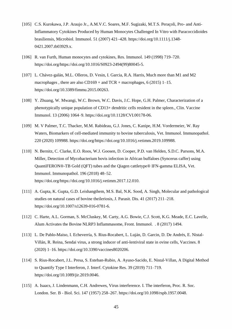
45
[105] C.S. Kurokawa, J.P. Araujo Jr., A.M.V.C. Soares, M.F. Sugizaki, M.T.S. Peraçoli, Pro- and Anti-
Inflammatory Cytokines Produced by Human Monocytes Challenged In Vitro with Paracoccidioides
brasiliensis, Microbiol. Immunol. 51 (2007) 421–428. https://doi.org/10.1111/j.1348-
0421.2007.tb03929.x.
[106] R. van Furth, Human monocytes and cytokines, Res. Immunol. 149 (1998) 719–720.
https://doi.org/https://doi.org/10.1016/S0923-2494(99)80045-5.
[107] L. Chávez-galán, M.L. Olleros, D. Vesin, I. Garcia, R.A. Harris, Much more than M1 and M2
macrophages , there are also CD169 + and TCR + macrophages, 6 (2015) 1–15.
https://doi.org/10.3389/fimmu.2015.00263.
[108] Y. Zhuang, W. Mwangi, W.C. Brown, W.C. Davis, J.C. Hope, G.H. Palmer, Characterization of a
phenotypically unique population of CD13+ dendritic cells resident in the spleen., Clin. Vaccine
Immunol. 13 (2006) 1064–9. https://doi.org/10.1128/CVI.00178-06.
[109] M. V Palmer, T.C. Thacker, M.M. Rabideau, G.J. Jones, C. Kanipe, H.M. Vordermeier, W. Ray
Waters, Biomarkers of cell-mediated immunity to bovine tuberculosis, Vet. Immunol. Immunopathol.
220 (2020) 109988. https://doi.org/https://doi.org/10.1016/j.vetimm.2019.109988.
[110] N. Bernitz, C. Clarke, E.O. Roos, W.J. Goosen, D. Cooper, P.D. van Helden, S.D.C. Parsons, M.A.
Miller, Detection of Mycobacterium bovis infection in African buffaloes (Syncerus caffer) using
QuantiFERON®-TB Gold (QFT) tubes and the Qiagen cattletype® IFN-gamma ELISA, Vet.
Immunol. Immunopathol. 196 (2018) 48–52.
https://doi.org/https://doi.org/10.1016/j.vetimm.2017.12.010.
[111] A. Gupta, K. Gupta, G.D. Leishangthem, M.S. Bal, N.K. Sood, A. Singh, Molecular and pathological
studies on natural cases of bovine theileriosis, J. Parasit. Dis. 41 (2017) 211–218.
https://doi.org/10.1007/s12639-016-0781-6.
[112] C. Harte, A.L. Gorman, S. McCluskey, M. Carty, A.G. Bowie, C.J. Scott, K.G. Meade, E.C. Lavelle,
Alum Activates the Bovine NLRP3 Inflammasome, Front. Immunol. . 8 (2017) 1494.
[113] L. De Pablo-Maiso, I. Echeverría, S. Rius-Rocabert, L. Luján, D. Garcin, D. De Andrés, E. Nistal-
Villán, R. Reina, Sendai virus, a strong inducer of anti-lentiviral state in ovine cells, Vaccines. 8
(2020) 1–16. https://doi.org/10.3390/vaccines8020206.
[114] S. Rius-Rocabert, J.L. Presa, S. Esteban-Rubio, A. Ayuso-Sacido, E. Nistal-Villan, A Digital Method
to Quantify Type I Interferon, J. Interf. Cytokine Res. 39 (2019) 711–719.
https://doi.org/10.1089/jir.2019.0046.
[115] A. Isaacs, J. Lindenmann, C.H. Andrewes, Virus interference. I. The interferon, Proc. R. Soc.
London. Ser. B - Biol. Sci. 147 (1957) 258–267. https://doi.org/10.1098/rspb.1957.0048.

46
[116] E. Voigt, B. Inankur, A. Baltes, J. Yin, A quantitative infection assay for human type I, II, and III
interferon antiviral activities, Virol. J. 10 (2013) 224. https://doi.org/10.1186/1743-422X-10-224.
[117] D.A. Magee, K.M. Conlon, N.C. Nalpas, J.A. Browne, C. Pirson, C. Healy, K.E. McLoughlin, J.
Chen, H.M. Vordermeier, E. Gormley, D.E. MacHugh, S. V. Gordon, Innate cytokine profiling of
bovine alveolar macrophages reveals commonalities and divergence in the response to
Mycobacterium bovis and Mycobacterium tuberculosis infection, Tuberculosis. 94 (2014) 441–450.
https://doi.org/10.1016/j.tube.2014.04.004.
[118] J. Hellemans, J. Vandesompele, Selection of Reliable Reference Genes for RT-qPCR Analysis, in:
2014: pp. 19–26. https://doi.org/10.1007/978-1-4939-0733-5_3.
[119] C. Puech, L. Dedieu, I. Chantal, V. Rodrigues, Design and evaluation of a unique SYBR Green real-
time RT-PCR assay for quantification of five major cytokines in cattle, sheep and goats, BMC Vet.
Res. 11 (2015) 65. https://doi.org/10.1186/s12917-015-0382-0.
[120] S.A. Bustin, V. Benes, J.A. Garson, J. Hellemans, J. Huggett, M. Kubista, R. Mueller, T. Nolan,
M.W. Pfaffl, G.L. Shipley, J. Vandesompele, C.T. Wittwer, The MIQE guidelines: minimum
information for publication of quantitative real-time PCR experiments., Clin. Chem. 55 (2009) 611–
22. https://doi.org/10.1373/clinchem.2008.112797.
[121] A.S. Stephens, S.R. Stephens, N.A. Morrison, Internal control genes for quantitative RT-PCR
expression analysis in mouse osteoblasts, osteoclasts and macrophages, BMC Res. Notes. 4 (2011).
https://doi.org/10.1186/1756-0500-4-410.
[122] M.B. Maeß, S. Sendelbach, S. Lorkowski, Selection of reliable reference genes during THP-1
monocyte differentiation into macrophages, BMC Mol. Biol. 11 (2010) 1–8.
https://doi.org/10.1186/1471-2199-11-90.
[123] C. Lecchi, F. Dilda, P. Sartorelli, F. Ceciliani, Widespread expression of SAA and Hp RNA in bovine
tissues after evaluation of suitable reference genes, Vet. Immunol. Immunopathol. 145 (2012) 556–
562. https://doi.org/10.1016/j.vetimm.2011.12.017.
[124] M.W. Pfaffl, A. Tichopad, C. Prgomet, T.P. Neuvians, Determination of stable housekeeping genes,
differentially regulated target genes and sample integrity: BestKeeper--Excel-based tool using pair-
wise correlations, Biotechnol. Lett. 26 (2004) 509–515.
https://doi.org/10.1023/b:bile.0000019559.84305.47.
[125] D. Garcia-Crespo, R.A. Juste, A. Hurtado, Selection of ovine housekeeping genes for normalisation
by real-time RT-PCR; analysis of PrP gene expression and genetic susceptibility to scrapie, BMC
Vet. Res. 1 (2005) 3. https://doi.org/10.1186/1746-6148-1-3.
[126] J. Jarczak, J. Kaba, E. Bagnicka, The validation of housekeeping genes as a reference in quantitative

47
Real Time PCR analysis: Application in the milk somatic cells and frozen whole blood of goats
infected with caprine arthritis encephalitis virus, Gene. 549 (2014) 280–285.
https://doi.org/https://doi.org/10.1016/j.gene.2014.07.063.
[127] J. Yperman, G. De Visscher, P. Holvoet, W. Flameng, Beta-actin cannot be used as a control for gene
expression in ovine interstitial cells derived from heart valves, J. Heart Valve Dis. 13 (2004) 848–
853.
[128] M.M. Elnaggar, G.S. Abdellrazeq, M. Sester, S.A. Khaliel, M. Singh, H.A. Torky, W.C. Davis,
Development of an improved ESAT-6 and CFP-10 peptide-based cytokine flow cytometric assay for
bovine tuberculosis, Comp. Immunol. Microbiol. Infect. Dis. 42 (2015) 1–7.
https://doi.org/10.1016/j.cimid.2015.07.005.
[129] P. Lovelace, H.T. Maecker, Multiparameter intracellular cytokine staining., Methods Mol. Biol. 699
(2011) 165–78. https://doi.org/10.1007/978-1-61737-950-5_8.
[130] Y. Corripio-Miyar, J. Hope, C.J. McInnes, S.R. Wattegedera, K. Jensen, Y. Pang, G. Entrican, E.J.
Glass, Phenotypic and functional analysis of monocyte populations in cattle peripheral blood
identifies a subset with high endocytic and allogeneic T-cell stimulatory capacity, Vet. Res. 46 (2015)
112. https://doi.org/10.1186/s13567-015-0246-4.
[131] B. Pomeroy, A. Sipka, J. Hussen, M. Eger, Y. Schukken, H.-J. Schuberth, Counts of bovine monocyte
subsets prior to calving are predictive for postpartum occurrence of mastitis and metritis, Vet. Res. 48
(2017) 13. https://doi.org/10.1186/s13567-017-0415-8.
[132] A.N. Barclay, M.H. Brown, The SIRP family of receptors and immune regulation., Nat. Rev.
Immunol. 6 (2006) 457–64. https://doi.org/10.1038/nri1859.
[133] A.M. Nagi, L.A. Babiuk, Characterization of surface markers of bovine gut mucosal leukocytes using
monoclonal antibodies., Vet. Immunol. Immunopathol. 22 (1989) 1–14. https://doi.org/10.1016/0165-
2427(89)90159-1.
[134] A. Saalmüller, J.K. Lunney, C. Daubenberger, W. Davis, U. Fischer, T.W. Göbel, P. Griebel, E.
Hollemweguer, T. Lasco, R. Meister, H.-J. Schuberth, K. Sestak, P. Sopp, F. Steinbach, W. Xiao-
Wei, B. Aasted, Summary of the animal homologue section of HLDA8., Cell. Immunol. 236 (n.d.)
51–8. https://doi.org/10.1016/j.cellimm.2005.08.009.
[135] B. Brando, D. Barnett, G. Janossy, F. Mandy, B. Autran, G. Rothe, B. Scarpati, G. D’Avanzo, J.L.
D’Hautcourt, R. Lenkei, G. Schmitz, A. Kunkl, R. Chianese, S. Papa, J.W. Gratama,
Cytofluorometric methods for assessing absolute numbers of cell subsets in blood. European Working
Group on Clinical Cell Analysis., Cytometry. 42 (2000) 327–46. https://doi.org/10.1002/1097-
0320(20001215)42:6<327::aid-cyto1000>3.0.co;2-f.

48
[136] F. Grandoni, M.M. Elnaggar, G.S. Abdellrazeq, F. Signorelli, L.M. Fry, C. Marchitelli, V. Hulubei,
S.A. Khaliel, H.A. Torky, W.C. Davis, Characterization of leukocyte subsets in buffalo ( Bubalus
bubalis ) with cross-reactive monoclonal antibodies specific for bovine MHC class I and class II
molecules and leukocyte differentiation molecules, Dev. Comp. Immunol. 74 (2017) 101–109.
https://doi.org/10.1016/j.dci.2017.04.013.
[137] M.M. Elnaggar, G.S. Abdellrazeq, S.K. Venn-Watson, E.D. Jensen, V. Hulubei, L.M. Fry, R.E.
Sacco, W.C. Davis, Identification of monoclonal antibodies cross-reactive with bottlenose dolphin
orthologues of the major histocompatibility complex and leukocyte differentiation molecules, Vet.
Immunol. Immunopathol. 192 (2017) 54–59. https://doi.org/10.1016/j.vetimm.2017.09.007.
[138] S. Thornton, R. Tan, A. Sproles, T. Do, J. Schick, A.A. Grom, M. DeLay, G.S. Schulert, A
Multiparameter Flow Cytometry Analysis Panel to Assess CD163 mRNA and Protein in Monocyte
and Macrophage Populations in Hyperinflammatory Diseases, J. Immunol. 202 (2019) 1635–1643.
https://doi.org/10.4049/jimmunol.1800765.
[139] A. Saalmüller, B. Aasted, Summary of the animal homologue section of HLDA8., Vet. Immunol.
Immunopathol. 119 (2007) 2–13. https://doi.org/10.1016/j.vetimm.2007.06.009.
[140] P. Berthon, J. Hopkins, Ruminant cluster CD14., Vet. Immunol. Immunopathol. 52 (1996) 245–8.
https://doi.org/10.1016/0165-2427(96)05568-7.
[141] P. Sopp, D. Werling, C. Baldwin, Cross-reactivity of mAbs to human CD antigens with cells from
cattle., Vet. Immunol. Immunopathol. 119 (2007) 106–14.
https://doi.org/10.1016/j.vetimm.2007.06.014.
[142] N. Goldfinch, P. Reinink, T. Connelley, A. Koets, I. Morrison, I. Van Rhijn, Conservation of mucosal
associated invariant T (MAIT) cells and the MR1 restriction element in ruminants, and abundance of
MAIT cells in spleen, Vet. Res. 41 (2010) 62. https://doi.org/10.1051/vetres/2010034.
[143] P. Sopp, L.S. Kwong, C.J. Howard, Identification of bovine CD14., Vet. Immunol. Immunopathol. 52
(1996) 323–8. https://doi.org/10.1016/0165-2427(96)05583-3.
[144] V.K. Gupta, I. McConnell, R.G. Dalziel, J. Hopkins, Identification of the sheep homologue of the
monocyte cell surface molecule — CD14, Vet. Immunol. Immunopathol. 51 (1996) 89–99.
https://doi.org/10.1016/0165-2427(95)05512-6.
[145] P. Boysen, G. Gunnes, D. Pende, M. Valheim, A.K. Storset, Natural killer cells in lymph nodes of
healthy calves express CD16 and show both cytotoxic and cytokine-producing properties, Dev.
Comp. Immunol. 32 (2008) 773–783. https://doi.org/10.1016/j.dci.2007.11.006.
[146] M.M. Elnaggar, G.S. Abdellrazeq, V. Mack, L.M. Fry, W.C. Davis, K.T. Park, Characterization and
use of new monoclonal antibodies to CD11c, CD14, and CD163 to analyze the phenotypic

49
complexity of ruminant monocyte subsets, Vet. Immunol. Immunopathol. 178 (2016) 57–63.
https://doi.org/10.1016/j.vetimm.2016.06.010.
[147] C.J. Howard, G.P. Brooke, D. Werling, P. Sopp, J.C. Hope, K.R. Parsons, R.A. Collins, Dendritic
cells in cattle: phenotype and function., Vet. Immunol. Immunopathol. 72 (1999) 119–24.
https://doi.org/10.1016/s0165-2427(99)00124-5.
[148] A.M. Nagi, L.A. Babiuk, Peanut agglutinin (PNA): binding and stimulation of bovine intestinal and
peripheral blood leukocytes., Vet. Immunol. Immunopathol. 22 (1989) 67–78.
https://doi.org/10.1016/0165-2427(89)90164-5.
[149] E.M. Ibeagha-Awemu, A.E. Ibeagha, X. Zhao, The influence of different anticoagulants and sample
preparation methods on measurement of mCD14 on bovine monocytes and polymorphonuclear
neutrophil leukocytes, BMC Res. Notes. 5 (2012) 93. https://doi.org/10.1186/1756-0500-5-93.
[150] F. Mandy, B. Brando, Enumeration of Absolute Cell Counts Using Immunophenotypic Techniques,
Curr. Protoc. Cytom. 13 (2000). https://doi.org/10.1002/0471142956.cy0608s13.
[151] L.C. Davies, P.R. Taylor, Tissue-resident macrophages: then and now., Immunology. 144 (2015)
541–8. https://doi.org/10.1111/imm.12451.
[152] M. Hesketh, K.B. Sahin, Z.E. West, R.Z. Murray, Macrophage Phenotypes Regulate Scar Formation
and Chronic Wound Healing, Int. J. Mol. Sci. 18 (2017) 1545. https://doi.org/10.3390/ijms18071545.
[153] P. Chandrasekaran, S. Izadjoo, J. Stimely, S. Palaniyandi, X. Zhu, W. Tafuri, D.M. Mosser,
Regulatory Macrophages Inhibit Alternative Macrophage Activation and Attenuate Pathology
Associated with Fibrosis., J. Immunol. 203 (2019) 2130–2140.
https://doi.org/10.4049/jimmunol.1900270.
[154] J. Dunn, ed., Manual of Diagnostic Cytology of the Dog and Cat, Wiley, 2014.
https://doi.org/10.1002/9781118823040.
[155] E. Piaton, M. Fabre, I. Goubin-Versini, M.-F. Bretz-Grenier, M. Courtade-Saïdi, S. Vincent, G.
Belleannée, F. Thivolet, J. Boutonnat, H. Debaque, J. Fleury-Feith, P. Vielh, C. Egelé, J.-P. Bellocq,
J.-F. Michiels, B. Cochand-Priollet, Guidelines for May-Grünwald-Giemsa staining in haematology
and non-gynaecological cytopathology: recommendations of the French Society of Clinical Cytology
(SFCC) and of the French Association for Quality Assurance in Anatomic and Cytologic Pathology
(AFA, Cytopathology. 27 (2016) 359–68. https://doi.org/10.1111/cyt.12323.
[156] M.V. Palmer, T.C. Thacker, W.R. Waters, Analysis of Cytokine Gene Expression using a Novel
Chromogenic In-situ Hybridization Method in Pulmonary Granulomas of Cattle Infected
Experimentally by Aerosolized Mycobacterium bovis, J. Comp. Pathol. 153 (2015) 150–159.
https://doi.org/10.1016/j.jcpa.2015.06.004.

50
[157] H.-E. Park, H.-T. Park, Y.H. Jung, H.S. Yoo, Gene expression profiles of immune-regulatory genes in
whole blood of cattle with a subclinical infection of Mycobacterium avium subsp. paratuberculosis,
PLoS One. 13 (2018) e0196502. https://doi.org/10.1371/journal.pone.0196502.
[158] M. V. Palmer, T.C. Thacker, W.R. Waters, Multinucleated giant cell cytokine expression in
pulmonary granulomas of cattle experimentally infected with Mycobacterium bovis, Vet. Immunol.
Immunopathol. 180 (2016) 34–39. https://doi.org/10.1016/j.vetimm.2016.08.015.
[159] M. V. Palmer, J. Wiarda, C. Kanipe, T.C. Thacker, Early Pulmonary Lesions in Cattle Infected via
Aerosolized Mycobacterium bovis, Vet. Pathol. 56 (2019) 544–554.
https://doi.org/10.1177/0300985819833454.
[160] G. Ampem, H. Azegrouz, Á. Bacsadi, L. Balogh, S. Schmidt, J. Thuróczy, T. Röszer, Adipose tissue
macrophages in non-rodent mammals: a comparative study., Cell Tissue Res. 363 (2016) 461–78.
https://doi.org/10.1007/s00441-015-2253-1.
[161] A.L. Rivas, F.W. Quimby, J. Blue, O. Coksaygan, Longitudinal evaluation of bovine mammary gland
health status by somatic cell counting, flow cytometry, and cytology., J. Vet. Diagn. Invest. 13 (2001)
399–407. https://doi.org/10.1177/104063870101300506.
[162] P. Ganjei-Azar, M. Nadji, eds., Color Atlas of Immunocytochemistry in Diagnostic Cytology,
Springer US, Boston, MA, 2006. https://doi.org/10.1007/978-0-387-32122-6.
[163] H.M. Golbar, T. Izawa, V. Juniantito, C. Ichikawa, M. Tanaka, M. Kuwamura, J. Yamate,
Immunohistochemical characterization of macrophages and myofibroblasts in fibrotic liver lesions
due to Fasciola infection in cattle., J. Vet. Med. Sci. 75 (2013) 857–65.
https://doi.org/10.1292/jvms.12-0536.
[164] M. V Palmer, J. Wiarda, C. Kanipe, T.C. Thacker, Early Pulmonary Lesions in Cattle Infected via
Aerosolized Mycobacterium bovis., Vet. Pathol. 56 (2019) 544–554.
https://doi.org/10.1177/0300985819833454.
[165] M. Fernández, J. Benavides, P. Castaño, N. Elguezabal, M. Fuertes, M. Muñoz, M. Royo, M.C.
Ferreras, V. Pérez, Macrophage Subsets Within Granulomatous Intestinal Lesions in Bovine
Paratuberculosis., Vet. Pathol. 54 (2017) 82–93. https://doi.org/10.1177/0300985816653794.
[166] M. Fernández, M. Fuertes, N. Elguezabal, P. Castaño, M. Royo, M.C. Ferreras, J. Benavides, V.
Pérez, Immunohistochemical expression of interferon-γ in different types of granulomatous lesions
associated with bovine paratuberculosis., Comp. Immunol. Microbiol. Infect. Dis. 51 (2017) 1–8.
https://doi.org/10.1016/j.cimid.2017.01.002.
[167] A.W. Newman, A. Miller, F.A. Leal Yepes, E. Bitsko, D. Nydam, S. Mann, The effect of the
transition period and postpartum body weight loss on macrophage infiltrates in bovine subcutaneous

51
adipose tissue, J. Dairy Sci. 102 (2019) 1693–1701. https://doi.org/10.3168/jds.2018-15362.
[168] T. Yamada, M. Kamiya, M. Higuchi, N. Nakanishi, Fat depot-specific differences of macrophage
infiltration and cellular senescence in obese bovine adipose tissues., J. Vet. Med. Sci. 80 (2018)
1495–1503. https://doi.org/10.1292/jvms.18-0324.
[169] L.J. Oliveira, P.J. Hansen, Phenotypic characterization of macrophages in the endometrium of the
pregnant cow., Am. J. Reprod. Immunol. 62 (2009) 418–26. https://doi.org/10.1111/j.1600-
0897.2009.00761.x.
[170] L.J. Oliveira, P.J. Hansen, Deviations in populations of peripheral blood mononuclear cells and
endometrial macrophages in the cow during pregnancy., Reproduction. 136 (2008) 481–90.
https://doi.org/10.1530/REP-08-0218.
[171] Y. Shimamoto, J. Nio-Kobayashi, H. Watarai, M. Nagano, N. Saito, E. Takahashi, H. Higuchi, A.
Kobayashi, T. Kimura, H. Kitamura, Generation and validation of novel anti-bovine CD163
monoclonal antibodies ABM-1A9 and ABM-2D6., Vet. Immunol. Immunopathol. 198 (2018) 6–13.
https://doi.org/10.1016/j.vetimm.2018.02.004.
[172] C.J. Jenvey, J.M. Hostetter, A.L. Shircliff, J.R. Stabel, Relationship between the pathology of bovine
intestinal tissue and current diagnostic tests for Johne’s disease., Vet. Immunol. Immunopathol. 202
(2018) 93–101. https://doi.org/10.1016/j.vetimm.2018.06.012.
[173] C.J. Jenvey, A.L. Shircliff, J.P. Bannantine, J.R. Stabel, Phenotypes of macrophages present in the
intestine are impacted by stage of disease in cattle naturally infected with Mycobacterium avium
subsp. paratuberculosis., PLoS One. 14 (2019) e0217649.
https://doi.org/10.1371/journal.pone.0217649.
[174] C.J. Jenvey, J.M. Hostetter, A.L. Shircliff, J.P. Bannantine, J.R. Stabel, Quantification of
Macrophages and Mycobacterium avium Subsp. paratuberculosis in Bovine Intestinal Tissue During
Different Stages of Johne’s Disease., Vet. Pathol. 56 (2019) 671–680.
https://doi.org/10.1177/0300985819844823.
[175] J. De Koster, C. Strieder-Barboza, J. de Souza, A.L. Lock, G.A. Contreras, Short communication:
Effects of body fat mobilization on macrophage infiltration in adipose tissue of early lactation dairy
cows., J. Dairy Sci. 101 (2018) 7608–7613. https://doi.org/10.3168/jds.2017-14318.
[176] R.K. Nelli, J. De Koster, J.N. Roberts, J. de Souza, A.L. Lock, W. Raphael, D. Agnew, G.A.
Contreras, Impact of uterine macrophage phenotype on placental retention in dairy cows.,
Theriogenology. 127 (2019) 145–152. https://doi.org/10.1016/j.theriogenology.2019.01.011.
[177] A. Sterner-Kock, W. Haider, F. Sacchini, A. Liljander, J. Meens, J. Poole, M. Guschlbauer, M.
Heller, J. Naessens, J. Jores, Morphological characterization and immunohistochemical detection of

52
the proinflammatory cytokines IL-1β, IL-17A, and TNF-α in lung lesions associated with contagious
bovine pleuropneumonia., Trop. Anim. Health Prod. 48 (2016) 569–76.
https://doi.org/10.1007/s11250-016-0994-9.
[178] C.-J. Li, L.-Y. Sun, C.-Y. Pang, Synergistic protection of N-acetylcysteine and ascorbic acid 2-
phosphate on human mesenchymal stem cells against mitoptosis, necroptosis and apoptosis., Sci.
Rep. 5 (2015) 9819. https://doi.org/10.1038/srep09819.
Figure Captions
Fig. 1 Bovine peripheral blood smears.
A. Normal round monocyte with a deeply indented nucleus and light blue homogeneous cytoplasm
is present in a background of normal erythrocytes. Note the non-vacuolated cytoplasm, not all
peripheral blood monocytes contain cytoplasmic vacuoles. Bar=7,2 m.
B. Large peripheral blood monocyte with light blue non-vacuolated cytoplasm with an irregularly
bean-shaped indented nucleus. Bar= 6 m.
C. Large peripheral blood monocyte with irregular membrane contour and abundant light blue
cytoplasm characterized by multiple, variably sized discrete cytoplasmic vacuoles. Bar= 4,9 m.
May-Grünwald Giemsa stain.
Fig. 2 Phase-contrast photomicrographs of cultured monocytes following MACS purification.
A. Monocytes the first day in culture (10%FBS supplemented RPMI media). Bar= 20 m;
B. Monocytes maintained for two days demonstrating a slight increase in size and shape and size
variability. Bar=20 m.
C. Two monocytes maintained for four days in culture and characterized by variably increased size
and presenting with cytoplasmic processes indicative of activation and initial transformation into
macrophages. Bar= 10 m.
Fig. 3. Flow cytometry evaluation of monocytes, before and after sorting.
PBMC are showed in a representative dot plot FSC vs SSC based on size and granularity (complexity)
before (A) and after MACS separation (D). Monocytes staining with FITC-anti-CD14 antibody is
showed before (B) and after sorting (E). Representative histograms of the relative percentage of
lymphocytes and monocytes are showed before (C) and after sorting (F). MACS purification gives
an enrichment of CD14+ monocytes population > of 98%.
Fig. 4. Flow cytometry detection, by annexin V-FITC/PI, of apoptosis and necrosis on bovine
PBMC incubated for 1 h at 37 °C.

53
On dot plot FSC vs SSC, a gate around lymphocytes and monocytes regions was drawn and apoptosis
and necrosis in gated events were determined. Live lymphocytes (A) and monocytes (B) are Annexin
V and PI double negative (lower left quadrant). At the early stage of apoptosis, the cells bind annexin
V while still excluding PI (lower right quadrant). Double positive events are necrotic cells (upper
right quadrant). The PI-positive - annexin V negative cells (upper left quadrant) are considered
necroptotic cells [178].
Fig. 5. In vitro methods to measure chemotaxis
(A) Schematic illustration of the Boyden chamber assay. Cells seeded in the upper chamber migrate
through a porous membrane, towards the lower chamber in the response of a chemoattractant gradient
concentration. (B) Schematic illustration of a migration transwell plate. The multiwell plate is
packaged with permeable and removable cell culture inserts with a porous membrane that allows the
migration of the cells towards the medium/chemoattractant-filled lower well. Images were created
with BioRender.com".
Fig. 6 Morphological and Immunohistochemical features of bovine macrophages in
cytological and tissue specimens
Fig. 6 A Cytology of synovial fluid in a calf with arthritis. Moderately cellular cytological sample
illustrating numerous foamy reactive macrophages with variably abundant cytoplasm with
numerous cytoplasmic vacuoles admixed with non-degenerate neutrophils in a background of
confluent erythrocytes. Joint tap, May-Grünwald Giemsa stain. Bar= 25 µm
Fig. 6 B Cytology of synovial fluid in a calf with arthritis. Large reactive macrophage with heavily
vacuolated cytoplasm containing phagocytized apoptotic nuclear material. The macrophage is
surrounded by moderately degenerated erythrocytes (crenation) and non-degenerated neutrophils in
a granular background of synovial mucins. Joint tap, May-Grünwald Giemsa stain. Bar=7,5 µm
Fig. 6 C Histology of a bovine mesenteric lymph node in a case of paratuberculosis. Sheets of
reactive macrophages characterized by varying size, pink to grey cytoplasm and oval nuclei with
granular to reticular chromatin and evidence of 1-2 small, basophilic nucleoli are admixed with
mature lymphocytes and rare plasma cells. A single multinucleated macrophage with haphazardly
arranged nuclei is evident (arrowhead). Haematoxylin and eosin stain. Bar= 14 m
Fig. 6 D Histology of a bovine thoracic lymph node in a case of Tuberculosis. At the centre of the
image, there is a large multinucleated giant macrophage characterized by several nuclei arranged in
a horse-show fashion beneath the cell membrane. The cell is surrounded by occasional epithelioid

54
cells, foamy reactive macrophages with large cytoplasmic well distinct vacuoles and elevated
numbers of lymphocytes (left) and lesser plasma cells. Haematoxylin and eosin stain. Bar= 32 m
Fig. 6 E Bovine small intestine in a case of paratuberculosis. Lamina propria of intestinal villi is
substituted and expanded by massive infiltration of large reactive macrophages displacing
peripherally and compressing intestinal glands. Haematoxylin and eosin stain. Bar= 170 m
Fig. 6 F Bovine tongue in a case of wooden tongue (Actinobacillus lignieresii infection). Large
granuloma composed of reactive macrophages admixed with lesser numbers of neutrophils,
eosinophils and lymphocytes and surrounded by fibroblasts and collagen). Haematoxylin and eosin
stain. Bar= 75 m
Fig. 6 G Bovine small intestine in a case of paratuberculosis. Diffuse granulomatous inflammation
expanding and substituting the intestinal lamina propria with separation of the epithelial mucosal
lining. Large reactive macrophages predominate and associate with lesser numbers of lymphocytes.
In the left lower part of the image, an intestinal gland is compressed with gland epithelium lightly
infiltrated by eosinophils (arrowhead). Haematoxylin and eosin stain. Bar= 64 m
Fig. 6 H Bovine small intestine in a case of paratuberculosis. High numbers of acid-fast positive
mycobacteria (Mycobacterium avium sbsp. paratuberculosis) in the cytoplasm of macrophages and
free in the lamina propria. Ziehl – Neelsen stain. Bar= 22 m
Fig. 6 I Bovine lung in a case of tuberculosis. The image shows the typical stratigraphy of a
tuberculoid granuloma substituting the pulmonary parenchyma. The right and central parts of the
image represent the extensive central area of caseous necrosis characterized by amorphous
eosinophilic and granular basophilic tissue debris. In the left part of the image, necrosis is rimmed
by neutrophils, macrophages, multinucleated giant cells of the Langhans cell type and mature
lymphocytes. The external portion of the granuloma is composed of fibroblasts and collagen
delimiting the granuloma. Haematoxylin and eosin stain. Bar= 196 m
Fig. 6 J Bovine tongue in a case of wooden tongue (Actinobacillus lignieresii infection). Discrete
granuloma with central intensely eosinophilic material corresponding to the accumulation of
immunoglobulin/immunocomplexes and termed Splendore-Hoeppli phenomenon. Haematoxylin
and eosin stain. Bar= 110 m
Fig 6 K Immunohistochemical stain of a bovine reactive thoracic lymph node. Numerous Iba-1
positive reactive foamy macrophages in the medullary cords. Anti-Iba-1 immunoperoxidase stain,
AEC chromogen, hematoxylin counterstain. Bar= 70 m
Fig 6 L Immunohistochemical stain of a calf lung with bronchopneumonia. Alveolar lumen
containing numerous reactive macrophages and rare neutrophils. Two M1 type macrophages
intensely expressing TNF. Anti-TNF immunoperoxidase stain. AEC chromogen, hematoxylin

55
counterstain. Bar= 18 m
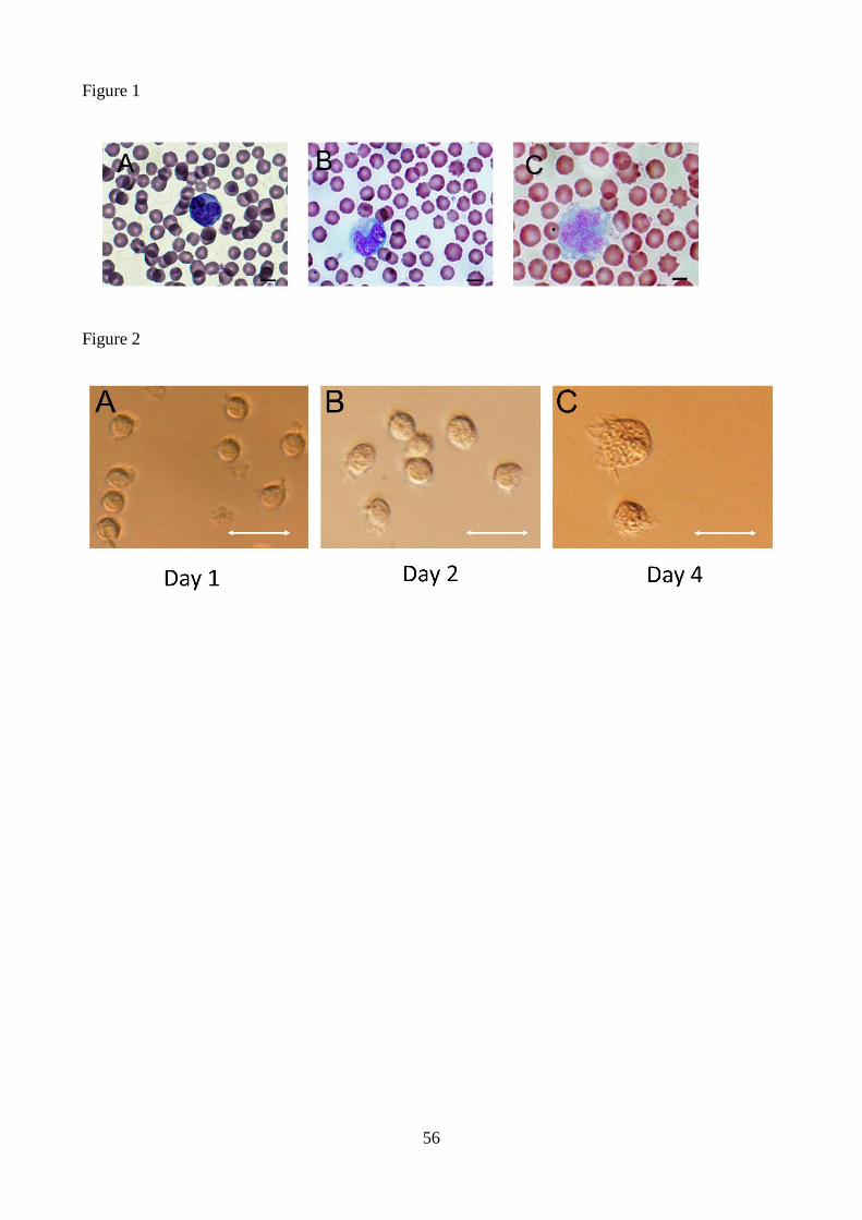
56
Figure 1
Figure 2

57
Figure 3

58
Figure 4

59
Figure 5

60
Figure 6