Embed Size (px)
Citation preview
1. INTRODUCTION
The word diabetes derived from the Greek means to siphon and refers to the marked loss
of water by urination, polyuria. The word mellitus derived from the Latin means sweet
and thus diabetes mellitus is known as sweet urine disease. It is regarded as a metabolic
disease of unknown cause resulting from a deficiency of the pancreatic hormone insulin
and an irregularity in the release of glucagon, a polypeptide hormone. Often, the sufferer
of this disease has been consuming large amounts of refined sweets such as cakes, pies,
ice cream, candies, pastries etc. Under these conditions, the pancreas is continually
stressed to secrete its hormone in order to eliminate the excess glucose from the blood.
This results in enervation of the gland and exhaustion leads to decreased insulin output.
The fact is other organs are involved as observed in many other symptoms manifested by
the diabetic such as arteriosclerosis, blindness etc. One problem with refined sugar is that
it goes immediately into the blood without digestion. This flood of sugar is very
enervating to the pancreas.
Diabetes is a disease characterized by abnormal metabolism of blood sugar and defective
insulin production. Blood sugar level is an important parameter for the diagnosis,
treatment and prognosis of diabetes. Blood sugar level is the level of sugar circulating in
blood at a given time. Blood glucose levels vary with time and some factors that affect
blood sugar levels are body composition, age, physical activity and sex.
1.1. Definition
Diabetes mellitus (DM), often simply referred to as diabetes, is a group of metabolic
diseases in which a person has high blood sugar, either because the body does not
produce enough insulin, or because cells do not respond to the insulin that is produced.
This high blood sugar produces the classical symptoms of polyuria (frequent urination),
polydipsia (increased thirst) and polyphagia (increased hunger). In other words, DM is a
set of related diseases in which the body cannot regulate the amount of sugar
(specifically, glucose) in the blood. The blood delivers glucose to provide the body with
energy to perform all of a person's daily activities.
1.2. History
Diabetes was one of the first diseases described,1 with an Egyptian manuscript from 1500
BCE mentioning „too great emptying of the urine‟.2 Indian physicians around the same
time identified the disease and classified it as madhumeha or "honey urine", noting the
urine would attract ants.2 The word diabetes comes from Latin „diabētēs’, which in turn
comes from Ancient Greek „diabētēs‟ which literally means „a passer through; a siphon.‟3
The term "diabetes" or "to pass through" was first used in 230 BCE by the Greek
Appollonius of Memphis.2 Ancient Greek physician Aretaeus of Cappadocia (1st century
CE) used that word, with the intended meaning "excessive discharge of urine", as the
name for the disease.4,5
Ultimately, the word comes from Greek „diabainein‟, meaning "to
pass through,"3 which is composed of „dia-„, meaning "through" and „bainein’, meaning
"to go".4 The word "diabetes" is first recorded in English, in the form diabete, in a
medical text written around 1425. The word mellitus comes from the classical Latin word
mellītus, meaning "mellite"6 (i.e. sweetened with honey;
6 honey-sweet
7). The Latin word
comes from mell-, which comes from mel, meaning „honey‟;6,7
sweetness;7 pleasant
thing,7 and the suffix -ītus,
6 whose meaning is the same as that of the English suffix"-
ite".8 It was Thomas Willis who in 1675 added "mellitus" to the word „diabetes‟ as a
designation for the disease, when he noticed the urine of a diabetic had a sweet taste
(glycosuria).5
1.3. Classification
Diabetes mellitus is classified into four broad categories: type 1, type 2, gestational
diabetes and "other specific types".9 The "other specific types" are a collection of a few
dozen individual causes.9
The term "diabetes", without qualification, usually refers to
diabetes mellitus. The rare disease diabetes insipidus has similar symptoms as diabetes
mellitus, but without disturbances in the sugar metabolism (insipidus means "without
taste" in Latin).
The term "type 1 diabetes" has replaced several former terms, including childhood-onset
diabetes, juvenile diabetes, and insulin-dependent diabetes mellitus (IDDM). Likewise,
the term "type 2 diabetes" has replaced several former terms, including adult-onset
diabetes, obesity-related diabetes, and noninsulin-dependent diabetes mellitus (NIDDM).
Beyond these two types, there is no agreed-upon standard nomenclature. Various sources
have defined "type 3 diabetes" as: gestational diabetes,10
insulin-resistant type 1 diabetes
(or "double diabetes"), type 2 diabetes which has progressed to require injected insulin,
and latent autoimmune diabetes of adults (or LADA or "type 1.5" diabetes).11
The three main types of diabetes mellitus (DM) are:
Type 1 DM results from the body's failure to produce insulin, and presently
requires the person to inject insulin. (Also referred to as insulin-dependent
diabetes mellitus (IDDM) or "juvenile" diabetes)
Type 2 DM results from insulin resistance, a condition in which cells fail to use
insulin properly, sometimes combined with an absolute insulin deficiency.
(Formerly referred to as noninsulin-dependent diabetes mellitus (NIDDM) or
"adult-onset" diabetes)
Gestational diabetes is when pregnant women, who have never had diabetes
before, have a high blood glucose level during pregnancy. It may precede
development of type 2 DM.
Other forms of diabetes mellitus include congenital diabetes, which is due to genetic
defects of insulin secretion, cystic fibrosis-related diabetes, steroid diabetes induced by
high doses of glucocorticoids, and several forms of monogenic diabetes.
1.3.1. Type 1 diabetes
Type 1 diabetes mellitus is characterized by loss of the insulin-producing beta cells of the
islets of Langerhans in the pancreas, leading to insulin deficiency. The body stops
producing insulin or produces too little insulin to regulate blood glucose level. This type
can be further classified as immune-mediated or idiopathic. The majority of type 1
diabetes is of the immune-mediated nature, in which beta cell loss is a T-cell-mediated
autoimmune attack.12
There is no known preventive measure against type 1 diabetes,
which causes approximately 10% of DM cases in North America and Europe. Most
affected people are otherwise healthy and of a healthy weight when onset occurs.
Sensitivity and responsiveness to insulin are usually normal, especially in the early stages.
Type 1 diabetes can affect children or adults, but was traditionally termed "juvenile
diabetes" because a majority of these diabetes cases were in children.
Type 1 diabetes is typically diagnosed during childhood or adolescence. It used to
be referred to as juvenile-onset diabetes or insulin-dependent diabetes mellitus.
Type 1 diabetes can occur in an older individual due to destruction of the pancreas
by alcohol, disease, or removal by surgery. It also results from progressive failure
of the pancreatic beta cells, the only cell type that produces significant amounts of
insulin.

People with type 1 diabetes require insulin treatment daily to sustain life.
"Brittle" diabetes, also known as unstable diabetes or labile diabetes, is a term that was
traditionally used to describe to dramatic and recurrent swings in glucose levels, often
occurring for no apparent reason in insulin-dependent diabetes.13, 14
1.3.2. Type 2 diabetes
Type 2 diabetes mellitus is characterized by insulin resistance, which may be combined
with relatively reduced insulin secretion.9 Although the pancreas still secretes insulin, the
body of someone with type 2 diabetes is partially or completely unable to use this insulin.
This is sometimes referred to as insulin resistance. The pancreas tries to overcome this
resistance by secreting more and more insulin. The defective responsiveness of body
tissues to insulin is believed to involve the insulin receptor. However, the specific defects
are not known. Diabetes mellitus cases due to a known defect are classified separately.
Type 2 diabetes is the most common type.
In the early stage of type 2, the predominant abnormality is reduced insulin sensitivity. At
this stage, hyperglycemia can be reversed by a variety of measures and medications that
improve insulin sensitivity or reduce glucose production by the liver.
People with insulin resistance develop type 2 diabetes when they fail to secrete enough
insulin to cope with their higher demands.
At least 90% of adult individuals with diabetes have type 2 diabetes.
Type 2 diabetes is typically diagnosed in adulthood, usually after age 45 years. It
used to be called adult-onset diabetes mellitus, or non-insulin-dependent diabetes
mellitus. These names are no longer used because type 2 diabetes does occur in
younger people, and some people with type 2 diabetes require insulin therapy.
Type 2 diabetes is usually controlled with diet, weight loss, exercise, and oral
medications. However, more than half of all people with type 2 diabetes require insulin to
control their blood sugar levels at some point in the course of their illness.
1.3.3. Metabolic syndrome
Metabolic syndrome (also referred to as syndrome X) is a set of abnormalities in which
insulin-resistant diabetes (type 2 diabetes) is almost always present along with
hypertension (high blood pressure), high fat levels in the blood (increased serum lipids,
predominant elevation of LDL cholesterol, decreased HDL cholesterol, and elevated
triglycerides), central obesity, and abnormalities in blood clotting (fibrinolysis,
procoagulation) and inflammatory responses. A high rate of cardiovascular disease is
associated with metabolic syndrome.
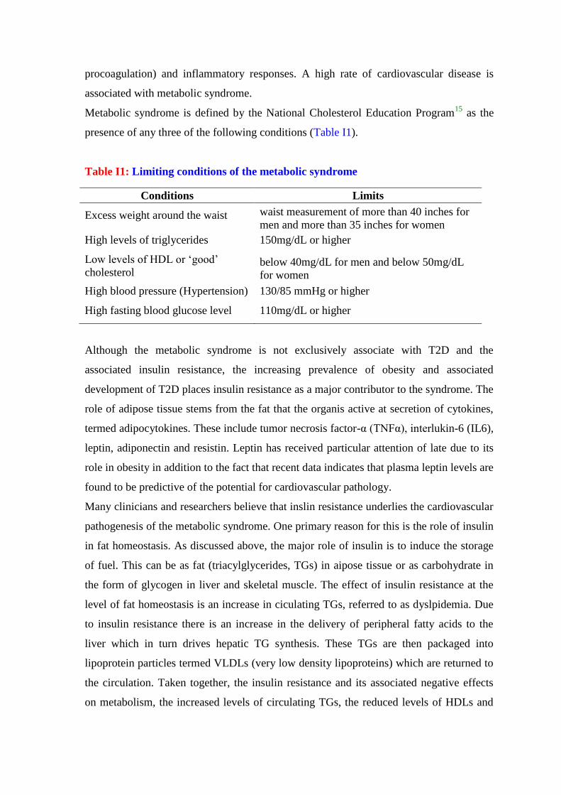
Metabolic syndrome is defined by the National Cholesterol Education Program15
as the
presence of any three of the following conditions (Table I1).
Table I1: Limiting conditions of the metabolic syndrome
Conditions Limits
Excess weight around the waist waist measurement of more than 40 inches for
men and more than 35 inches for women
High levels of triglycerides 150mg/dL or higher
Low levels of HDL or „good‟
cholesterol
below 40mg/dL for men and below 50mg/dL
for women
High blood pressure (Hypertension) 130/85 mmHg or higher
High fasting blood glucose level 110mg/dL or higher
Although the metabolic syndrome is not exclusively associate with T2D and the
associated insulin resistance, the increasing prevalence of obesity and associated
development of T2D places insulin resistance as a major contributor to the syndrome. The
role of adipose tissue stems from the fat that the organis active at secretion of cytokines,
termed adipocytokines. These include tumor necrosis factor-α (TNFα), interlukin-6 (IL6),
leptin, adiponectin and resistin. Leptin has received particular attention of late due to its
role in obesity in addition to the fact that recent data indicates that plasma leptin levels are
found to be predictive of the potential for cardiovascular pathology.
Many clinicians and researchers believe that inslin resistance underlies the cardiovascular
pathogenesis of the metabolic syndrome. One primary reason for this is the role of insulin
in fat homeostasis. As discussed above, the major role of insulin is to induce the storage
of fuel. This can be as fat (triacylglycerides, TGs) in aipose tissue or as carbohydrate in
the form of glycogen in liver and skeletal muscle. The effect of insulin resistance at the
level of fat homeostasis is an increase in ciculating TGs, referred to as dyslpidemia. Due
to insulin resistance there is an increase in the delivery of peripheral fatty acids to the
liver which in turn drives hepatic TG synthesis. These TGs are then packaged into
lipoprotein particles termed VLDLs (very low density lipoproteins) which are returned to
the circulation. Taken together, the insulin resistance and its associated negative effects
on metabolism, the increased levels of circulating TGs, the reduced levels of HDLs and
hypertension, all contribute to the progression of atherosclerosis. With associated
coagulation and fibrinolysis pathogenesis, the cardiovascular events of the metabolic
syndrome can be devastating.
Since many of these pathogeneses can be reversed with proper diet and exercise, it is in a
person‟s best interest to take responsibility for the role their lifestyle choices play in the
development of the metabolic syndrome.16
1.3.4. Prediabetes
It is a common condition related to diabetes. In people with prediabetes, the blood sugar
level is higher than normal but not yet high enough to be considered diagnostic of
diabetes.
Prediabetes increases a person's risk of developing type 2 diabetes, heart disease,
or stroke.
Prediabetes can typically be reversed (without insulin or medication) with lifestyle
changes such as losing a modest amount of weight and increasing physical activity
levels. Weight loss can prevent, or at least delay, the onset of type 2 diabetes.
An international expert committee of the American Diabetes Association
redefined the criteria for prediabetes, lowering the blood sugar level cut-off point
for prediabetes. Approximately 20% more adults are now believed to have this
condition and may develop diabetes within 10 years if they do make lifestyle
changes such as exercising more and maintaining a healthy weight.
About 17 million Americans (6.2% of adults in North America) are believed to have
diabetes. It has been estimated that about one third of adults with diabetes do not know
they have diabetes.
About 1 million new cases of diabetes is diagnosed occur each year, and diabetes
is the direct or indirect cause of at least 200,000 deaths each year.
The incidence of diabetes is increasing rapidly. This increase is due to many
factors, but the most significant are the increasing incidence of obesity associated
with the prevalence of a sedentary lifestyle.
Some cases of diabetes are caused by the body's tissue receptors not responding to insulin
(even when insulin levels are normal, which is what separates it from type 2 diabetes);
this form is very uncommon. Genetic mutations (autosomal or mitochondrial) can lead to
defects in beta cell function. Abnormal insulin action may also have been genetically
determined in some cases. Any disease that causes extensive damage to the pancreas may
lead to diabetes (for example, chronic pancreatitis and cystic fibrosis). Diseases
associated with excessive secretion of insulin-antagonistic hormones can cause diabetes
(which is typically resolved once the hormone excess is removed). Many drugs impair
insulin secretion and some toxins damage pancreatic beta cells. The ICD-10 (1992)
diagnostic entity, malnutrition-related diabetes mellitus (MRDM or MMDM, ICD-10
code E12), was deprecated by the World Health Organization when the current taxonomy
was introduced in 1999.17
1.4. Relation among Insulin Resistance, Pre-diabetes and T2DM
If someone has insulin resistance his/her muscle, fat and liver cells do not use insulin
properly. The pancreas tries to keep up with the demand for insulin by producing more.
Eventually, the pancreas cannot keep up with the body‟s need for insulin, and excess
glucose builds up in the bloodstream. Many people with insulin resistance have high level
of blood glucose and high levels of insulin circulating in their blood at the same time.
People with blood glucose levels that are higher than normal but not yet in the diabetic
range have “pre-diabetic”. Doctors sometimes call this condition impaired fasting glucose
(IFG) or impaired glucose tolerance (IGT), depending on the test used to diagnose it.
If someone has pre-diabetes, he/she has a higher risk of developing type 2 diabetes,
formerly called adult-onset diabetes or non insulin dependent diabetes. Studies have
shown that that most people with pre-diabetes go on to develop type 2 diabetes within 10
years, unless they loose 5 to 7 percent of their body weight – which is about 10 to 15
pounds for someone who weighs 200 pounds – by making modest changes in their diet
and level of physical activity. People with pre-diabetes also have a higher risk of heart
disease. Type 2 diabetes is sometimes defined as the form of diabetes that develops when
the body does not respond properly to insulin, as opposed to type 1 diabetes, in which the
pancreas makes no insulin at all. At first, the pancreas keeps up with the added demand
by producing more insulin. In times, however, it loses the ability to secrete enough insulin
in response to meals. Insulin resistance can also occur in people who have type 1
diabetes, especially if they are overweight.15
Also Latent Autoimmune Diabetes of Adults (LADA) is another condition in which type
1 DM develops in adults. Adults with LADA are frequently initially misdiagnosed as
having type 2 DM, based on age rather than etiology. 17
1.5. Causes
The cause of diabetes depends on the type.
Type 1 diabetes is believed to be an autoimmune disease. The body's immune system
specifically attacks the cells in the pancreas that produce insulin.
A predisposition to develop type 1 diabetes may run in families, but genetic
causes (a postitive family history) are much more common for type 2 diabetes.
Environmental factors, including common unavoidable viral infections, may also
contribute to type 1 diabetes.
Type 1 diabetes is most common in people of non-Hispanic, Northern European
descent (especially Finland and Sardinia), followed by African Americans, and
Hispanic Americans. It is relatively rare in those of Asian descent.
Type 1 diabetes is slightly more common in men than in women.
Type 1 diabetes is partly inherited, and then triggered by certain infections, with some
evidence pointing at Coxsackie B4 virus. A genetic element in individual susceptibility to
some of these triggers has been traced to particular HLA genotypes (i.e., the genetic "self"
identifiers relied upon by the immune system). However, even in those who have
inherited the susceptibility, T1DM seems to require an environmental trigger.
Type 2 diabetes is due primarily to lifestyle factors and genetics.18
T2DM as a common
and complex disease has been characterized by the following causes:
• Obesity: obesity is also considered a key risk factor for T2DM. The association between
increasing body mass index (BMI) and greater weight gain and risk of diabetes is most
pronounced among Asians, suggesting that lower cut off BMI values are needed to
identify. Asians at a higher risk of diabetes.19
BMI cut point for Indians for any
cardiometabolic risk factors is 23 kg/m2 in both sexes.
• Abdominal adiposity: there is also a probable indication that there is a preferential
abdominal adiposity in Indians irrespective of the degree of general adiposity.20
• Imbalance of human metabolism is associated with T2DM: Changes in work patterns
from heavy labour to sedentary, the increase in computerization and mechanization, and
improved transport are just a few of the changes that have had an impact on human
metabolism.
• Genes: since 2007, genome-wide association studies has catalogued around 20 genes
(like TCF7L2, HHEX, CDKAL1, SLC30A8 etc.) showing a strong association (with
modest odds ratio ranges between 1.2 and 1.5) with T2DM.
• Ethnicity: the interethnic differences (like differences in prevalence of T2DM among
Europeans, Americans, Chinese, and Asian Indians) in insulin resistance may have an
environmental or genetic explanation. The main acquired factors that seemingly increase
insulin resistance in all ethnic groups include obesity, sedentary lifestyle, diet rich in
animal products, and aging.21
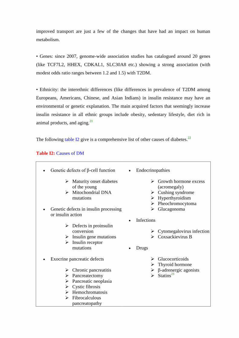
The following table I2 give is a comprehensive list of other causes of diabetes.22
Table I2: Causes of DM
Genetic defects of β-cell function
Maturity onset diabetes
of the young
Mitochondrial DNA
mutations
Genetic defects in insulin processing
or insulin action
Defects in proinsulin
conversion
Insulin gene mutations
Insulin receptor
mutations
Exocrine pancreatic defects
Chronic pancreatitis
Pancreatectomy
Pancreatic neoplasia
Cystic fibrosis
Hemochromatosis
Fibrocalculous
pancreatopathy
Endocrinopathies
Growth hormone excess
(acromegaly)
Cushing syndrome
Hyperthyroidism
Pheochromocytoma
Glucagonoma
Infections
Cytomegalovirus infection
Coxsackievirus B
Drugs
Glucocorticoids
Thyroid hormone
β-adrenergic agonists
Statins23
Insulin is the principal hormone that regulates uptake of glucose from the blood into most
cells (primarily muscle and fat cells, but not central nervous system cells). Therefore,
deficiency of insulin or the insensitivity of its receptors plays a central role in all forms of
diabetes mellitus.
Humans are capable of digesting some carbohydrates, in particular those most common in
food; starch, and some disaccharides such as sucrose, are converted within a few hours to
simpler forms, most notably the monosaccharide glucose, the principal carbohydrate
energy source used by the body. The rest are passed on for processing by gut flora largely
in the colon. Insulin is released into the blood by beta cells (β-cells), found in the islets of
Langerhans in the pancreas, in response to rising levels of blood glucose, typically after
eating. Insulin is used by about two-thirds of the body's cells to absorb glucose from the
blood for use as fuel, for conversion to other needed molecules, or for storage.
Insulin is also the principal control signal for conversion of glucose to glycogen for
internal storage in liver and muscle cells. Lowered glucose levels result both in the
reduced release of insulin from the β-cells and in the reverse conversion of glycogen to
glucose when glucose levels fall. This is mainly controlled by the hormone glucagon,
which acts in the opposite manner to insulin. Glucose thus forcibly produced from
internal liver cell stores (as glycogen) re-enters the bloodstream; muscle cells lack the
necessary export mechanism. Normally, liver cells do this when the level of insulin is low
(which normally correlates with low levels of blood glucose).
Higher insulin levels increase some anabolic ("building up") processes, such as cell
growth and duplication, protein synthesis, and fat storage. Insulin (or its lack) is the
principal signal in converting many of the bidirectional processes of metabolism from a
catabolic to an anabolic direction, and vice versa. In particular, a low insulin level is the
trigger for entering or leaving ketosis (the fat-burning metabolic phase).
If the amount of insulin available is insufficient, if cells respond poorly to the effects of
insulin (insulin insensitivity or resistance), or if the insulin itself is defective, then glucose
will not have its usual effect, so it will not be absorbed properly by those body cells that
require it, nor will it be stored appropriately in the liver and muscles. The net effect is
persistent high levels of blood glucose, poor protein synthesis, and other metabolic
derangements, such as acidosis.
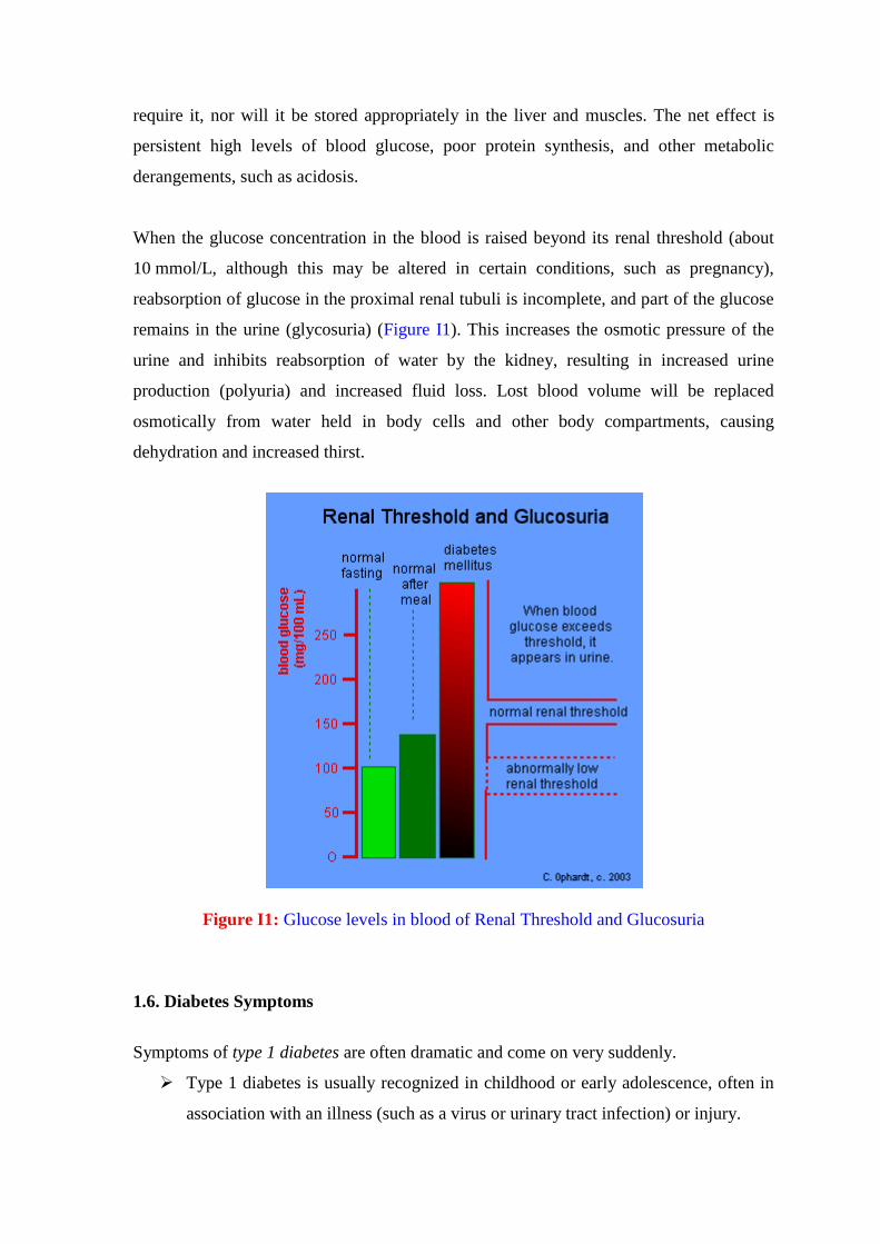
When the glucose concentration in the blood is raised beyond its renal threshold (about
10 mmol/L, although this may be altered in certain conditions, such as pregnancy),
reabsorption of glucose in the proximal renal tubuli is incomplete, and part of the glucose
remains in the urine (glycosuria) (Figure I1). This increases the osmotic pressure of the
urine and inhibits reabsorption of water by the kidney, resulting in increased urine
production (polyuria) and increased fluid loss. Lost blood volume will be replaced
osmotically from water held in body cells and other body compartments, causing
dehydration and increased thirst.
Figure I1: Glucose levels in blood of Renal Threshold and Glucosuria
1.6. Diabetes Symptoms
Symptoms of type 1 diabetes are often dramatic and come on very suddenly.
Type 1 diabetes is usually recognized in childhood or early adolescence, often in
association with an illness (such as a virus or urinary tract infection) or injury.
The extra stress can cause diabetic ketoacidosis.
Symptoms of ketoacidosis include nausea and vomiting. Dehydration and
often-serious disturbances in blood levels of potassium follow.
Without treatment, ketoacidosis can lead to coma and death.
Symptoms of type 2 diabetes are often subtle and may be attributed to aging or obesity.
A person may have type 2 diabetes for many years without knowing it.
People with type 2 diabetes can develop hyperglycemic hyperosmolar nonketotic
syndrome.
Type 2 diabetes can be precipitated by steroids and stress.
If not properly treated, type 2 diabetes can lead to complications such as
blindness, kidney failure, heart disease, and nerve damage.
Common symptoms of both type 1 and type 2 diabetes include:
Fatigue, constantly tired: In diabetes, the body is inefficient and sometimes unable
to use glucose for fuel. The body switches over to metabolizing fat, partially or
completely, as a fuel source. This process requires the body to use more energy.
The end result is feeling fatigued or constantly tired.
Unexplained weight loss: People with diabetes are unable to process many of the
calories in the foods they eat. Thus, they may lose weight even though they eat an
apparently appropriate or even an excessive amount of food. Losing sugar and
water in the urine and the accompanying dehydration also contributes to weight
loss.
Excessive thirst (polydipsia): A person with diabetes develops high blood sugar
levels, which overwhelms the kidney's ability to reabsorb the sugar as the blood is
filtered to make urine. Excessive urine is made as the kidney spills the excess
sugar. The body tries to counteract this by sending a signal to the brain to dilute
the blood, which translates into thirst. The body encourages more water
consumption to dilute the high blood sugar back to normal levels and to
compensate for the water lost by excessive urination.
Excessive urination (polyuria): Another way the body tries to rid the body of the
extra sugar in the blood is to excrete it in the urine. This can also lead to
dehydration because a large amount of water is necessary to excrete the sugar.
Excessive eating (polyphagia): If the body is able, it will secrete more insulin in
order to try to manage the excessive blood sugar levels. Moreover, the body is
resistant to the action of insulin in type 2 diabetes. One of the functions of insulin
is to stimulate hunger. Therefore, higher insulin levels lead to increased hunger.
Despite increased caloric intake, the person may gain very little weight and may
even lose weight.
Poor wound healing: High blood sugar levels prevent white blood cells, which are
important in defending the body against bacteria and also in cleaning up dead
tissue and cells, from functioning normally. When these cells do not function
properly, wounds take much longer to heal and become infected more frequently.
Long-standing diabetes also is associated with thickening of blood vessels, which
prevents good circulation, including the delivery of enough oxygen and other
nutrients to body tissues.
Infections: Certain infections, such as frequent yeast infections of the genitals, skin
infections, and frequent urinary tract infections, may result from suppression of
the immune system by diabetes and by the presence of glucose in the tissues
which allow bacteria to grow. These infections can also be an indicator of poor
blood sugar control in a person known to have diabetes.
Altered mental status: Agitation, unexplained irritability, inattention, extreme
lethargy, or confusion can all be signs of very high blood sugar, ketoacidosis,
hyperosmolar hyperglycemia nonketotic syndrome, or hypoglycemia (low sugar).
Thus, any of these merit the immediate attention of a medical professional.
Blurry vision: Blurry vision is not specific for diabetes but is frequently present
with high blood sugar levels.
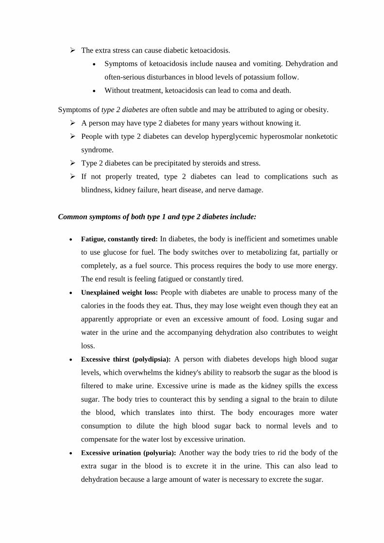
A pictorial representation of the organs (with their location in our body) involved with
the symptoms of diabetes in given in figure I2.
Figure I2: Body organs involved with symptoms of diabetes
1.7. Diagnosis of Diabetes
Physicians prescribe special tests in diagnosing diabetes and also in monitoring blood
sugar level control in known diabetics.
A number of laboratory tests are available to confirm the diagnosis of diabetes.
Finger stick blood glucose: This is a rapid screening test that may be performed anywhere,
including community-based screening programs.
Although a not as accurate as testing the patient's blood in the hospital laboratory,
a fingerstick blood glucose test but is easy to perform, and the result is available
right away.
The test involves sticking the patient's finger for a blood sample, which is then
placed on a strip. The strip goes into a machine that reads the blood sugar level.
These machines are only accurate to within about 10%-20% of true laboratory
values.
Fingerstick blood glucose values tend to be most inaccurate at very high or very
low levels, so this test is only a preliminary screening study. Fingerstick is the
way most people with diabetes monitor their blood sugar levels at home.
Fasting plasma glucose: The patient will be asked to eat or drink nothing for 8 hours
before having blood drawn (usually first thing in the morning). If the blood glucose level
is greater than or equal to 126 mg/dL (without eating anything), they probably have
diabetes.
If the result is abnormal, the fasting plasma glucose test may be repeated on a
different day to confirm the result, or the patient may undergo an oral glucose
tolerance test or a glycosylated hemoglobin test (often called "hemoglobin A1c")
as a confirmatory test.
If fasting plasma glucose level is greater than 100 but less than 126 mg/dL, then
the patient has what is called impaired fasting glucose, or IFG. This is considered
to be prediabetes. These patients do not have diabetes, but they are at high risk of
developing diabetes in the near future.
Oral glucose tolerance test: This test involves drawing blood for a fasting plasma glucose
test, then drawing blood for a second test at two hours after drinking a very sweet drink
containing up to 75 grams of sugar.
If the blood sugar level after the sugar drink is greater than or equal to 200 mg/dL,
the patient has diabetes.
If the blood glucose level is between 140 and 199, then the patient has impaired
glucose tolerance (IGT), which is also a prediabetic condition.
Glycosylated hemoglobin or hemoglobin A1c: This test is a measurement of how high the
blood sugar levels have been over approximately the last 120 days (the average life-span
of the red blood cells on which the test is based). Glycated hemoglobin is better than
fasting glucose for determining risks of cardiovascular disease and death from any cause.
Excess blood glucose hooks itself on to the hemoglobin in red blood cells and
stays there for the life of the red blood cell.
The percentage of hemoglobin that has had excess blood sugar attached to it can
be measured in the blood. The test involves having a small amount of blood
drawn.
A hemoglobin A1c test is the best measurement of blood sugar control in people
known to have diabetes. A hemoglobin A1c result of 7% or less indicates good
glucose control. A result of 8% or greater indicates that blood sugar levels are too
high, too much of the time.
The hemoglobin A1c test is the best test for diabetes follow-up care, than to
diagnose diabetes. Still, a hemoglobin A1c result greater than 6.1% is highly
suggestive of diabetes. Generally, a confirmatory test would be needed before
diagnosing diabetes.
The hemoglobin A1c test is generally measured about every 3 to 6 months for
people with known diabetes, although it may be done more frequently for people
who are having difficulty achieving and maintaining good blood sugar control.
This test is not used for people who do not have diabetes or are not at increased
risk of diabetes.
Normal values may vary from laboratory to laboratory, although an effort is under
way to standardize how measurements are performed.
Special attention is paid to history including information about the patient's symptoms,
risk factors for diabetes, past medical problems, current medications, allergies to
medications, family history of diabetes, or other medical problems such as high
cholesterol or heart disease, and personal habits and lifestyle.
The limiting values of the parameters of the pathological tests for the diagnosis of DM are
listed in table I3.
Table I3: Diagnosis criteria for Diabetes Mellitus
Condition 2 hour glucose
mmol/l(mg/dl)
Fasting glucose
mmol/l(mg/dl)
HbA1c
%
Normal <7.8 (<140) <6.1 (<110) <6.0
Prediabetes
Impaired
fasting
glycaemia
<7.8 (<140) ≥ 6.1(≥110) &
<7.0(<126) 6.0-6.4
Impaired
glucose
tolerance
≥7.8 (≥140) <7.0 (<126) 6.0-6.4
Diabetes mellitus ≥11.1 (≥200) ≥7.0 (≥126) ≥6.5
1.8. Complications of diabetes
Both type 1 and type 2 diabetes ultimately lead to high blood sugar levels, a condition
called hyperglycemia. Over a long period of time, hyperglycemia damages the retina of
the eye, the blood vessels of the kidneys, the nerves, and other blood vessels.
Damage to the retina from diabetes (diabetic retinopathy) is a leading cause of
blindness.
Damage to the kidneys from diabetes (diabetic nephropathy) is a leading cause of
kidney failure.
Damage to the nerves from diabetes (diabetic neuropathy) is a leading cause of
foot wounds and ulcers, which frequently lead to foot and leg amputations.
Damage to the nerves in the autonomic nervous system can lead to paralysis of the
stomach (gastroparesis), chronic diarrhea, and an inability to control heart rate and
blood pressure during postural changes.
Diabetes accelerates atherosclerosis, (the formation of fatty plaques inside the
arteries), which can lead to blockages or a clot (thrombus). Such changes can then
lead to heart attack, stroke, and decreased circulation in the arms and legs
(peripheral vascular disease).
Diabetes predisposes people to elevated blood pressure, high levels of cholesterol
and triglycerides. These conditions both independently and together with
hyperglycemia, increase the risk of heart disease, kidney disease, and other blood
vessel complications.
Diabetes can contribute to a number of acute (short-lived) medical problems.
Many infections are associated with diabetes, and infections are frequently more
dangerous in someone with diabetes because the body's normal ability to fight
infections is impaired. To compound the problem, infections may worsen glucose
control, which further delays recovery from infection.
Hypoglycemia or low blood sugar, occurs intermittently in most people with
diabetes. It can result from taking too much diabetes medication or insulin
(sometimes called an insulin reaction), missing a meal, exercising more than
usual, drinking too much alcohol, or taking certain medications for other
conditions. It is very important to recognize hypoglycemia and be prepared to
treat it at all times. Headache, feeling dizzy, poor concentration, tremor of the
hands, and sweating are common symptoms of hypoglycemia. A person can faint
or have a seizure if blood sugar level becomes too low.
Diabetic ketoacidosis (DKA) is a serious condition in which uncontrolled
hyperglycemia (usually due to complete lack of insulin or a relative deficiency of
insulin) over time creates a buildup of ketones (acidic waste products) in the
blood. High levels of ketones can be very harmful. This typically happens to
people with type 1 diabetes who do not have good blood glucose control. Diabetic
ketoacidosis can be precipitated by infection, stress, trauma, missing medications
like insulin, or medical emergencies such as a stroke and heart attack.
Hyperosmolar hyperglycemic nonketotic syndrome is a serious condition in
which the blood sugar level gets very high. The body tries to get rid of the excess
blood sugar by eliminating it in the urine. This increases the amount of urine
significantly, and often leads to dehydration so severe that it can cause seizures,
coma, and even death. This syndrome typically occurs in people with type 2
diabetes who are not controlling their blood sugar levels, who have become
dehydrated, or who have stress, injury, stroke, or are taking certain medications,
like steroids.
1.9. Diabetes Prognosis
Diabetes is a leading cause of death in all industrialized nations. Overall, the risk of
premature death of people with diabetes is twice that of people who do not have diabetes.
Prognosis depends on the type of diabetes, degree of blood sugar control, and
development of complications.
With India having the highest number of diabetic patients in the world, the sugar disease
is posing an enormous health problem in the country.23,24,25
Calling India the „diabetes
capital of the world‟, the International Journal of Diabetes in Developing Countries has
said that there is alarming rise in prevalence of diabetes, which has gone beyond epidemic
form to a pandemic one.
The International Diabetes Federation estimates that the number of diabetic patients in
India more than doubled from 19 million in 1995 to 40.9 million in 2007. It is projected
to increase to 69.9 million by 2025. Currently, up to 11 per cent of India‟s urban
population and 3 per cent of rural population above the age of 15 has diabetes. Diabetes
affects all people in the society, not just those who live with it. The World Health
Organization estimates that mortality from diabetes and heart disease cost India about
$210 billion every year and is expected to increase to $335 billion in the next ten years.
These estimates are based on lost productivity, resulting primarily from premature death.
The most prevalent is the Type 2 diabetes, which constitutes 95 per cent of the diabetic
population in the country. 26
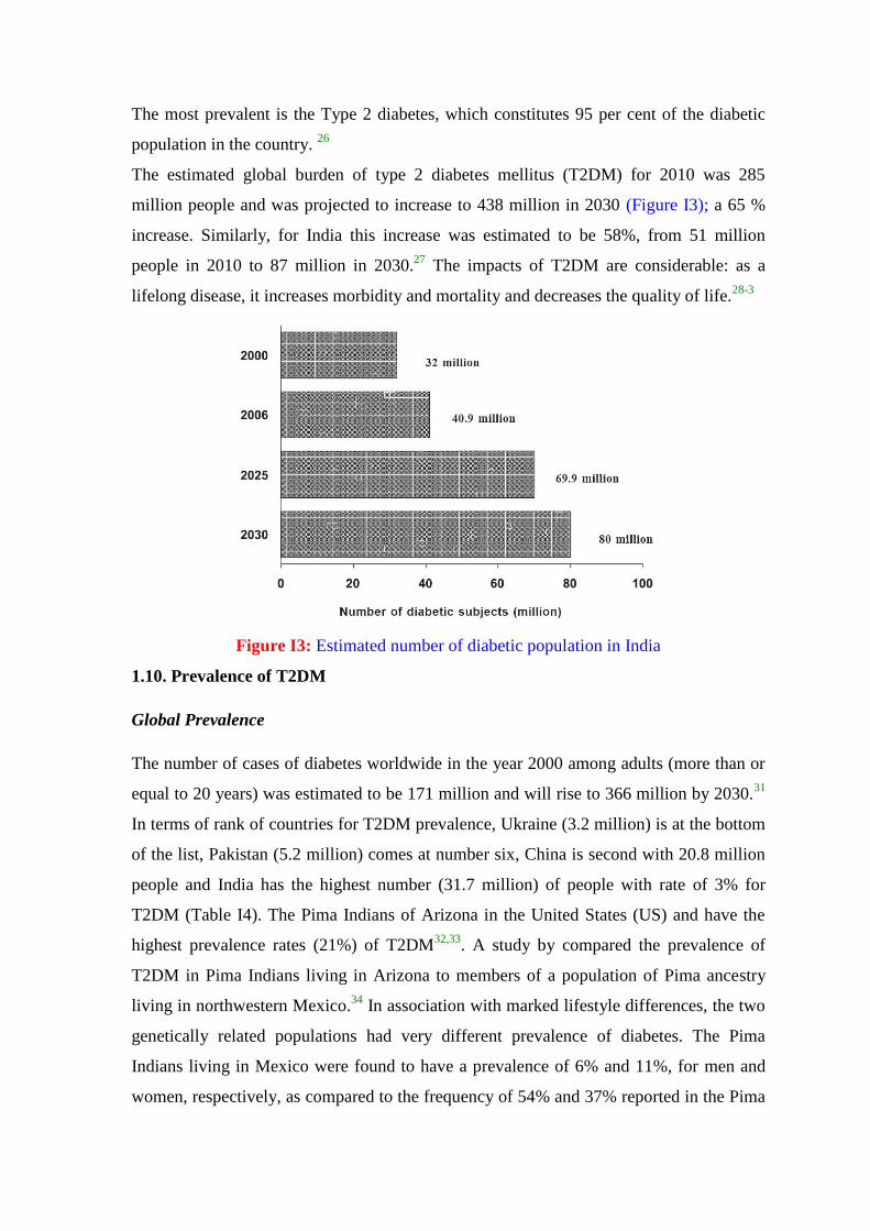
The estimated global burden of type 2 diabetes mellitus (T2DM) for 2010 was 285
million people and was projected to increase to 438 million in 2030 (Figure I3); a 65 %
increase. Similarly, for India this increase was estimated to be 58%, from 51 million
people in 2010 to 87 million in 2030.27
The impacts of T2DM are considerable: as a
lifelong disease, it increases morbidity and mortality and decreases the quality of life.28-3
Figure I3: Estimated number of diabetic population in India
1.10. Prevalence of T2DM
Global Prevalence
The number of cases of diabetes worldwide in the year 2000 among adults (more than or
equal to 20 years) was estimated to be 171 million and will rise to 366 million by 2030.31
In terms of rank of countries for T2DM prevalence, Ukraine (3.2 million) is at the bottom
of the list, Pakistan (5.2 million) comes at number six, China is second with 20.8 million
people and India has the highest number (31.7 million) of people with rate of 3% for
T2DM (Table I4). The Pima Indians of Arizona in the United States (US) and have the
highest prevalence rates (21%) of T2DM32,33
. A study by compared the prevalence of
T2DM in Pima Indians living in Arizona to members of a population of Pima ancestry
living in northwestern Mexico.34
In association with marked lifestyle differences, the two
genetically related populations had very different prevalence of diabetes. The Pima
Indians living in Mexico were found to have a prevalence of 6% and 11%, for men and
women, respectively, as compared to the frequency of 54% and 37% reported in the Pima
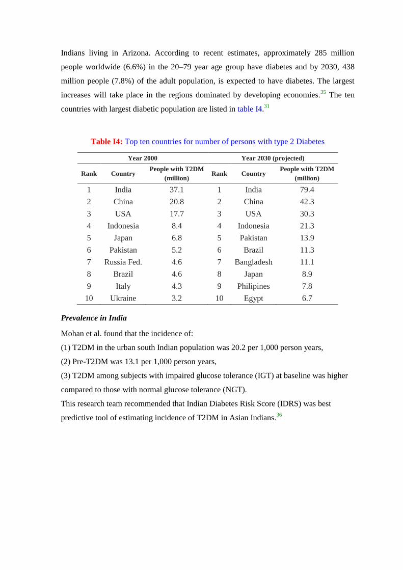
Indians living in Arizona. According to recent estimates, approximately 285 million
people worldwide (6.6%) in the 20–79 year age group have diabetes and by 2030, 438
million people (7.8%) of the adult population, is expected to have diabetes. The largest
increases will take place in the regions dominated by developing economies.35
The ten
countries with largest diabetic population are listed in table I4.31
Table I4: Top ten countries for number of persons with type 2 Diabetes
Year 2000 Year 2030 (projected)
Rank Country People with T2DM
(million) Rank Country
People with T2DM
(million)
1 India 37.1 1 India 79.4
2 China 20.8 2 China 42.3
3 USA 17.7 3 USA 30.3
4 Indonesia 8.4 4 Indonesia 21.3
5 Japan 6.8 5 Pakistan 13.9
6 Pakistan 5.2 6 Brazil 11.3
7 Russia Fed. 4.6 7 Bangladesh 11.1
8 Brazil 4.6 8 Japan 8.9
9 Italy 4.3 9 Philipines 7.8
10 Ukraine 3.2 10 Egypt 6.7
Prevalence in India
Mohan et al. found that the incidence of:
(1) T2DM in the urban south Indian population was 20.2 per 1,000 person years,
(2) Pre-T2DM was 13.1 per 1,000 person years,
(3) T2DM among subjects with impaired glucose tolerance (IGT) at baseline was higher
compared to those with normal glucose tolerance (NGT).
This research team recommended that Indian Diabetes Risk Score (IDRS) was best
predictive tool of estimating incidence of T2DM in Asian Indians.36
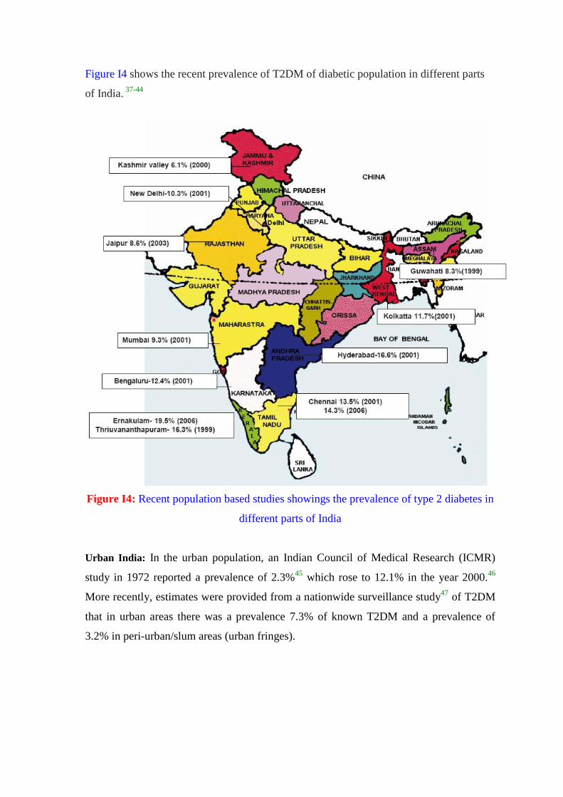
Figure I4 shows the recent prevalence of T2DM of diabetic population in different parts
of India. 37-44
Figure I4: Recent population based studies showings the prevalence of type 2 diabetes in
different parts of India
Urban India: In the urban population, an Indian Council of Medical Research (ICMR)
study in 1972 reported a prevalence of 2.3%45
which rose to 12.1% in the year 2000.46
More recently, estimates were provided from a nationwide surveillance study47
of T2DM
that in urban areas there was a prevalence 7.3% of known T2DM and a prevalence of
3.2% in peri-urban/slum areas (urban fringes).
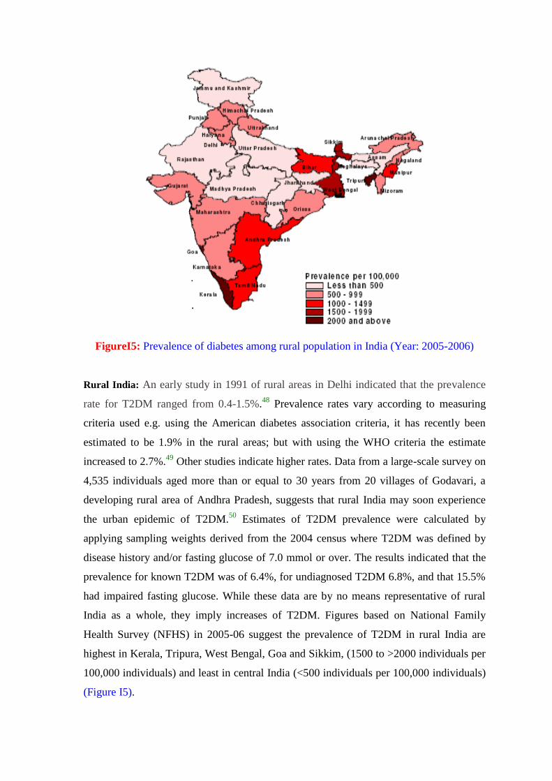
FigureI5: Prevalence of diabetes among rural population in India (Year: 2005-2006)
Rural India: An early study in 1991 of rural areas in Delhi indicated that the prevalence
rate for T2DM ranged from 0.4-1.5%.48
Prevalence rates vary according to measuring
criteria used e.g. using the American diabetes association criteria, it has recently been
estimated to be 1.9% in the rural areas; but with using the WHO criteria the estimate
increased to 2.7%.49
Other studies indicate higher rates. Data from a large-scale survey on
4,535 individuals aged more than or equal to 30 years from 20 villages of Godavari, a
developing rural area of Andhra Pradesh, suggests that rural India may soon experience
the urban epidemic of T2DM.50
Estimates of T2DM prevalence were calculated by
applying sampling weights derived from the 2004 census where T2DM was defined by
disease history and/or fasting glucose of 7.0 mmol or over. The results indicated that the
prevalence for known T2DM was of 6.4%, for undiagnosed T2DM 6.8%, and that 15.5%
had impaired fasting glucose. While these data are by no means representative of rural
India as a whole, they imply increases of T2DM. Figures based on National Family
Health Survey (NFHS) in 2005-06 suggest the prevalence of T2DM in rural India are
highest in Kerala, Tripura, West Bengal, Goa and Sikkim, (1500 to >2000 individuals per
100,000 individuals) and least in central India (<500 individuals per 100,000 individuals)
(Figure I5).
1.11. Economic Burden of Diabetes in India
Despite diabetes being a life-long disorder and is expensive to manage and treat for the
large proportion of subjects in developing societies, there is lack of data on its economic
burden in India. In the Indian context the financial burden is often shared by relatives of
the patients.51
The health care budget of the government in India is a meager 2%52
compared to 14% to defense.53
The total amount needed for India to treat T2DM is
estimated to around 2.2 billion USD.51
In India the direct medical cost to identify one
subject with insulin glucose tolerance is INR 5,278.54
The cost of insulin amounts to
350.00 USD (16,000 Indian Rupees) per year, while medication for non-insulin-requiring
patients costs about 70.00 USD per year.55
In the Indian context these costs are
prohibitive: 75.5% of the Indian population is earning less than $2 per day and 41.6% less
than $1.25 per day.55
To determine the direct cost of ambulatory diabetes care, to evaluate
the socio-demographic associates of spending, and to ascertain the relationship of
spending with the delivered quality of diabetes care; the community based data available
from the middle and high income groups in Delhi (DEDICOM survey) was analyzed and
it was concluded from the study that a majority of diabetes patients spend a significant
proportion of their family income on diabetes related expenditure (~Rs. 6000 i.e. ~US$
150) per year.56
The cost is higher for subjects with longer duration since diagnosis, those
with higher education or income, those with co-morbidities and those requiring oral
hypoglycemic agents or insulin. In developing countries like India, the brunt of diabetes
and cardiovascular disease occurs among the economically productive age group (20-45
year olds).57
Diabetes mellitus is responsible for 1157 thousand years of life lost due to
the disease, and for 2263 thousand DALYs during 2004.58
The World Health
Organization estimates that mortality from diabetes and heart disease cost India about
$210 billion every year and is expected to increase to $335 billion in the next ten years.
These estimates are based on lost productivity, resulting primarily from premature
death.26
Overall cost during the course of treatment of T2DM borne by in and out-patient subjects
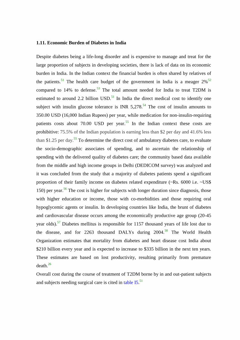
and subjects needing surgical care is cited in table I5.51
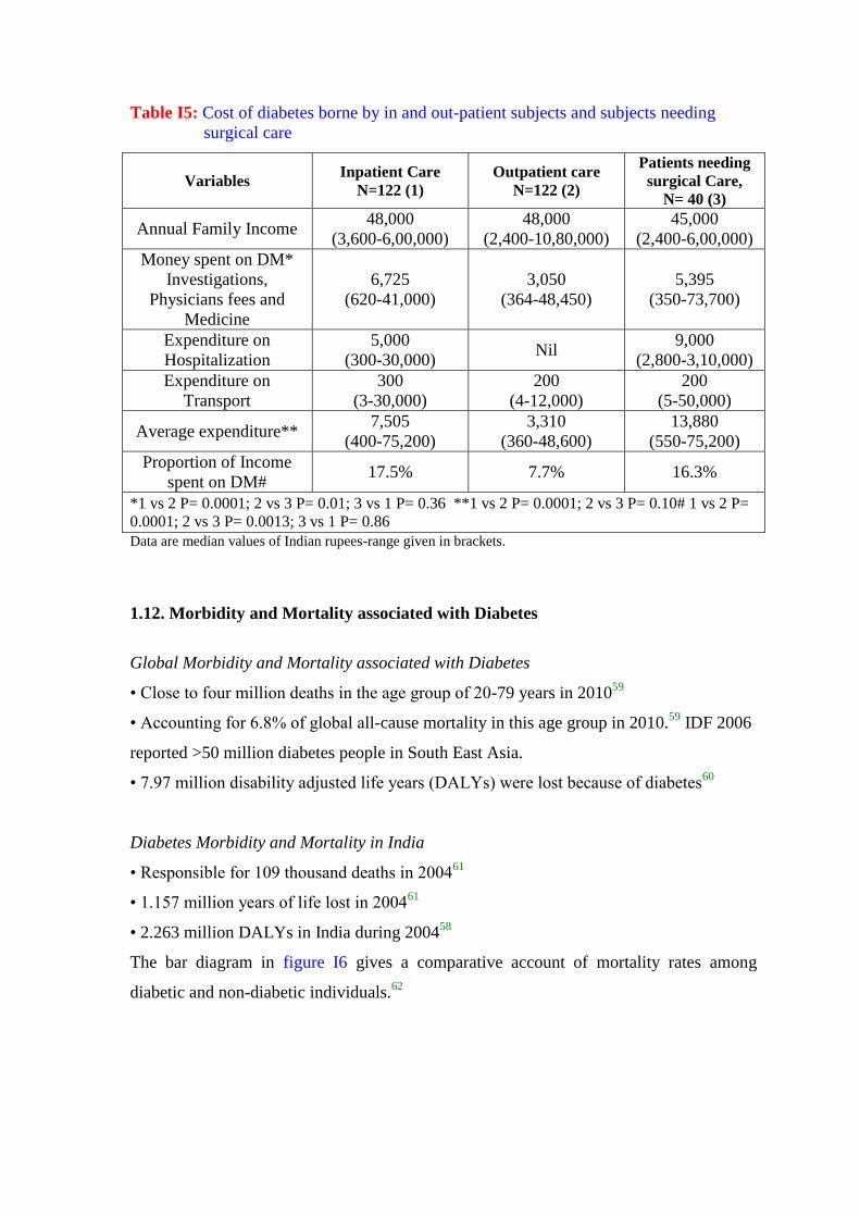
Table I5: Cost of diabetes borne by in and out-patient subjects and subjects needing
surgical care
Variables Inpatient Care
N=122 (1)
Outpatient care
N=122 (2)
Patients needing
surgical Care,
N= 40 (3)
Annual Family Income 48,000
(3,600-6,00,000)
48,000
(2,400-10,80,000)
45,000
(2,400-6,00,000)
Money spent on DM*
Investigations,
Physicians fees and
Medicine
6,725
(620-41,000)
3,050
(364-48,450)
5,395
(350-73,700)
Expenditure on
Hospitalization
5,000
(300-30,000) Nil
9,000
(2,800-3,10,000)
Expenditure on
Transport
300
(3-30,000)
200
(4-12,000)
200
(5-50,000)
Average expenditure** 7,505
(400-75,200)
3,310
(360-48,600)
13,880
(550-75,200)
Proportion of Income
spent on DM# 17.5% 7.7% 16.3%
*1 vs 2 P= 0.0001; 2 vs 3 P= 0.01; 3 vs 1 P= 0.36 **1 vs 2 P= 0.0001; 2 vs 3 P= 0.10# 1 vs 2 P=
0.0001; 2 vs 3 P= 0.0013; 3 vs 1 P= 0.86
Data are median values of Indian rupees-range given in brackets.
1.12. Morbidity and Mortality associated with Diabetes
Global Morbidity and Mortality associated with Diabetes
• Close to four million deaths in the age group of 20-79 years in 201059
• Accounting for 6.8% of global all-cause mortality in this age group in 2010.59
IDF 2006
reported >50 million diabetes people in South East Asia.
• 7.97 million disability adjusted life years (DALYs) were lost because of diabetes60
Diabetes Morbidity and Mortality in India
• Responsible for 109 thousand deaths in 200461
• 1.157 million years of life lost in 200461
• 2.263 million DALYs in India during 200458
The bar diagram in figure I6 gives a comparative account of mortality rates among
diabetic and non-diabetic individuals.62
Figure I6: Differences in mortality rates among diabetic and non-diabetic individuals
1.13. Treatment of Diabetes
While many measures were tried, effective treatment was not developed until the early
part of the 20th century, when Canadians Frederick Banting and Charles Best developed
insulin in 1921 and 1922.63
This was followed by the development of the long-acting
insulin NPH in the 1940s.2
The treatment of diabetes is highly individualized, depending on the type of diabetes,
whether the patient has other active medical problems, whether the patient has
complications of diabetes, and age and general health of the patient at time of diagnosis.
A health care professional will set goals for lifestyle changes, blood sugar control,
and treatment.
Together, the patient and the health care professional will formulate a plan to help
meet those goals.
Education about diabetes and its treatment is essential in all types of diabetes.
When the patient is first diagnosed with diabetes, the diabetes care team will
spend a lot of time with the patient, teaching them about their condition,
treatment, and everything they need to know to care for themselves on a daily
basis.
The diabetes care team includes the health care professional and his or her staff. It
may include specialists in foot care, neurology, kidney diseases, and eye diseases.
A professional dietitian and a diabetes educator also may be part of the team.
1.13.1. Type 1 diabetes
Treatment of diabetes almost always involves the daily injection of insulin, usually a
combination of short-acting insulin (for example, lispro [Humalog] or aspart [NovoLog])
and longer acting insulin (for example, NPH, Lente, glargine [Lantus], detemir
[Levemir]).
Insulin must be given as an injection just under the skin. If taken by mouth,
insulin would be destroyed in the stomach before it could get into the blood where
it is needed.
Most people with type 1 diabetes give these injections to themselves. Even if
someone else usually gives the patient injections, it is important that the patient
knows how to do it in case the other person is unavailable.
The patient should be trained to store and inject the insulin. Insulin is usually
given in two or three injections per day, generally around mealtimes. Dosage is
individualized and is tailored to the patient's specific needs by the health care
professional. Longer acting insulins are typically administered one or two times
per day.
Some people have their insulin administered by continuous infusion pumps to
provide adequate blood glucose control. Supplemental mealtime insulin is
programmed into the pump by the individual as recommended by his or her health
care professionals.
It is very important to eat after the taking insulin, as the insulin will lower blood
sugar regardless of whether the person has eaten. If insulin is taken without eating,
the result may be hypoglycemia. This is called an insulin reaction.
There is an adjustment period while the patient learns how insulin affects them,
and how to time meals and exercise with insulin injections to keep blood sugar
levels as even as possible.
Keeping accurate records of blood sugar levels and insulin dosages is crucial for
the patient's diabetes management.
Eating a consistent, healthy diet appropriate for the patient's size and weight is
essential in controlling blood sugar level.
1.13.2. Type 2 diabetes
Depending on how elevated the patient's blood sugar and glycosylated hemoglobin
(HbA1c) are at the time of diagnosis, they may be given a chance to lower blood sugar
levels through lifestyle changes, without medication.
The best way to do this is to lose weight if the patient is obese, and begin an
exercise program.
This will generally be tried for 3 to 6 months, then blood sugar and glycosylated
hemoglobin will be rechecked. If they remain high, the patient will be started on
an oral medication, usually a sulfonylurea or biguanide (metformin
[Glucophage]), to help control blood sugar levels.
Even if the patient is on medication, it is still important to eat a healthy diet, lose
weight if they are overweight, and engage in moderate physical activity as often as
possible.
The health care professional will initially monitor the patient's progress on
medication very carefully. It is important to receive just the right dose of the right
medication, to regulate blood sugar levels in the recommended range with the
fewest side effects.
The doctor may decide to combine two types of medications to achieve blood
sugar levels control.
Gradually, even people with type 2 diabetes may require insulin injections to
control their blood sugar levels.
It is becoming more common for people with type 2 diabetes to take a
combination of oral medication and insulin injections to control blood sugar
levels.
1.14. Diabetic Medications (Therapeutic Intervention for Hyperglycemia)
Many, if not all, of the vascular consequences of insulin resistance are due to the
persistent hyperglycemia seen in T2D. For these reason a major goal of therapeutic
intervention in T2D is to reduce circulating glucose levels. Many different types of
medications are available to help lower blood sugar levels in people with type 2 diabetes.
Each type works in a different way. There are many pharmacologic strategies to
accomplish these goals. It is very common to combine two or more types to get the best
effect with fewest side effects.
Sulfonylureas: They are oral hypoglycemic drugs and are referred to as endogenous
insulin secretagogues because they induce pancreatic release of endogenous insulin.16
The first generation sulfonylureas (tolbutamide, acetohexamide, chlorpropamide and
tolazamide) are not routinely prescribed in the US.16
The second generation
sulfonylureas include glipizide, glimipiride and glyburide. Because all of these drugs
can induce pronounced hypoglycemia, treatment is initiated with the lowest possible
dose and carefully monitored until the dose is found that results in a fasting plasma
glucose (FPG) of 110-140 mg/dL. Sulfonylureas function by binding to and inhibiting
the pancreatic ATP-dependent potassium channel that is normally involved in glucose
mediated insulin secretion. They have no significant effects on circulating
triglycerides lipoproteins or cholesterol.
Biguanides: They are a class of oral hypoglycemic drugs that function to lower serum
glucose levels by enhancing insulin-mediated suppression of hepatic glucose
production and enhancing insulin-stimulated glucose uptake by skeletal muscle.
Metformin (Glucophage) is a member of this class and is currently the most widely
prescribed insulin-sensitizing drug in current clinical use. Metformin administration
does not lead to increased insulin release from the pancreas and as such the risk of
hypoglycemia is minimal. Because the major site of action for metformin is the liver
its use can be contraindicated in patients with liver dysfunction. The drug is ideal for
obese patients and for younger T2 diabetics.
Evidence on the mode of action (MOA) of metformin shows that it improves insulin
sensitivity by increasing insulin receptor tyrosine kinase activity, enhancing glycogen
synthesis and increasing recruitment and transport of GLUT4 transporters to the
plasma membrane. Additionally, it has been shown that metformin affects
mitochondrial activities dependent upon the model system studies. Metformin has a
mild inhibitory effect on complex I of oxidative phosphorylation, has antioxidant
properties, and activates both glucose 6-phospahte dehydrogenase (G6PDH) and
AMP-activated protein kinase (AMPK). The importance of AMPK metformin action
stems from the role of AMPK in the regulation of both lipid and carbohydrate
metabolism. In adipose tissue, metformin inhibits lipolysis while enhancing re-
esterification of fatty acids.
In adolescent females with T2D, the use if metformin is highly recommended to
reduce the incidence of as well as the potential for polycystic ovarian syndrome,
(PCOS). PCOS is brought on by the hyperinsulinemia if T2D. insulin effects on the
ovary drive conversion of progesterone to testosterone and a reduction in serum
hormone globulin (SHBG). Taken together, the effects of hyperinsulinemia leads to a
hyperandrogenic state in the ovary resulting in follicular atresis and ovulatory
dysfunction.
Alpha-glucosidase inhibitors: These oral hypoglycemic agents slow absorption of the
starches a person eats. This slows down glucose production. Alpha-glucosidase
inhibitors like acarbose (Precose) and miglitol (Glyset) function by interfering with
the action of the α-glucosidases present in the small intestinal brush border. The
consequence of this inhibition is a reduction in digestion and the consequent
absorption of glucose into the systemic circulation. The reduction in glucose uptake
allows the pancreatic β-cells to more effectively regulate insulin secretion. The
advantage to the use of the α-glucosidase inhibitors is that they function locally in the
intestine and have no major systemic action. Hypoglycemia does not usually occur
with the use of α-glucosidase inhibitors but they are effective at reducing fasting
plasma glucose (FPG) levels and levels of glycosylated haemoglobin (HbA1c). the
adverse side effects of these inhibitors are abdominal bloating and discomfort,
diarrhea and flatulence.16
Thiazolidinediones (TZDs): These oral hypoglycemic agents increase sensitivity to
insulin. The TZDs such as troglitazone (Rezulin: Warner Lambert Co. but this drug
was oluntarily removed from the market due to liver damage risk64
), rosiglitazone
(Avandia: Glaxo Smithkline) and pioglitazone (Actos: Eli Lilly and Co.) have been
proven useful in treating the hyperglycemia associated with insulin-resistance in both
T2D and nondiabetic conditions (though these have also been withdrawn in many
countries owing to there cardiovascular adverse effects65
). The TZDs function as
agonist for the nuclear receptor peroxisome proliferator activated repceptor-γ
(PPARγ). The net effect of the TZDs is a potentiation of the actions of insulin in liver,
adipose tissue and skeletal muscle, increased peripheral glucose disposal and a
decrase in glucose output by the liver.
Meglitinides: These oral hypoglycemic agents stimulate the pancreas to make more
insulin. The meglitinides repaglinide (Prandin) and nateglinide (Starlix) are non
sulfonylurea insulin secretogogues that are both fast acting and of short duration. Like
the sulfonylureas meglitinide therapy results in significant reduction in FPG as well as
HbA1c. The MOA of meglitinide is initiated by binding to a receptor on the pancreatic
β-cell that is distinct from the receptors for the sulfonylureas. However, meglitinides
do exert effects on potassium conductance like the sulfonylureas, the meglitinides
have no direct effects on the circulating levels of plasma lipids.
D-phenylalanine derivatives: These agents stimulate the pancreas to produce more
insulin more quickly.
Amylin synthetic derivatives: Amylin is a naturally occurring hormone secreted by the
pancreas along with insulin. An amylin derivative, such as pramlintide (Symlin), is
indicated when blood sugar control is not achieved despite optimal insulin therapy.
Pramlintide is administered as a subcutaneous injection along with insulin and helps
achieve lower blood sugar levels after meals, helps reduce fluctuation of blood sugar
levels throughout the day, and improves hemoglobin A1C levels.
Insulins: Synthetic human insulin is now the only type of insulin available in the
United States; it is less likely to cause allergic reactions than animal-derived varieties
of insulin used in the past. The type of insulin chosen to customize treatment for an
individual is based on the goal of providing optimal blood sugar control. Different
types of insulin are available and categorized according to their times of action onset
and duration. Commercially prepared mixtures of insulin may also be used to provide
constant (basal) control and immediate control.
Examples of rapid-acting insulins
Regular insulin (Humulin R, Novolin R)
Insulin lispro (Humalog)
Insulin aspart (Novolog)
Insulin glulisine (Apidra)
Prompt insulin zinc (Semilente, slightly slower acting)
Examples of intermediate-acting insulins
Isophane insulin, neutral protamine Hagedorn(NPH) (HumulinN, NovolinN)
Insulin zinc (Lente)
Examples of long-acting insulins
Extended insulin zinc insulin (Ultralente)
Insulin glargine (Lantus)
Insulin detemir (Levemir)
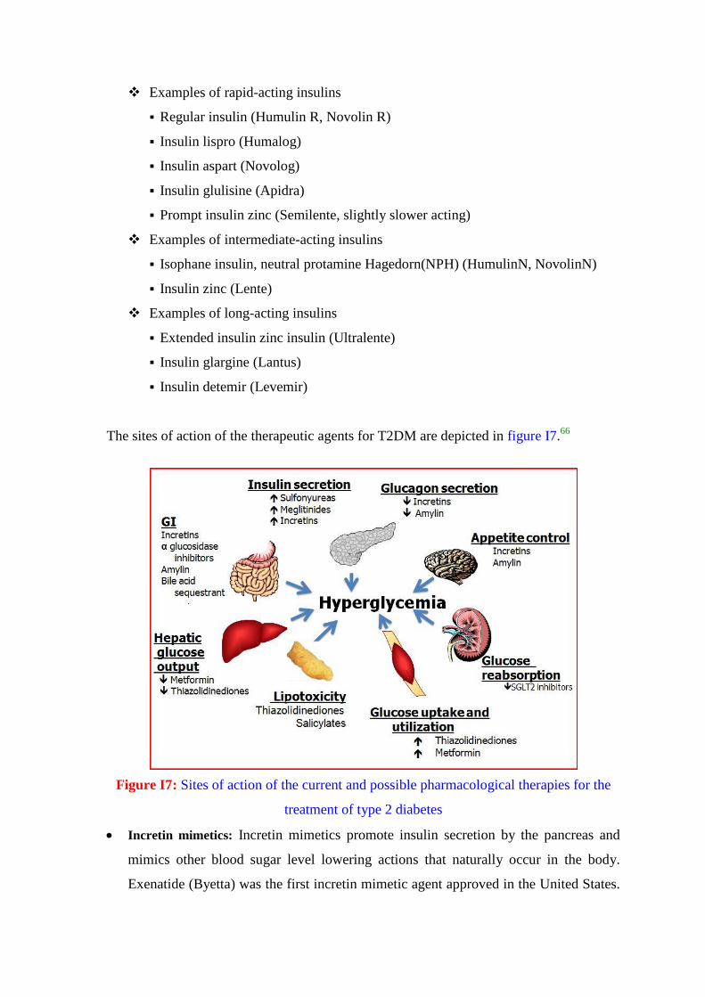
The sites of action of the therapeutic agents for T2DM are depicted in figure I7.66
Figure I7: Sites of action of the current and possible pharmacological therapies for the
treatment of type 2 diabetes
Incretin mimetics: Incretin mimetics promote insulin secretion by the pancreas and
mimics other blood sugar level lowering actions that naturally occur in the body.
Exenatide (Byetta) was the first incretin mimetic agent approved in the United States.
It is indicated for diabetes mellitus type 2 in addition to metformin or a sulfonylurea
when these agents have not attained blood sugar level control alone.
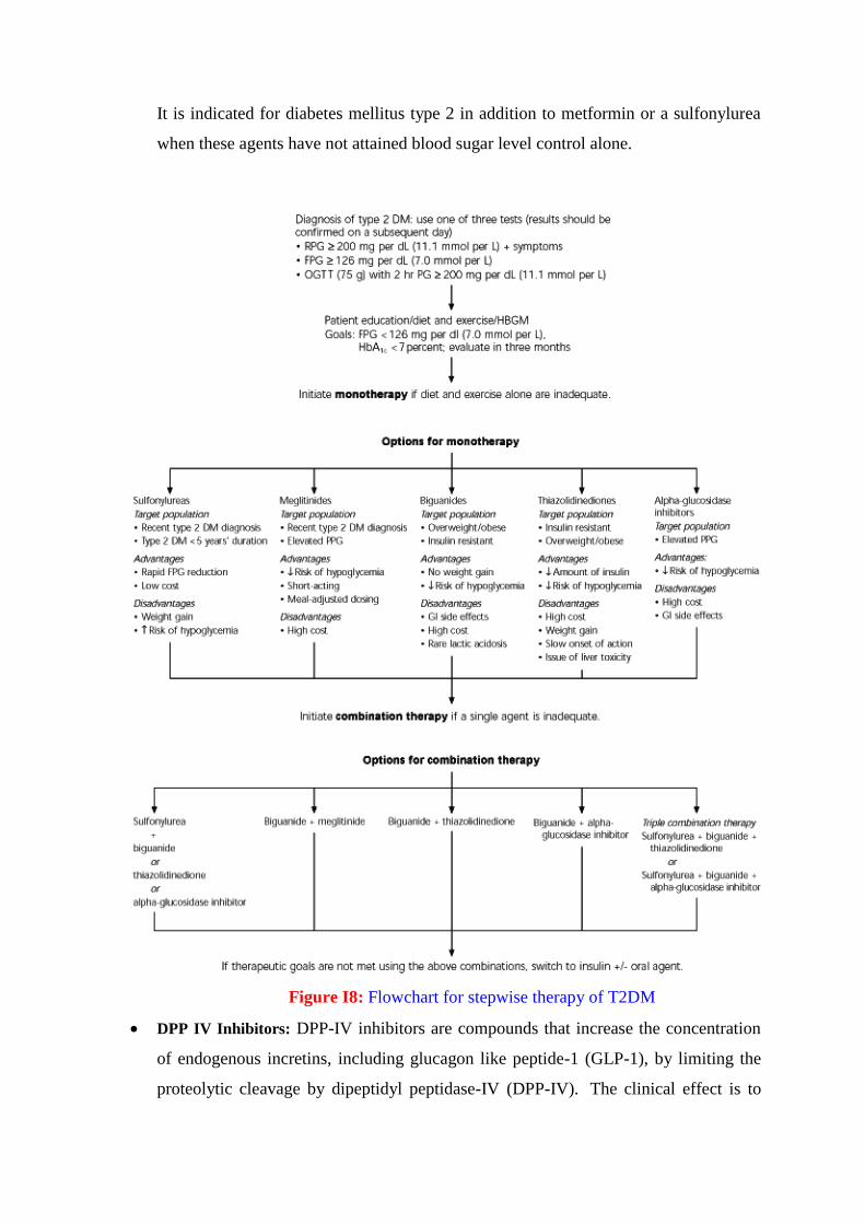
Figure I8: Flowchart for stepwise therapy of T2DM
DPP IV Inhibitors: DPP-IV inhibitors are compounds that increase the concentration
of endogenous incretins, including glucagon like peptide-1 (GLP-1), by limiting the
proteolytic cleavage by dipeptidyl peptidase-IV (DPP-IV). The clinical effect is to
stimulate insulin secretion in a glucose-specific manner and suppress glucagon
secretion. DPP4 inhibitors such as sitagliptin and vildagliptin are novel agents for
treatment of type 2 diabetes. They target both prandial and fasting glucose
concentrations, and work by improving β-cell sensitivity to glucose, whereby it
increases glucose-dependent insulin secretion. Gliptins can be used as monotherapy or
combined with metformin or SUs. Gliptins are largely weight neutral. No serious
adverse events were noted during the clinical trials. Vildagliptin is not recommended
in patients with hepatic impairment. Long-term safety regarding cardiovascular
outcomes needs to be assessed.67
A flowchart of the currently available stepwise clinical therapy for T2DM is given in
figure I8.68
1.15. Peroxisome Proleferator Activated Receptors and T2DM
Type 2 diabetes is characterized by hyperglycemia, insulin resistance, and defects in
insulin secretion and is usually associated with dyslipidemia, hypertension, and obesity.
Although the detailed pathophysiology of this disease remains incompletely understood,
metabolic defects in the liver, pancreatic β-cells, adipose tissue, and skeletal muscle all
contribute to the development of type 2 diabetes. Though long thought to be mainly a
disorder of carbohydrate metabolism, today a great deal of evidence suggests that
abnormalities in fat metabolism play a central role in the pathogenesis of this disease.69-71
Peroxisome proliferator-activated receptors (PPARs) are orphan receptors belonging to
the steroid/thyroid/ retinoid receptor superfamily of ligand-activated transcription factors.
Although cloned only a few decade ago,72,73
the rapid progress in functional analysis of
these receptors has established that the PPARs play a central role in regulating the storage
and catabolism of lipids in both animals and humans (Figure I9). Therefore, much
attention has been paid in the research of structural determination and ligand activation of
these receptors. There are three PPAR subtypes, which are the products of distinct genes
and are commonly designated PPARα [NR1C1], PPARγ [NR1C3], and PPARδ
[NR1C2].74
The PPARs have a protein domain structure (Figure I9) common to other
members of the nuclear receptor gene family. This consists of a variable N-terminal
region that contains the transcriptional activation function 1 domain (AF-1), a highly
conserved DNA-binding domain (DBD), and a ligand-binding domain (LBD) within
which lies a C-terminal region that contains the transcriptional activation function 2
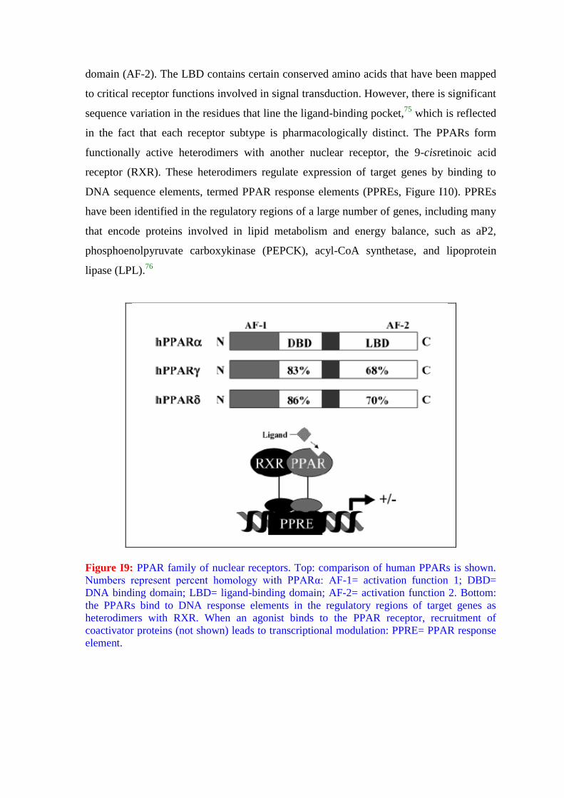
domain (AF-2). The LBD contains certain conserved amino acids that have been mapped
to critical receptor functions involved in signal transduction. However, there is significant
sequence variation in the residues that line the ligand-binding pocket,75
which is reflected
in the fact that each receptor subtype is pharmacologically distinct. The PPARs form
functionally active heterodimers with another nuclear receptor, the 9-cisretinoic acid
receptor (RXR). These heterodimers regulate expression of target genes by binding to
DNA sequence elements, termed PPAR response elements (PPREs, Figure I10). PPREs
have been identified in the regulatory regions of a large number of genes, including many
that encode proteins involved in lipid metabolism and energy balance, such as aP2,
phosphoenolpyruvate carboxykinase (PEPCK), acyl-CoA synthetase, and lipoprotein
lipase (LPL).76
Figure I9: PPAR family of nuclear receptors. Top: comparison of human PPARs is shown.
Numbers represent percent homology with PPARα: AF-1= activation function 1; DBD=
DNA binding domain; LBD= ligand-binding domain; AF-2= activation function 2. Bottom:
the PPARs bind to DNA response elements in the regulatory regions of target genes as
heterodimers with RXR. When an agonist binds to the PPAR receptor, recruitment of
coactivator proteins (not shown) leads to transcriptional modulation: PPRE= PPAR response
element.
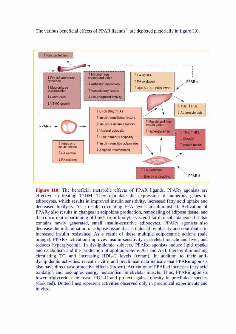
The various beneficial effects of PPAR ligands77
are depicted pictorially in figure I10.
Figure I10: The beneficial metabolic effects of PPAR ligands. PPARγ agonists are
effective in treating T2DM. They modulate the expression of numerous genes in
adipocytes, which results in improved insulin sensitivity, increased fatty acid uptake and
decreased lipolysis. As a result, circulating FFA levels are diminished. Activation of
PPARγ also results in changes in adipokine production, remodeling of adipose tissue, and
the concurrent repartioning of lipids from lipolytic visceral fat into subcutaneous fat that
contains newly generated, small insulin-sensitive adipocytes. PPARγ agonists also
decrease the inflammation of adipose tissue that is induced by obesity and contributes to
increased insulin resistance. As a result of these multiple adipocentric actions (pale
orange), PPARγ activation improves insulin sensitivity in skeletal muscle and liver, and
reduces hyperglycemia. In dyslipidemic subjects, PPARα agonists induce lipid uptake
and catabolism and the production of apolipoproteins A-I and A-II, thereby diminishing
circulating TG and increasing HDL-C levels (cream). In addition to their anti-
dyslipidemic activities, recent in vitro and preclinical data indicate that PPARα agonists
also have direct vasoprotective effects (brown). Activation of PPAR-d increases fatty acid
oxidation and uncouples energy metabolism in skeletal muscle. Thus, PPARδ agonists
lower triglycerides, increase HDL-C and protect against obesity in preclinical species
(dark red). Dotted lines represent activities observed only in preclinical experiments and
in vitro.
1.15.1. PPARα
PPARα plays a pivotal role in the uptake and oxidation of fatty acids and also in
lipoprotein metabolism.78
PPARα is expressed highly in liver, heart and skeletal muscle,
tissues that extract a high level of their energy requirements from lipids.79
During
prolonged fasting that results in hypoglycemia, fatty acids are released from fat depots
and travel to the liver where they are taken up, oxidized and metabolized into ketone
bodies to provide fuel for peripheral tissues. The crucial role of PPARα in mediating
these metabolic processes and, ultimately, energy homeostasis is demonstrated by the
phenotype of PPARα null mice, which, on fasting, are characterized by hypoglycemia,
hypoketonemia, hyperlipidemia and hepatic steatosis.80
Dyslipidemia, characterized by
elevated circulating levels of triglycerides (TGs) in combination with decreased levels of
high-density lipoprotein cholesterol (HDL-C), is often a forerunner of cardiovascular
disease.81
PPARα agonists decrease plasma TG levels and increase HDL-C levels.82
The
former action is mediated by increasing lipid uptake, activation and catabolism through
the transcriptional modulation of numerous genes that control these processes.83
The latter
is mediated, in part, by augmenting hepatic production of apolipoprotein A-I (apoA-I)84
and apoA-II,85
which are major proteinaceous components of HDL-C. Additionally,
PPARα agonists cause favorable changes in the particle size and subclass distribution of
lipoproteins.86
The effects of PPARα agonists (Figure I11) on circulating lipid parameters and, perhaps,
vascular cells are beneficial because these ligands reduce the progression of
atherosclerosis and the incidence of coronary events in major clinical studies, including
the Helsinki Heart Study and the Veterans Affairs High-Density Lipoprotein Cholesterol
Intervention Trial (VA-HIT).87
The antiatherosclerotic efficacy of PPARα agonists is
particularly pronounced in diabetic patients, as demonstrated in the VA-HIT88
and the
Diabetes Atherosclerosis Intervention Study.89
Such results are noteworthy because
cardiovascular disease is the major cause of mortality in T2DM patients, a cohort in
which the prevalence of dyslipidemia is 2–3 times higher than in the general population.

Cl
OO
O
Clofibrate (1)
Cl
O
OO
O
Fenofibrate (2)
OOH
O
Cl
NH
O
Benzafibrate (3)
Figure I11: Some Standard PPARα agonists
1.15.2. PPARγ
PPARγ is present in high concentrations in adipocytes.90
Seminal studies in vitro have
demonstrated that this receptor is both necessary and sufficient for adipocyte
differentiation, and that it promotes lipid accumulation by adipocytes.91
The importance
of PPARγ in adipocyte biology is underscored further by studies in vivo in which
adipose-specific ablation of PPARγ expression in mice results in adipocyte
hypocellularity,92
and heterozygous PPARγ knockout mice have reduced adiposity.93-95
Consonant with the idea that PPARγ ligands (Figure I12) mediate their effects primarily
through adipose tissue, it has been demonstrated that they alter the expression of genes
that are involved in lipid uptake, lipid metabolism and insulin action in adipocytes.96
As a
result, they enhance adipocyte insulin signaling, lipid uptake and anabolic lipid
metabolism, and attenuate lipolysis and free fatty acid (FFA) release. Consequently, lipid
levels in adipose tissue rise whereas circulating FFAs diminish.97
It has been proposed
that by repartitioning lipids away from liver and muscle, the two primary tissues that are
responsible for insulin-mediated glucose disposal and metabolism, PPARγ agonists
ameliorate hyperglycemia by reversing lipotoxicity-induced insulin resistance. Thus far,
TZD treatment has been shown to diminish the lipid content of liver98-99
but not skeletal
muscle.98
Data from patients with type 2 diabetes mellitus (T2DM) and preclinical
species also demonstrate that PPARγ agonists function as „adipose remodeling factors‟
that redistribute lipids from insulin-resistant, lipolytic visceral-fat depots into
subcutaneous fat99-102
that contains small, newly differentiated, insulin-responsive
adipocytes.103,104
Because fatty acids that are released from visceral adipose tissue drain
into the portal vein and can serve as gluconeogenic substrates in the liver,105
such
anatomical changes are thought to decrease their availability, thereby reducing the hepatic
production of glucose and further improving glucose homeostasis. In support of this
hypothesis, human probands with inhibitory PPARγ mutations suffer from partial
lipodystrophy, which is characterized by decreased subcutaneous fat, increased visceral
fat, hyperglycemia and insulin resistance.106,107
In addition to altering fat deposition,
PPARγ agonists modulate the endocrine activity of adipose tissue by regulating the
synthesis of secreted adipocyte proteins („adipokines‟) that affect insulin signaling in
hepatic and peripheral tissue.108
For example, adiponectin, which potentiates insulin
sensitivity in liver109
and skeletal muscle,110
is upregulated in response to PPARγ
activation.111,112
Other studies reveal that PPARγ agonists reduce elevated levels of pro-
inflammatory cytokines and chemokines that result from the excessive accumulation of
macrophages in adipose tissue of obese, insulin-resistant rodents.113
Because pro-
inflammatory cytokines derived from adipocytes and macrophages can inhibit
insulinstimulated signal transduction and, thereby, induce insulin resistance,114,115
PPARγ
agonists might also improve insulin sensitivity through this immunosuppressant
mechanism.
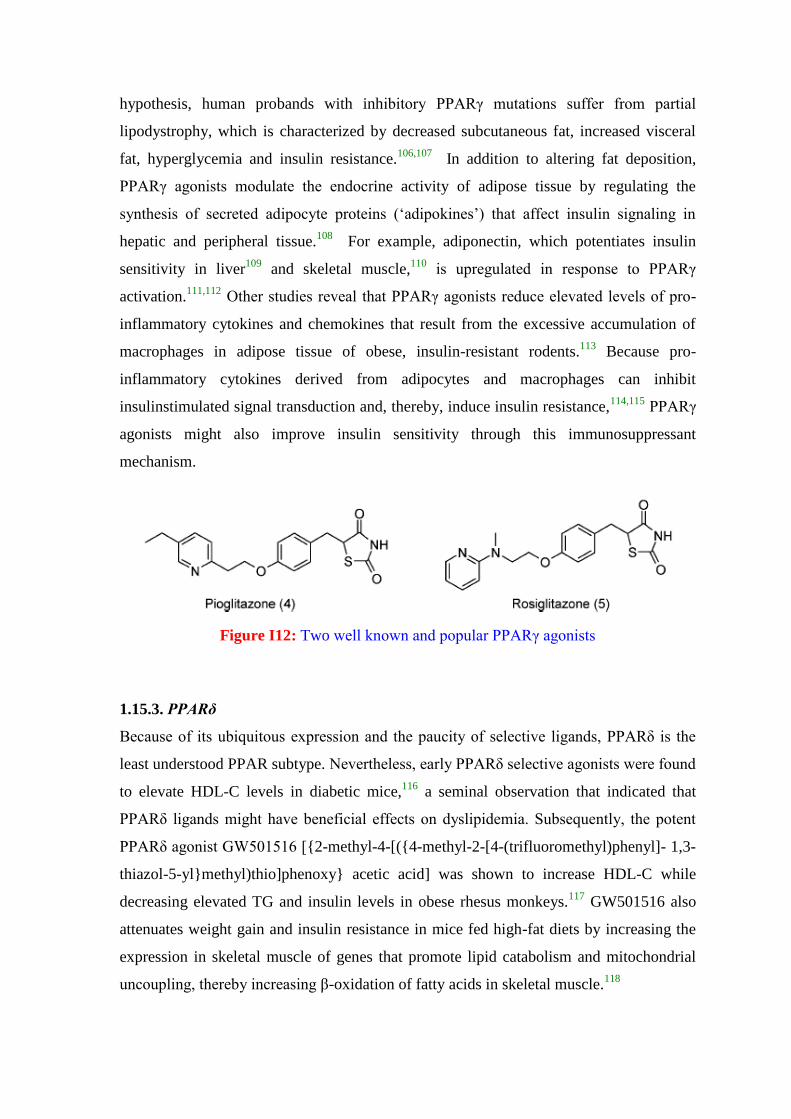
Figure I12: Two well known and popular PPARγ agonists
1.15.3. PPARδ
Because of its ubiquitous expression and the paucity of selective ligands, PPARδ is the
least understood PPAR subtype. Nevertheless, early PPARδ selective agonists were found
to elevate HDL-C levels in diabetic mice,116
a seminal observation that indicated that
PPARδ ligands might have beneficial effects on dyslipidemia. Subsequently, the potent
PPARδ agonist GW501516 [{2-methyl-4-[({4-methyl-2-[4-(trifluoromethyl)phenyl]- 1,3-
thiazol-5-yl}methyl)thio]phenoxy} acetic acid] was shown to increase HDL-C while
decreasing elevated TG and insulin levels in obese rhesus monkeys.117
GW501516 also
attenuates weight gain and insulin resistance in mice fed high-fat diets by increasing the
expression in skeletal muscle of genes that promote lipid catabolism and mitochondrial
uncoupling, thereby increasing β-oxidation of fatty acids in skeletal muscle.118
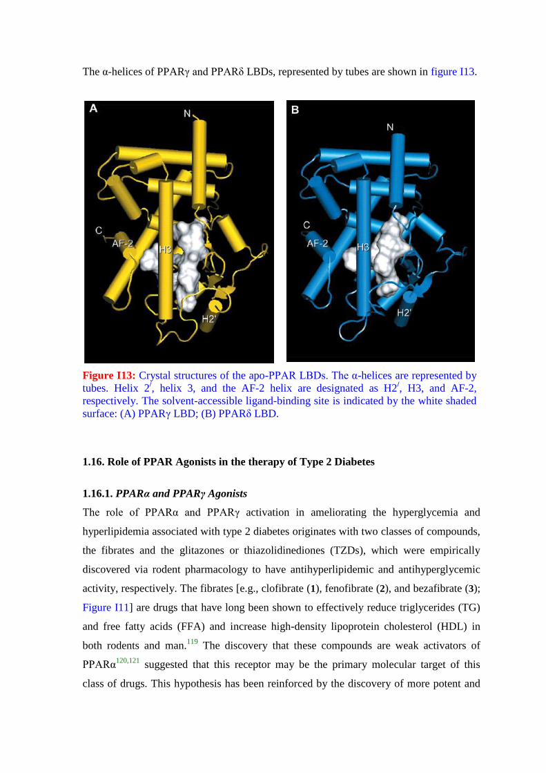
The α-helices of PPARγ and PPARδ LBDs, represented by tubes are shown in figure I13.
Figure I13: Crystal structures of the apo-PPAR LBDs. The α-helices are represented by
tubes. Helix 2/, helix 3, and the AF-2 helix are designated as H2
/, H3, and AF-2,
respectively. The solvent-accessible ligand-binding site is indicated by the white shaded
surface: (A) PPARγ LBD; (B) PPARδ LBD.
1.16. Role of PPAR Agonists in the therapy of Type 2 Diabetes
1.16.1. PPARα and PPARγ Agonists
The role of PPARα and PPARγ activation in ameliorating the hyperglycemia and
hyperlipidemia associated with type 2 diabetes originates with two classes of compounds,
the fibrates and the glitazones or thiazolidinediones (TZDs), which were empirically
discovered via rodent pharmacology to have antihyperlipidemic and antihyperglycemic
activity, respectively. The fibrates [e.g., clofibrate (1), fenofibrate (2), and bezafibrate (3);
Figure I11] are drugs that have long been shown to effectively reduce triglycerides (TG)
and free fatty acids (FFA) and increase high-density lipoprotein cholesterol (HDL) in
both rodents and man.119
The discovery that these compounds are weak activators of
PPARα120,121
suggested that this receptor may be the primary molecular target of this
class of drugs. This hypothesis has been reinforced by the discovery of more potent and
selective ligands for PPARR that display improved lipidlowering activity compared to the
fibrates.122
In addition, fibrates have also been shown to improve glucose tolerance in
type 2 diabetic patients,123
although this activity may not be attributable to activation of
PPARα because some these compounds also have appreciable PPARγ activity.119
Fibrates
are generally well-tolerated drugs; however, they are associated with a number of side
effects, the most common of which are gastrointestinal side effects such as nausea and
diarrhea, and elevations in liver enzymes.124,125
Skeletal myopathy and acute
rhabdomyolysis have also been reported during treatment with all the currently marketed
fibrates. It is not clear whether the effects on muscle are mediated by PPARα, but it will
be important to carefully monitor these side effects with the more potent, selective
PPARα agonists currently in clinical development. In addition, fibrates are excreted via
the kidneys and thus should be avoided in patients with renal failure. Finally, fibrates
have a propensity to cause drug-drug interactions because of their inhibition of
cytochrome P450 enzymes and thus must be used with great caution in combination with
other lipid-lowering drugs, particularly statins. While the combination of stains and
fibrates has shown improved control of lipoprotein risk factors relative to either agent
alone, this combination has shown an increase in renal failure, myopathy, and severe
rhabodomyolysis.
The TZDs have been shown to enhance the sensitivity of target tissues to insulin and to
reduce plasma glucose, lipid, and insulin levels in animal models of type 2 diabetes and in
humans.104
The TZDs pioglitazone [Actos (4)] and rosiglitazone [Avandia (5)] (Figure
I12) are currently marketed for the treatment of type 2 diabetes and represent important
agents in the treatment of this disease both as monotherapy or in combination with
existing therapies. These drugs display significant glucose-lowering efficacy, generally
achieving mean decreases in haemoglobin A1C (HbA1C) of approximately 1-1.5% and
mean decreases in fasting glucose of 60-80 mg/dL in type 2 diabetic patients. These drugs
also display modest beneficial effects on TGs, FFAs, and HDL cholesterol.126
As was the
case with fibrates and PPARR, the discovery that the TZDs are potent, selective agonists
of PPARγ127,128
provided a key link to understanding the molecular mechanism of these
drugs. These discoveries have alsoin the effort to optimize selective PPARγ agonists as
effective antidiabetic agents and selective PPARα agonists as antihyperlipidemic agents.
While rosiglitazone and pioglitazone have many beneficial effects in type 2 diabetics,
they also have some undesirable effects. A gain in weight of 3-5 kg is seen in most
patients, with a minority gaining an inordinate amount of weight.129
This weight gain is
accompanied by an increase in subcutaneous fat mass. The clinical significance of this
weight gain requires further evaluation, but in a treatment population that generally is
already overweight this can minimally lead to negative psychological effects. In some
patients this weight gain is accompanied by an increase in plasma volume leading to
edema that is often resistant to diuretic therapy. This plasma volume expansion may
precipitate or exacerbate congestive heart failure, and hence the currently marketed TZDs
are not recommended in diabetic patients with NYHA class III and class IV cardiac status
and should be used with caution in class I and class II patients. While the mechanism of
the fluid retention seen with the marketed TZDs has not been elucidated, it is likely to be
a PPARγ mediated effect because structurally unrelated selective PPARγ agonists also
promote fluid retention.
Given the importance of controlling both glucose and lipid levels in type 2 diabetes, the
concept of identifying ligands that bind and activate both PPARα and PPARγ represents a
logical continuation in the field of PPAR research. In addition to their benefit on lipids,
reports in the literature that fibrates reduce body weight gain in rodents without affecting
food intake130,131
offer hope that activation of PPARα may mitigate the weight gain
induced by PPARγ activation seen in humans. The hypothesis that PPARα/γ dual
agonism should provide additive, and possibly synergistic, pharmacology has resulted in
an intensive effort within the pharmaceutical industry to develop and evaluate these
agents (Figure II4).132
1.16.2. The PPAR dual agonists
PPARα/γ dual agonists
PPARα/γ dual agonists have been postulated as a combination strategy to achieve a broad
spectrum of metabolic effects and reduce mortality rates associated with type 2 diabetes
by improving insulin resistance, hyperglycemia and alleviating atherogenic dyslipidemia.
In addition, PPARα agonists stimulate lipid oxidation and decrease adiposity in rodent
obesity models. Thus, the propensity of PPARγ activation to induce weight gain through
its adipogenic affects may be offset by the ability of PPARα activation to stimulate lipid
catabolism thereby providing a compound with reduced propensity for inducing undesired
weight gain.133,134
In recent years, a number of structurally diverse PPARα/γ dual agonists
have been reported. Many of these have been evaluated in the clinic and some have
advanced into late stage development. The clinical efficacy of the initially discovered
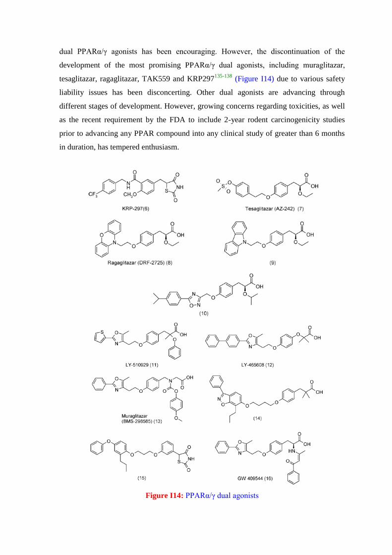
dual PPARα/γ agonists has been encouraging. However, the discontinuation of the
development of the most promising PPARα/γ dual agonists, including muraglitazar,
tesaglitazar, ragaglitazar, TAK559 and KRP297135-138
(Figure I14) due to various safety
liability issues has been disconcerting. Other dual agonists are advancing through
different stages of development. However, growing concerns regarding toxicities, as well
as the recent requirement by the FDA to include 2-year rodent carcinogenicity studies
prior to advancing any PPAR compound into any clinical study of greater than 6 months
in duration, has tempered enthusiasm.
Figure I14: PPARα/γ dual agonists
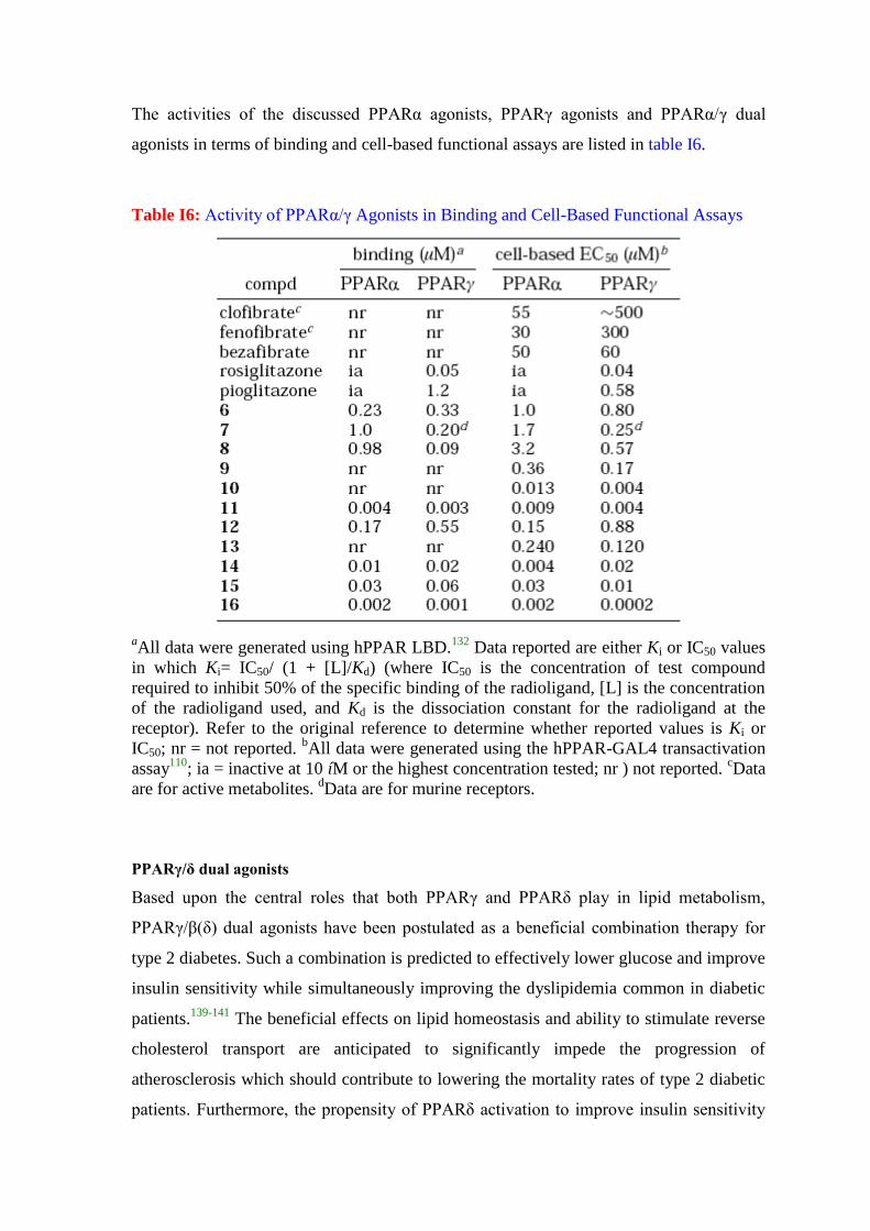
The activities of the discussed PPARα agonists, PPARγ agonists and PPARα/γ dual
agonists in terms of binding and cell-based functional assays are listed in table I6.
Table I6: Activity of PPARα/γ Agonists in Binding and Cell-Based Functional Assays
aAll data were generated using hPPAR LBD.
132 Data reported are either Ki or IC50 values
in which Ki= IC50/ (1 + [L]/Kd) (where IC50 is the concentration of test compound
required to inhibit 50% of the specific binding of the radioligand, [L] is the concentration
of the radioligand used, and Kd is the dissociation constant for the radioligand at the
receptor). Refer to the original reference to determine whether reported values is Ki or
IC50; nr = not reported. bAll data were generated using the hPPAR-GAL4 transactivation
assay110
; ia = inactive at 10 íM or the highest concentration tested; nr ) not reported. cData
are for active metabolites. dData are for murine receptors.
PPARγ/δ dual agonists
Based upon the central roles that both PPARγ and PPARδ play in lipid metabolism,
PPARγ/β(δ) dual agonists have been postulated as a beneficial combination therapy for
type 2 diabetes. Such a combination is predicted to effectively lower glucose and improve
insulin sensitivity while simultaneously improving the dyslipidemia common in diabetic
patients.139-141
The beneficial effects on lipid homeostasis and ability to stimulate reverse
cholesterol transport are anticipated to significantly impede the progression of
atherosclerosis which should contribute to lowering the mortality rates of type 2 diabetic
patients. Furthermore, the propensity of PPARδ activation to improve insulin sensitivity
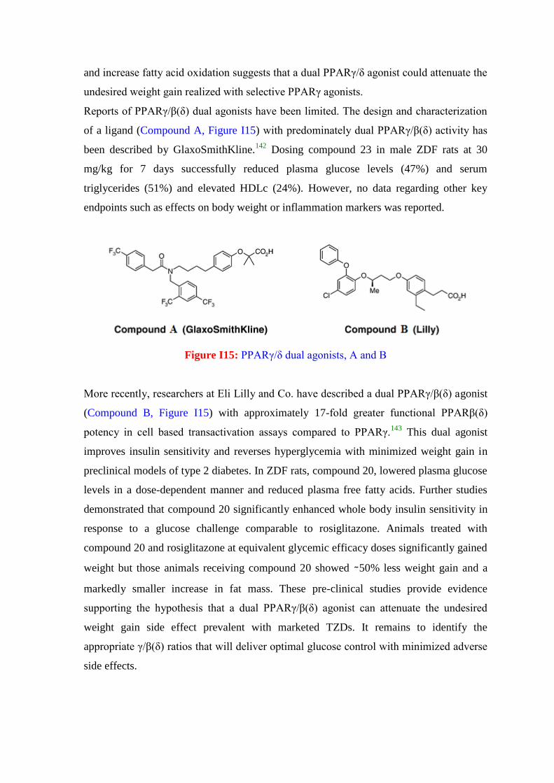
and increase fatty acid oxidation suggests that a dual PPARγ/δ agonist could attenuate the
undesired weight gain realized with selective PPARγ agonists.
Reports of PPARγ/β(δ) dual agonists have been limited. The design and characterization
of a ligand (Compound A, Figure I15) with predominately dual PPARγ/β(δ) activity has
been described by GlaxoSmithKline.142
Dosing compound 23 in male ZDF rats at 30
mg/kg for 7 days successfully reduced plasma glucose levels (47%) and serum
triglycerides (51%) and elevated HDLc (24%). However, no data regarding other key
endpoints such as effects on body weight or inflammation markers was reported.
Figure I15: PPARγ/δ dual agonists, A and B
More recently, researchers at Eli Lilly and Co. have described a dual PPARγ/β(δ) agonist
(Compound B, Figure I15) with approximately 17-fold greater functional PPARβ(δ)
potency in cell based transactivation assays compared to PPARγ.143
This dual agonist
improves insulin sensitivity and reverses hyperglycemia with minimized weight gain in
preclinical models of type 2 diabetes. In ZDF rats, compound 20, lowered plasma glucose
levels in a dose-dependent manner and reduced plasma free fatty acids. Further studies
demonstrated that compound 20 significantly enhanced whole body insulin sensitivity in
response to a glucose challenge comparable to rosiglitazone. Animals treated with
compound 20 and rosiglitazone at equivalent glycemic efficacy doses significantly gained
weight but those animals receiving compound 20 showed ∼50% less weight gain and a
markedly smaller increase in fat mass. These pre-clinical studies provide evidence
supporting the hypothesis that a dual PPARγ/β(δ) agonist can attenuate the undesired
weight gain side effect prevalent with marketed TZDs. It remains to identify the
appropriate γ/β(δ) ratios that will deliver optimal glucose control with minimized adverse
side effects.
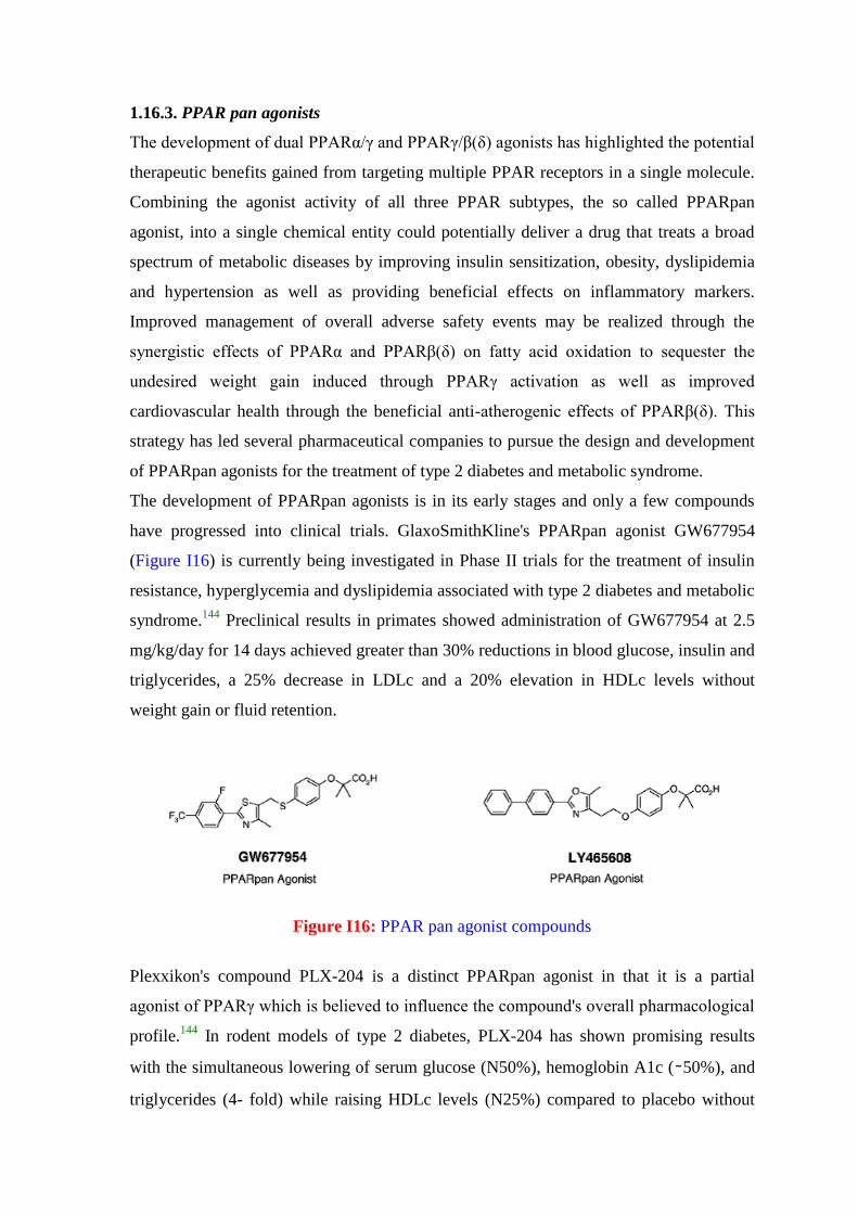
1.16.3. PPAR pan agonists
The development of dual PPARα/γ and PPARγ/β(δ) agonists has highlighted the potential
therapeutic benefits gained from targeting multiple PPAR receptors in a single molecule.
Combining the agonist activity of all three PPAR subtypes, the so called PPARpan
agonist, into a single chemical entity could potentially deliver a drug that treats a broad
spectrum of metabolic diseases by improving insulin sensitization, obesity, dyslipidemia
and hypertension as well as providing beneficial effects on inflammatory markers.
Improved management of overall adverse safety events may be realized through the
synergistic effects of PPARα and PPARβ(δ) on fatty acid oxidation to sequester the
undesired weight gain induced through PPARγ activation as well as improved
cardiovascular health through the beneficial anti-atherogenic effects of PPARβ(δ). This
strategy has led several pharmaceutical companies to pursue the design and development
of PPARpan agonists for the treatment of type 2 diabetes and metabolic syndrome.
The development of PPARpan agonists is in its early stages and only a few compounds
have progressed into clinical trials. GlaxoSmithKline's PPARpan agonist GW677954
(Figure I16) is currently being investigated in Phase II trials for the treatment of insulin
resistance, hyperglycemia and dyslipidemia associated with type 2 diabetes and metabolic
syndrome.144
Preclinical results in primates showed administration of GW677954 at 2.5
mg/kg/day for 14 days achieved greater than 30% reductions in blood glucose, insulin and
triglycerides, a 25% decrease in LDLc and a 20% elevation in HDLc levels without
weight gain or fluid retention.
Figure I16: PPAR pan agonist compounds
Plexxikon's compound PLX-204 is a distinct PPARpan agonist in that it is a partial
agonist of PPARγ which is believed to influence the compound's overall pharmacological
profile.144
In rodent models of type 2 diabetes, PLX-204 has shown promising results
with the simultaneous lowering of serum glucose (N50%), hemoglobin A1c (∼50%), and
triglycerides (4- fold) while raising HDLc levels (N25%) compared to placebo without
significant changes in body weight or hematocrit. Phase II clinical trials with PLX-204
are in progress.
LY465608 (Figure I16) was originally reported as a dual PPARα/γ agonist by researchers
at Eli Lilly Pharmaceuticals.145
Further detailed studies demonstrated that this compound
also possesses significant PPARδ activity and now LY465608 is classified as a PPARpan
agonist. Data compiled from various preclinical studies in rodent models of type 2
diabetes shows that LY465608 effectively lowers plasma glucose levels, improves insulin
sensitivity and exerts a positive effect on lipid and cholesterol homeostasis by reducing
serum triglycerides and elevating HDL cholesterol. Weight gain was observed in long
term treatment of ZDF rats with LY465608, but the increase was significantly less than
observed with the full PPARγ agonist rosiglitazone despite equivalent levels of glycemic
control.
The prospect of PPARpan agonists to combat multiple aspects of the diabetic phenotype
has high expectations. The key issue for the development of PPARpan agonists, as well as
PPAR dual agonist combinations, is whether they can deliver an improved therapeutic
index compared to the existing PPAR subtype selective agonists. More clinical
information on these PPARpan agonists is eagerly anticipated and will be closely
scrutinized as the first generation of these compounds advance through development.146
1.17. Partial Agonism to the Rescue
Disappointingly, full PPAR agonists are plagued by certain adverse side effects. On the
up side, partial PPAR agonists have the potential to retain the desired efficacy and
beneficial effects of full PPAR agonists while diminishing the unwanted effects.
Especially in case of PPARγ; partial agonists are being developed with the goal of
retaining the beneficial effects of this class of agents, while diminishing their adverse
effects (For example metaglidasen by Metabolex).147
Studies with partial agonists of
PPARγ suggests that a focus on partial PPAR agonists may be a way of developing
agents that have the desired efficacy of PPAR agonists and are devoid of, at least, some of
their potential adverse effects.147
The hypothesis “Partial agonism at both PPARα and PPARγ receptors by dual partial
activators may provide a solution - resulting in the desirable responses and reducing the
adverse effects” caused by the individual agonists for the treatment of T2DM attracted
our attention and prompted us to develop a research protocol to design, predict, select and
synthesise such new chemical entities. Development of such agents is a very challenging
endeavor, as the molecules should have affinity simultaneously for all the aimed
macromolecular targets with similar binding pockets and also the fine tuning of the
substituents required to obtain the optimum potency, which may finally result in desired
activity profile and lesser or no adverse effects as compared to the selective ligands.
1.18. Computational methods in the design of multiple receptor activating ligands
Computational methods which has emerged as very useful and money saving tool in drug
design, can be effectively employed in designing multiple receptor activating ligands. The
design of multiple receptor activating ligands is very challenging as it requires structural
fine tuning so that the designed molecule should bind to and activate the targeted
receptors of structural similarity. The x-ray crystal structural information of the 3D-
geometry of targeted receptors is advantageous for this special effort. 3D-QSAR (three
dimensional structure activity relationship) techniques are applied to derive indirect
binding information from the correlation between the biological activity of a training set
of molecules and their 3D structures which could facilitate in designing newer molecules
„yet to be synthesized‟ as novel agents having desired therapeutic potential and safety
profile. The importance of steric and electrophilic characteristics is revealed by aligning
structurally similar analogues using pharmacophoric features as structural
superimposition guides.148
The comparative molecular field analysis (CoMFA) calculates
steric and electrostatic properties in the space surrounding each of the aligned molecules
in a data set according to Lennard-Jones and Coulomb potentials, respectively.149
CoMFA
develops the 3D QSAR model based on the relationship between the steric/electrostatic
properties and biological activities. Comparative Molecular Similarity Indices Analysis
(CoMSIA) calculates similarity indices around the molecules, with the similarity
expressed in the terms of different physicochemical properties, such as steric occupancy,
partial atomic charges, local hydrophobicity and hydrogen bond donor and acceptor
properties. The similarities are calculated using much smoother potentials that are not as
steep as the Lennard-Jones and Coulombic functions and have a finite value even at the
atomic positions. Docking of designed molecules into the x-ray crystal structure of the
active site of the target protein may reveal valuable approximation regarding binding of
the molecules to the protein in biological system. Finally the analysis of the results
obtained from 3D QSAR and Docking experiments enable us to screen and select the best
target molecules with optimum efficacies to be taken forward for synthesis and
characterization.
AIM AND OBJECTIVES
The aim is to work on the hypothesis that partial PPAR / dual agonism may provide
additive and positive synergistic pharmacology with less or no adverse effects so as to
meet and overcome the issues facing with the development of dual acting agonists.
Accordingly it was planned to synthesize and validate computationally heterocyclyl
linked acyclic analogs of isoxazolidinediones i.e. 1,3-dicarbonyl compounds in general
and β-ketoesters in particular, after designing on the basis of 3D-QSAR studies, which
have almost all the structural features to act as effective insulin sensitizer and are
expected to bind and activate PPARα and PPARγ, but to partial extent relative to full
agonists which will eventually lead to Partial PPARα and PPARγ dual agonists for the
management of hyperglycemia and hyperlipidemia.
● To build robust and significantly good predictive 3D QSAR (Comparative Molecular
Field Analysis- CoMFA) models for prediction of PPAR α, γ activities.
● Design of New Chemical Entities based on the contour analysis resulted from the
generated CoMFA models.
● To predict the activities and binding affinities (Docking studies) of the designed NCEs
computationally.
● Final selection of the molecules for synthesis on the basis of prediction of activities and
binding affinities obtained computationally with an aim to achieve partial agonism.
● Devising new synthetic strategies, which meet the requirement of “Ideal Synthesis” and
also take into consideration the concept of connectivity analysis, thereby, providing new
insights towards the skeletal and functionality requirement of these molecules.
● Synthesis and structural characterization of the final selected molecules.
Dual PPARα/γ agonists (tyrosine and α-aryl/alkoxy propanoic acid based molecules for
which the PPARα and PPARγ activities are reported for single enantiomer) were taken
from the literature to obtain a dataset which was divided into training and test set to
develop 3D QSAR models. The energy optimized conformation of the crystal structure of
farglitazar (a potent PPARα/γ dual agonist) extracted from the crystal structure of PPARγ
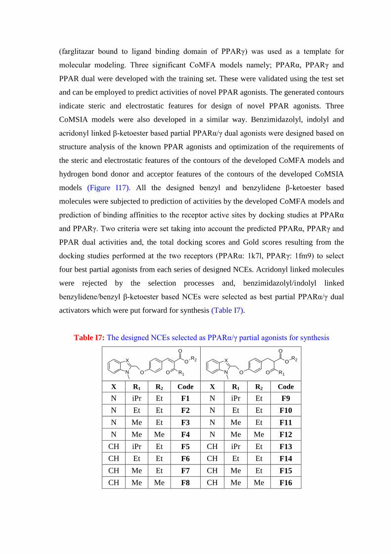
(farglitazar bound to ligand binding domain of PPARγ) was used as a template for
molecular modeling. Three significant CoMFA models namely; PPARα, PPARγ and
PPAR dual were developed with the training set. These were validated using the test set
and can be employed to predict activities of novel PPAR agonists. The generated contours
indicate steric and electrostatic features for design of novel PPAR agonists. Three
CoMSIA models were also developed in a similar way. Benzimidazolyl, indolyl and
acridonyl linked β-ketoester based partial PPARα/γ dual agonists were designed based on
structure analysis of the known PPAR agonists and optimization of the requirements of
the steric and electrostatic features of the contours of the developed CoMFA models and
hydrogen bond donor and acceptor features of the contours of the developed CoMSIA
models (Figure I17). All the designed benzyl and benzylidene β-ketoester based
molecules were subjected to prediction of activities by the developed CoMFA models and
prediction of binding affinities to the receptor active sites by docking studies at PPARα
and PPARγ. Two criteria were set taking into account the predicted PPARα, PPARγ and
PPAR dual activities and, the total docking scores and Gold scores resulting from the
docking studies performed at the two receptors (PPARα: 1k7l, PPARγ: 1fm9) to select
four best partial agonists from each series of designed NCEs. Acridonyl linked molecules
were rejected by the selection processes and, benzimidazolyl/indolyl linked
benzylidene/benzyl β-ketoester based NCEs were selected as best partial PPARα/γ dual
activators which were put forward for synthesis (Table I7).
Table I7: The designed NCEs selected as PPARα/γ partial agonists for synthesis
X
N O
O
O
O R1
R2
X
N O
O
O
O R1
R2
X R1 R2 Code X R1 R2 Code
N iPr Et F1 N iPr Et F9
N Et Et F2 N Et Et F10
N Me Et F3 N Me Et F11
N Me Me F4 N Me Me F12
CH iPr Et F5 CH iPr Et F13
CH Et Et F6 CH Et Et F14
CH Me Et F7 CH Me Et F15
CH Me Me F8 CH Me Me F16
1.17. Partial Agonism to the Rescue
Disappointingly, full PPAR agonists are plagued by certain adverse side effects. On the
up side, partial PPAR agonists have the potential to retain the desired efficacy and
beneficial effects of full PPAR agonists while diminishing the unwanted effects.
Especially in case of PPARγ; partial agonists are being developed with the goal of
retaining the beneficial effects of this class of agents, while diminishing their adverse
effects (For example metaglidasen by Metabolex).147
Studies with partial agonists of
PPARγ suggests that a focus on partial PPAR agonists may be a way of developing
agents that have the desired efficacy of PPAR agonists and are devoid of, at least, some of
their potential adverse effects.147
The hypothesis “Partial agonism at both PPARα and PPARγ receptors by dual partial
activators may provide a solution - resulting in the desirable responses and reducing the
adverse effects” caused by the individual agonists for the treatment of T2DM attracted
our attention and prompted us to develop a research protocol to design, predict, select and
synthesise such new chemical entities. Development of such agents is a very challenging
endeavor, as the molecules should have affinity simultaneously for all the aimed
macromolecular targets with similar binding pockets and also the fine tuning of the
substituents required to obtain the optimum potency, which may finally result in desired
activity profile and lesser or no adverse effects as compared to the selective ligands.
1.18. Computational methods in the design of multiple receptor activating ligands
Computational methods which has emerged as very useful and money saving tool in drug
design, can be effectively employed in designing multiple receptor activating ligands. The
design of multiple receptor activating ligands is very challenging as it requires structural
fine tuning so that the designed molecule should bind to and activate the targeted
receptors of structural similarity. The x-ray crystal structural information of the 3D-
geometry of targeted receptors is advantageous for this special effort. 3D-QSAR (three
dimensional structure activity relationship) techniques are applied to derive indirect
binding information from the correlation between the biological activity of a training set
of molecules and their 3D structures which could facilitate in designing newer molecules
„yet to be synthesized‟ as novel agents having desired therapeutic potential and safety
profile. The importance of steric and electrophilic characteristics is revealed by aligning
structurally similar analogues using pharmacophoric features as structural
superimposition guides.148
The comparative molecular field analysis (CoMFA) calculates
steric and electrostatic properties in the space surrounding each of the aligned molecules
in a data set according to Lennard-Jones and Coulomb potentials, respectively.149
CoMFA
develops the 3D QSAR model based on the relationship between the steric/electrostatic
properties and biological activities. Comparative Molecular Similarity Indices Analysis
(CoMSIA) calculates similarity indices around the molecules, with the similarity
expressed in the terms of different physicochemical properties, such as steric occupancy,
partial atomic charges, local hydrophobicity and hydrogen bond donor and acceptor
properties. The similarities are calculated using much smoother potentials that are not as
steep as the Lennard-Jones and Coulombic functions and have a finite value even at the
atomic positions. Docking of designed molecules into the X-ray crystal structure of the
active site of the target protein may reveal valuable approximation regarding binding of
the molecules to the protein in biological system. Finally the analysis of the results
obtained from 3D QSAR and Docking experiments enable us to screen and select the best
target molecules with optimum efficacies to be taken forward for synthesis and
characterization.
AIM AND OBJECTIVES
The aim is to work on the hypothesis that partial PPAR / dual agonism may provide
additive and positive synergistic pharmacology with less or no adverse effects so as to
meet and overcome the issues facing with the development of dual acting agonists.
Accordingly it was planned to synthesize and validate computationally heterocyclyl
linked acyclic analogs of oxazolidinediones i.e. α-alkoxy carboxylic acids, after designing
on the basis of 3D-QSAR studies, which have almost all the structural features to act as
effective insulin sensitizer and are expected to bind and activate PPARα and PPARγ, but
to partial extent relative to full agonists which will eventually lead to Partial PPARα
and PPARγ dual agonists for the management of hyperglycemia and hyperlipidemia.
● To build robust and significantly good predictive 3D QSAR (Comparative Molecular
Similarity Field Analysis- CoMSIA) models for prediction of PPAR α, γ activities.
● Design of New Chemical Entities based on the contour analysis resulted from the
generated CoMSIA models.
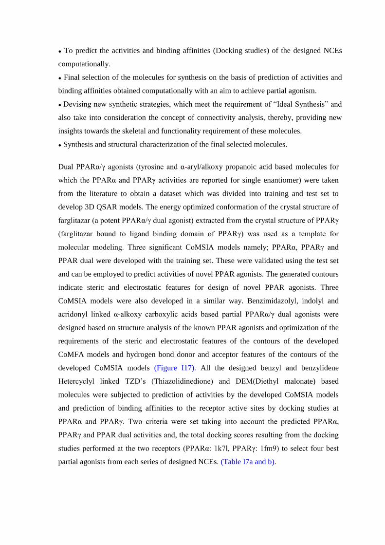
● To predict the activities and binding affinities (Docking studies) of the designed NCEs
computationally.
● Final selection of the molecules for synthesis on the basis of prediction of activities and
binding affinities obtained computationally with an aim to achieve partial agonism.
● Devising new synthetic strategies, which meet the requirement of “Ideal Synthesis” and
also take into consideration the concept of connectivity analysis, thereby, providing new
insights towards the skeletal and functionality requirement of these molecules.
● Synthesis and structural characterization of the final selected molecules.
Dual PPARα/γ agonists (tyrosine and α-aryl/alkoxy propanoic acid based molecules for
which the PPARα and PPARγ activities are reported for single enantiomer) were taken
from the literature to obtain a dataset which was divided into training and test set to
develop 3D QSAR models. The energy optimized conformation of the crystal structure of
farglitazar (a potent PPARα/γ dual agonist) extracted from the crystal structure of PPARγ
(farglitazar bound to ligand binding domain of PPARγ) was used as a template for
molecular modeling. Three significant CoMSIA models namely; PPARα, PPARγ and
PPAR dual were developed with the training set. These were validated using the test set
and can be employed to predict activities of novel PPAR agonists. The generated contours
indicate steric and electrostatic features for design of novel PPAR agonists. Three
CoMSIA models were also developed in a similar way. Benzimidazolyl, indolyl and
acridonyl linked α-alkoxy carboxylic acids based partial PPARα/γ dual agonists were
designed based on structure analysis of the known PPAR agonists and optimization of the
requirements of the steric and electrostatic features of the contours of the developed
CoMFA models and hydrogen bond donor and acceptor features of the contours of the
developed CoMSIA models (Figure I17). All the designed benzyl and benzylidene
Hetercyclyl linked TZD‟s (Thiazolidinedione) and DEM(Diethyl malonate) based
molecules were subjected to prediction of activities by the developed CoMSIA models
and prediction of binding affinities to the receptor active sites by docking studies at
PPARα and PPARγ. Two criteria were set taking into account the predicted PPARα,
PPARγ and PPAR dual activities and, the total docking scores resulting from the docking
studies performed at the two receptors (PPARα: 1k7l, PPARγ: 1fm9) to select four best
partial agonists from each series of designed NCEs. (Table I7a and b).

Table I7a: The designed NCEs selected as PPARα/γ partial agonists for synthesis
Table I7b: The Other NCEs selected as PPARα/γ partial agonists for synthesis
O
N
N
O
O
H
OR
CH3
O
N
O
O
H
OR
CH3
ON
O
OH
OR
O
R Code R Code R Code
Me F1a
Et F1b
i-Pr F1c i-Pr F2c i-Pr F3c
i-Bu F1d i-BuOH F2d i-BuOH F3d
O
N
X
Me
S
NH
O
O
O
N
X
Me
S
NH
O
O
O
N
X
Me
OEt
O
O OEt
O
N
X
Me
OEt
O
O OEt
X Code X Code X Code X Code
N F4a N F4b N F6a N F6b
CH F5a CH F5b CH F7a CH F7b

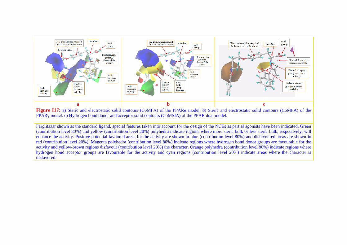
a b c
Figure I17: a) Steric and electrostatic solid contours (CoMFA) of the PPARα model. b) Steric and electrostatic solid contours (CoMFA) of the
PPARγ model. c) Hydrogen bond donor and acceptor solid contours (CoMSIA) of the PPAR dual model.
Farglitazar shown as the standard ligand, special features taken into account for the design of the NCEs as partial agonists have been indicated. Green
(contribution level 80%) and yellow (contribution level 20%) polyhedra indicate regions where more steric bulk or less steric bulk, respectively, will
enhance the activity. Positive potential favoured areas for the activity are shown in blue (contribution level 80%) and disfavoured areas are shown in
red (contribution level 20%). Magenta polyhedra (contribution level 80%) indicate regions where hydrogen bond donor groups are favourable for the
activity and yellow-brown regions disfavour (contribution level 20%) the character. Orange polyhedra (contribution level 80%) indicate regions where
hydrogen bond acceptor groups are favourable for the activity and cyan regions (contribution level 20%) indicate areas where the character is
disfavored.
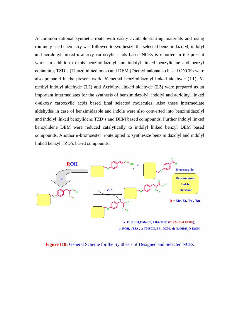
A common rational synthetic route with easily available starting materials and using
routinely used chemistry was followed to synthesize the selected benzimidazolyl, indolyl
and acridonyl linked α-alkoxy carboxylic acids based NCEs is reported in the present
work. In addition to this benzimidazolyl and indolyl linked benzylidene and benzyl
containing TZD‟s (Thiazolidinediones) and DEM (Diethylmalonates) based ONCEs were
also prepared in the present work. N-methyl benzimidazolyl linked aldehyde (L1), N-
methyl indolyl aldehyde (L2) and Acridinyl linked aldehyde (L3) were prepared as an
important intermediates for the synthesis of benzimidazolyl, indolyl and acridinyl linked
α-alkoxy carboxylic acids based final selected molecules. Also these intermediate
aldehydes in case of benzimidazole and indole were also converted into benzimidazolyl
and indolyl linked benzylidene TZD‟s and DEM based compounds. Further indolyl linked
benzylidene DEM were reduced catalytically to indolyl linked benzyl DEM based
compounds. Another α-bromoester route opted to synthesize benzimidazolyl and indolyl
linked benzyl TZD‟s based compounds.
a: Ph3P+CH2OMe Cl
-, LDA-THF, (DIPA-nBuLi/THF),
b: ROH, pTSA , c: TMSCN, BF3-DCM, d: NaOH/H2O-EtOH
O
NH
N
O
O
H
OR
N
NO
CH3
H
O
O
NH
N
OCH3
O
NH
N
OR
OR
O
NH
N
CN
OR
a
Heterocycle
c, d
Benzimidazole
Indole
Acridone
b
ROH
R = Me, Et, iPr ,
iBu
Figure I18: General Scheme for the Synthesis of Designed and Selected NCEs
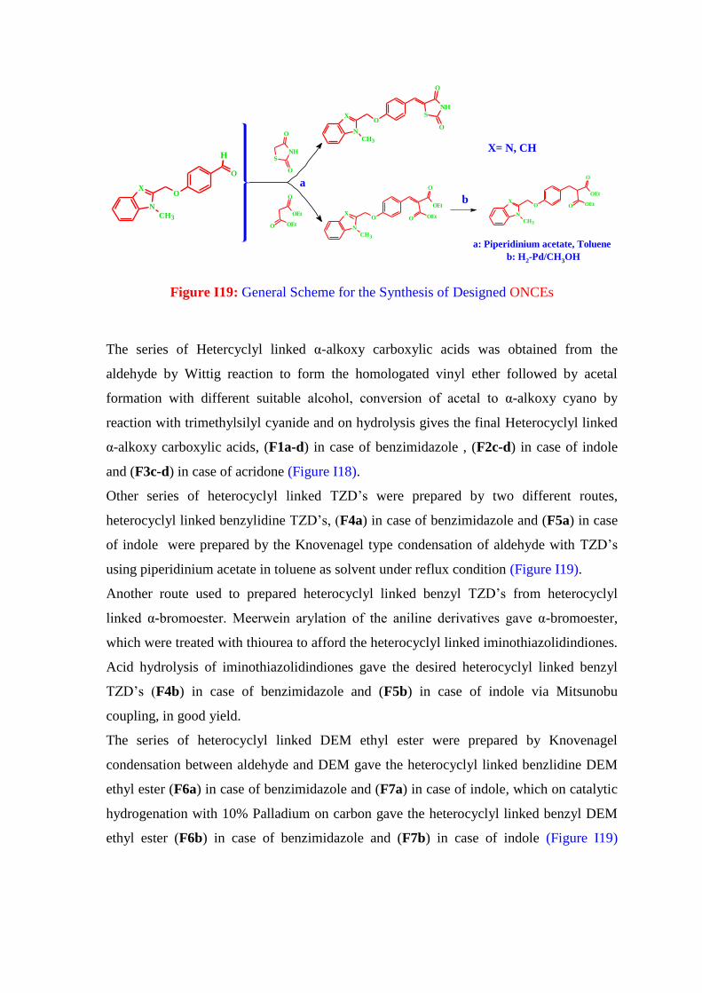
O
N
X
CH3
OEt
O
O OEt
O
N
X
CH3
OEt
O
O OEt
O
N
X
CH3
SNH
O
O
O
N
X
CH3
O
H
OEt
O
O OEt
SNH
O
O
a
b
a: Piperidinium acetate, Toluene
b: H2-Pd/CH3OH
X= N, CH
Figure I19: General Scheme for the Synthesis of Designed ONCEs
The series of Hetercyclyl linked α-alkoxy carboxylic acids was obtained from the
aldehyde by Wittig reaction to form the homologated vinyl ether followed by acetal
formation with different suitable alcohol, conversion of acetal to α-alkoxy cyano by
reaction with trimethylsilyl cyanide and on hydrolysis gives the final Heterocyclyl linked
α-alkoxy carboxylic acids, (F1a-d) in case of benzimidazole , (F2c-d) in case of indole
and (F3c-d) in case of acridone (Figure I18).
Other series of heterocyclyl linked TZD‟s were prepared by two different routes,
heterocyclyl linked benzylidine TZD‟s, (F4a) in case of benzimidazole and (F5a) in case
of indole were prepared by the Knovenagel type condensation of aldehyde with TZD‟s
using piperidinium acetate in toluene as solvent under reflux condition (Figure I19).
Another route used to prepared heterocyclyl linked benzyl TZD‟s from heterocyclyl
linked α-bromoester. Meerwein arylation of the aniline derivatives gave α-bromoester,
which were treated with thiourea to afford the heterocyclyl linked iminothiazolidindiones.
Acid hydrolysis of iminothiazolidindiones gave the desired heterocyclyl linked benzyl
TZD‟s (F4b) in case of benzimidazole and (F5b) in case of indole via Mitsunobu
coupling, in good yield.
The series of heterocyclyl linked DEM ethyl ester were prepared by Knovenagel
condensation between aldehyde and DEM gave the heterocyclyl linked benzlidine DEM
ethyl ester (F6a) in case of benzimidazole and (F7a) in case of indole, which on catalytic
hydrogenation with 10% Palladium on carbon gave the heterocyclyl linked benzyl DEM
ethyl ester (F6b) in case of benzimidazole and (F7b) in case of indole (Figure I19)