Embed Size (px)
Citation preview

1. Eukurym. Microbial.. 5 l(3). 2004 pp. 325-332 0 2004 by the Society of Protozoologists
Preferential PCR Amplification of Parasitic Protistan Small Subunit rDNA from Metazoan Tissues
SUSAN M. BOWER,”’ RYAN B. CARNEGIE,”2 BENJAMIN GOH,*j SIMON R. M. JONES,” GEOFFREY J. LOWEd and MICHELLE W. S. MAW.4
“Fisheries and Oceans Canada, Pacific Biological Stution, 3190 Hammond Bay Rd.. Nanuimo. British Columbiu V9T 6N7, Canada
ABSTRACT. A “universal non-metazoan” polymerase chain reaction (UNonMet-PCR) that selectively amplifies a segment of non- metazoan Small Subunit (SSU) rDNA gene was validated. The primers used were: 18s-EUK581 -F (5’-GTGCCAGCAGCCGCG-3‘) and 18s-EUKI 134-R (5’-TlTAAGT’ITCAGCCTTGCG-3’) with specificity provided by the 19-base reverse primer. Its target site is highly conserved across the Archaea, Bacteria, and eukaryotes (including fungi), but not most Metazoa (except Porifera, Ctenophora, and Myxozoa) which have mismatches at bases 14 and 19 resulting in poor or failed amplification. During validation, UNonMet-PCR amplified SSU rDNA gene fragments from all assayed protists (n = 16 from 7 higher taxa, including two species of marine phytoplank- ton) and Fungi (n = 3) but amplified very poorly or not at all most assayed Metazoa (n = 13 from 8 higher taxa). When a non- metazoan parasite was present in a metazoan host, the parasite DNA was preferentially amplified. For example, DNA from the parasite Trypanosomu danilewskyi was preferentially amplified in mixtures containing up to I ,000X more goldfish Carassius auratus (host) DNA. Also, the weak amplification of uninfected host (Chionoeceres tanneri) SSU rDNA did not occur in the presence of a natural infection with a parasite (Hematodinium sp.). Only Hemarodinium sp. SSU rDNA was amplified in samples from infected C. ranneri. This UNonMet-PCR is a powerful tool for amplifying SSU rDNA from non-metazoan pathogens or symbionts that have not been isolated from metazoan hosts. Key Words. Molecular tool, preferential amplification of parasite DNA, SSU rDNA fragment.
HE polymerase chain reaction (PCR) is proving to be a T significant research and diagnostic tool for the detection of protistan pathogens in commercially important aquatic or- ganisms (e.g. Carnegie et al. 2000; Cunningham 2002; Stokes, Siddall, and Burreson 1995). Its specificity and often exquisite sensitivity make it a powerful complement to conventional his- topathological techniques already used for parasite detection. However, the crucial first step to the development of specific molecular assays is the characterisation of a diagnostic gene such as that coding for SSU rDNA (Hillis and Dixon 199 1) and this step is often difficult. In some cases, the characterisation of parasite genes has failed because pathogen DNA could not be separated from host DNA. The application of “universal” eukaryotic PCR primers may amplify both pathogen and host DNA, but because host DNA often predominates in bulk mix- tures, it is preferentially amplified to the exclusion of parasite DNA. Alternatively, amplification of parasite genes may fail because the target sequences for “universal” eukaryotic PCR primers are divergent in the genome of the pathogen. Pure iso- lates of many pathogens are unavailable as axenic cultures, fur- ther confounding the ability to characterise these organisms. Thus, molecular analyses, with all the associated advantages including the development of sensitive and specific diagnostic assays, are not possible or proceed slowly.
This dilemma was exemplified by Mikrocytos mackini, a pro- tistan parasite of unknown taxonomic affiliation, which causes Denman Island disease in wild and aquacultured oysters (Cras- sostrea gigas, Crassostrea virginica, Ostrea edulis, and Ostrea conchaphila) (Bower, Hervio, and Meyer 1997; Farley, Wolf, and Elston 1988; Hervio, Bower, and Meyer 1996; Hine et al. 2001 ; Quayle 1961). After various unsuccessful attempts to identify M. mackini DNA using universal eukaryotic PCR prim- ers, a set of PCR primers designed not to amplify host DNA generated a 544-base pair (bp) 18s rDNA fragment from M. mackini-infected oysters and enriched M. mackini isolates, but
Corresponding Author: S. Bower-Telephone number 250-756-7077;
’ Authors are listed in alphabetical order. FAX number 250-756-7053; E-mail: [email protected].
Virginia Institute of Marine Science, Gloucester Point, Virginia
Fisheries and Oceans Canada, West Vancouver Laboratory, 4160
Dept. of Biological Sciences, University of Alberta, Edmonton, Al-
23062, USA
Marine Drive, West Vancouver, British Columbia V7V 1 N6, Canada.
berta, T6G 2E9, Canada.
not from uninfected control oysters. The gene fragment with subsequent elongation was confirmed to be M. mackini 18s rDNA by fluorescent in situ hybridization (Carnegie et al. 2003). Both forward and reverse primers (18s-EUK581-F: 5 ’ - GTGCCAGCAGCCGCG-3’ and 18s-EUK- 1 134-R: 5‘-TTTA AGTTTCAGCCTTGCG-3’, hereafter referred to as the UNonMet-PCR), were designed to preferentially amplify para- site DNA. These primers targeted regions highly conserved across all sequences in an alignment of eukaryotes, Archaea and Bacteria. The reverse primer, however, was mismatched to the target sequence in metazoan 18s rDNA at the 14th and 19th (final) positions (Fig. 1). It was therefore expected to amplify metazoan rDNA inefficiently, if at all. The success achieved by Camegie et al. (2003) indicated the potential application of this new UNonMet-PCR to identify SSU rDNA of pathogens in metazoa.
The purpose of the present study was to identify the wider application of the UNonMet-PCR. In addition to comparing the primer target sites with SSU rDNA sequences available in GenBank, the capability of the primers to amplify a segment of the SSU rDNA gene from 32 representative species of 16 higher eukaryotic taxa was tested. In addition, the suitability of the UNonMet-PCR to amplify bacterial DNA was informally assessed.
MATERIALS AND METHODS General approach. Gene segments of the predicted length
resulting from the UNonMet-PCR of test samples were se- quenced and the sequences compared with those available in GenBank to determine the homology of the amplified segment. The quality of the DNA in metazoan samples that produced no or poor products (i.e. that could not be sequenced) with the UNonMet-PCR was verified using known specific or universal primers. The limitation of the UNonMet-PCR was tested on separate mixtures of DNA from two haemoflagellate parasites with DNA from a cell line derived from their respective fish hosts (Trypanosoma danilewskyi with the Cyprinus carpio EPC cell line and. Cryptobia salmositica with the Oncorhynchus rshawytscha CHSE 214 cell line) and on serial dilutions of T. danilewskyi DNA with host (Carassius auratus) DNA. In ad- dition, the relevance of the proposed UNonMet-PCR was tested on natural infections of Hematodinium sp. in Chionocetes tan- neri and results compared with histological examination and a Hematodinium genus-specific PCR.
325

326 1. EUKARYOT. MICROBIOL.. VOL. 51, NO. 3, MAY-JUNE 2004
5 ' 4 8s-Euk581-F-3' + Metazoa GTGCCAGCAGCC GCG Protista GTGCCAGCAGCCGCG Archaea - GTGt CAGCCGCCGCG Bacteria GTGCCAGCAGCXGCG
1 4 r IQ 13
t 3'-18S-E~k1134-R-5' Metazoa T G C A A A s C T GAAACTllLAR
a C G T - S indicates a g or C - I I I 1100%
90-99% I 80-89% c 80%
Fig. 1. Conservation of 18s-581-F (top) and 18s-1134-R (bottom) SSU rDNA target regions in the Metazoa, protists, Archaea, and Bac- teria used in the UNonMet-PCR. For clarity, sense sequences are shown for both target sites. Arrows indicate the directionality of the primers and numbering denotes the position in the primers. Frequencies indicate the estimated level of conservation at each target position among the 294 metazoan, protistan, archaeal, and bacterial species that were aligned. In the 19th position of the 18s-Euk1134-R target site, for ex- ample, the metazoan "T"-not bold or underlined-indicates that 8& 89% of metazoan species in the alignment displayed a T at this position.
Collection and isolation of DNA. DNA was obtained from a total of 32 species of eukaryotes represented by one isolate in most cases (Table 1). These isolates represented 16 higher taxa in the superkingdom Eukaryota according to taxonomic information obtained from the GenBank database of the Na- tional Center for Biotechnology Information (July 2003). DNA was purified from the organisms using the D N e a s P Tissue Kit (Qiagen Operon Technologies, Inc., Mississauga, Ontario, Can- ada) following the instructions provided for isolation of DNA from animal tissue, cultured cells, whole nucleated blood or yeast depending on the nature of the sample (Table 1). The concentration of isolated DNA was determined using a Gene- quant pro RNA/DNA Calculator (Biochrom Ltd., Cambridge, U.K.) and all samples were diluted to between 4 and 40 ng/pl in Tris-EDTA (TE) buffer (pH 7.8) for use as a PCR template. All primers used during this study were obtained from either Qiagen Operon Technologies, Inc. or Invitrogen Canada Inc. (Burlington, Ontario, Canada).
In addition, DNA was obtained from cultures of two Gram- negative (Aeromonas salmonicida and Escherichia coli) and two Gram-positive (Renibacterium Dr143, Nocardia crassos- treae) bacteria. Each sample was assayed with the UNonMet- PCR at various annealing temperatures between 47 "C and 66 "C. Also, DNA integrity of the bacteria was confirmed using the 16s rDNA universal primers 2 9 4 7 forward and Univ1492r
described by Rainey et al. (1992) and Jackson et al. (2001), respectively.
Assaying the specificity of the UNonMet-PCR. The PCR reaction mixture contained PCR buffer at 1 X concentration; MgCl, at 2.5 mM; nucleotides at 0.2 mM; primers 581F and 1134R at 0.05 pM; Platinum Taq DNA polymerase at 0.05 units/pI (all reagents purchased from Invitrogen Canada Inc.); and M O ng of template DNA. The reaction vol. was 15 pl. The temperature profile included an initial denaturation step at 94 "C for 10 min, followed by 40 cycles of amplification (94 "C for 1 min, annealing temperature (optimised for each reac- tion; see below) for 1 min, and extension at 72 "C for 1 min) and a final extension at 72 "C for 10 min on a PTC200 ther- mocycler (MJ Research, Waltham, Massachusetts, USA). The optimal annealing temperature (Table 2) was determined by running a PCR with an annealing temperature gradient of 49- 62 "C. The products were electrophoresed on a 1.5% agarose (in 1 X Tris-borate EDTA buffer) gel containing 0.1 Fg/ml eth- idium bromide and were visualised using UV light.
The DNA from Mikrocytos mackini, Alexandrium catenella (a photosynthetic dinoflagellate) or Saccharomyces cerevisiae (baker's yeast) was used as a positive control in the PCR. The negative control was PCR-grade water used in place of template DNA.
Identification of UNonMet-PCR products. The UNonMet- PCR reactions immediately preceding sequencing were per- formed in larger 50-pl vol. with 5 )*l of template DNA. If amplification generated a single band of around the predicted size (based on the alignment of the PCR primers with the target or a related sequence in Genbank), the PCR product was se- quenced directly. If multiple distinct bands were produced, the band of interest was cut from the gel, immersed in 0.2 ml of TE buffer and subjected to three freeze (-20 "C) and thaw (room temperature) cycles. The resulting elution was re-ampli- fied and sequenced. If the multiple bands could not be ade- quately separated electrophoretically, the components of the mixed amplification product were isolated by PCR cloning us- ing a TOP0 TA Cloning Kit (Invitrogen Canada Inc.) and then sequenced.
If amplification using UNonMet-PCR failed, the quality of the DNA in the sample was assayed by PCR with specific prim- ers (for Oncorhynchus tshawytscha, Salmo salar, Caenorhab- ditis elegans, Loma sulrnonae, and Kudoa thyrsites) and pro- tocols as required. Samples from species for which specific primers were not available (Carassius auratus, Chionoecetes tanneri, Cancer magister, Rhopalura ophiocomae, and Crus- sostrea gigas) were assayed using universal primers ( 1 8E and 18G) described by Hillis and Dixon (1991). With these primers, the above PCR procedure was used except that the final nucle- otide concentration was increased to 0.25 mM and the anneal- ing and extension times for each cycle were increased to 90s from 60s.
Bidirectional sequences of the isolated UNonMet-PCR prod- ucts were determined using BigDye Terminator (Invitrogen Canada Inc.) or BigDye Primer (Invitrogen Canada Inc.) se- quencing reactions with the 58 lF/1134R-primer set and sub- sequent electrophoresis on an ABI 377 Automated DNA Se- quencer (Applied Biosystems, Foster City, California, USA). The percent homology of each product was determined using a Genbank BLAST search (Altschul et al. 1997) for a correspond- ing organism.
Analysis of bands produced by metazoa. In order to ana- lyse the UNonMet-PCR amplification of DNA from metazoa, DNA samples from 40 Cancer magister and 32 Crassostrea gigas were tested using the procedure described above. The

BOWER ET AL.-UNIVERSAL NON-METAZOAN PCR 327
Table 1. Taxonomic affiliation (used in GenBank), material received for analysis, source and DNA extraction protocol (if applicable) of organisms tested with the UNonMet-PCR.
D N e a s F Species (number of Taxonomic (Qiagen) sampled examined) affiliation Materials Source protocol used
Alexandrium catenella (1)
Hematodinium sp. (1 3)
Perkinsus m a r i n u s 4 14- 13, clone of ACC50738 (1)
Rhodomonas sp. (1)
Cryptobia salmositica-T4
Trypanosoma dcinilewskyi ( 1)
Chrysochromulina sp. (1)
Isochrysis sp.-CCMP 1324
Pavlova l u t h e r i 4 C M P 1235
Bangia fuscopurpurea (1)
Colaconema caespitosum-
Chaetoceros gracilis (1)
Heterosigma akushiwo (1)
Thalassiosira pseudonana (1)
Arabidopsis thaliana (1)
Tetraselmis suecica (1)
vaccine (1)
(1)
(1)
GWSC 3582 (1)
Saccharomyces cerevisiae (1)
Cronartium ribicola (1)
b m a salmonae (1)
Pleurobrachia bachei (1)
Suberites ficus (1)
Homarus americanus (2)
Cancer magister (40)
Chionoecetes tanneri (67)
Cyprinus carpi-EPC cell
Carassius auratus (3) Oncorhynchus tshawytscha-
CHSE 214 cell line (1) Oncorhynchus tshawytscha (2)
Salmo salar (1)
Rhopalura ophiocomae (1)
Crassostrea gigas (32)
Kudoa thyrsites ( 1)
Caenorhabditis elegans-N2
line (1)
genomic (1)
EUKARYOTES exclusive of FUNGI and METOZOA Alveolata
Alveolata
Alveolata
Cr yptophyta
Euglenozoa
Euglenozoa
Haptophyceae
Haptophyceae
Haptophyceae
Rhodophyta
Rhodophyta
stramenopiles
stramenopiles
stramenopiles
Vidiplantae
Viridiplantae
Fungi
Fungi
Fungi
Ctenophora
Porifera
Arthropoda
ArthrOpOda
Arthropoda
Chordata
Chordata Chordata
Chordata
Chordata
Mesozoa
Mollusca
My x o z o a
Nematoda
live, axenic culture
alcohol preserved sample
alcohol preserved culture
live, axenic culture
alcohol preserved culture
alcohol preserved culture
live, axenic culture
live, axenic culture
live, axenic culture
live, axenic culture
DNA
live, axenic culture
live, axenic culture
live, axenic culture
DNA
live, axenic culture
FUNGI live, axenic culture
DNA
alcohol preserved spores
METAZOA fresh tissue
DNA
alcohol preserved tissue
alcohol preserved tissue and haemolymph
alcohol preserved haemo-
live culture
fresh tissue live culture
DNA
DNA
alcohol preserved tissue
alcohol preserved tissue
alcohol preserved spores
DNA
lymph
J.N.C. Whyte, Pacific Biological
S. Bower, Pacific Biological Sta-
D. Bushek, Virginia Institute of
J.N.C. Whyte, Pacific Biological
ET.K. Woo, University of
M. Belosevic, University of Al-
J.N.C. Whyte, Pacific Biological
J.N.C. Whyte, Pacific Biological
J.N.C. Whyte, Pacific Biological
K. Muller, University of Water-
G. Saunders, University of New
J.N.C. Whyte, Pacific Biological
J.N.C. Whyte, Pacific Biological
J.N.C. Whyte, Pacific Biological
B. Yu, Pacific Forestry Centre,
J.N.C. Whyte, Pacific Biological
Station, Canada
tion, Canada
Marine Science, USA
Station, Canada
Guelph, Canada
berta, Canada
Station, Canada
Station, Canada
Station, Canada
loo, Canada
Brunswick, Canada
Station, Canada
Station, Canada
Station, Canada
Canada
Station, Canada
commercial product, Fermipan",
B. Yu, Pacific Forestry Centre,
S. Jones, Pacific Biological Sta-
Lallemand Inc. USA
Canada
tion, Canada
B. Goh, West Vancouver Labs, Canada
A. Collins, Institut fur Tierokolo- gie und Zellbiologie, Germany
S. Bower, Pacific Biological Sta- tion, Canada
S. Bower, Pacific Biological Sta- tion, Canada
S. Bower, Pacific Biological Sta- tion, Canada
J. Robinson, Animal Health Cen- tre, Canada
local pet store J. Robinson, Animal Health Cen-
tre, Canada K.M. Miller, Pacific Biological
Station, Canada K.M. Miller, Pacific Biological
Station, Canada C. Adema, University of New
Mexico, USA S. Bower, Pacific Biological Sta-
tion, Canada S. Jones, Pacific Biological Sta-
tion, Canada D. Baillie, Simon Fraser Univer-
sity. Canada
animal tissue
animal tissue
animal tissue
animal tissue
animal tissue
animal tissue
animal tissue
animal tissue
animal tissue
animal tissue
not applicable
animal tissue
animal tissue
animal tissue
not applicable
animal tissue
yeast
not applicable
animal tissue
animal tissue
not applicable
animal tissue
animal tissue or whole nucleated blood
blood whole nucleated
cultured cells
animal tissue cultured cells
not applicable
not applicable
animal tissue
animal tissue
animal tissue
not applicable

328 J. EUKARYOT. MICROBIOL., VOL. 51, NO. 3, MAY-JUNE 2004
Table 2. The number of bands (in a 1.5% agarose gel stained with ethidium bromide) resulting from UNonMet-PCR products produced at optimum annealing temperatures and the percent homology obtained by BLAST analysis of sequenced PCR product with sequences given in GenBank. For source of species tested see Table 1.
Optimal Number of annealing % Homology with
Species (number of bands temperature sequence in GenBank isolates sequenced) produced ("C) (accession number)
Alexandrium catenella (1)
Hematodinium sp. (2) Perkinsus marinus (1) Rhodomonas sp. (1)
Cryprobia salmositica (1) Trypunosoma denilewskyi (1 Chrysochromulina sp. ( I ) Isochrysis sp. (CCMP 1324) ( 1 ) Pavlova lutheri (CCMP 1235) ( 1 ) Bungio fuscopurpurea (1) Colaconema cuespitosum ( I ) Chueroceros gracilis (1) Heterosigma akashiwo (I) Thalassiosira pseudonuna (1 ) Arabidopsis thalianu (1) Tetruselmis suecica
Saccharomyces cerevisiae ( 1 ) Conrurrium ribicola ( I )
Lomu salmonae (1 )'
Pleurohruchia bachci ( I ) Suberites ficus (1 ) Homurus americanus (2) Cuncer magister (2)' Chionoecetes tanneri ( 1 )' Cyprinus carpio (EPC) (0) Carussius auratus (1 )' Oncorhynchus tshawytscha (CHSE-2 14) (0) Oncorhynchus tshawytscha (1) Salmo sular (0) Rhopalura ophiocomae (0) Crassostrea gigas (1)
Kudoa thyrsites (1 )' Cuenorhubditis elegans ( 1 )
EUKARYOTES exclusive of FUNGI and METOZOA one
one one threea
one one twoh twoh one threeu one one one one one twob
one two'
two-h
one one one variable" variableh three to ten one* three to ten one none none variableh
tW0U.h none
57
55 55 56
49 50 57 49 49 62 49 49 49 49 49 57
FUNGI 61 56
56
METAZOA 55 61 57 49 56 49-62 49 49-62 51 49-62 49-62 49
56 5 1-62
98 with A. catenella (AB088335). 98 with Alexandrium
97 (AF421) 98 (X75762) 79 with Rhdomonas sp. (AJ420694). 97 with Tetraselmis
95 (AF080225) 95 with Trypunosoma cobitis (AJ009143) 100 with Chrysochromulina hirta (AJ246272) 99 with Isochrysis galhana (AJ246266) 100 (AB058362) 97 (AF169336) 99 (AF079787) 94 with Chaeroceros sp. (AF145226 and X85390) 99 (AB001287) 99 (AF374481) 100 (AC006837) 98 (U41900)
tamurense (AF022191)
striafa (X70802)
98 (275578) 75 with C. ribicola (M94338), 96 with finus elliottii
(D38245) 95 with Candida coipomoensis (ABOI 3561)", sequence
unreadableb
1 0 0 (AF293677) 100 (AFI00947) 96 (AF235971) 97 with Hepatus epheliticus (AF436004) sequence unreadable not sequenced sequence unreadable not sequenced sequence unreadable not applicable not applicable 42 with C. gigas (AB064942), 90 with Ephelota sp.
99 with Festuca rubra (AF168844)" sequence unreadableh not applicable
(AF326357) but containing large gaps
_ _ Amplicon of interest isolated for DNA sequencing using a TOP0 TA Cloning Kit (Invitrogen Canada Inc.). Amplicon of interest isolated for DNA sequencing using gel extraction procedure described in Materials and Methods. None or only partial SSU rDNA sequence data available in GenBank for species tested.
* Band was about 200 bp in length.
600-bp band produced in two crabs and one oyster were se- quenced as above.
Detection of non-metazoan DNA in a mixture. Trypano- soma danilewskyi DNA was serially diluted from 10 ng/p1 to lo-'" ng/p1 and each dilution was mixed with 10 ng/p1 C. car- pi0 DNA (isolated from an EPC cell line). The reverse dilution series was also made by diluting C. carpio DNA from 500 ng/ p1 to lo-'" ng/p1 and mixing these dilutions with 10ng/pl T. danilewskyi DNA. Each mixture was then assayed in a PCR reaction using the UNonMet-PCR as described above. The ex- periment was repeated using C. salmositica DNA and 0. tshaw- ytscha DNA (isolated from the CHSE-214 cell line) except the 0. tshawytscha DNA concentration ranged from 75 ng/pl to lo-'" ng/pl. These procedures were performed three times to
confirm detection sensitivity. In addition, each of the above serial dilutions of T. dunilewskyi DNA was mixed with 10 ngl p1 Carussius auratus DNA isolated directly from the muscle of a goldfish. To confirm and refine the minimum amount of T. danilewskyi DNA detectable in the mixture, the procedure was repeated eight times using narrower dilution series ( 1 0-* ng/pl to ng/p,l).
Application of assay to natural parasitic infections. The utility of the UNonMet-PCR for amplifying parasite DNA in infected animals was tested on natural parasitic dinoflagellate (Hematodinium sp.) infections in Tanner crabs (Chionoecetes tanneri). Tissue samples were obtained from 67 selected C. tanneri collected about 10 miles off the west coast of Vancou- ver Island, British Columbia, Canada during the months of Jan-

BOWER ET AL.-UNIVERSAL NON-METAZOAN PCR 329
Fig. 2. Effects of annealing temperature on the amplification of SSU rDNA by UNonMet-PCR as visualised with ultraviolet light in agarose gel (1.5%) stained with ethidium bromide. For both gels, the annealing temperatures of each PCR increased from lanes 2 to 7 (49.4, 52.7, 55.6, 57.8, 59.4, 60.6 "C respectively), while the annealing temperature for lanes 1 and 8 were 49 "C. In both gels, the first and last lanes are molecular markers, lane I contains DNA from the positive control (Al- exandrium catenella), and lane 8 contains the negative control (ddH,O). In gel A, lanes 2 to 7 contain DNA from Cryptobia salmosiricu (T-4 Vaccine, 10 ng in a 15-pI PCR), and in gel B, lanes 2 to 7 contain DNA from Curussius aururus (10 ng in a 15-pl PCR).
uary, March, and April 2001. Haemolymph samples were pre- served in 95% ethanol and samples of the hepatopancreas, gills, heart and/or muscle tissue from each crab were fixed in David- son's solution. For DNA analysis, cells in the haemolymph samples were concentrated by centrifugation and processed us- ing the animal tissue protocol of the D N e a s p Tissue Kit. The DNA in each sample was separately analysed using the UNonMet-PCR (predicted product size: 600 bp) and the genus- specific primers HEMAT 18SF (GAACCGAACCAAGCTCT GCTTGGCC) and HEMAT 18SR (CCAAAGGGTGCACCG ATCGCTTCAA) (predicted product size: 450 bp). The PCR conditions were as indicated above except for assays using the HEMAT 18s primers in which the initial 94 "C hold was shorter (2 min), the annealing temperature was higher (60 "C), the times for melting, annealing and extension were shorter (0.5 min for each) and the final concentration of MgCl, was lower (1.5 mM). For histopathological analysis, tissues preserved in Davidson's solution were processed and examined microscopically accord- ing to standard procedures (Howard and Smith 1983). Results obtained using the UNonMet-PCR were compared with those from histopathological examinations and PCR assays using ge- nus-specific primers.
RESULTS Amplification by UNonMet-PCR. The UNonMet-PCR gen-
erated an amplification product of predicted size (550-850 bp) from all eukaryotic non-metazoa at all annealing temperatures (49-62 "C). However, samples from some non-metazoa pro- duced one or two additional faint bands (ranging from about 400-700 bp) that were different from the predicted size for that species (Table 2). These faint bands were not analysed. The metazoa (exclusive of the fish cell lines) inconsistently pro- duced several bands ranging in size from 400-850 bp at lower annealing temperatures and occasional faint bands at higher temperatures (Table 2). Many bands ranging in size from about 200-2,000 bp were amplified from the two fish cell lines (EPC and CHSE 214). As the annealing temperature was increased, the banding patterns for the non-metazoa tended to remain con- sistent (Fig. 2A) but those derived from metazoan samples fad-
ed and appeared inconsistently at various temperatures (Fig. 2B). The optimum annealing temperature for the various or- ganisms tested (Table 2) was determined by the presence of the least number of secondary bands and the strongest band in the predicted size range on the gel for each species.
The DNA from all four species of bacteria assayed with the UNonMet-PCR produced two strong and several weak bands over a range of annealing temperatures. One strong band larger than 2,000 bp was produced at all annealing temperatures (47- 66 "C) and the other strong band of about 600 bp was produced at lower annealing temperatures (below 58 "C). The band of expected size (about 400 bp) was one of the weaker bands produced at lower annealing temperatures, and none of the bands was further analysed. Nevertheless, the integrity of the bacterial DNA was confirmed by production of expected bands of slightly more than 1,500 base pairs in size for each isolate using thel6S rDNA 29-47 forward and Univ1492r primer set.
Sequencing of UNonMet-PCR amplicons from non-meta- zoa. Sequencing of PCR products of predicted size and sub- sequent BLAST analysis confirmed that the UNonMet-PCR had amplified a segment of the SSU rDNA with 94% to 100% ho- mology to the sequence for conspecific or closely related eu- karyotic species in GenBank in most cases (Table 2). The se- quence from the sample received as Rhodomonas sp. matched more closely with that of Tetraselmis striata. The supplier of the sample indicated that in the 17 yr since the sample had been collected it had probably become contaminated with T. striata which was commonly cultured in the laboratory (Whyte, J. N. C., pers. commun.). The samples of Isochrysis sp. and Chry- sochromulina sp. that were tested aligned most closely with I. galbana (99% homology), and C. hirta ( 1 00% homology), re- spectively. There were no published SSU rDNA sequences for Trypanosoma danilewskyi and Chaetoceros gracilis. The se- quenced product for T. danilewskyi aligned most closely (95% homology) with Trypanosoma cobitis, another parasite of fish; C. gracilis aligned most closely (94% homology) with two un- identified Chaetoceros sp. isolates.
Of the three fungi assayed, only one (Saccharomyces cer- evisiae) produced a clear band that was confirmed by sequenc- ing to be the predicted amplification product (Table 2). Unex- pected sequencing results from the other two species were at- tributable to contamination in the original samples (see discus- sion). The sequence of Loma salmonae did not match that of L. salmonae in GenBank but was 95% homologous to the yeast Candida coipomoensis. Also, the sequence of Cronartium ri- bicola (a fungal parasite of trees) was only 75% homologous to the C. ribicola sequence in GenBank and the sequence most closely resembled the SSU rDNA gene of Pinus elliottii (96% homology, no SSU rDNA sequence available for the host tree, Pinus monticola, in GenBank).
Analysis of bands produced by metazoa. Only 3 of the 13 metazoa assayed, a Ctenophora, a Porifera and one Arthropoda, produced a clear band that when sequenced resulted in at least 96% homology to the sequence of the organism assayed (Table 2). The other 10 species did not produce amplicons (with the UNonMet-PCR) that could be sequenced or gave aberrant re- sults. The integrity of the DNA in these samples (except of the fish cell lines) was verified using other primers (Table 3). Spe- cific primers applied to five of the samples resulted in PCR amplicons of ,expected size. The other five samples assayed with the universal SSU rDNA primers 18E and 18G described by Hillis and Dixon (1991) also gave good results. Samples from two species resulted in sequences greater than 96% ho- mology to sequences for respective species in GenBank. There were no published SSU rDNA sequences for the other three metazoans (Cancer magister, Chionoecetes tanneri and Car-

330 J. EUKARYOT. MICROBIOL., VOL. 51, NO. 3, MAY-JUNE 2004
Table 3. Confirmation of DNA integrity in samples of a fungus ( L a m salmonae) and the metazoa that did not produce amplicons that could be sequenced or gave aberrant results with the UNonMet-PCR.
Optimal annealing % Homology with
temperature Amplicon sequence in GenBank Species Primers used ("C) size (bp) (accession number)
Lama salmonae Species specific LS1 and LS2 56 272 Not sequenced.
Cancer magister Universal 18E and 18G (Hillis and 60 1600 98 with Raninoides louisianensis
Chionoecetes tanneri Universal 18E and 18G (Hillis and 60 1600 99 with Helice tridens tientsinensis
Carassius auratus Universal 18E and 18G (Hillis and 49 800 99 with Cyprinus carpio (AF133089)
Oncorhynchus tshawytscha Salmon specific AlRcl and Alef 51 250 Not sequenced"
Salmo salar Salmon specific AlRcl and Alef 51 250 Not sequenced"
Rhopalura ophiocomae Universal 18E and 18G (Hillis and 60 1600 97.5 (U58361)
Crassostrea gigas Universal 18E and 18G (Hillis and 60 1600 96.5 (AB064942)
Kudoa rhyrsites Species specific Kt18S-3F and Kud- 56 531 Not sequenced"
(Docker et al. 1997)
Dixon 1991) (AF436005)
Dixon 1991) (HlTR18SRR)
Dixon 1991)
(K.M. Miller pers. commun.)
(K.M. Miller pers. commun.)
Dixon 1991)
Dixon 1991)
3R (Jones, Goh, and Prosperi-Por- ta 2003)
and C50F4.11B reverse (Kitagawa and Rose 1999)
Caenorhabdiris elegans Species specific C50F4.11 A forward 49 659 Not sequence@
a PCR amplicon produced band of expected size on gel.
assius auratus). However, the resulting sequences were most similar to that of the crab Raninoides louisianensis, the crab Helice tridens tientsinensis and the carp Cyprinus carpio, re- spectively.
The inconsistent amplification of metazoan DNA with the UNonMet-PCR was investigated in detail with two inverte- brates, Crassostrea gigas (Pacific oyster) and Cancer magister (Dungeness crab). Within the range of annealing temperatures tested (49-62 "C) various sized bands were inconsistently pro- duced from a sample of each species. Thus, many individuals of each species were tested using an annealing temperature of 49 "C. Of the 32 samples of C. gigas tested, five failed to show any amplification of DNA while the other 27 samples produced several banding patterns. Eight of these produced a faint band of approximately 600 bp, 12 produced strong bands of about 550 bp, four produced two bands of both sizes (of which the 550-bp band was stronger), and three produced multiple bands ranging from 400-600 bp. One of the 12 samples that produced a single strong band (550 bp) was sequenced and the sequence aligned most closely with a ciliate (Suctoria) (Table 2). The quality of the DNA in this sample was verified using primers 18E and 18G of Hillis and Dixon (1991). The resulting se- quence was 96.5% homologous to the sequence for C. gigas in GenBank.
Of the 40 samples of C. magister tested, nine failed to show any amplification of DNA while the other 3 1 samples produced several banding patterns. Nine of these produced one strong band of approximately 600 bp. The other 22 produced multiple bands (three to five) ranging in size from 300-700 bp with the strongest band being 600 bp. One of the nine samples that gave a single strong 600-bp band was sequenced, and the 600-bp sequence was found to be 97% homologous to the SSU rDNA gene of Hepatus epheliticus, the calico box crab (there was no sequence for C. magister in GenBank). A product obtained from the same sample using primers 18E and 18G of Hillis and Dixon (1991) had 98% homology to another species of crab
Raninoides louisianensis. Also, 189 bps of the 5' end of this amplicon overlapped with the 3' end of the amplicon produced using UNonMet-PCR (99% homology within the overlapping segment).
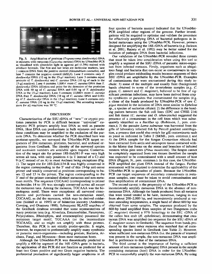
Detection of non-metazoan DNA in a mixture. A parasite- specific PCR product was produced from a mixture of T. dan- ilewskyi DNA and EPC cell line DNA when the concentration of EPC DNA was not greater than 250X that of T. danilewskyi DNA. The same results were obtained using C. salrnositica DNA and CHSE 214 cell line DNA. In both cases, as little as 1 pg (in a 15-y1 PCR) of parasite DNA was detected in the absence of cell line DNA. However, in the presence of cell line DNA (15 ng in a 15-pl PCR), at least 60 pg of parasite DNA was required for detection by UNonMet-PCR. However when T. danilewskyi DNA was mixed with C. auratus DNA, the UNonMet-PCR detected T. danilewskyi DNA when the con- centration of C. auratus DNA was not greater than 1,000X that of T. danilewskyi DNA (Fig. 3). In this case, 10 pg of T. dun- ilewskyi DNA was detected in 10 ng (in a 15-yl PCR) of C. auratus DNA.
Assay of natural parasitic infections. Histopathological ex- aminations indicated that the intensity of natural Hernatodiniurn sp. infections varied among C. tanneri. The parasite was de- tected in 13 of 67 C. tanneri via histopathology and samples from all 13 positive crabs produced a strong 450-bp band with the Hematodiniurn-specific PCR assay. The UNonMet-PCR produced a single strong 600-bp band in all samples from the 13 infected C. tanneri. The UNonMet-PCR products from two of the 13 samples were sequenced and both were found to be 97% identical to the Hematodiniurn sp. sequence provided in GenBank (Table 2). In the remaining 54 samples two or more bands ranging from 400 bp to 600 bp were produced by the UNonMet-PCR. Of these, 19 C. tanneri had faint 450-bp bands with the Hernatodiniurn-specific PCR probably indicating that light infections (not detectable by histopathology) were present.
BOWER ET AL.-UNIVERSAL NON-METAZOAN PCR 33 1
Fig. 3. Amplification of protistan (Ttypanosoma danilewskyi) DNA in mixtures with metazoan (Carassius auratus) DNA by UNonMet-PCR as visualised with ultraviolet light in agarose gel (1.5%) stained with ethidium bromide. The first and last lanes are molecular markers, lane 1 contains DNA from the positive control (Alexandrium cutenella), and lane 7 contains the negative control (ddH,O). Lane 2 contains only T. danilewskyi DNA (10 ng in the 15-pI reaction). Lane 3 contains equal amounts of T. danilewskyi and C. aurafus DNA (10 ng of each in the 15-pI reaction). Lane 4 contains 1,OOOX more C. aurafus DNA than T. danilewskyi DNA (dilution end point for the detection of the protistian DNA with 10 ng of C. aurafus DNA and 0.01 ng of T. danilewskyi DNA in the 15-(~1 reaction). Lane 5 contains 10,OOOX more C. aurafus DNA than T. dtznilewskyi DNA (10 ng of C. auratus DNA and 0.001 ng of T. danilewskyi DNA in the 15-(~1 reaction). Lane 6 contains only C. auratus DNA (10 ng in the 1.5-pl reaction). The annealing temper- ature for all reactions was 50 “C.
DISCUSSION Characterization of the SSU rDNA of “new” or cryptic pro-
tistan parasites by PCR is difficult because “universal” eu- karyotic PCR primers amplify host DNA as well as parasite DNA. Host DNA can predominate in bulk mixtures and under these conditions may be amplified to the exclusion of the par- asite DNA. To determine whether the PCR described here was protistan-specific, we surveyed the UNonMet-FTR target se- quences of 294 metazoan, protistan, bacterial, and archaeal or- ganisms from GenBank. The identity of the surveyed species and accession numbers are available upon request. Conserva- tion of the target site for primer 18s-EuW81-F was very high across all taxa, with only positions 4 (a T instead of a C) and 9 (a C instead of an A) in most Archaea being exceptions (Fig. 1). The target site for 18s-Euk1134-R was highly conserved in the region corresponding to the 5’ end of this 19-bp anti-sense primer and weakly conserved at positions corresponding to ba- ses 12 and 13 in the primer. The region corresponding to the 3’ end of the primer contained distinct metazoan and non-meta- zoan motifs. The sequence CGCAAG (corresponding to primer nucleotides 14 to 19) was strongly conserved across all except the metazoan taxa. Among the metazoa, TGCAAA was the ho- mologous motif. There were exceptions: all surveyed cteno- phores and poriferans carried the non-metazoan motif, CGCAAG, as did the myxozoa, which may be derived cnidar- ians (Siddall et al. 1995) or of bilaterian ancestry (Anderson, Canning, and Okamura 1998). Subsequent BLAST searches of the GenBank database revealed that some representatives of many protistan taxa (e.g. Alveolata, Entamoebidae, Paramyxea, Polycystinea, Rhodophyta, and stramenopiles) possessed the metazoan target motif, TGCAAA (or the intermediate TGCAAG), and so might not amplify with these primers. Therefore, this PCR is not strictly protist-specific. It might, however, be expected to preferentially amplify many symbiotic or parasitic micro-organisms-including protists, Bacteria, Ar- chaea, Fungi, and Myxozoa-from most metazoan hosts.
Although the UNonMet-PCR was theoretically predicted to amplify a 400-bp segment of the 16s rDNA gene in bacteria, the application of this PCR did not function as predicted for at least two Gram positive and two Gram negative bacteria. The preferential production of significantly larger amplicons in all
four species of bacteria assayed indicated that the UNonMet- PCR amplified other regions of the genome. Further investi- gations will be required to optimise and validate the procedure for effectively amplifying DNA of bacterial pathogens in in- fected metazoans using the UNonMet-PCR. However, primers designed for amplifying the 16s rDNA of bacteria (e.g. Jackson et al. 2001; Rainey et al. 1992) may be better suited for the isolation of pathogen DNA from bacterial infections.
Our validation of the UNonMet-PCR revealed three caveats that must be taken into consideration when using this tool to amplify a segment of the SSU rDNA of parasitic micro-organ- ism from infected metazoa. Firstly, organisms such as ubiqui- tous fungi and bacteria that can occur as contaminants in sam- ples could produce misleading results because segments of their SSU rDNA are amplifiable by the UNonMet-PCR. Examples of contaminants that were encountered during this study in- clude: 1) some of the multiple and usually faint (background) bands obtained in some of the invertebrate samples (e.g. C. gigas, C. tanneri and C. magister), believed to be free of sig- nificant protistan infections, may have been derived from cryp- tic symbionts or parasites that contaminated the samples (e.g. a clone of the bands produced by UNonMet-PCR of one C. gigas resulted in the isolation of DNA most similar to Ephelota sp., a species of suctorian ciliate); 2) the difference in the band- ing pattern between the fish cell lines (EPC and CHSE 214) and fish tissue (C. auratus and 0. tshawytscha) suggested the presence of a contaminant in the cell lines which was subse- quently identified as a Bacillus sp. (Robinson, J., pers. com- mun.); 3) the parasite Lama salmonae was harvested from the gills of laboratory infected fish by Percoll gradient centrifuga- tion, a process that could also enrich for gill contaminants such as yeast as indicated in Table 2 (SRMJ., pers. commun.); and 4) the DNA sample for the fungus Cronartium ribicola had been extracted from aecia and aeciospore tissue contained with- in the blister that forms on the stems and branches of infected western white pine trees (Pinus monticola). The fungus cannot be completely separated from tree tissues therefore the sample was expected to be contaminated with a small amount of host DNA (Piggott, N., pers. commun.). In this case, the UNonMet- PCR amplified the plant DNA instead of the fungus. Further assessment will be required to determine the applicability of the UNonMet-PCR to parasites of plants. Because the UNonMet- PCR can target sequences of secondary contaminants in meta- zoan samples, care must be taken to avoid Contamination and thus amplification of contaminant DNA.
The second caveat is the propensity of the UNonMet-PCR to inconsistently amplify metazoan DNA in the absence of non- metazoan DNA. Although the bands produced from all metazoa that were tested tended to be faint, numerous, ranging in size from about 200-2,000 bp, and inconsistently produced at var- ious annealing temperatures, a single band of about 600 bp was obtained from some samples. The sequence produced by the 600-bp band amplified from samples of two Dungeness crabs (C. magister) showed a 97% homology to the SSU rDNA of the calico box crab (H. epheliticus), demonstrating that CIUS- tacean DNA was amplified (no sequence for the SSU rDNA of C. magister occurs in Genbank). The sequence from bands pro- duced by the three other metazoa also matched that of corre- sponding species listed in GenBank (see Table 2). However, when sufficient non-metazoan DNA (i.e. the parasite of interest) was present in the sample, the non-metazoan DNA was ampli- fied in preference to that of the metazoan DNA.
The third caveat is the importance of having a sufficient amount of non-metazoan (pathogen) DNA present in the sample containing metazoan (host) DNA in order for the UNonMet- PCR to successfully amplify the non-metazoan DNA. By using
332 J . EUKARYOT. MICROBIOL., VOL. 5 1, NO. 3, MAY-JUNE 2004
mixtures of parasite DNA and DNA from host cell lines (i.e. T. danilewskyi DNA with EPC (C. carpio) cell line DNA and C. sulmositica DNA with CHSE-214 (0. tshawytscha) cell line DNA), we determined that a band corresponding to amplifica- tion of parasite DNA was produced when the concentration of the cell line DNA did not exceed approximately 250X that of the parasite DNA. However, T. danilewskyi DNA was detected by UNonMet-PCR in mixtures with four times as much DNA obtained directly from the goldfish host. The presence of a con- taminating Bacillus sp. in the EPC cell line possibly provided competition for the UNonMet primers thereby reaffirming the first caveat and exemplifying the impact that contaminants may have on results. Alternately, DNA for C. carpio and C. auratus may be differentially amplified by the UNonMet-PCR. Exam- ination of crabs naturally infected with Hematodinium provided further support for the third caveat. Amplification of DNA from heavily infected C. tanneri resulted in a strong 600-bp band identified as Hematodinium, in contrast to the faint or multiple bands produced from lightly infected or non-infected crabs.
In conjunction with the three caveats described above, it is necessary to have appropriate uninfected controls when using this primer set in order to characterize bands that may be pro- duced by non-specific amplification of metazoan DNA. In ad- dition, characterization of the PCR product(s) by techniques such as sequencing or restriction analysis is essential in con- firming the identity of the amplicon(s). Nevertheless, during our validation process, the UNonMet-PCR proved useful in specif- ically identifying two phytoplankton cultures (Isochrysis sp. (CCMP 1324) as Isochrysis galbana and Chrysochromulina sp. as Chrysochrornulina hirra) and indicated the contamination of another phytoplankton culture (the isolate thought to be Rho- domonas sp. was a species of Tetraselmis sp.). In addition, par- tial sequences of the SSU rDNA of five organisms (Cancer magister, Trypanosoma danilewskyi, Pinus monticola, Chaeto- ceros gracilis, and Chionoecetes tanneri) not previously avail- able in GenBank were added to the database (accession numbers AY527220, AY52722 I , AY527222, AY527223, AY527224, respectively). Future research could include the characterisation of secondary bands obtained in many of the samples. In addition, further refinement of the UNonMet-PCR could reduce the production of the secondary bands and in- crease the specificity of the primer set.
The application of PCR using the UNonMet-PCR could be a significant research tool as new fungal and protistan parasitic diseases of metazwa emerge. The taxonomic affinities of a new or cryptic parasite are normally deduced only after expensive morphological analysis often including time-consuming ultra- structural examinations by electron microscopy. Genetic char- acterisation may lag behind because of problems associated with isolating the organism or amplifying its DNA from host tissue. Using this PCR assay, parasite SSU rDNA could be rap- idly amplified for sequencing and its phylogenetic affiliation deduced within days. Inferences about a potential parasite life cycle could follow based on the life histories of other members of its taxon. Thus, this non-metazoan PCR assay might expedite the description of new species and emerging pathogens.
ACKNOWLEDGMENTS
We are grateful to the suppliers of materials for testing as indicated in Table 1 . We also thank Karia Kaukinen, Angela Schulze and Amy Tabata for sequencing our samples on the automated DNA sequencer and Gary Meyer for assistance in preparing the figures. This project was financially supported by
the Canadian Regulatory Systems for Biotechnology compo- nent of the Canadian Biotechnology Strategy Funds.
LITERATURE CITED Altschul, S. F., Madden, T. L., Schaffer, A. A., Zhang, J., Zhang, Z.,
Miller, W. & Lipman, D. J. 1997. Gapped BLAST and PSI-BLAST a new generation of protein database search programs. Nucl. Acids Res.. 25:3389-3402.
Anderson, C. L., Canning, E. U. & Okamura, B. 1998. A triploblastic origin for Myxozoa? Nature, 392:346-347.
Bower, S . M., Hervio, D. & Meyer, G. R. 1997. Infectivity of Mikro- cyros mackini, the causative agent of Denman Island disease in Pa- cific oysters Crassostrea gigas, to various species of oysters. Dis. Aquar. Org.. 29:lll-116.
Carnegie, R. B., Barber, B. J., Culloty, S. C., Figueras. A. J. & Distel, D. L. 2000. Development of a PCR assay for detection of the oyster pathogen Bonamia osrreae and support for its inclusion in the Hap- losporidia. Dis. Aqua?. Org.. 42:9Y-206.
Carnegie, R. B., Meyer, G. R., Blackbourn, J. , Cochennec-Laureau, N., Berthe, F. C. J. & Bower, S. M. 2003. Molecular detection of the oyster parasite Mikrocytos mackini, and a preliminary phylogenetic analysis. Dis. Aquar. Org.. 54:2 19-227.
Corpet, F. 1988. Multiple sequence alignment with hierarchical cluster- ing. Nucl. Acids Res., 16: 1 OX8 1-1 0890.
Cunningham, C. 0. 2002. Molecular diagnosis of salmonid diseases. Kluwer Academic Publishers, Dordrecht, Netherlands.
Docker, M. F., Devlin, R. H., Richard, J., Khattra, J. & Kent, M. L. 1997. Sensitive and specific polymerase chain reaction assay for de- tection of Loma salmonae (Microsporea). Dis. Aquat. Org., 29:4 1- 48.
Farley, C. A., Wolf, P. H. & Elston, R. A. 1988. A long-term study of “microcell” disease in oysters with description of a new genus, Mik- rocytos (g. n.), and two new species, Mikrocytos mackini (sp. n.) and Mikrocytos roughleyi (sp. n.). Fish. Bull., 8658 1-593.
Hervio, D., Bower, S. M. & Meyer, G . R. 1996. Detection, isolation, and experimental transmission of Mikrocytos mackini, a microcell parasite of Pacific oysters Crassostrea gigus (Thunberg). J. Invert. Pathol.. 67:72-79.
Hillis, D. M. & Dixon, M. T. 199 1. Ribosomal DNA: molecular evo- lution and phylogenetic inference. Quart. Rev. Biol.. 66:4 I 1453 .
Hine, P. M., Bower, S. M., Meyer, G. R., Cochennec-Laureau, N. & Berthe. F. C. J. 2001. Ultrastructure of Mikrocytos muckini, the cause of Denman Island disease in oysters Crussostrea spp. and Ostrea spp. in British Columbia, Canada. Dis. Aquat. Org., 45:215-227.
Howard, D. W. & Smith, C. S. 1983. Histological techniques for bivalve mollusks. NOAA Technical Memorandum NMFS-FINEC-25.
Jackson, C. R., Langner, H. W., Donahoe-Christiansen, J., Inskeep, W. P. & McDermott, T. R. 2001. Molecular analysis of microbial corn- munity structure in an arsenite-oxidizing acidic thermal spring. En- viron. Microbiol.. 3532-542.
Jones, S. R. M., Goh, B. & Prosperi-Porta, G. 2003. Duration and meth- od of fixation affects the sensitivity of a digoxygenin-labelled DNA probe in detecting Kudoa thyrsites in Atlantic salmon skeletal muscle. Aquaculrure, 220: 157-164.
Kitagawa, R. & Rose, A. M. 1999. Components of the spindle-assembly checkpoint are essential in Caenorhabditis elegans. Nut. Cell Biol.. 1:5 16521.
Quayle, D. B. 1961. Denman Island oyster disease and mortality, 1960. Fish. Res. Bd. Can. Manu. Rep., 71336~.
Rainey, F.A., Dorsch, M., Morgan, H. W. & Stackebrandt, E. 1992. 16s rDNA analysis of Spirochaeta thermophila: its phylogenetic position and implications for the systematics of the order Spirochaetales. Syst. Appl. Microbiol. 15: 197-202.
Siddall, M. E., Martin, D. S., Bridge, D., Desser, S. S. & Cone, D. K. 1995. The demise of a phylum of protists: phylogeny of the Myxozoa and other parasitic cnidaria. J. Parasitol.. 81:96 1-967.
Stokes, N. A., Siddall, M. E. & Burreson, E. M. 1995. Detection of Haplosporidium nelsoni (Haplosporidia: Haplosporidiidae) in oysters by PCR amplification. Dis. Aquat. Org., 23: 145-152.
Received I I / ] 7/03, 03/32/04; accepted 03/13/04


![Staal and Tupene and Western Sydney Area Health Service [2004] NSWIRComm 325 (4 November 2004)](https://img.pdfslide.us/doc/110x75/5464f62fb4af9fb96e8b48f9/staal-and-tupene-and-western-sydney-area-health-service-2004-nswircomm-325-4-november-2004.jpg)