Embed Size (px)
Citation preview
1
Neuroepigenetic Regulation and Down Syndrome
Richard C. Deth, PhD Department of Pharmaceutical Sciences Northeastern University
The image cannot be displayed. Your computer may not have enough memory to
Key Points: • Epigenetic regulation of gene expression is the driver of development
• DS (trisomy of Chr21) increases “gene dosage” • Genes on Chr21 can affect development via their effect on cellular oxidative state and methylation status
• Altered methylation may also contribute to DS risk • Metabolic support of methylation may help optimize the abilities of DS individuals
4
• 1. Epigenetic regulation of gene expression is the driver of development
5
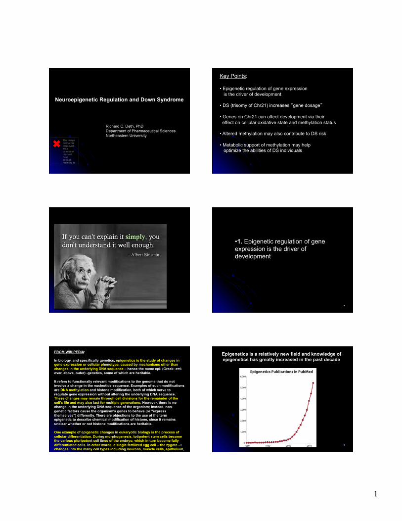
FROM WIKIPEDIA: In biology, and specifically genetics, epigenetics is the study of changes in gene expression or cellular phenotype, caused by mechanisms other than changes in the underlying DNA sequence – hence the name epi- (Greek: επί- over, above, outer) -genetics, some of which are heritable. It refers to functionally relevant modifications to the genome that do not involve a change in the nucleotide sequence. Examples of such modifications are DNA methylation and histone modification, both of which serve to regulate gene expression without altering the underlying DNA sequence. These changes may remain through cell divisions for the remainder of the cell's life and may also last for multiple generations. However, there is no change in the underlying DNA sequence of the organism; instead, non-genetic factors cause the organism's genes to behave (or "express themselves") differently. There are objections to the use of the term epigenetic to describe chemical modification of histone, since it remains unclear whether or not histone modifications are heritable. One example of epigenetic changes in eukaryotic biology is the process of cellular differentiation. During morphogenesis, totipotent stem cells become the various pluripotent cell lines of the embryo, which in turn become fully differentiated cells. In other words, a single fertilized egg cell – the zygote – changes into the many cell types including neurons, muscle cells, epithelium, endothelium of blood vessels, etc. as it continues to divide. It does so by activating some genes while inhibiting others.
6
Epigenetics is a relatively new field and knowledge of epigenetics has greatly increased in the past decade
2
7
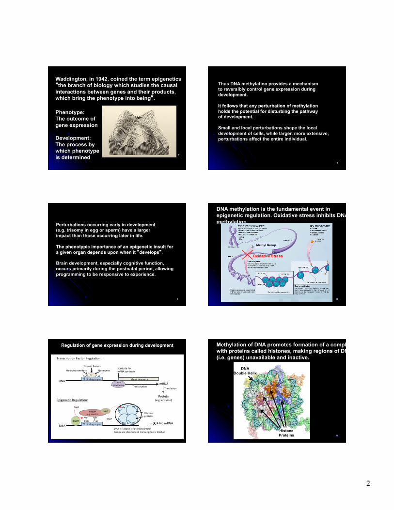
Waddington, in 1942, coined the term epigenetics “the branch of biology which studies the causal interactions between genes and their products, which bring the phenotype into being”.
Phenotype: The outcome of gene expression Development: The process by which phenotype is determined 8
Thus DNA methylation provides a mechanism to reversibly control gene expression during development. It follows that any perturbation of methylation holds the potential for disturbing the pathway of development. Small and local perturbations shape the local development of cells, while larger, more extensive, perturbations affect the entire individual.
9
Perturbations occurring early in development (e.g. trisomy in egg or sperm) have a larger impact than those occurring later in life. The phenotypic importance of an epigenetic insult for a given organ depends upon when it “develops”. Brain development, especially cognitive function, occurs primarily during the postnatal period, allowing programming to be responsive to experience.
10
DNA methylation is the fundamental event in epigenetic regulation. Oxidative stress inhibits DNA methylation.
Methyl Group
Oxidative Stress
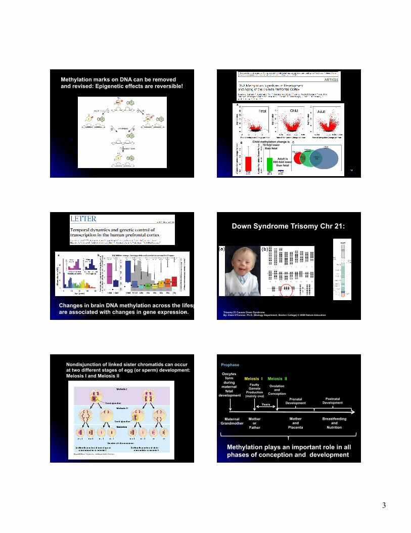
Regulation of gene expression during development
X
Gene sequence
Start site for mRNA synthesis
TF
Growth Factors Neurotransmi:ers Hormones
RNA polymerase mRNA
Protein (e.g. enzyme)
DNA
TranscripEon TranslaEon
DNA DNA + Histone = HeterochromaEn Genes are silenced and transcripEon is blocked
Me Me
MBDP (e.g. MeCP2) Histone
proteins
HMT
Me Me
Me Me DNMT
SAM
SAM CpG CpG
TranscripEon Factor RegulaEon:
EpigeneEc RegulaEon:
TF binding region
TF binding region
CpG CpG
No mRNA X
12
Methylation of DNA promotes formation of a complex with proteins called histones, making regions of DNA (i.e. genes) unavailable and inactive.
DNA Double Helix
Histone Proteins
3
Methylation marks on DNA can be removed and revised: Epigenetic effects are reversible!
14
Child methylation change is 70-fold lower
than fetal
Adult is 600-fold lower
than fetal
15
Changes in brain DNA methylation across the lifespan are associated with changes in gene expression.
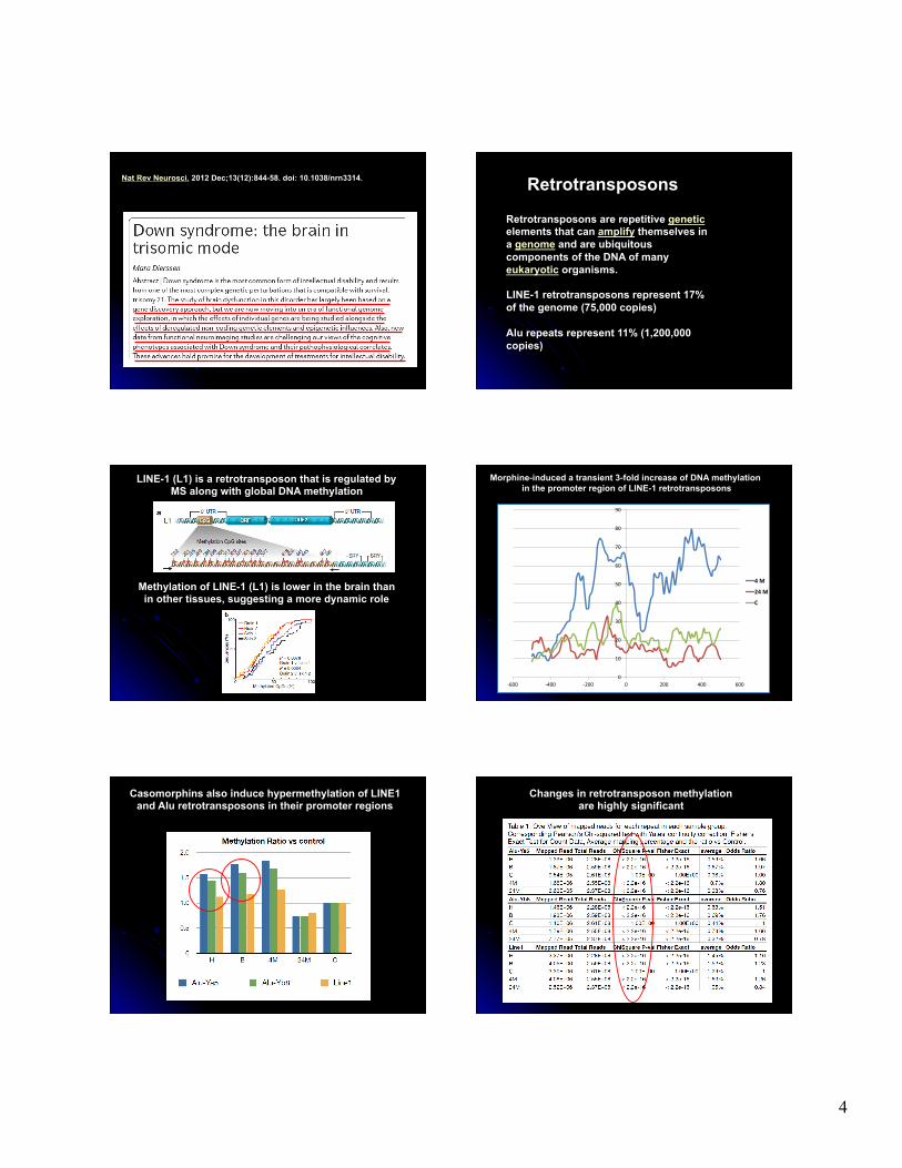
Down Syndrome Trisomy Chr 21:
Trisomy 21 Causes Down Syndrome By: Clare O'Connor, Ph.D. (Biology Department, Boston College) © 2008 Nature Education
Nondisjunction of linked sister chromatids can occur at two different stages of egg (or sperm) development: Meiosis I and Meiosis II
Faulty Gamete
Production (mainly ova)
Ovulation and
Conception Prenatal
Development Postnatal
Development
Mother and
Placenta
Breastfeeding and
Nutrition
Methylation plays an important role in all phases of conception and development
Meiosis I Meiosis II Oocytes
form during
maternal fetal
development
Mother or
Father
Maternal Grandmother
Years
Prophase
4
Nat Rev Neurosci. 2012 Dec;13(12):844-58. doi: 10.1038/nrn3314.
Retrotransposons are repetitive genetic elements that can amplify themselves in a genome and are ubiquitous components of the DNA of many eukaryotic organisms. LINE-1 retrotransposons represent 17% of the genome (75,000 copies) Alu repeats represent 11% (1,200,000 copies)
Retrotransposons
LINE-1 (L1) is a retrotransposon that is regulated by MS along with global DNA methylation
Methylation of LINE-1 (L1) is lower in the brain than in other tissues, suggesting a more dynamic role
0
10
20
30
40
50
60
70
80
90
-‐600 -‐400 -‐200 0 200 400 600
4 M
24 M
C
Morphine-induced a transient 3-fold increase of DNA methylation in the promoter region of LINE-1 retrotransposons
Casomorphins also induce hypermethylation of LINE1 and Alu retrotransposons in their promoter regions
Changes in retrotransposon methylation are highly significant
5
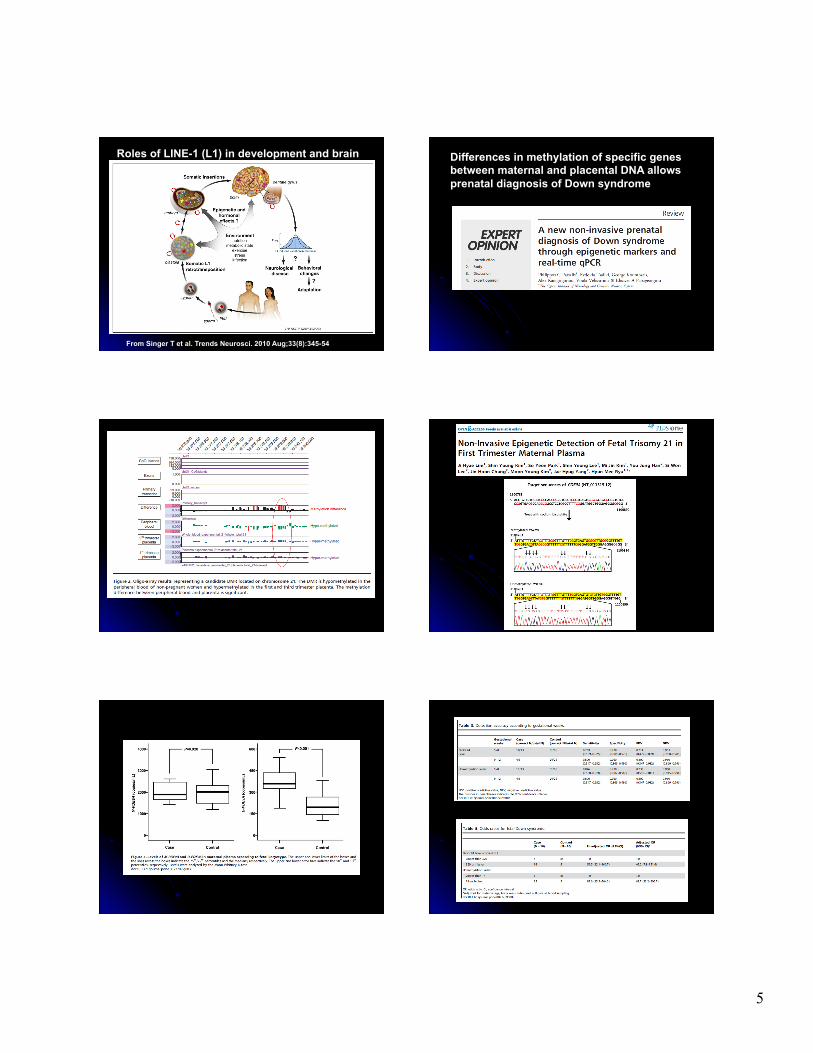
From Singer T et al. Trends Neurosci. 2010 Aug;33(8):345-54
Roles of LINE-1 (L1) in development and brain Differences in methylation of specific genes between maternal and placental DNA allows prenatal diagnosis of Down syndrome
6
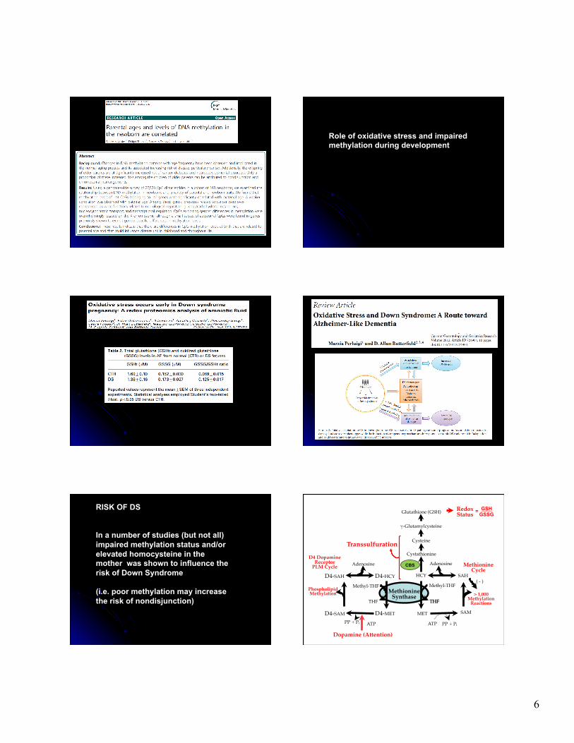
Role of oxidative stress and impaired methylation during development
RISK OF DS In a number of studies (but not all) impaired methylation status and/or elevated homocysteine in the mother was shown to influence the risk of Down Syndrome (i.e. poor methylation may increase the risk of nondisjunction)
Glutathione (GSH)! Redox!Status!
"-Glutamylcysteine!
Cysteine!
Cystathionine!
HCY!
Methionine!Cycle!
SAH!
Adenosine!
> 1,000!Methylation!
Reactions!
SAM!
( - )!
ATP! PP + Pi!
THF!
Methyl-THF!Methionine!
Synthase!
MET!
Adenosine!
D4-SAH!
ATP!PP + Pi!
Dopamine (Attention)!
Phospholipid!Methylation!
D4-HCY!
D4-MET!D4-SAM!
Methyl-THF!
THF! THF!
D4 Dopamine!Receptor !
PLM Cycle! CBS
GSH GSSG =
Transsulfuration!
7
24% decrease in HCY in DS 47% decrease in MET in DS 350% increase in cystathionine
15% decrease in cysteine in DS
Consistent with a decrease in methionine synthase activity and an increase in CBS activity
25% decrease in combined SAM and SAH in DS 33% increase in adenosine in DS
Consistent with a decrease in methionine synthase activity and an increase in CBS activity
Glutathione (GSH)! Redox!Status!
"-Glutamylcysteine!
Cysteine!
Cystathionine!
HCY!
↓Methionine!
Cycle!SAH!
Adenosine!
> 1,000!Methylation!
Reactions!
SAM!
( - )!
ATP! PP + Pi!
THF!
Methyl-THF!Methionine!
Synthase!
MET!
Adenosine!
D4-SAH!
ATP!PP + Pi!
Dopamine (Attention)!
Phospholipid!Methylation!
D4-HCY!
D4-MET!D4-SAM!
Methyl-THF!
THF! THF!
D4 Dopamine!Receptor !
PLM Cycle! CBS
GSH GSSG =
↑Transsulfuratio
n!
In DS: Increased transsulfuration Decreased methylation
• Chromosome 21 has about 400 genes. Increased gene dosage from each of them probably contributes to Down Syndrome characteristics.
• Certain genes deserve special attention for their relationship to oxidative stress and methylation, which are the foundation of development, especially brain development.
• These genes is located in the region of chromosome 21 (21q21-22) that has been implicated as being most important for DS.
• Increased activity of the proteins produced by these genes is likely to contribute to DS.
Amyloid precursor protein (APP) Cystathionine-beta-synthase (CBS) DNA methyltransferase 3L (DNMT3L) Formiminotransferase cyclodeaminase (FTCD) Superoxide dismutase 1 (SOD1)
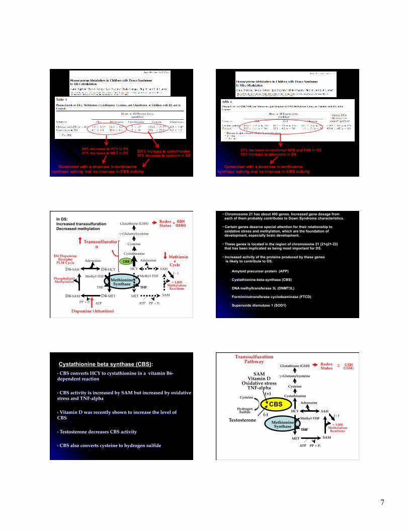
Cystathionine beta synthase (CBS): • CBS converts HCY to cystathionine in a vitamin B6-dependent reaction!!• CBS activity is increased by SAM but increased by oxidative stress and TNF-alpha!
• Vitamin D was recently shown to increase the level of CBS !
• Testosterone decreases CBS activity !
• CBS also converts cysteine to hydrogen sulfide!
Transsulfuration"Pathway!
Glutathione (GSH)! Redox!Status!
#-Glutamylcysteine!
Cysteine!
Cystathionine!
HCY! SAH!
Adenosine!
> 1,000!Methylation!
Reactions!
SAM!
( - )!
ATP! PP + Pi!
THF!
Methyl-THF!Methionine!
Synthase!
MET!
THF!
GSH!GSSG!=!~!
CBS Cysteine!
Hydrogen!Sulfide!!
SAM!Vitamin D!
Oxidative stress!TNF-alpha!
(+)!
Testosterone!(-)!
8
Excessive CBS activity in DS limits methylation by removing HCY from the methionine cycle. The critical balance between methylation and transsulfuration is therefore altered.
AMYLOID PRECURSOR PROTEIN (APP): A cleavage product of amyloid precursor protein (APP), known as Aβ, is though to be the primary cause of Alzheimer’s disease (AD). Amyloid plaques rich in Aβ are found at autopsy in AD brain, but the neurodegeneration is thought to be caused by small Aβ oligomers, starting decades before the onset of AD symptoms. AD is much more common in DS, presumably because the extra APP gene leads to increased Aβ.
Amyloid Processing
http://www.bath.ac.uk/bio-sci/research/profiles/brown-d.html
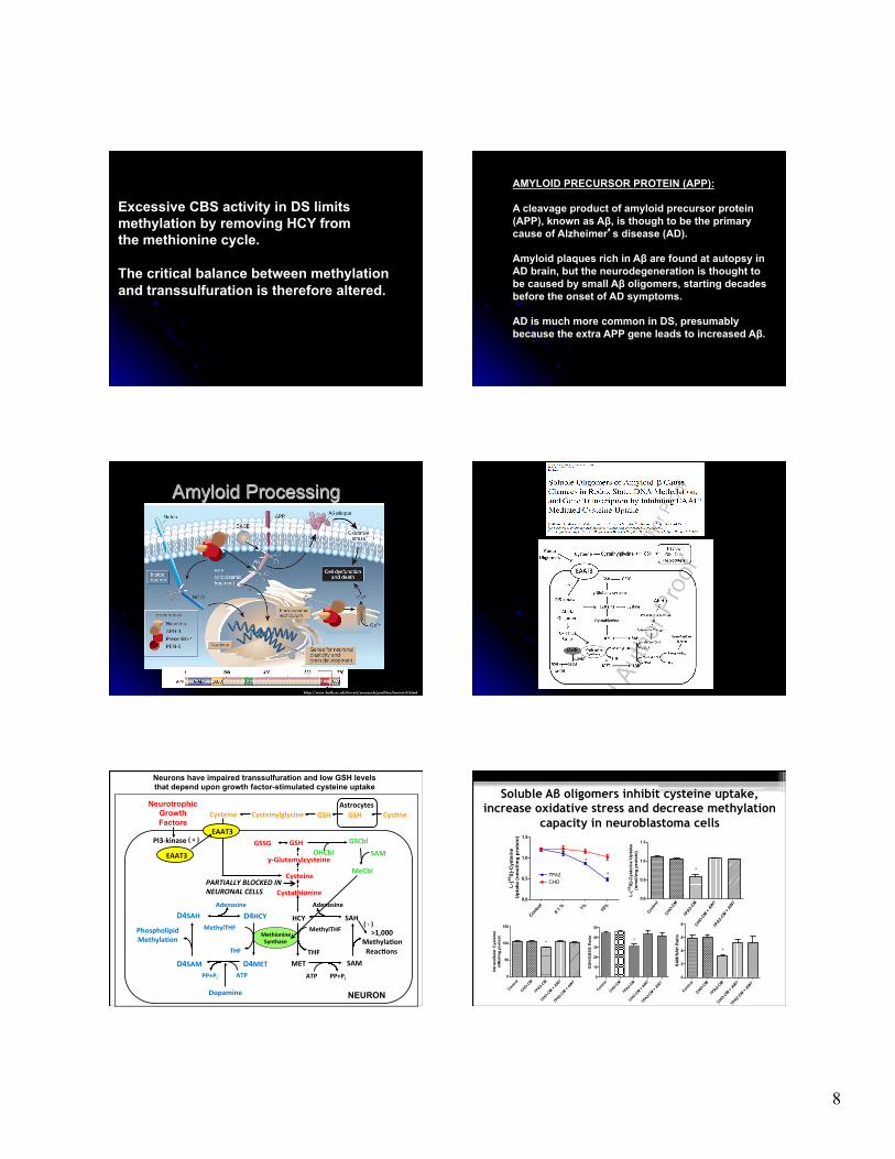
Neurons have impaired transsulfuration and low GSH levels that depend upon growth factor-stimulated cysteine uptake
Methionine Synthase
HCY
MET
SAH
SAM
>1,000 Methyla8on Reac8ons
ATP PP+Pi
Adenosine
MethylTHF
THF
Cystathionine
Cysteine
GSH
γ-‐Glutamylcysteine
D4HCY
D4SAM
D4SAH
D4MET ATP PP+Pi
MethylTHF
THF
Phospholipid Methyla8on
Adenosine
Dopamine
Cysteine
( -‐ )
PI3-‐kinase ( + )
PARTIALLY BLOCKED IN NEURONAL CELLS
EAAT3
Astrocytes Cysteinylglycine GSH
GSSG
Neurotrophic Growth Factors
GSCbl
MeCbl
SAM OHCbl
GSH
NEURON
Cys8ne
EAAT3
Soluble Aβ oligomers inhibit cysteine uptake, increase oxidative stress and decrease methylation
capacity in neuroblastoma cells
Contro
l
CHO-CM
7PA2-C
M
CHO-CM +
AW7
7PA2-C
M + AW7
0
50
100
150
*
Intr
acel
lula
r C
yste
ine
nMol
/mg
prot
ein
Contro
l
CHO-CM
7PA2-C
M
CHO-CM +
AW7
7PA2-C
M + AW7
0
10
20
30
40
50
*
GS
H/G
SS
G R
atio
Contro
l
CHO-CM
7PA2-C
M
CHO-CM +
AW7
7PA2-C
M + AW7
0
2
4
6
8
*
SA
M/S
AH
Rat
io
Contro
l0.1
% 1% 10%
0.0
0.5
1.0
1.5
*
*
7PA2CHO
L-[35
S]-
Cys
tein
e U
ptak
e (n
mol
/mg
prot
ein)
Contro
l
CHO-CM
7PA2-
CMCHO-C
M + AW
77P
A2-CM +
AW7
0.0
0.5
1.0
1.5
*
L-[35
S]-
Cys
tein
e U
ptak
e(n
mol
/mg
prot
ein)
9
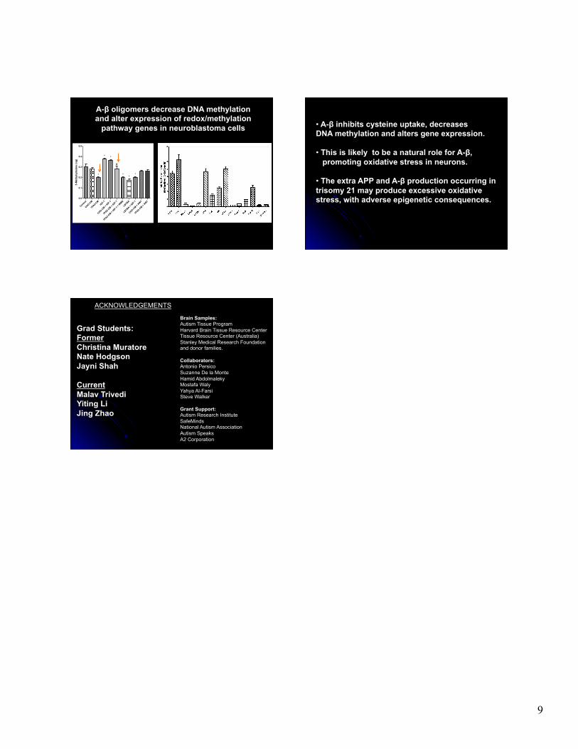
A-β oligomers decrease DNA methylation and alter expression of redox/methylation
pathway genes in neuroblastoma cells
Control
CHO-CM
7PA2-C
MIG
F-1
CHO-CM +
IGF-1
7PA2-C
M + IGF-1
7PA2-C
M + IGF-1
+ WMN
LBTBA
LBTBA + IG
F-1
CHO-CM +
AW7
7PA2-C
M + AW7
0.0
0.1
0.2
0.3
0.4
0.5
*
*
*
#
*
**
5-M
ethy
lcyt
osin
e (n
g)
• A-β inhibits cysteine uptake, decreases DNA methylation and alters gene expression. • This is likely to be a natural role for A-β, promoting oxidative stress in neurons. • The extra APP and A-β production occurring in trisomy 21 may produce excessive oxidative stress, with adverse epigenetic consequences.
Brain Samples: Autism Tissue Program Harvard Brain Tissue Resource Center Tissue Resource Center (Australia) Stanley Medical Research Foundation and donor families. Collaborators: Antonio Persico Suzanne De la Monte Hamid Abdolmaleky Mostafa Waly Yahya Al-Farsi Steve Walker Grant Support: Autism Research Institute SafeMinds National Autism Association Autism Speaks A2 Corporation
ACKNOWLEDGEMENTS
Grad Students: Former Christina Muratore Nate Hodgson Jayni Shah Current Malav Trivedi Yiting Li Jing Zhao