Embed Size (px)
Citation preview
Research ArticleEffect of High Protein Diet and Probiotic Lactobacillus caseiShirota Supplementation in Aflatoxin B1-Induced Rats
Z. Nurul Adilah, Winnie-Pui-Pui Liew, S. Mohd Redzwan , and I. Amin
Department of Nutrition and Dietetics, Faculty of Medicine and Health Sciences, Universiti Putra Malaysia,43400 Serdang, Selangor, Malaysia
Correspondence should be addressed to S. Mohd Redzwan; [email protected]
Received 16 November 2017; Revised 12 March 2018; Accepted 5 April 2018; Published 23 May 2018
Academic Editor: Koichiro Wada
Copyright © 2018 Z. Nurul Adilah et al.This is an open access article distributed under the Creative Commons Attribution License,which permits unrestricted use, distribution, and reproduction in any medium, provided the original work is properly cited.
Probiotic Lactobacillus casei Shirota (LcS) is a potential decontaminating agent of aflatoxin B1 (AFB1). However, few studies haveinvestigated the influence of diet, especially a high protein (HP) diet, on the binding of AFB1 by probiotics. This research wasconducted to determine the effect of HP diet on the ability of LcS to bind AFB1 and reduce aflatoxin M1 (AFM1) in AFB1-inducedrats. Sprague Dawley rats were randomly divided into three groups: A (HP only), B (HP + 108 CFU LcS + 25 𝜇g AFB1/kg BW), andC (HP + 25 𝜇gAFB1/kg BW). Levels of AST and ALP were higher in all groups but other liver function’s biomarkers were in thenormal range, and the liver’s histology showed no structural changes. The urea level of rats in group B (10.02 ± 0.73mmol/l) wassignificantly lower (𝑝 < 0.05) than that of rats in group A (10.82 ± 0.26mmol/l). The presence of carcinoma in the small intestineand colon was more obvious in group C than in group B. Moreover, rats in group B had significantly (𝑝 < 0.05) lower AFM1concentration (0.39 ± 0.01 ng/ml) than rats in group C (5.22 ± 0.28 ng/ml). Through these findings, LcS supplementation with HPdiet alleviated the adverse effects of AFB1 by preventing AFB1 absorption in the small intestine and reducing urinary AFM1.
1. Introduction
Aflatoxin is produced by Aspergillus flavus and Aspergillusparasiticus [1]. This food contaminant can be found com-monly in agricultural and food commodities such as maize,grains, peanuts, cereal, and animal feeds [1]. Food legislationand food processing are becoming more advanced, yet theyare still unable to prevent the occurrence of aflatoxin in foodcommodities. Due to high stability of aflatoxin, it has becomea problem not only during harvesting but also at every stageof food production, starting from harvesting of raw material,storage, and processing until the food reaches the consumers[2]. Aflatoxin should be removed, as long-term consumptionof aflatoxin-contaminated food can cause carcinogenic andtoxicity effects on humans and animals [3].
One of the main adverse effects of dietary aflatoxin expo-sure is aflatoxicosis. Aflatoxicosis is a foodborne disease dueto aflatoxin ingestion in the diet and it can be categorized intoacute and chronic aflatoxicosis. Acute aflatoxicosis resultsin death, whereas chronic aflatoxicosis can cause immune
suppression, cancer, and other “slow” pathohistological con-ditions [4].The liver is themain target of aflatoxicosis. In fact,one of the metabolites of aflatoxin, aflatoxin B1 (AFB1), hasbeen classified by the International Agency for Research onCancer (IARC) as group 1 carcinogen and is linked to thedevelopment of the liver cancer [5].
The recent alternative approach to remove AFB1 fromthe body is through the consumption of probiotic bacteria,as some studies have shown that probiotic bacteria mightbe a potential adsorbent of aflatoxin in the gastrointestinaltract by reducing aflatoxin bioavailability [6, 7].WorldHealthOrganization (WHO) defined probiotics as “live microor-ganisms” which are able to provide advantages to the hostwhen consumed in an adequate amount [8]. It is evident thatprobiotic consumption improved gastrointestinal health andimmune system [9]. As an adsorbent of aflatoxin, probioticbacteria remove AFB1 through noncovalent binding of AFB1molecule to the bacterial cell wall [10]. Besides, polysaccha-rides and peptidoglycan components as well as teichoic acidsof bacterial cell wall are involved in the binding process
HindawiBioMed Research InternationalVolume 2018, Article ID 9568351, 10 pageshttps://doi.org/10.1155/2018/9568351
2 BioMed Research International
of AFB1 by probiotics [7]. The authors indicated also thatteichoic acids play a major role in the binding of AFB1 byLactobacillus reuteri and Lactobacillus casei Shirota (LcS) [7].
A randomized, double-blind, cross-over, placebo-con-trolled study with two 4-week intervention phases wasconducted [11] to investigate the effectiveness of probioticsin reducing circular production of aflatoxin biomarkers in apopulation exposed to aflatoxin. The authors found that pro-biotic intervention reduced AFB1-lysine adduct (AFB1-lys)and urinary aflatoxin M1 (AFM1) in certain subjects [11].Interestingly, it was found that diets may be one of theconfounding factors that can affect the ability of probioticLcS to bind AFB1 in the gastrointestinal tract. In addition,high intake of macronutrients can influence the metabolismof aflatoxin and subsequently affect the circular productionof AFB1 metabolites [11].
In a recent review article [12], the authors mentioned thata high protein (HP) diet can affect aflatoxicosis. For example,rats fedwithHPdiet and exposed toAFB1 hadno focal hyper-plasia and less ductular reaction of liver [13].These symptomsare commonly observed in the early stages of hepatocellularcarcinoma (HCC). As such, the findings [13] may explainthe effect of diets on the metabolism of aflatoxin as reportedby Mohd Redzwan et al. [11] and to some extent slow theprogression of HCC associated with aflatoxin exposure. Onthe other hand, it is still unclear whether diet manipulationcan have influence on the activity of probiotics in reducingAFB1 bioavailability. A study by Nikbakht Nasrabadi et al.[14] found that LcS supplementation in aflatoxin-induced ratsreduced serum AFB1. Nevertheless, the authors [14] did nottake into consideration the influence of diet in the studyprotocol. Furthermore, diet was found as a confoundingfactor that can affect LcS activity and aflatoxin metabolism[11]. Therefore, this research was conducted to elucidate theeffect of HP diet on the ability of probiotic LcS to reduceurinary AFM1 and certain aflatoxicosis symptoms in AFB1-induced rats.
2. Materials and Methods
Yakult fermented milk drink was purchased from a localsupermarket in Serdang, Selangor, Malaysia. AFB1 wasacquired from Trilogy Analytical Laboratory, Inc. (VossbrinkDrive, Washington). MRS broth, MRS agar, and sodiumchloride were purchased fromMerck (Darmstadt, Germany).Glycerol solution was procured from Sigma-Aldrich (St.Louis, MO, USA). ELISA kit for the detection of AFM1 inurine was purchased from Helica Biosystems, Inc. (SantaAna, CA, USA).
2.1. Culturing Probiotic Bacteria. The source of LcS was fromthe Yakult cultured milk drink (contained live LcS) [14]. Onehundred microliters (100 𝜇L) of Yakult was aseptically spreadontoMRS agar and incubated for 48 hours aerobically at 37∘C.Then, one colony of LcS was transferred into MRS broth andfurther cultured for another 24 hours aerobically at 37∘C.The growth of LcS was recorded every two hours, and thecorresponding CFU was monitored optically at 600 nm. Onehundred microliters (100 𝜇L) was withdrawn from the broth
and spread on the MRS agar to acquire the CFU. LcS’s abilityto bind AFB1 also depends on bacterial concentration. For asignificant removal of 50% of aflatoxin, the minimum con-centration of Lactobacillus bacteria needed is 2 × 10⁹ CFU/ml[14]. Based on the absorbance value (OD) and CFU that wererecorded during the 24 h of incubation, the bacterial cell wasincubated at least 19 h in order to reach concentration of109 CFU.The bacterial cells were harvested via centrifugationat 2200𝑔 using Kubota 2810 centrifuge (Tokyo, Japan) for 15minutes and the supernatant was discarded. The bacterialpellet was resuspended with 50% (v/v) glycerol [14]. Priorto the usage, the glycerol liquid was replaced with salinesolution. The identity of LcS cultured from Yakult culturedmilk drink was confirmed with the 16S rRNA sequencingservice provided by First BASE Laboratories Sdn Bhd (SeriKembangan, Selangor, Malaysia).
2.2. Animals. Twenty-four (𝑛 = 24) male Sprague Dawleyrats (7-8 weeks old, 290–300 g) were purchased from theAnimal Resource Unit (ARU), Department of VeterinaryPathology andMicrobiology, Faculty of VeterinaryMedicine,University Putra Malaysia (UPM). The rats were kept atroom temperature under standard conditions of light (12 hlight-dark cycle) and regulated temperature (20–22∘C) andventilation in the animal research house of ComparativeMedicine and Technology Unit (COMeT), Institute of Bio-science, UPM. Two rats were housed in a cage with woodshavings. The cleaning process of the cages was performedtwo times per week and water supply was changed on a dailybasis. Ethical approval for this animal study was given by theInstitutional Animal Care and Use Committee, UPM (UPM/IACUC/AUP-R098/2016).
2.3. Preparation of Diet. A high protein (HP) diet wasprepared based on Envigo recipe [15]. This diet was approxi-mately 40% protein in terms of calories.
2.4. Experimental Study. Twenty-four rats (𝑛 = 24) wererandomly divided into three groups of diets. Rats in groupA (𝑛 = 8) received HP diet only (HP only); rats in groupB (𝑛 = 8) were fed with HP diet supplemented with LcS(108-109 CFU) and AFB1 (LcS + HP + AFB1), while rats ingroup C (𝑛 = 8) were only provided with HP diet and AFB1(HP + AFB1). For rats in group B, immediately after thefifth probiotic dose, AFB1 was given at a complete dosage.The complete dosage of AFB1 for rats in group B was 25 𝜇gAFB1/kg body weight (BW). The dose selected in this studyis equivalent to 0.03 to 0.45mg/kg (30–450 ppb) of AFB1 infood. This range is commonly found in contaminated foodsthat are consumed daily by many populations, especially indeveloping countries [16, 17]. The rats were dosed five daysper week and sacrificed 24 h following the last dose [16]. Asfor the rats in group C (𝑛 = 8), they were fed with HPdiet and given the same AFB1 dose as previously described.Since rats of group A and C were not supplemented withLcS, they were gavaged with a saline solution. The repeateddose of AFB1 given in this study is a standard protocol foran animal study [18]. Overall, the experiment ran for 25 daysand HP diet and water were provided ad libitum. Water and
BioMed Research International 3
diet consumption as well as the body weight of all rats wererecorded at the beginning and every three days. The bodyweight of rats was recorded using an electronic balance (A&DCo., Ltd., Tokyo, Japan). At the end of the intervention, all ratswere anesthetized using Ketamine and Xylazine and bloodsample was taken by cardiac puncture from the artery.
2.5. Urine Collection. Following the last dose of AFB1, all ratswere kept individually in metabolic cage for the collectionof urine. The urine samples were then stored at −80∘C untilanalysis.
2.6. Blood Withdrawal. About 3–10ml of blood was with-drawn and collected using blood collection tube with serumseparator (Becton, Dickinson andCompany (BD), Plymouth,UK). The blood serum was separated using Kubota 2810centrifuge (Tokyo, Japan) at 4∘C for 13min at 2000𝑔. Serumwas collected and stored at −80∘C until analysis.
2.7. Liver and Kidney Function Test. The levels of alanineaminotransferase (ALT), aspartate aminotransferase (AST),alkaline phosphatase (ALP), total protein, and albumin weremeasured for liver function, while the levels of urea (UREA)and blood creatinine (CREA) weremeasured to assess kidneyfunction. These tests were analyzed using fully automatedclinical analyzer BiOLiS 24i Premium at the Haematologyand Biochemistry Clinical Laboratory of Faculty of Veteri-nary Medicine, UPM.
2.8. Analysis of Urinary AFM1. Urine was analyzed for thepresence of AFM1 using ELISA kit, specifically for thedetermination of urinary AFM1 (Helica Biosystems, Inc.,Santa Ana, CA, USA) [19].
2.9. Histopathological Examination. The entire small intes-tine, colon, liver, and spleen were removed and fixed informalin solution 10%, Neutral Buffered (R&M Chemicals,UK), for 3 days at room temperature. Fixed tissue sampleswere washed several times with 80–95% ethanol, followed bydehydration in absolute ethanol before clearing with xyleneand embedding in paraffin. The paraffin-embedded tissueswere sectioned serially at 4 𝜇m thickness. The sections werestained with hematoxylin and eosin (H&E) for qualitativehistological analysis.
2.10. Statistical Analysis. Data were analyzed using SPSSversion 20 software (SPSS Inc., Chicago, IL). The meandifferences of liver and kidney biomarkers were analyzedusingANOVAbetween groups and post hoc analysis (Tukey’stest) was conducted for every significant ANOVA output. Onthe other hand, the difference of urinary AFM1 level wasdetermined by independent 𝑡-test. Results were expressed interms of mean ± SD.The level of significance was assigned at𝑝 < 0.05.
3. Result and Discussion
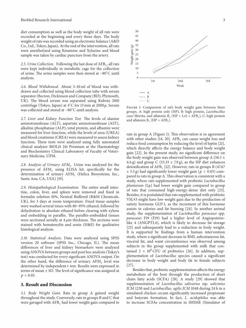
3.1. Body Weight Gain. Rats in group A gained weightthroughout the study. Conversely, rats in groups B and C thatwere gavaged with AFB1 had lower weight gain compared to
05
101520253035404550
1st
ABC
2nd 3rd
Wei
ght g
ain
(g)
Week
Figure 1: Comparison of rat’s body weight gain between threegroups. A: high protein only (HP), B: high protein, Lactobacilluscasei Shirota, and aflatoxin B1 (HP + LcS + AFB1), C: high proteinand aflatoxin B1 (HP + AFB1).
rats in group A (Figure 1). This observation is in agreementwith other studies [14, 20]. AFB1 can cause weight loss andreduce food consumption by reducing the level of leptin [21],which directly affects the energy balance and body weightgain [22]. In the present study, no significant difference onthe body weight gain was observed between group A (58.5 ±4.6 g) and group C (53.33 ± 7.9 g), as the HP diet enhancesdetoxification of AFB1 [12]. However, rats in groups B (47.67± 5.5 g) had significantly lower weight gain (𝑝 < 0.05) com-pared to rats in group A.This observation is consistent with astudy, where rats supplemented with probiotic Lactobacillusplantarum (Lp) had lower weight gain compared to groupof rats that consumed high-energy-dense diet only [23].Besides, it is postulated that rats supplemented with probioticVSL#3 might have low weight gain due to the production ofsatiety hormone GLP-1, as the increment of this hormoneassists in calories and fat burning [24]. In another animalstudy, the supplementation of Lactobacillus paracasei spp.paracasei F19 (F19) had a higher level of Angiopoietin-like 4 (ANGPTL4), which is likely to decrease fat storage[25] and subsequently lead to a reduction in body weight.It is supported by findings from a human interventionstudy, where a significant decrease in BMI, subcutaneous fat,visceral fat, and waist circumference was observed amongsubjects in the group supplemented with milk that con-tained 2 × 10⁸ CFU of probiotics [26]. In addition, sup-plementation of Lactobacillus species caused a significantdecrease in body weight and body fat in female subjects[27].
Besides that, probiotic supplementation affects the energymetabolism of the host through the production of shortchain fatty acids (SCFA) [28]. A study [29] showed thatsupplementation of Lactobacillus salivarius ssp. saliciniusJCM 1230 and Lactobacillus. agilis JCM 1048 during 24 h in asimulated chicken cecum significantly increased propionateand butyrate formation. In fact, L. acidophilus was ableto increase SCFAs concentration in SHIME (Simulator of
4 BioMed Research International
Human Microbial Ecosystem) reactor [30]. An increase ofSCFAs is associatedwith the increment of the circulating con-centrations of anorectic gut hormones such as peptide (PYY)and glucagon-like peptide-1 (GLP-1), and these gut hormoneshave been shown to cause a reduction in energy intake [31].Other than that, SCFA reduces weight by increasing energyexpenditure and enhances fat oxidation and thermogenesisby increasing the rate of oxygen consumption [32].
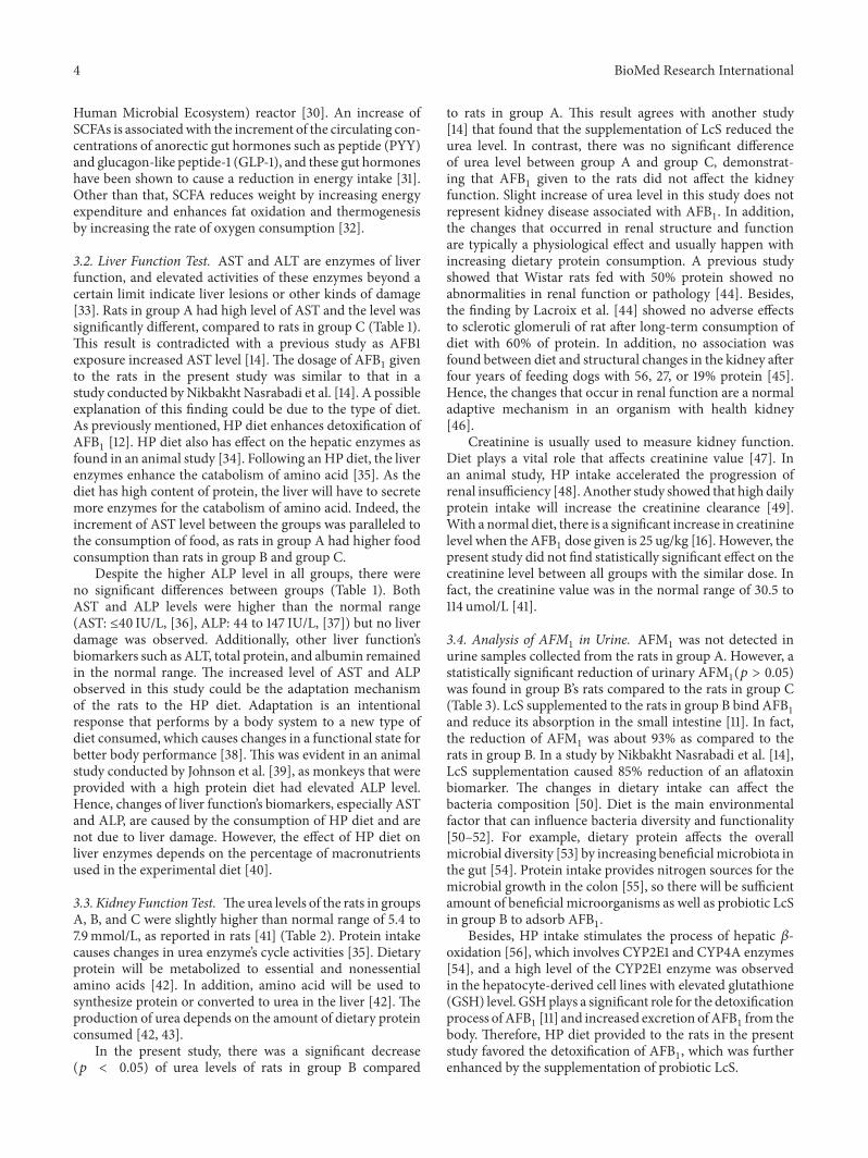
3.2. Liver Function Test. AST and ALT are enzymes of liverfunction, and elevated activities of these enzymes beyond acertain limit indicate liver lesions or other kinds of damage[33]. Rats in group A had high level of AST and the level wassignificantly different, compared to rats in group C (Table 1).This result is contradicted with a previous study as AFB1exposure increased AST level [14]. The dosage of AFB1 givento the rats in the present study was similar to that in astudy conducted by Nikbakht Nasrabadi et al. [14]. A possibleexplanation of this finding could be due to the type of diet.As previously mentioned, HP diet enhances detoxification ofAFB1 [12]. HP diet also has effect on the hepatic enzymes asfound in an animal study [34]. Following anHP diet, the liverenzymes enhance the catabolism of amino acid [35]. As thediet has high content of protein, the liver will have to secretemore enzymes for the catabolism of amino acid. Indeed, theincrement of AST level between the groups was paralleled tothe consumption of food, as rats in group A had higher foodconsumption than rats in group B and group C.
Despite the higher ALP level in all groups, there wereno significant differences between groups (Table 1). BothAST and ALP levels were higher than the normal range(AST: ≤40 IU/L, [36], ALP: 44 to 147 IU/L, [37]) but no liverdamage was observed. Additionally, other liver function’sbiomarkers such as ALT, total protein, and albumin remainedin the normal range. The increased level of AST and ALPobserved in this study could be the adaptation mechanismof the rats to the HP diet. Adaptation is an intentionalresponse that performs by a body system to a new type ofdiet consumed, which causes changes in a functional state forbetter body performance [38]. This was evident in an animalstudy conducted by Johnson et al. [39], as monkeys that wereprovided with a high protein diet had elevated ALP level.Hence, changes of liver function’s biomarkers, especially ASTand ALP, are caused by the consumption of HP diet and arenot due to liver damage. However, the effect of HP diet onliver enzymes depends on the percentage of macronutrientsused in the experimental diet [40].
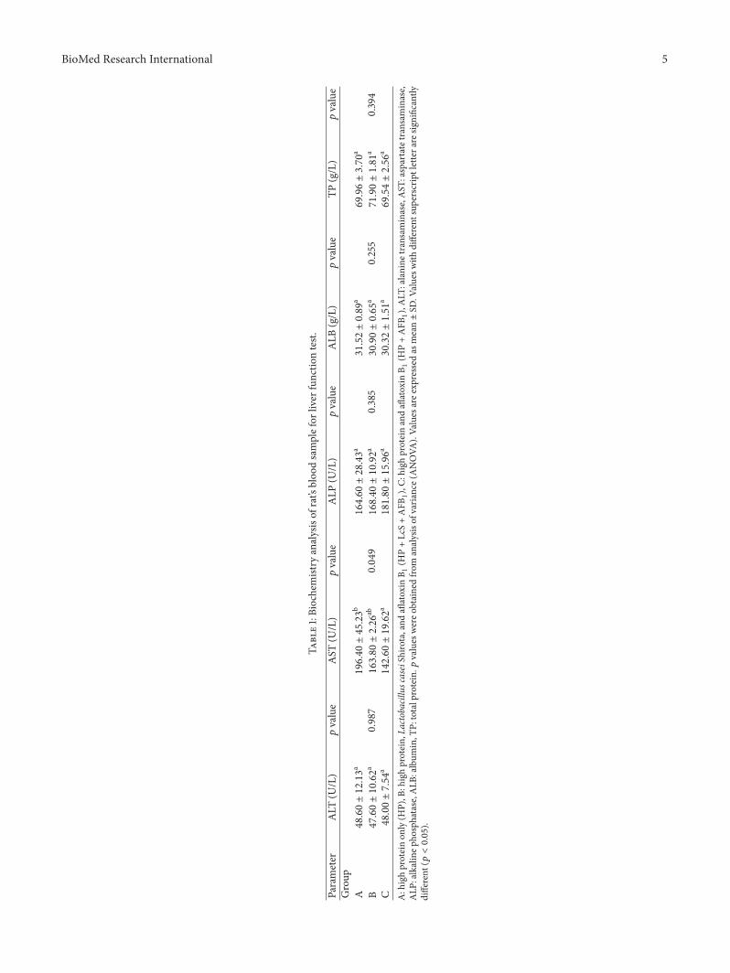
3.3. Kidney Function Test. Theurea levels of the rats in groupsA, B, and C were slightly higher than normal range of 5.4 to7.9mmol/L, as reported in rats [41] (Table 2). Protein intakecauses changes in urea enzyme’s cycle activities [35]. Dietaryprotein will be metabolized to essential and nonessentialamino acids [42]. In addition, amino acid will be used tosynthesize protein or converted to urea in the liver [42]. Theproduction of urea depends on the amount of dietary proteinconsumed [42, 43].
In the present study, there was a significant decrease(𝑝 < 0.05) of urea levels of rats in group B compared
to rats in group A. This result agrees with another study[14] that found that the supplementation of LcS reduced theurea level. In contrast, there was no significant differenceof urea level between group A and group C, demonstrat-ing that AFB1 given to the rats did not affect the kidneyfunction. Slight increase of urea level in this study does notrepresent kidney disease associated with AFB1. In addition,the changes that occurred in renal structure and functionare typically a physiological effect and usually happen withincreasing dietary protein consumption. A previous studyshowed that Wistar rats fed with 50% protein showed noabnormalities in renal function or pathology [44]. Besides,the finding by Lacroix et al. [44] showed no adverse effectsto sclerotic glomeruli of rat after long-term consumption ofdiet with 60% of protein. In addition, no association wasfound between diet and structural changes in the kidney afterfour years of feeding dogs with 56, 27, or 19% protein [45].Hence, the changes that occur in renal function are a normaladaptive mechanism in an organism with health kidney[46].
Creatinine is usually used to measure kidney function.Diet plays a vital role that affects creatinine value [47]. Inan animal study, HP intake accelerated the progression ofrenal insufficiency [48]. Another study showed that high dailyprotein intake will increase the creatinine clearance [49].With a normal diet, there is a significant increase in creatininelevel when the AFB1 dose given is 25 ug/kg [16]. However, thepresent study did not find statistically significant effect on thecreatinine level between all groups with the similar dose. Infact, the creatinine value was in the normal range of 30.5 to114 umol/L [41].
3.4. Analysis of AFM1 in Urine. AFM1 was not detected inurine samples collected from the rats in group A. However, astatistically significant reduction of urinary AFM1(𝑝 > 0.05)was found in group B’s rats compared to the rats in group C(Table 3). LcS supplemented to the rats in group B bind AFB1and reduce its absorption in the small intestine [11]. In fact,the reduction of AFM1 was about 93% as compared to therats in group B. In a study by Nikbakht Nasrabadi et al. [14],LcS supplementation caused 85% reduction of an aflatoxinbiomarker. The changes in dietary intake can affect thebacteria composition [50]. Diet is the main environmentalfactor that can influence bacteria diversity and functionality[50–52]. For example, dietary protein affects the overallmicrobial diversity [53] by increasing beneficialmicrobiota inthe gut [54]. Protein intake provides nitrogen sources for themicrobial growth in the colon [55], so there will be sufficientamount of beneficial microorganisms as well as probiotic LcSin group B to adsorb AFB1.
Besides, HP intake stimulates the process of hepatic 𝛽-oxidation [56], which involves CYP2E1 and CYP4A enzymes[54], and a high level of the CYP2E1 enzyme was observedin the hepatocyte-derived cell lines with elevated glutathione(GSH) level. GSHplays a significant role for the detoxificationprocess ofAFB1 [11] and increased excretion ofAFB1 from thebody. Therefore, HP diet provided to the rats in the presentstudy favored the detoxification of AFB1, which was furtherenhanced by the supplementation of probiotic LcS.
BioMed Research International 5
Table1:Biochemistry
analysisof
rat’sbloo
dsamplefor
liver
functio
ntest.
Parameter
ALT
(U/L)
𝑝value
AST
(U/L)
𝑝value
ALP
(U/L)
𝑝value
ALB
(g/L)
𝑝value
TP(g/L)
𝑝value
Group
A48.60±12.13a
0.987
196.40±45.23b
0.049
164.60±28.43a
0.385
31.52±0.89
a
0.255
69.96±3.70
a
0.394
B47.60±10.62a
163.80±2.26
ab168.40±10.92a
30.90±0.65
a71.90±1.81
a
C48.00±7.54
a142.60±19.62a
181.80±15.96a
30.32±1.51
a69.54±2.56
a
A:highproteinon
ly(H
P),B
:highprotein,La
ctobacillus
caseiShirota,and
aflatoxin
B 1(H
P+LcS+AFB1),C:
high
proteinandaflatoxin
B 1(H
P+AFB1),ALT
:alanine
transaminase,AST
:aspartatetransaminase,
ALP
:alkalinep
hosphatase,A
LB:album
in,T
P:totalprotein.𝑝
values
wereo
btainedfro
manalysisof
varia
nce(ANOVA
).Va
lues
aree
xpressed
asmean±SD
.Valuesw
ithdifferent
superscriptlettera
resig
nificantly
different
(𝑝<0.05).
6 BioMed Research International
Table 2: Biochemistry analysis of rat’s blood sample for kidney function test.
Parameter Urea (mmol/l) 𝑝 value Creatinine (umol/L) 𝑝 valueGroupA 10.82 ± 0.26b
0.03246.2 ± 3.34a
0.772B 10.02 ± 0.73a 45.0 ± 2.54a
C 10.10 ± 0.14ab 45.0 ± 3.08a
A: high protein only (HP), B: high protein, Lactobacillus casei Shirota, and aflatoxin (HP + LcS + AFB1), C: high protein and aflatoxin (HP + AFB1). 𝑝 valueswere obtained from analysis of variance (ANOVA). Values are expressed as mean ± SD. Values with different superscript letter are significantly different (𝑝 <0.05).
Table 3: The concentration of AFM1 in urine of aflatoxin-inducedrats.
Parameter AFM1 (ng/ml) 𝑝 valueGroupB 0.39 ± 0.01
<0.001C 5.22 ± 0.28B: high protein, Lactobacillus casei Shirota, and aflatoxin B1 (HP + LcS +AFB1), C: high protein and aflatoxin B1 (HP + AFB1). Values are expressedas mean ± SD. 𝑝 value was obtained by independence 𝑡-test.
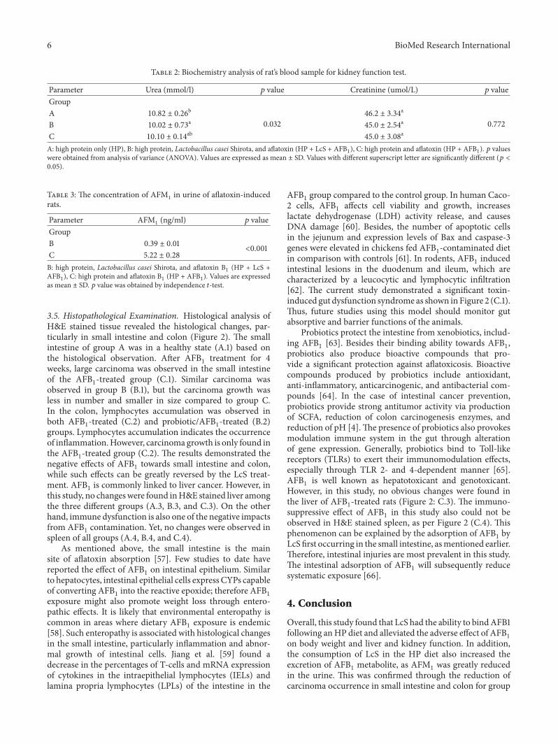
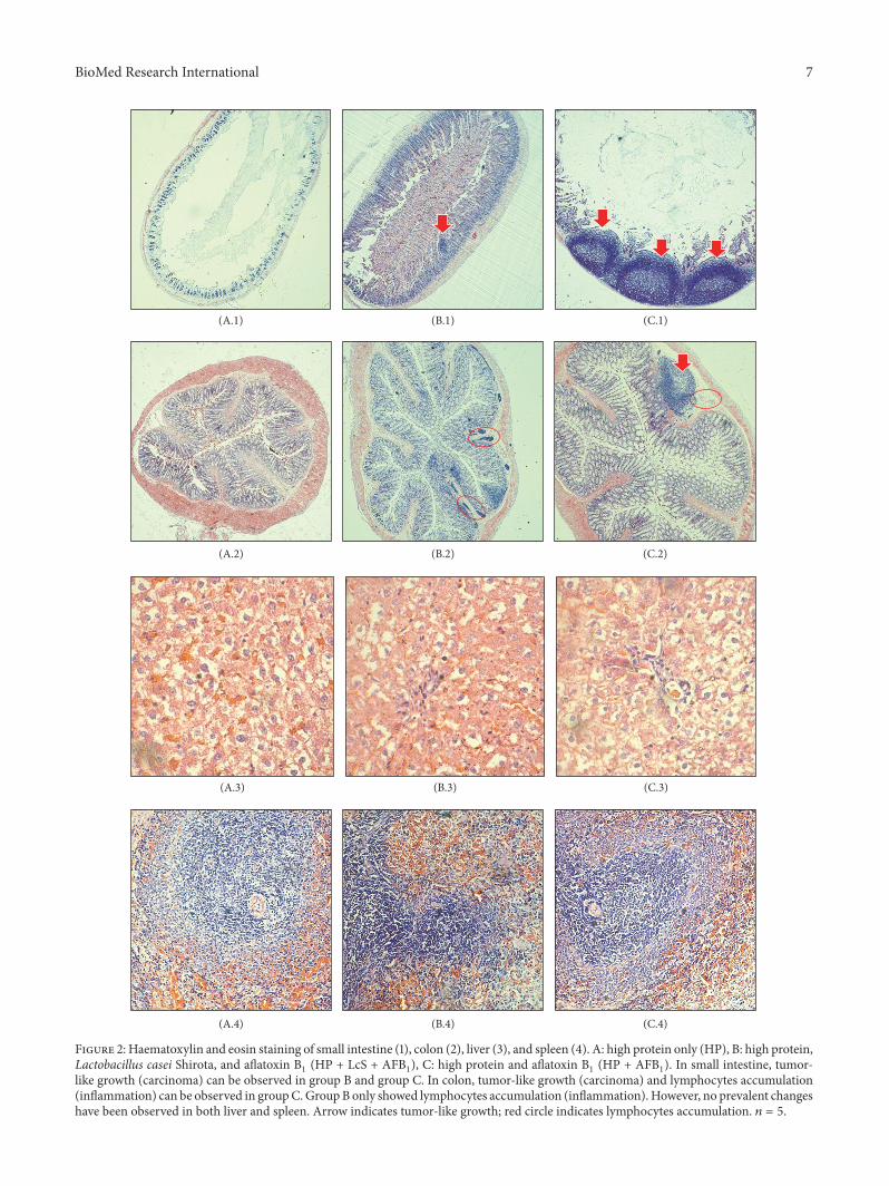
3.5. Histopathological Examination. Histological analysis ofH&E stained tissue revealed the histological changes, par-ticularly in small intestine and colon (Figure 2). The smallintestine of group A was in a healthy state (A.1) based onthe histological observation. After AFB1 treatment for 4weeks, large carcinoma was observed in the small intestineof the AFB1-treated group (C.1). Similar carcinoma wasobserved in group B (B.1), but the carcinoma growth wasless in number and smaller in size compared to group C.In the colon, lymphocytes accumulation was observed inboth AFB1-treated (C.2) and probiotic/AFB1-treated (B.2)groups. Lymphocytes accumulation indicates the occurrenceof inflammation.However, carcinoma growth is only found inthe AFB1-treated group (C.2). The results demonstrated thenegative effects of AFB1 towards small intestine and colon,while such effects can be greatly reversed by the LcS treat-ment. AFB1 is commonly linked to liver cancer. However, inthis study, no changeswere found inH&E stained liver amongthe three different groups (A.3, B.3, and C.3). On the otherhand, immune dysfunction is also one of the negative impactsfrom AFB1 contamination. Yet, no changes were observed inspleen of all groups (A.4, B.4, and C.4).
As mentioned above, the small intestine is the mainsite of aflatoxin absorption [57]. Few studies to date havereported the effect of AFB1 on intestinal epithelium. Similarto hepatocytes, intestinal epithelial cells express CYPs capableof converting AFB1 into the reactive epoxide; therefore AFB1exposure might also promote weight loss through entero-pathic effects. It is likely that environmental enteropathy iscommon in areas where dietary AFB1 exposure is endemic[58]. Such enteropathy is associated with histological changesin the small intestine, particularly inflammation and abnor-mal growth of intestinal cells. Jiang et al. [59] found adecrease in the percentages of T-cells and mRNA expressionof cytokines in the intraepithelial lymphocytes (IELs) andlamina propria lymphocytes (LPLs) of the intestine in the
AFB1 group compared to the control group. In human Caco-2 cells, AFB1 affects cell viability and growth, increaseslactate dehydrogenase (LDH) activity release, and causesDNA damage [60]. Besides, the number of apoptotic cellsin the jejunum and expression levels of Bax and caspase-3genes were elevated in chickens fed AFB1-contaminated dietin comparison with controls [61]. In rodents, AFB1 inducedintestinal lesions in the duodenum and ileum, which arecharacterized by a leucocytic and lymphocytic infiltration[62]. The current study demonstrated a significant toxin-induced gut dysfunction syndrome as shown in Figure 2 (C.1).Thus, future studies using this model should monitor gutabsorptive and barrier functions of the animals.
Probiotics protect the intestine from xenobiotics, includ-ing AFB1 [63]. Besides their binding ability towards AFB1,probiotics also produce bioactive compounds that pro-vide a significant protection against aflatoxicosis. Bioactivecompounds produced by probiotics include antioxidant,anti-inflammatory, anticarcinogenic, and antibacterial com-pounds [64]. In the case of intestinal cancer prevention,probiotics provide strong antitumor activity via productionof SCFA, reduction of colon carcinogenesis enzymes, andreduction of pH [4].The presence of probiotics also provokesmodulation immune system in the gut through alterationof gene expression. Generally, probiotics bind to Toll-likereceptors (TLRs) to exert their immunomodulation effects,especially through TLR 2- and 4-dependent manner [65].AFB1 is well known as hepatotoxicant and genotoxicant.However, in this study, no obvious changes were found inthe liver of AFB1-treated rats (Figure 2: C.3). The immuno-suppressive effect of AFB1 in this study also could not beobserved in H&E stained spleen, as per Figure 2 (C.4). Thisphenomenon can be explained by the adsorption of AFB1 byLcS first occurring in the small intestine, asmentioned earlier.Therefore, intestinal injuries are most prevalent in this study.The intestinal adsorption of AFB1 will subsequently reducesystematic exposure [66].
4. Conclusion
Overall, this study found that LcS had the ability to bindAFB1following anHP diet and alleviated the adverse effect of AFB1on body weight and liver and kidney function. In addition,the consumption of LcS in the HP diet also increased theexcretion of AFB1 metabolite, as AFM1 was greatly reducedin the urine. This was confirmed through the reduction ofcarcinoma occurrence in small intestine and colon for group
BioMed Research International 7
(A.1)
(A.2)
(A.3)
(A.4)
(B.1)
(B.2)
(B.3)
(B.4)
(C.1)
(C.2)
(C.3)
(C.4)
Figure 2: Haematoxylin and eosin staining of small intestine (1), colon (2), liver (3), and spleen (4). A: high protein only (HP), B: high protein,Lactobacillus casei Shirota, and aflatoxin B1 (HP + LcS + AFB1), C: high protein and aflatoxin B1 (HP + AFB1). In small intestine, tumor-like growth (carcinoma) can be observed in group B and group C. In colon, tumor-like growth (carcinoma) and lymphocytes accumulation(inflammation) can be observed in groupC.GroupB only showed lymphocytes accumulation (inflammation). However, no prevalent changeshave been observed in both liver and spleen. Arrow indicates tumor-like growth; red circle indicates lymphocytes accumulation. 𝑛 = 5.
8 BioMed Research International
of rats fed with AFB1 and LcS, compared to those fed withAFB1 alone. However, this study was limited by the lack of anormal diet group. In addition, broader research is needed todetermine the effect of different percentage of protein on theability of LcS andother probiotics to reduce the negative effectof AFB1 as this study only provided the rats with 40% of HPdiet. Different macronutrients such as carbohydrates and fatmay also have effect on probiotics and aflatoxin metabolismand hence warrant further investigation to determine theefficiency of probiotics as an adsorbent of aflatoxin.
Conflicts of Interest
The authors declare that there are no conflicts of interest.
Authors’ Contributions
Z. Nurul Adilah and Winnie-Pui-Pui Liew conducted theexperiment, carried out data analysis, and wrote the manu-script. S. Mohd Redzwan and I. Amin contributed by provid-ing technical support for the project and proofread the finalmanuscript.
Acknowledgments
This research was funded by Putra Research Grants fromUniversiti Putra Malaysia (UPM) [GP-IPS/2017/9517000 andGP-IPM/2016/9480100]. Z. Nurul Adilah and Winnie-Pui-Pui Liew are recipients of Graduate Research Fellowship(GRF) from School of Graduate Studies, UPM. Winnie-Pui-Pui Liew would like to acknowledge Ministry of Higher Edu-cation (MoHE), Malaysia, for the MyBrain15 Scholarship.
References
[1] M. Tola, B. Kebede, and F. Yildiz, “Occurrence, importance andcontrol of mycotoxins: A review,” Cogent Food & Agriculture,vol. 2, no. 1, pp. 1–12, 2016.
[2] P. Udomkun, A. N. Wiredu, M. Nagle, J. Muller, B. Vanlauwe,and R. Bandyopadhyay, “Innovative technologies to manageaflatoxins in foods and feeds and the profitability of application– A review,” Food Control, vol. 76, pp. 127–138, 2017.
[3] M. E. Zain, “Impact of mycotoxins on humans and animals,”Journal of Saudi Chemical Society, vol. 15, no. 2, pp. 129–144,2011.
[4] K. Siva Kumar, “Colon cancer prevention through probiotics:An overview,” Journal of Cancer Science &Therapy, vol. 7, no. 3,pp. 81–92, 2015.
[5] World Health Organization, “IARCmonographs on the evalua-tion of carcinogenic risks to humans,” in some traditional herbalmedicines, some mycotoxins, naphthalene and styrene, vol. 82,pp. 1–556, IARC Press, 2002.
[6] S.Gratz,M. Taubel, R.O. Juvonen et al., “Lactobacillus rhamno-sus strain GG modulates intestinal absorption, fecal excretion,and toxicily of aflatoxin B1 in rats,” Applied and EnvironmentalMicrobiology, vol. 72, no. 11, pp. 7398–7400, 2006.
[7] A. Hernandez-Mendoza, D. Guzman-De-Pena, and H. S. Gar-cia, “Key role of teichoic acids on aflatoxin B1 binding by probi-otic bacteria,” Journal of AppliedMicrobiology, vol. 107, no. 2, pp.395–403, 2009.
[8] FAO/WHO, “Food safety risk analysis: A guide for nationalfood safety authorities,” FAO Food and Nutrition Paper, vol. 87,pp. 1–119, 2006.
[9] A.A.Amara andA. Shibl, “Role of Probiotics in health improve-ment, infection control and disease treatment and manage-ment,” Saudi Pharmaceutical Journal, vol. 23, no. 2, pp. 107–114,2013.
[10] E. Damayanti, L. Istiqomah, J. E. Saragih, T. Purwoko, and T.Sardjono, “Characterization of lactic acid bacteria as poultryprobiotic candidates with aflatoxin B1 binding activities,” IOPConference Series: Earth and Environmental Science, vol. 101,Article ID 012030, 2017.
[11] S.MohdRedzwan,M. S. AbdMutalib, J.-S.Wang et al., “Effect ofsupplementation of fermented milk drink containing probioticLactobacillus casei Shirota on the concentrations of aflatoxinbiomarkers among employees of Universiti Putra Malaysia:A randomised, double-blind, cross-over, placebo-controlledstudy,”British Journal of Nutrition, vol. 115, no. 1, pp. 39–54, 2016.
[12] Z. Nurul Adilah and S. Mohd Redzwan, “Effect of dietarymacronutrients on aflatoxicosis: a mini-review,” Journal of theScience of Food and Agriculture, vol. 97, no. 8, pp. 2277–2281,2017.
[13] A. E. Rogers and P.M.Newberne, “Diet and aflatoxin B1 toxicityin rats,” Toxicology and Applied Pharmacology, vol. 20, no. 1, pp.113–121, 1971.
[14] E. Nikbakht Nasrabadi, R. Jamaluddin, M. S. Abdul Mutalib,H. Khaza’ai, S. Khalesi, and S. Mohd Redzwan, “Reductionof aflatoxin level in aflatoxin-induced rats by the activity ofprobiotic Lactobacillus casei strain Shirota,” Journal of AppliedMicrobiology, vol. 114, no. 5, pp. 1507–1515, 2013.
[15] ENVIGO: Custom Research Diet, “Envigo.com, 2017,” http://www.envigo.com.
[16] G. Qian, F. Wang, L. Tang et al., “Integrative toxicopathologicalevaluation of aflatoxin B,” Toxicologic Pathology, vol. 41, no. 8,pp. 1093–1105, 2013.
[17] J. H. Daniel, L. W. Lewis, Y. A. Redwood et al., “Comprehensiveassessment of maize aflatoxin levels in eastern Kenya, 2005-2007,” Environmental Health Perspectives, vol. 119, no. 12, pp.1794–1799, 2011.
[18] J. Wang, L. Tang, T. C. Glenn, and J.-S. Wang, “Aflatoxin B1induced compositional changes in gut microbial communitiesof male F344 rats,” Toxicological Sciences, vol. 150, no. 1, ArticleID kfv259, pp. 54–63, 2016.
[19] S. M. Redzwan, J. Rosita, A.M.M. Sokhini, and A. R. N. Aqilah,“Association between aflatoxin M1 excreted in human urinesamples with the consumption of milk and dairy products,”Bulletin of Environmental Contamination and Toxicology, vol.89, no. 6, pp. 1115–1119, 2012.
[20] A. S. Hathout, S. R.Mohamed, A. A. El-Nekeety, N. S. Hassan, S.E. Aly, and M. A. Abdel-Wahhab, “Ability of Lactobacillus caseiand Lactobacillus reuteri to protect against oxidative stress inrats fed aflatoxins-contaminated diet,”Toxicon, vol. 58, no. 2, pp.179–186, 2011.
[21] M. A. Abdel-Wahhab and S. E. Aly, “Antioxidants and radicalscavenging properties of vegetable extracts in rats fed aflatoxin-contaminated diet,” Journal of Agricultural and Food Chemistry,vol. 51, no. 8, pp. 2409–2414, 2003.
[22] R. Monalisa, “Role of leptin in obesity,” Research Journal ofPharmacy and Technology, vol. 8, no. 8, pp. 1073–1076, 2015.
[23] C. L. J. Karlsson, G. Molin, F. Fak et al., “Effects on weight gainand gut microbiota in rats given bacterial supplements and a
BioMed Research International 9
high-energy-dense diet from fetal life through to 6 months ofage,” British Journal of Nutrition, vol. 106, no. 6, pp. 887–895,2011.
[24] H. Yadav, J. H. Lee, J. Lloyd, P.Walter, and S. G. Rane, “Beneficialmetabolic effects of a probiotic via butyrate-induced GLP-1hormone secretion,” The Journal of Biological Chemistry, vol.288, no. 35, pp. 25088–25097, 2013.
[25] L. Aronsson, Y. Huang, P. Parini et al., “Decreased fat storageby Lactobacillus paracasei is associated with increased levels ofangiopoietin-like 4 protein (ANGPTL4),” PLoS ONE, vol. 5, no.9, Article ID e13087, pp. 1–7, 2010.
[26] Y. Kadooka, M. Sato, A. Ogawa et al., “Effect of Lactobacillusgasseri SBT2055 in fermented milk on abdominal adiposityin adults in a randomised controlled trial,” British Journal ofNutrition, vol. 110, no. 9, pp. 1696–1703, 2013.
[27] S. Park and J.-H. Bae, “Probiotics for weight loss: A systematicreview and meta-analysis,” Nutrition Research, vol. 35, no. 7, pp.566–575, 2015.
[28] J. G. LeBlanc, F. Chain, R. Martın, L. G. Bermudez-Humaran,S. Courau, and P. Langella, “Beneficial effects on host energymetabolism of short-chain fatty acids and vitamins producedby commensal and probiotic bacteria,”Microbial Cell Factories,vol. 16, no. 1, article no. 79, pp. 1–10, 2017.
[29] A. Meimandipour, A. F. Soleimani, M. Houshmand et al.,“Effects of rough handling on short chain fatty acid productionand gastrointestinal pH in broilers and modulatory role ofLactobacilli,”African Journal of Biotechnology, vol. 10, no. 74, pp.17030–17037, 2011.
[30] K. Sivieri, M. L. V. Morales, M. A. T. Adorno, I. K. Sakamoto, S.M. I. Saad, and E. A. Rossi, “Lactobacillus acidophilus CRL 1014improved “gut health” in the SHIME� reactor,” BMC Gastro-enterology, vol. 13, no. 1, article no. 100, 2013.
[31] C. S. Byrne, E. S. Chambers, D. J. Morrison, and G. Frost, “Therole of short chain fatty acids in appetite regulation and energyhomeostasis,” International Journal of Obesity, vol. 39, no. 9, pp.1331–1338, 2015.
[32] I. Kimura, D. Inoue, T. Maeda et al., “Short-chain fatty acidsand ketones directly regulate sympathetic nervous system viaG protein-coupled receptor 41 (GPR41),” Proceedings of theNational Acadamy of Sciences of the United States of America,vol. 108, no. 19, pp. 8030–8035, 2011.
[33] M. Heeringa, A. Hastings, S. Yamazaki, and P. De Koning,“Serum biomarkers in nonalcoholic steatohepatitis: Value forassessing drug effects?” Biomarkers in Medicine, vol. 6, no. 6,pp. 743–757, 2012.
[34] M. Michael, C. Anyakudo, O. F. Onuwabhagbe, and O. O.Ademidun, “Ec nutrition research article hepatotoxicity of highprotein diet in diabetic rats: An indication for necessary dietaryprecaution, vol. 5, pp. 195–202, 2017”.
[35] C. Jean, S. Rome, V. Mathe et al., “Metabolic evidence for adap-tation to a high protein diet in rats,” The Journal of Nutrition,vol. 131, no. 1, pp. 91–98, 2001.
[36] I. Cacciola, R. Scoglio, A. Alibrandi et al., “Evaluation of liverenzyme levels and identification of asymptomatic liver diseasepatients in primary care,” Internal and EmergencyMedicine, vol.12, no. 2, pp. 181–186, 2017.
[37] F. H. Schroder, B. Tombal, K. Miller et al., “Changes in alkalinephosphatase levels in patients with prostate cancer receivingdegarelix or leuprolide: Results from a 12-month, comparative,phase III study,” BJU International, vol. 106, no. 2, pp. 182–187,2010.
[38] A. Ogawa, T. Kobayashi, F. Sakai, Y. Kadooka, and Y. Kawasaki,“Lactobacillus gasseri SBT2055 suppresses fatty acid releasethrough enlargement of fat emulsion size in vitro and promotesfecal fat excretion in healthy Japanese subjects,” Lipids in Healthand Disease, vol. 14, no. 1, 2015.
[39] Q. Johnson, W. J. Veith, and T. Mouton, “The impact of dietaryprotein intake on serum biochemical and haematological pro-files in vervet monkeys,” Journal of Medical Primatology, vol. 30,no. 1, pp. 61–69, 2001.
[40] D. H. Pesta and V. T. Samuel, “A high-protein diet for reducingbody fat:Mechanisms and possible caveats,” Journal of Nutritionand Metabolism, vol. 11, no. 1, pp. 1–8, 2014.
[41] H. J. Baker, J. R. Lindsey, and S. H. Wesibroth, The LaboratoryRat: Biology and diseases, vol. 1, Academic Press Inc, New York,2013.
[42] I. D. Weiner, W. E. Mitch, and J. M. Sands, “Urea and ammoniametabolism and the control of renal nitrogen excretion,” Clini-cal Journal of the American Society of Nephrology, vol. 10, no. 8,pp. 1444–1458, 2015.
[43] M. C. Sales, A. de Azevedo Paiva, D. de Queiroz, R. A. F. Costa,M. A. L. da Cunha, and D. F. Pedraza, “Nutritional status of ironin children from6 to 59months of age and its relation to vitaminA deficiency,”Nutricion Hospitalaria, vol. 28, no. 3, pp. 734–740,2013.
[44] M. Lacroix, C. Gaudichon, A. Martin et al., “A long-term high-protein dietmarkedly reduces adipose tissue withoutmajor sideeffects in Wistar male rats,” American Journal of Physiology-Regulatory, Integrative and Comparative Physiology, vol. 287, no.4, pp. R934–R942, 2004.
[45] G. Robertson, I. Leclercq, and G. C. Farrell, “NonalcoholicSteatosis and Steatohepatitis. II. Cytochrome P-450 enzymesand oxidative stress,” American Journal of Physiology-Gastro-intestinal and Liver Physiology, vol. 281, no. 5, pp. G1135–G1139,2001.
[46] W. F. Martin, L. E. Armstrong, and N. R. Rodriguez, “Dietaryprotein intake and renal function,”NutritionMetabolism, vol. 2,no. 1, p. 25, 2005.
[47] G. Banfi and M. Del Fabbro, “Serum creatinine values in eliteathletes competing in 8 different sports: Comparison withsedentary people,” Clinical Chemistry, vol. 52, no. 2, pp. 330-331,2006.
[48] K. Lentine and E. M. Wrone, “New insights into protein intakeand progression of renal disease,” Current Opinion in Nephro-logy and Hypertension, vol. 13, no. 3, pp. 333–336, 2004.
[49] S. P. Juraschek, L. J. Appel, C. A. M. Anderson, and E. R. MillerIII, “Effect of a high-protein diet on kidney function in healthyadults: Results from the omniheart trial,” American Journal ofKidney Diseases, vol. 61, no. 4, pp. 547–554, 2013.
[50] M. I. Queipo-Ortuno, M. Boto-Ordonez, M. Murri et al.,“Influence of red wine polyphenols and ethanol on the gutmicrobiota ecology and biochemical biomarkers,” AmericanJournal of Clinical Nutrition, vol. 95, no. 6, pp. 1323–1334, 2012.
[51] I. Nadal, A. Santacruz, and A. Marcos, “Shifts in clostridia, bac-teroides and immunoglobulin-coating fecal bacteria associatedwith weight loss in obese adolescents,” International Journal ofObesity, vol. 33, no. 7, pp. 758–767, 2009.
[52] I. Gomez-Hurtado, A. Santacruz, G. Peiro et al., “Gut micro-biota dysbiosis is associated with inflammation and bacterialtranslocation in mice with CCl 4-Induced fibrosis,” PLoS ONE,vol. 6, no. 7, Article ID e23037, 2011.
10 BioMed Research International
[53] D. W. Lescheid, “Probiotics as regulators of inflammation: Areview,” Functional Foods in Health and Disease, vol. 4, no. 7,pp. 299–311, 2014.
[54] P. Lopez-Legarrea, N. R. Fuller, M. A. Zulet, J. A. Martinez, andI. D. Caterson, “The influence of Mediterranean, carbohydrateand high protein diets on gut microbiota composition in thetreatment of obesity and associated inflammatory state,” AsiaPacific Journal of Clinical Nutrition, vol. 23, no. 3, pp. 360–368,2014.
[55] S. H. Duncan, G. E. Lobley, G. Holtrop et al., “Human colonicmicrobiota associated with diet, obesity and weight loss,” Inter-national Journal of Obesity, vol. 32, no. 11, pp. 1720–1724, 2008.
[56] M. Bortolotti, R. Kreis, C. Debard et al., “High protein intakereduces intrahepatocellular lipid deposition in humans,” Amer-ican Journal of Clinical Nutrition, vol. 90, no. 4, pp. 1002–1010,2009.
[57] C. Dogi, A. Cristofolini, M. L. Gonzalez Pereyra et al., “Afla-toxins and Saccharomyces cerevisiae: Yeastmodulates the intes-tinal effect of aflatoxins, while aflatoxin B1 influences yeastultrastructure,”World Mycotoxin Journal, vol. 10, no. 2, pp. 171–181, 2017.
[58] B. Knipstein, J. Huang, E. Barr et al., “Dietary aflatoxin-inducedstunting in a novel rat model: Evidence for toxin-inducedliver injury and hepatic growth hormone resistance,” PediatricResearch, vol. 78, no. 2, pp. 120–127, 2015.
[59] M. Jiang, X. Peng, J. Fang, H. Cui, Z. Yu, and Z. Chen, “Effects ofaflatoxin B1 on t-cell subsets andmRNAexpression of cytokinesin the intestine of broilers,” International Journal of MolecularSciences, vol. 16, no. 4, pp. 6945–6959, 2015.
[60] J. Zhang, N. Zheng, J. Liu, F. D. Li, S. L. Li, and J. Q. Wang,“Aflatoxin B1 and aflatoxin M1 induced cytotoxicity and DNAdamage in differentiated and undifferentiated Caco-2 cells,”Food and Chemical Toxicology, vol. 83, pp. 54–60, 2015.
[61] X. Peng, K. Zhang, S. Bai et al., “Histological lesions, cellcycle arrest, apoptosis and T cell subsets changes of spleen inchicken fed aflatoxin-contaminated corn,” International Journalof Environmental Research and Public Health, vol. 11, no. 8, pp.8567–8580, 2014.
[62] F. J. Akinrinmade, A. S. Akinrinde, and A. Amid, “Changesin serum cytokine levels, hepatic and intestinal morphology inaflatoxin B1-induced injury: modulatory roles of melatonin andflavonoid-rich fractions from Chromolena odorata,”MycotoxinResearch, vol. 32, no. 2, pp. 53–60, 2016.
[63] S.H.Ahlberg, V. Joutsjoki, andH. J. Korhonen, “Potential of lac-tic acid bacteria in aflatoxin risk mitigation,” International Jour-nal of Food Microbiology, vol. 207, pp. 87–102, 2015.
[64] A. Di Cerbo, B. Palmieri, M. Aponte, J. C. Morales-Medina,and T. Iannitti, “Mechanisms and therapeutic effectiveness oflactobacilli,” Journal of Clinical Pathology, vol. 69, no. 3, pp. 187–203, 2016.
[65] A. Finamore, M. Roselli, A. Imbinto, J. Seeboth, I. P. Oswald,and E. Mengheri, “Lactobacillus amylovorus inhibits the TLR4inflammatory signaling triggered by enterotoxigenic Escher-ichia coli via modulation of the negative regulators and involve-ment of TLR2 in intestinal Caco-2 cells and pig explants,” PLoSONE, vol. 9, no. 4, Article ID e94891, 2014.
[66] L. Gambacorta, P. Pinton, G. Avantaggiato, I. P. Oswald, andM.Solfrizzo, “Grape Pomace, an Agricultural Byproduct ReducingMycotoxin Absorption: In Vivo Assessment in Pig Using Uri-nary Biomarkers,” Journal of Agricultural and Food Chemistry,vol. 64, no. 35, pp. 6762–6771, 2016.

Hindawiwww.hindawi.com
International Journal of
Volume 2018
Zoology
Hindawiwww.hindawi.com Volume 2018
Anatomy Research International
PeptidesInternational Journal of
Hindawiwww.hindawi.com Volume 2018
Hindawiwww.hindawi.com Volume 2018
Journal of Parasitology Research
GenomicsInternational Journal of
Hindawiwww.hindawi.com Volume 2018
Hindawi Publishing Corporation http://www.hindawi.com Volume 2013Hindawiwww.hindawi.com
The Scientific World Journal
Volume 2018
Hindawiwww.hindawi.com Volume 2018
BioinformaticsAdvances in
Marine BiologyJournal of
Hindawiwww.hindawi.com Volume 2018
Hindawiwww.hindawi.com Volume 2018
Neuroscience Journal
Hindawiwww.hindawi.com Volume 2018
BioMed Research International
Cell BiologyInternational Journal of
Hindawiwww.hindawi.com Volume 2018
Hindawiwww.hindawi.com Volume 2018
Biochemistry Research International
ArchaeaHindawiwww.hindawi.com Volume 2018
Hindawiwww.hindawi.com Volume 2018
Genetics Research International
Hindawiwww.hindawi.com Volume 2018
Advances in
Virolog y Stem Cells International
Hindawiwww.hindawi.com Volume 2018
Hindawiwww.hindawi.com Volume 2018
Enzyme Research
Hindawiwww.hindawi.com Volume 2018
International Journal of
MicrobiologyHindawiwww.hindawi.com
Nucleic AcidsJournal of
Volume 2018
Submit your manuscripts atwww.hindawi.com