Embed Size (px)
Citation preview
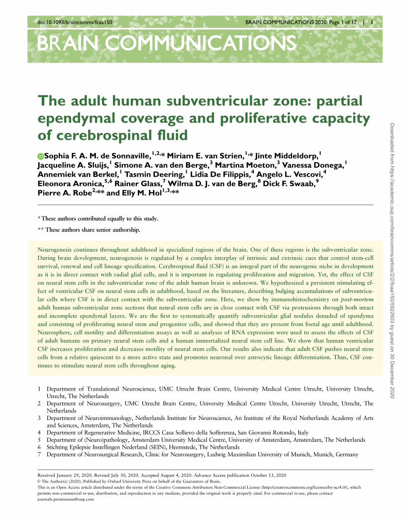
The adult human subventricular zone: partialependymal coverage and proliferative capacityof cerebrospinal fluid
Sophia F. A. M. de Sonnaville,1,2,* Miriam E. van Strien,1,* Jinte Middeldorp,1
Jacqueline A. Sluijs,1 Simone A. van den Berge,3 Martina Moeton,3 Vanessa Donega,1
Annemiek van Berkel,1 Tasmin Deering,1 Lidia De Filippis,4 Angelo L. Vescovi,4
Eleonora Aronica,5,6 Rainer Glass,7 Wilma D. J. van de Berg,8 Dick F. Swaab,9
Pierre A. Robe2,** and Elly M. Hol1,3,**
*These authors contributed equally to this study.
** These authors share senior authorship.
Neurogenesis continues throughout adulthood in specialized regions of the brain. One of these regions is the subventricular zone.
During brain development, neurogenesis is regulated by a complex interplay of intrinsic and extrinsic cues that control stem-cell
survival, renewal and cell lineage specification. Cerebrospinal fluid (CSF) is an integral part of the neurogenic niche in development
as it is in direct contact with radial glial cells, and it is important in regulating proliferation and migration. Yet, the effect of CSF
on neural stem cells in the subventricular zone of the adult human brain is unknown. We hypothesized a persistent stimulating ef-
fect of ventricular CSF on neural stem cells in adulthood, based on the literature, describing bulging accumulations of subventricu-
lar cells where CSF is in direct contact with the subventricular zone. Here, we show by immunohistochemistry on post-mortem
adult human subventricular zone sections that neural stem cells are in close contact with CSF via protrusions through both intact
and incomplete ependymal layers. We are the first to systematically quantify subventricular glial nodules denuded of ependyma
and consisting of proliferating neural stem and progenitor cells, and showed that they are present from foetal age until adulthood.
Neurosphere, cell motility and differentiation assays as well as analyses of RNA expression were used to assess the effects of CSF
of adult humans on primary neural stem cells and a human immortalized neural stem cell line. We show that human ventricular
CSF increases proliferation and decreases motility of neural stem cells. Our results also indicate that adult CSF pushes neural stem
cells from a relative quiescent to a more active state and promotes neuronal over astrocytic lineage differentiation. Thus, CSF con-
tinues to stimulate neural stem cells throughout aging.
1 Department of Translational Neuroscience, UMC Utrecht Brain Centre, University Medical Centre Utrecht, University Utrecht,Utrecht, The Netherlands
2 Department of Neurosurgery, UMC Utrecht Brain Centre, University Medical Centre Utrecht, University Utrecht, Utrecht, TheNetherlands
3 Department of Neuroimmunology, Netherlands Institute for Neuroscience, An Institute of the Royal Netherlands Academy of Artsand Sciences, Amsterdam, The Netherlands
4 Department of Regenerative Medicine, IRCCS Casa Sollievo della Sofferenza, San Giovanni Rotondo, Italy5 Department of (Neuro)pathology, Amsterdam University Medical Centre, University of Amsterdam, Amsterdam, The Netherlands6 Stichting Epilepsie Instellingen Nederland (SEIN), Heemstede, The Netherlands7 Department of Neurosurgical Research, Clinic for Neurosurgery, Ludwig Maximilian University of Munich, Munich, Germany
Received January 29, 2020. Revised July 30, 2020. Accepted August 4, 2020. Advance Access publication October 13, 2020VC The Author(s) (2020). Published by Oxford University Press on behalf of the Guarantors of Brain.
This is an Open Access article distributed under the terms of the Creative Commons Attribution Non-Commercial License (http://creativecommons.org/licenses/by-nc/4.0/), which
permits non-commercial re-use, distribution, and reproduction in any medium, provided the original work is properly cited. For commercial re-use, please contact
BBRAIN COMMUNICATIONSAIN COMMUNICATIONSdoi:10.1093/braincomms/fcaa150 BRAIN COMMUNICATIONS 2020: Page 1 of 17 | 1
Dow
nloaded from https://academ
ic.oup.com/braincom
ms/article/2/2/fcaa150/5922603 by guest on 30 D
ecember 2020

8 Department of Anatomy and Neurosciences, Section Clinical Neuroanatomy, Amsterdam University Medical Centre, Location VU,Amsterdam, The Netherlands
9 Department of Neuropsychiatric Disorders, Netherlands Institute for Neuroscience, An Institute of the Royal Netherlands Academyof Arts and Sciences, Amsterdam, The Netherlands
Correspondence to: Elly M. Hol, PhD
Department of Translational Neuroscience
UMC Utrecht Brain Centre, University Medical Centre Utrecht
Internal postal address Str. 4.205/P.O. Box 85060
Utrecht, The Netherlands.
E-mail: [email protected]
Keywords: cerebrospinal fluid; glial nodules; human; neural stem cells; subventricular zone
Abbreviations: ASCL1 ¼ achaete-scute family bHLH transcription factor 1; BSA ¼ bovine serum albumin; DAB ¼ 3,3’-diamino-bezidine tetrahydrochloride; DCX ¼ doublecortin; DMEM ¼ Dulbecco’s modified Eagle’s medium; EF1a ¼ elongation factor 1-alpha; EGF(R) ¼ epidermal growth factor (receptor); FGF(R) ¼ fibroblast growth factor receptor; FOXJ1 ¼ forkhead box proteinJ1; FCS ¼ foetal calf serum; GAPDH ¼ glyceraldehyde-3-phosphate dehydrogenase; GFAP ¼ glial fibrillary acidic protein; GLUL ¼glutamate-ammonia ligase; HES ¼ hes family bHLH transcription factor; HLA-DR ¼ human leucocyte antigen – DR isotype; JAG1¼ jagged canonical Notch ligand 1; NBB ¼ Netherlands Brain Bank; NCAM1 ¼ neural cell adhesion molecule 1; NDC ¼ non-de-mented control; NES ¼ nestin; NOTCH1 ¼ notch receptor 1; NPC ¼ neural progenitor cell; NSC ¼ neural stem cell; PCNA ¼ pro-liferating cell nuclear antigen; PMD ¼ post-mortem delay; P/S ¼ penicillin/streptomycin; PSEN1 ¼ presenilin 1; S100b ¼ S100calcium-binding protein B; SGN ¼ subventricular glial nodule; SLC1A2 ¼ Solute carrier family 1 member 2; SOX2 ¼ Sex determin-ing region box 2; SVZ ¼ subventricular zone; TBS ¼ Tris-buffered saline; TUBB ¼ tubulin; VIM ¼ vimentin
Graphical Abstract
2 | BRAIN COMMUNICATIONS 2020: Page 2 of 17 S. F. A. M. deSonnaville et al.
Dow
nloaded from https://academ
ic.oup.com/braincom
ms/article/2/2/fcaa150/5922603 by guest on 30 D
ecember 2020
IntroductionThe subventricular zone (SVZ) is a neurogenic brain area
that lines the walls of the ventricles. Subventricular zone
neural stem cells (NSCs) are able to proliferate and differ-
entiate into neurons and glial cells (Doetsch et al., 1999;
Kukekov et al., 1999; Sanai et al., 2004). During brain
development, radial glia are in direct contact with cerebro-
spinal fluid (CSF), which provides important growth fac-
tors and cues for directional migration (Sawamoto, 2006;
Johansson et al., 2010; Zappaterra and Lehtinen, 2012;
Ortega et al., 2018). In rodents, NSCs are in direct con-
tact with CSF through apical processes (Mirzadeh et al.,2008). In humans, adult NSCs are arranged in a ribbon,
which is separated from the CSF by a single layer of epen-
dymocytes and a hypocellular gap (Sanai et al., 2004; van
den Berge et al., 2010; Jimenez et al., 2014). Human NSC
protrusions extend through the ependymal cell layer to-
wards the ventricle as shown by Sanai et al. (2004). The
ependyma forms a semi-permeable CSF–brain barrier sup-
ported by incomplete apical tight junctions, lateral adhe-
rens and gap junctions, a luminal sialic acid coating and
aquaporin-4 channels on the basolateral membrane
(Nielsen et al., 1997; Schauer, 2009; Zhang et al., 2010;
Lehtinen and Walsh, 2011; Jimenez et al., 2014). At
young age, this barrier is largely intact; however, increased
penetrance of CSF into the parenchyma has been described
in aged rodents (Whish et al., 2015), and some ependymal
loss has been shown in human adults (Del Bigio, 2010).
The effect of increased direct contact of NSCs with CSF
in the adult human SVZ is largely unknown.
Cerebrospinal fluid is primarily produced by the chor-
oid plexus (Orekovi�c and Klarica, 2010), and contains a
broad spectrum of proteins including growth factors that
are key for neurogenic signalling (for a review, see
Zappaterra and Lehtinen, 2012). The few studies apply-
ing rodent and, in some cases, human-derived CSF to
neural stem and progenitor cells mainly suggest that CSF
enhances survival and stimulates proliferation of NSCs,
but the effect is controversial. In addition, from these
studies the effect of CSF on stem cell differentiation and
migration remains unclear (Gato et al., 2005; Buddensiek
et al., 2009, 2010; Lehtinen et al., 2011; Ma et al.,2013; Zhu et al., 2015; Alonso et al., 2017). Human
CSF composition changes with age (Zhang et al., 2005;
Baird et al., 2012), with a concomitant decrease in NSC
number in the SVZ and a significant reduction of the
rostral migratory stream (Sanai et al., 2005, 2011;
Conover and Shook, 2011; van Strien et al., 2011). A
study on the effect of young versus aged rodent choroid
plexus secretome shows that NSC proliferation and dif-
ferentiation is sensitive to age-related changes in CSF
composition (Silva-Vargas et al., 2016).
Direct contact with CSF also seems to affect subependy-
mal cells in vivo. Upon induced ependymal damage,
rodents develop subventricular glial nodules (SGNs)—
defined as collections of cells, bulging into the ventricle
lumen and denuded of ependyma (Gomez-Roldan et al.,
2008). The identity of these cells is still unclear. Some stud-
ies suggest that they are transient amplifying progenitor
cells, whereas others propose neuroblasts or reactive astro-
cytes (Kuo et al., 2006; Gomez-Roldan et al., 2008; Luo
et al., 2008; Jimenez et al., 2009; Roales-Bujan et al.,
2012). These SGNs—also referred to as subependymal nod-
ules or granular ependymitis—have been described in
humans with multiple sclerosis or schizophrenia, and are
associated with ependymal damage after viral infections
and ventriculomegaly (Johnson and Johnson, 1972; Adams
et al., 1987; Gray et al., 1992; Sarnat, 1995; Honer et al.,
1996; Domınguez-Pinos et al., 2005). Assumingly, SGN
form when ependymal cells provide an insufficient barrier
between growth factors and inflammatory mediators in the
CSF and NSCs in the SVZ.
Here, we aimed to study the effect of ventricular CSF
of adult humans on NSCs, by studying post-mortem
human SVZ tissue, primary NSCs and a human immor-
talized NSC line. We hypothesized a stimulating effect of
ventricular CSF on NSCs based on the literature, describ-
ing SGNs where CSF is in direct contact with the SVZ.
Our data confirm that adult human NSCs remain in dir-
ect contact with CSF via protrusions penetrating the
ependyma, but also due to significant lack of ependymal
coverage. We are the first to systematically quantify
SGNs in the human SVZ, to show that they are present
from foetal until old age and that they express prolifer-
ation, NSC and progenitor markers, presumably due to
direct CSF–NSC contact. This is supported by in vitro
experiments, showing that human ventricular CSF induces
NSC proliferation, inhibits motility and potentiates neur-
onal differentiation. Altogether, our results show that
adult human ventricular CSF still promotes neurogenesis.
This knowledge is important for initiatives exploring
stimulation of endogenous NSCs as a therapeutic option
for promoting brain repair in neurodegenerative diseases,
but also raises the question whether CSF could play a
role in the formation of brain tumours originating from
mutated NSCs within the SVZ.
Materials and methods
Immunohistochemistry of humanpost-mortem tissue sections
Human brain material was obtained from the
Netherlands Brain Bank (NBB). The NBB was given
informed consent by brain donors for tissue use and
accessing neuropathological and clinical information for
scientific research, in compliance with ethical and legal
guidelines (Netherlands Brain Bank, 2020).
For analysis of ependymal coverage, formalin-fixed,
paraffin-embedded human post-mortem tissue was col-
lected from 14 controls, classified as donors without a
neurological or neuropsychiatric disorder (clinically and
Cerebrospinal fluid and neural stem cells BRAIN COMMUNICATIONS 2020: Page 3 of 17 | 3
Dow
nloaded from https://academ
ic.oup.com/braincom
ms/article/2/2/fcaa150/5922603 by guest on 30 D
ecember 2020

pathologically confirmed) (Supplementary Table 1). Seven
males and seven females were included, not significantly
different in age (meanmales ¼ 80.6 6 2.0 years, meanfemales
¼ 85.9 6 2.9 years; Mann–Whitney U test, U¼ 15.50,
P¼ 0.2733). SVZ material was sampled from three lateral
ventricular areas (SVZ1-3; Fig. 1A, F) and cut into 8 lm
sections. One section per area per donor was deparaffi-
nized and rehydrated. To visualize the ependymal layer,
sections were incubated with 0.5% thionin (Merck) in
0.96% acetic acid for 10 min and embedded in Entellan
(Merck). Thionin staining was shown to correlate
significantly with staining of ependymal marker forkhead
box protein J1 (FOXJ1) in quantification of the epen-
dymal coverage (Supplementary Fig. 1).
For the analysis of SGNs, formalin-fixed paraffin-
embedded human hypothalamus tissue containing part of
the SVZ of the lateral and third ventricles was obtained.
We analysed two to four donors per age decennium:
from 2 to 94 years of age, with equal sex distribution (12
males, 14 females; two-tailed t-test, t24 ¼ 1.221,
P¼ 0.2339) (Supplementary Table 1). Serial sections of
6 lm were made and every 100th section were thionin-
Figure 1 Contact of neural stem cells with cerebrospinal fluid through protrusions and due to the absence of ependyma
covering the subventricular zone (SVZ) of adult donors. (A, F) White dashed lines in two 1 cm thick formalin-fixed human brain
slices indicate the three dissected SVZ areas. SVZ1 was dissected in the brain slice containing the most frontal part of the caudate nucleus
(cn) and SVZ2 was dissected from the same brain slice, or 1 slice more caudal, under the cingulate gyrus (cg), including part of the corpus
callosum (cc). SVZ3 was dissected from the most posterior part of the lateral ventricle (LV). (B, G) Immunofluorescent double labelling of a
fresh frozen SVZ tissue section of a control donor (NBB 2005-073, male, 87 years) for GFAPd (green) and nestin (magenta). White rectangles
and asterisks indicate protrusion(s) of NSCs towards the ventricle, of which magnifications are shown in the different channels (C–E and H–
J). Nuclei are counterstained with Hoechst. (K) Example of thionin staining showing ependymal cell coverage and absence (transition
indicated by the arrow). (L) Ependymal layer coverage in the SVZ of healthy controls is quantified in SVZ1-3 (n¼ 13 donors of SVZ1 and
SVZ3, n¼ 14 donors of SVZ2), with significantly less ependymal cell coverage in SVZ2 (one-way ANOVA, F(2,37) ¼ 7.417, P¼ 0.0020; Sidak
post-hoc analysis, **P< 0.01). Data are expressed as mean 6 standard deviation. (M) Sex does not correlate with the average ependymal layer
coverage over SVZ1-3 (Mann–Whitney U test, U¼ 13, P¼ 0.1649). Data are expressed as median 6 interquartile range. (N) Age does not
correlate with the average ependymal layer coverage in this age range (Pearson’s R2 ¼ 0.1601, P¼ 0.1564). Scale bars ¼ 10 lm.
4 | BRAIN COMMUNICATIONS 2020: Page 4 of 17 S. F. A. M. deSonnaville et al.
Dow
nloaded from https://academ
ic.oup.com/braincom
ms/article/2/2/fcaa150/5922603 by guest on 30 D
ecember 2020

stained. Additional single thionin-stained sections of 13
foetuses ranging in age from gestational week 9 to 40
were obtained from Bloemenhove clinic and Service
Histologie-Embryologie-Cytogenetique Hospital Necker-
Enfants Malades, Paris, France. This material was from
spontaneous or medically induced abortions, with mater-
nal informed consent for use of material and access to
medical records for research purposes. Tissue was col-
lected in accordance with the Declaration of Helsinki and
the Academic Medical Centre Research Code.
Immunofluorescent staining was performed as described
previously (van den Berge et al., 2010). Briefly, epitope
retrieval was performed with citrate buffer (10 mM citric
acid, 0.05% Tween-20, pH ¼ 6.0); to block non-specific
staining, sections were incubated with blocking solution
(1� Tris-buffered saline [TBS, pH ¼ 7.6], 2% horse
serum, 0.1% bovine serum albumin [BSA], 0.1%
TritonX-100, 0.05% Tween-20). Subsequently, sections
were incubated with primary antibody (Supplementary
Table 2), diluted in 1% TBS–BSA overnight at 4�C.
Incubation with fluorescently labelled species-specific sec-
ondary antibodies (1:400; Jackson Immuno-Research
Laboratories) was combined with Hoechst33258 (1:1000;
BioRad) in TBS during 1 h. To quench autofluorescence,
sections were incubated in 0.3% Sudan Black in 70%
ethanol for 7 min. Sections were embedded in Mowiol
(0.1 M Tris-HCl [pH ¼ 8.5], 25% glycerol, 10% w/v
Mowiol4-88 (Sigma-Aldrich)).
For 3,30-diaminobezidine tetrahydrochloride (DAB) stain-
ing, sections were incubated with biotinylated secondary
antibody (1:400; Vector Laboratories) diluted in TBS–BSA,
followed by incubation with avidin–biotin complex-system
(Vector Laboratories; 1:800). Subsequently, sections were
incubated with DAB-solution (Sigma-Aldrich) (0.5 mg/ml in
50 mM Tris, 0.04% H2O2) and embedded in Entellan.
Image acquisition and quantification
All thionin-stained sections were coded for unbiased quanti-
fication. For the analysis of ependymal coverage, tiled
images of the whole SVZ were taken using the
AxioImagerM2 microscope (Zeiss) with a 20�/0.5 N-ach-
roplan objective (Zeiss), AxioCamMRm camera (Zeiss) and
software Zen2011 (Zeiss). Total length of SVZ bordering
the lateral ventricle and length covered by ependymal cells
was measured to calculate the percentage of ependymal
coverage using ImageJ1.47q software (Rasband, 2015). For
the analysis of SGNs, images were obtained on an
AxioSkop microscope (Zeiss) with 40�/0.75 Plan-Neofluar
objective (Zeiss), using an EvolutionMPColour camera
(MediaCybernetics) and Image-ProPlus6.3 software
(MediaCybernetics). For quantification of SGN occurrence,
one thionin-stained section per foetus and between 5–10
sections per donor were scored. Fluorescently stained SVZ
sections and cells were imaged using an Axioplan2 micro-
scope (Zeiss) with 40�/0.95 Plan-Apochromat objectives
(Zeiss), with Retiga2000DC camera (Qimaging) and Image-
ProPlus6.3 software.
Primary human NSC isolation andculture of an immortalized humanNSC line
Freshly resected SVZ material was obtained from the
NBB (Supplementary Table 3). Neural stem cells from
fresh SVZ tissue from three adult donors (aNSCs) were
obtained based on a previously published protocol (van
Strien et al., 2014). Briefly, SVZ tissue was dissociated
and digested with 0.2% trypsin (Invitrogen) and 0.1%
DNase (Sigma-Aldrich). Subsequently, 0.2% foetal calf
serum (FCS) (Gemini Bio-products) was added and cells
were collected by centrifugation. Pellet was re-suspended
in Dulbecco’s modified Eagle’s medium (DMEM) (Gibco)
with 10% FCS, 1% penicillin/streptomycin (P/S) (Gibco)
and 0.1% gentamycin, and filtered through a 100-lm
mesh filter. Cell fraction was isolated by adding Percoll
(Amersham/GE Healthcare) and centrifugation at high
speed. aNSCs were isolated by CD271-coupled
MicroBeads with a magnetic separation system (Miltenyi
Biotec) by following the manufacturer’s instruction.
Foetal NSCs (fNSCs) from four donors—obtained from
the AMC as described above—were isolated similarly
with CD133 beads (Miltenyi Biotec). Also, a v-myc-medi-
ated immortalized human NSC line (ihNSC) derived from
foetal NSCs was used at low passage numbers (De
Filippis et al., 2007).
Human CSF
During autopsy, post-mortem ventricular CSF was
obtained by the NBB of adult control donors (based on
the clinical data) (two males, nine females; mean age,
82 years [70–94]; Supplementary Table 4).
Retrospectively, two of these donors were labelled as con-
trols with Alzheimer pathology after neuropathological
examination; and two had Lewy body pathology.
Cerebrospinal fluid was centrifuged at 300g for 5 min
and pushed through a 20-lm filter to sterilize.
Supernatant was stored at �80�C. Protein concentration
of CSF was determined with a bicinchoninic acid assay
(Pierce BCA kit; ThermoFisher Scientific). One serum
sample was obtained from donor NBB 2011-082 during
autopsy, and stored at �20�C.
Ante-mortem ventricular CSF was obtained during
brain surgery of patients at (i) the UMC Utrecht, The
Netherlands, upon approval of the Biobank Research
Ethics Committee of the Central Biobank of the UMC
Utrecht, and (ii) during brain surgery of patients at the
Ludwig Maximilian University of Munich, Munich,
Germany, after informed consent and with approval of
the Ethics Committee of the Ludwig Maximilian
University (four males, four females; mean age, 55 years
[38–65]; Supplementary Table 4).
Cerebrospinal fluid and neural stem cells BRAIN COMMUNICATIONS 2020: Page 5 of 17 | 5
Dow
nloaded from https://academ
ic.oup.com/braincom
ms/article/2/2/fcaa150/5922603 by guest on 30 D
ecember 2020

Lumbar CSF was obtained from 10 healthy controls
(five males, five females; mean age, 65.8 years [56–79];
Supplementary Table 4) at an outpatient clinic of
Amsterdam UMC, location VU University Medical Centre
(VUmc), as described by Van Dijk et al. (2013, 2014).
Donors gave informed consent for use of CSF and clinic-
al data for research purposes, and the study protocol was
approved by the VUmc ethics committee.
CSF in neurosphere cultures
After dissociation of ihNSC neurospheres, cells were
plated either in ihNSC medium [Euromed-N medium
(EuroClone), 1% N2 (5.375 mL DMEM-F12 (Gibco),
0.75% BSA, 62.5 mg insulin (Sigma), 100 mg apo-trans-
ferrin (Sigma), 10 lL 3 mM Na-Selenite (Sigma), 16 mg
Putrescine (Sigma), 20 lg Progesterone (Sigma)), 1%
glutaMAX (Gibco), 1% L-glutamin (Gibco), 1% P/S,
20 ng/mL epidermal growth factor (EGF) and 10 ng/mL
fibroblast growth factor (FGF) (both from Tebu-Bio)], or
in 75% ihNSC medium with 25% CSF (v/v). Similarly,
aNSCs were cultured in stem-cell medium [DMEM-F12,
2% B27 (Gibco), 1% P/S, 20 ng/mL EGF, 20 ng/mL
FGF], or in 75% stem-cell medium with 25% CSF (v/v).
After 5 days, neurospheres were imaged (n ¼ average of
five fields per well of a 24-well plate) using a Plan-
NeoFluar 10�/0.30 objective on an Axiovert200 inverted
microscope (Zeiss), with EXiAqua camera (Qimaging)
and Image-ProPlus6.3 software. For the ihNSCs, mean
neurosphere number and diameter were determined with
Image-ProPlus6.3 software. Afterwards, ihNSC neuro-
spheres were either passaged in ihNSC medium without
CSF or harvested for RNA isolation.
Single-cell motility assay of ihNSCs
Immortalized human NSCs—pre-treated with or without
CSF for 5 days—were plated in DMEM-F12þglutaMAX,
supplemented with 2% FCS and allowed to adhere on
laminin-coated glass dishes for 6 h. Single-cell motility
was analysed under a Zeiss Axiovert200 inverted micro-
scope in a pre-heated and humidified incubation chamber
(OKO Labs, Italy) at 37�C and 5% CO2. Overnight, pic-
tures were taken every 10 min using a Plan-NeoFluar
10�/0.30 objective with an EXiAqua camera and Image-
ProPlus6.3 software. Cell motility was measured by track-
ing single cells throughout all frames of the sequence and
measuring average velocity in lm/min using a manual
tracking plugin from ImageJ (Rasband, 2015). Per condi-
tion, pictures were taken at five locations and at every lo-
cation five single cells were tracked; this was repeated for
three independent experiments.
RNA isolation and quantitative PCR
Cells were lysed using Trizol (Life Technologies) and
RNA was isolated by organic extraction method using
chloroform and ethanol precipitation and rehydration.
The RNA concentrations were measured using
Nanodrop1000 (ThermoFisher Scientific). Next, cDNA
was synthesized from 0.5 lg RNA using the Quantitect
reverse transcriptase kit (Qiagen). The relative quantity of
the different cDNA fragments was determined using
quantitative PCR (qPCR) as described previously
(Kamphuis et al., 2012; SYBR Green, Applied
Biosystems). Primers used for qPCR are listed in
Supplementary Table 2.
Immunofluorescent double labellingof ihNSCs
For immunocytochemistry, untreated and CSF-treated
ihNSC neurospheres were dissociated and plated onto
laminin-coated coverslips (3 h at 37�C, 10 lg/mL, Sigma-
Aldrich) in a 24-well plate in ihNSC medium. Cells were
allowed to adhere for 6 h, fixed with 4% paraformalde-
hyde and incubated with primary antibodies diluted in
supermix (50 mM Tris, 0.15 M NaCl, 0.25% gelatin,
0.5% TritonX-100, pH ¼ 7.4) at 4�C for 24 h. Then,
cells were incubated with secondary antibodies (1:1200)
and Hoechst 33258 (1:1000) in supermix at room tem-
perature for 2 h, followed by Mowiol-embedding.
Differentiation assay of fNSCs andihNSCs
To induce astrocytic differentiation, 2.5� 104 cells were
seeded on laminin-coated wells of a 24-well plate in
DMEM-F12þglutaMAX, supplemented with 1% FCS
and 1% P/S. To induce neuronal differentiation,
2.5� 104 cells were seeded on laminin-coated wells of a
24-well plate in DMEM-F12þglutaMAX and Neurobasal
medium (Invitrogen) (1:1) [0.5% N2, 12.5 ng/mL insulin,
20 mM L-glutamin, 0.25 lM non-essential amino acids
(Sigma-Aldrich, St. Louis, USA), 1% B27 and 1% P/S].
Statistical analysis
GraphPad Prism7.04 (GraphPad Software) and SPSS ver-
sion 23 (IBM) were used to analyse data for statistical
significance. No sample size calculations were done; the
pre-set n per experiment—as described in the legend of
each figure—was determined mainly by the limited
amount of human material available. The F test, respect-
ively, Bartlett’s test was applied to test for equality in
variances; the D’Agostina and Pearson test was used to
test normality. For normally distributed data sets with
equal variances, the two-tailed or paired t-test, or one-
way ANOVA with Sidak post-hoc analysis was used. For
data sets with unequal variances, the two-tailed t-test
with Welch’s correction was used. For data sets violating
the assumption of normality, the Mann–Whitney U test,
Wilcoxon matched-pairs test or Kruskal–Wallis test with
Dunn’s post-hoc analyses was used. To test correlation,
Pearson’s correlation, Fisher’s exact test or logistic
6 | BRAIN COMMUNICATIONS 2020: Page 6 of 17 S. F. A. M. deSonnaville et al.
Dow
nloaded from https://academ
ic.oup.com/braincom
ms/article/2/2/fcaa150/5922603 by guest on 30 D
ecember 2020
regression was used. Statistical significance was accepted
when P< 0.05. Data are expressed as mean 6 standard
deviation, or median 6 interquartile range; when fold
change is presented, the data are normalized against the
control group (no CSF).
Data availability
Raw data were generated at the Netherlands Institute for
Neuroscience (Amsterdam, The Netherlands) and the
UMC Utrecht Brain Centre (University Medical Centre
Utrecht, Utrecht, The Netherlands). Derived data support-
ing the findings of this study are available from the cor-
responding author on request.
Results
Neural stem cells in adult SVZ areexposed to CSF via protrusionsthrough ependyma or due to lackof ependyma
The ependymal cell layer forms the CSF–brain barrier.
To determine whether NSCs in the human SVZ of adult
donors are in direct contact with CSF, fresh frozen adult
human SVZ sections were double-labelled for NSC
markers glial fibrillary acidic protein delta (GFAPd) and
nestin. Ependymal cells were clearly visible based on nu-
clear staining. We observed many GFAPþd/nestinþ cells
lining the ventricle and several double-positive cells had
protrusions through the intact ependyma towards the
ventricle (Fig. 1B–E, G–J), suggesting that NSCs in the
SVZ of adult humans remain in close contact with CSF.
We also studied whether the CSF–brain barrier was in-
tact by visualizing the ependymal layer with thionin stain-
ing in three different SVZ regions (Fig. 1A and F) in
adult control donors and assessing the coverage percent-
age of the SVZ (Fig. 1K). There was profound absence
of ependymal cells in all donors, with an average lack of
around 50% and significantly less ependymal coverage in
SVZ2 compared to SVZ1 and 3 (Fig. 1L). Ependymal
coverage did not correlate with sex or age (Fig. 1M and
N) nor with donor’s post-mortem delay (Pearson R2 ¼0.0090, P¼ 0.7473), fixation time (Pearson R2 ¼0.001236, P¼ 0.9050), CSF pH (Pearson R2 ¼ 0.0642,
P¼ 0.4034) or brain weight (Pearson R2 ¼ 0.0216,
P¼ 0.6165).
Subventricular nodules are presentfrom birth until old age, andexpress NSC and progenitormarkers
Subventricular glial nodules are defined as hypercellular
nodules bulging into the ventricular space, denuded of
ependyma. The incidence of SGNs in the healthy human
brain has not been assessed before. Therefore, its pres-
ence was scored on thionin-stained human hypothalamic
sections of control donors ranging from 2 to 94 years
(Supplementary Table 1). Often, SGNs contained dense
fibrous material, which might represent either cellular
extensions or acid mucopolysaccharides in the extracellu-
lar matrix (Fig. 2A and B). The SGNs ranged in diameter
from 100 to 500 lm and were observed in 69.2% (18/
26) of the donors, mostly in the wall of the lateral ven-
tricle (Supplementary Table 1). The occurrence of SGN
was not related to sex (Fisher’s exact test, P¼ 0.4009).
Also, no significant correlation was found with age al-
though a positive trend was noted (Fig. 2C). Donor’s
post-mortem delay (Mann–Whitney U test, U¼ 40,
P¼ 0.2316) and brain weight (Mann–Whitney U test,
U¼ 38, P¼ 0.1869) were unrelated to SGN incidence,
fixation time was related to SGN score (Mann–Whitney
U test, U¼ 35.50, P¼ 0.0420) with lower fixation times
for the donors scored to have SGNs. Among the 13 foe-
tal sections, only one brain showed two SGNs.
The cellular composition of SGNs was unravelled with
immunofluorescence. The SGNs stained positive for NSC
markers nestin, GFAPd (Fig. 2D) and sex-determining re-
gion box 2 (SOX2, Fig. 2E), proliferation marker prolif-
erating cell nuclear antigen (PCNA, Fig. 2F) and transit-
amplifying progenitor marker epidermal growth factor re-
ceptor (EGFR, Supplementary Fig. 2A). The SGNs were
also immunopositive for vimentin (Fig. 2E)—a marker for
immature and reactive astrocytes—and S100b(Supplementary Fig. 2B)—a mature astrocyte marker—
but were devoid of immature and mature neuronal mark-
er bIII-tubulin, oligodendrocyte precursor marker bIV-
tubulin and microglia marker HLA-DR (Supplementary
Fig. 2C–E). As GFAPa is not highly expressed in SGNs
(Fig. 2G), reactive gliosis seems unlikely. Overall, this
indicates a local accumulation of proliferating NSCs and
progenitor cells without apparent gliosis.
Adult ventricular CSF increases andlumbar CSF decreases NSCproliferation in vitro
Our in vivo data may indicate that exposure to ventricular
CSF leads to accumulations of NSC and progenitor cells
in SVZ denuded of ependyma. Therefore, we next assessed
the effect of CSF on NSC proliferation in vitro. First,
aNSCs were cultured with 25% pooled post-mortem ven-
tricular CSF for 5 days and compared to non-treated
aNSCs in a neurosphere assay (n¼ 3). It was observed
that CSF-treated aNSCs formed larger and more neuro-
spheres compared to untreated aNSCs (Fig. 3A). As aNSC
isolation yield of brain donors is very low, remaining
experiments were performed with ihNSCs. Similarly, neu-
rospheres formed by CSF-treated ihNSCs were on average
twice as large as untreated neurospheres (Fig. 3B and C).
Cerebrospinal fluid and neural stem cells BRAIN COMMUNICATIONS 2020: Page 7 of 17 | 7
Dow
nloaded from https://academ
ic.oup.com/braincom
ms/article/2/2/fcaa150/5922603 by guest on 30 D
ecember 2020

Also, the number of neurospheres was significantly
increased (Fig. 3B and D). This indicates that human ven-
tricular CSF contains neurogenic factors. Although post-
mortem delay was short (<7 h) in our CSF donors, it was
tested whether ante-mortem ventricular CSF had a similar
effect on ihNSCs. In accordance with the post-mortem
CSF finding, ihNSCs exposed to ante-mortem ventricular
CSF formed larger neurospheres as compared to untreated
ihNSCs (Supplementary Fig. 3A). To investigate whether
the effect of ventricular CSF treatment is long lasting and
Figure 2 Subventricular glial nodules are commonly present in the human subventricular zone and express proliferation
and stem cell markers. (A, B) SGN in the lateral ventricle of a 2-year-old male donor (NBB 1997-017, scale bar 100 lm) and a 93-year-old
female donor (NBB 2005-063, scale bar 50 lm), being denuded of ependyma, hypercellular and showing accumulation of thionin-positive
fibrous tissue. (C) No significant correlation was found between age and SGN occurrence (Cox–Snell’s R2 ¼ 0.1004, v2(1) ¼ 2.750,
P¼ 0.0972). (D–F) SGNs express NSC (nestin, GFAPd and SOX2), astrocyte (vimentin) and proliferation (PCNA) markers. (G) GFAPa is not
specifically expressed by SGNs (NBB 2005-073, male, 87 years). Scale bars C–F¼ 10 lm.
8 | BRAIN COMMUNICATIONS 2020: Page 8 of 17 S. F. A. M. deSonnaville et al.
Dow
nloaded from https://academ
ic.oup.com/braincom
ms/article/2/2/fcaa150/5922603 by guest on 30 D
ecember 2020

remains after CSF withdrawal, CSF-treated ihNSC neuro-
spheres were dissociated, washed and plated in ihNSC me-
dium containing EGF and FGF without CSF. After 7 days,
neurosphere size in the CSF-withdrawn condition was still
significantly larger compared to ihNSCs not exposed to
CSF (Fig. 3E). In contrast, secondary neurosphere number
was decreased (Fig. 3F).
To exclude the possibility that contamination of ven-
tricular CSF with blood-derived factors—which might
have occurred during puncturing of the lateral ventricle—
could be responsible for the increased proliferation of
ihNSCs, 50% of post-mortem CSF, respectively, different
concentrations of serum of the same donor was added to
a neurosphere assay. After 5 days, the number of neuro-
spheres formed was lower when ihNSCs were treated
with serum compared to CSF (Supplementary Fig. 3B).
Also, protein concentration in CSF was lower than the
lowest concentration of serum used in the neurosphere
assay, implying that if there is contamination it can only
be a minute fraction.
The effect of lumbar CSF was also assessed, and it was
observed that adding 25% of lumbar CSF led to the for-
mation of smaller and a decreased number of neuro-
spheres after 5 days in culture. In addition, the lumbar
CSF-treated ihNSCs adhered more to the cell-culture
plate, compared to untreated ihNSCs (Fig. 4A–C).
Altogether these data show that ventricular CSF, but not
lumber CSF, has a specific composition which stimulates
NSC growth and survival.
Adult ventricular CSF decreasesNSC motility in vitro
Progenitors derived from SVZ NSCs normally migrate to-
wards the rostral migratory stream. Since SGNs are local
accumulations of NSCs, it was tested whether contact
with post-mortem ventricular CSF affects NSC motility
in vitro. Average velocity of untreated and CSF-pretreated
ihNSCs was measured overnight by tracking cell move-
ment with time-lapse imaging. Cerebrospinal fluid-pre-
treated ihNSCs showed a significant decrease in average
velocity compared to untreated ihNSCs (Fig. 5A).
Furthermore, the morphology of CSF-pretreated ihNSCs
was changed, as CSF-pretreated cells were more extended
and showed more protrusions compared to untreated
ihNSCs (Fig. 5B), suggesting an enhanced interaction
with the cell-culture surface potentially causing decreased
ability to migrate upon CSF exposure.
Figure 3 Increased neural stem cell (NSC) proliferation upon treatment with ventricular CSF. (A) Representative image of
primary adult NSC (aNSC) culture of donor NBB 2012-066. aNSCs treated with 25% pooled post-mortem ventricular CSF were observed to
form more and larger neurospheres than untreated aNSCs after 5 days in culture. (B) Representative image of immortalized human NSC
(ihNSC) culture without and with CSF of donor NBB 2011-096. ihNSCs treated with 25% post-mortem ventricular CSF (n¼ 11 wells treated
with CSF of individual donors) form larger and more neurospheres compared to untreated cells (n¼ 8 untreated wells) after 5 days. (C, D)
The ihNSC neurosphere diameter (two-tailed t-test with Welch’s correction, t13.39 ¼ 8.13, P< 0.0001) and sphere number (Mann–Whitney U
test, U¼ 6, P¼ 0.0018) were quantified after 5 days of CSF treatment. (E, F) Seven days after CSF withdrawal, passaged ihNSC neurosphere
diameter (two-tailed t-test, t17 ¼ 7.877, P< 0.0001) and number (two-tailed t-test with Welch’s correction, t7.558 ¼ 2.887, P¼ 0.0216) were
quantified. In grey, the data points belonging to the wells treated with CSF of Alzheimer patients are indicated; exclusion of these data points
did not change the statistical results. Data expressed as mean 6 standard deviation in C, E and F, and as median 6 interquartile range in D;
*P< 0.05, **P< 0.01, ****P< 0.0001; Scale bars ¼ 100 lm.
Cerebrospinal fluid and neural stem cells BRAIN COMMUNICATIONS 2020: Page 9 of 17 | 9
Dow
nloaded from https://academ
ic.oup.com/braincom
ms/article/2/2/fcaa150/5922603 by guest on 30 D
ecember 2020

Adult ventricular CSF initiates NSCactivation
Next, the effect of post-mortem ventricular CSF on NSC ac-
tivation was assessed by testing indicators of the Notch sig-
nalling pathway. It was observed that 5-day CSF treatment
of ihNSCs led to higher mRNA expression of Notch homo-
log 1 (NOTCH1), hes family bHLH transcription factor 5
(HES5), Jagged1 (JAG1) and presenilin 1 (PSEN1) and a
positive trend in the expression of hes family bHLH tran-
scription factor 1 (HES1) (Supplementary Fig. 4).
Subsequently, the effect of CSF on NSCs was deter-
mined by measuring mRNA expression changes of several
NSC, progenitor and mature neuronal and astrocytic
markers (Fig. 6A, Supplementary Table 5). Nestin (NES),
NSC/astrocyte marker GFAPa and GFAPd were signifi-
cantly highly expressed in ihNSCs exposed to CSF
(Fig. 6B and C). For comparison, fNSCs were allowed to
differentiate, and showed an increase of GFAPa and
GFAPd expression and a decrease in GFAPd/a ratio at
day 1 of differentiation (Supplementary Fig. 5A–C).
Accordingly, upon CSF exposure, GFAPd/a—high in
NSCs and lower in astrocytes (Roelofs et al., 2005;
Middeldorp et al., 2010)—is also lower in ihNSCs, which
suggests that NSCs are activated and started to differenti-
ate. Correspondingly, mRNA expression of nerve growth
factor receptor (NGFR)—expressed by NSCs in the adult
human brain—showed negative trend upon CSF treat-
ment. Accordingly, the expression of late precursor and
mature astrocyte marker glutamine synthetase (GLUL)
and mature astrocyte progenitor marker solute carrier
family 1 member 2 (SLC1A2) was increased after CSF
treatment. Mature astrocyte markers fibroblast growth
factor receptor 3 (FGFR3) and S100b were not signifi-
cantly altered. Furthermore, neural progenitor and pro-
neural marker achaete–scute family bHLH transcription
factor 1 (ASCL1), neuroblast marker doublecortin
(DCX), immature neuronal marker neural cell adhesion
molecule 1 (NCAM1) and immature and mature neuronal
marker bIII-tubulin (TUBB3) were significantly higher
expressed after CSF treatment. Altogether, these data
show that upon CSF treatment NSCs transit to a more
Figure 4 Decreased NSC proliferation upon treatment with lumbar CSF. (A) Immortalized human NSCs (ihNSCs) treated with 25%
lumbar CSF (n¼ 10 wells treated with CSF of individual healthy controls) form smaller neurospheres compared to untreated cells (n¼ 10
untreated wells) after 5 days (two-tailed t-test, t18 ¼ 5.364, P< 0.0001). Data are expressed as mean 6 standard deviation; ****P< 0.0001. (B)
ihNSCs tend to form less neurospheres when exposed to lumbar CSF (Mann–Whitney U test, U¼ 31.50, P¼ 0.1717). Data are expressed as
median 6 interquartile range. (C) After 5 days of treatment with lumbar CSF, ihNSCs adhere more to the bottom of the well and form smaller
and less neurospheres compared to untreated ihNSCs. Representative image of neurosphere culture of with CSF of donor G022. Scale bars ¼100 lm.
10 | BRAIN COMMUNICATIONS 2020: Page 10 of 17 S. F. A. M. deSonnaville et al.
Dow
nloaded from https://academ
ic.oup.com/braincom
ms/article/2/2/fcaa150/5922603 by guest on 30 D
ecember 2020
active state with up-regulation of Notch, astrocyte and
neuronal progenitor markers.
Cerebrospinal fluid-induced transition of NSCs to pro-
genitors was confirmed by immunofluorescence staining
for the presence of nestin and SOX2, and pan-GFAP and
bIII-tubulin protein. In untreated ihNSCs, the levels of
GFAP and bIII-tubulin protein were below detection
(Fig. 6E). After treatment with pooled CSF for 5 days
(Fig. 6D and E), ihNSCs were still immunopositive for
nestin and SOX2, but also showed a clear increase in
pan-GFAP and bIII-tubulin immunofluorescence. These
results corroborate the changes in RNA expression after
CSF treatment. Altogether, the data show that upon CSF
exposure, NSCs become more activated and primed to-
wards proliferating progenitor cells since they not only
express NSC markers, but also show increased expression
of Notch, astrocyte and neuronal progenitor markers.
Adult ventricular CSF suppressesdifferentiation of NSCs intoastrocytes
In the following experiment, differentiation of ihNSCs was
assessed by culturing cells after 5 days of CSF exposure, in
astrocyte or neuronal differentiation medium without CSF.
Pre-treated ihNSCs cultured for 2 days in astrocyte differen-
tiation medium showed unchanged mRNA levels of NESand NGFR and a positive trend in GFAPa and GFAPd ex-
pression (Supplementary Fig. 6A), mimicking the expression
pattern seen in differentiating fNSCs as discussed earlier.
The GFAPd/a ratio was not significantly altered compared
to untreated ihNSCs. Additionally, expression of astrocyte
markers FGFR3, SLC1A2 and S100b and neuronal markers
TUBB3 and NCAM1 did not change, whereas neuronal
precursor markers ASCL1 and DCX showed a positive
trend. It suggests that CSF pre-exposure primes NSCs to
differentiate into the astrocytic as well as neuronal lineage,
even within an astrocytic differentiation protocol. This is
supported by immunohistochemical stainings after 7 days of
astrocytic differentiation that shows similar expression of
GLUL and S100b, and increased immunoreactivity of
FGFR3, GFAPd and DCX, compared to ihNSCs not pre-
treated with CSF (Supplementary Fig. 6B–D).
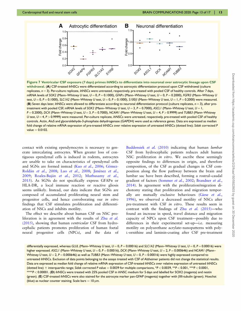
Yet, a longer differentiation time showed a repression
of astrocyte differentiation after CSF pre-exposure. In
ihNSCs that were allowed to differentiate after a 7-day
astrocytic differentiation protocol, all NSC and astrocytic
(progenitor) markers showed a clear negative trend, yet
not significant (Fig. 7A). The 7-day neuronal differenti-
ation protocol resulted in an unchanged mRNA expres-
sion of NSC and neuronal (progenitor) markers (Fig. 7B).
These results indicate that CSF pre-exposure primes
NSCs to differentiate into neuronal over astrocytic
lineage.
DiscussionIn this study, we used human in vitro models to show
that adult human ventricular CSF induces NSC prolifer-
ation, inhibits NSC motility and potentiates neuronal dif-
ferentiation. We also demonstrate that NSCs in the SVZ
of adult humans remain in contact with CSF via protru-
sions through an intact ependymal layer and even more
so due to extensive lack of ependymal coverage. We re-
veal accumulation of proliferating neural stem and pro-
genitor cells at places where the neurogenic niche is
denuded of ependyma, and are the first to quantify sys-
tematically the presence of these so-called SGNs in the
human SVZ and show that they are present from foetal
until old age. This indicates that CSF is an important
regulator of neurogenesis in humans throughout life.
An intact ependymal layer forms a semi-permeable bar-
rier, allowing a controlled exchange of factors between
CSF and brain parenchyma. Ependymocytes are post-mi-
totic cells that arise from radial glia during embryonic de-
velopment. It has been shown that these cells do not
have the capacity to regenerate (Spassky, 2005). In mice,
however, an increasing number of astrocytes—sharing
characteristics of ependymocytes—is interposed within the
ependymal layer with aging (Luo et al., 2006, 2008).
This form of ependymal repair is associated only with
mild denudation adjacent to an active neurogenic niche
(Luo et al., 2008), suggesting that an active neurogenic
niche needs an intact CSF–brain barrier for normal func-
tioning. Also, it suggests that an active SVZ and/or
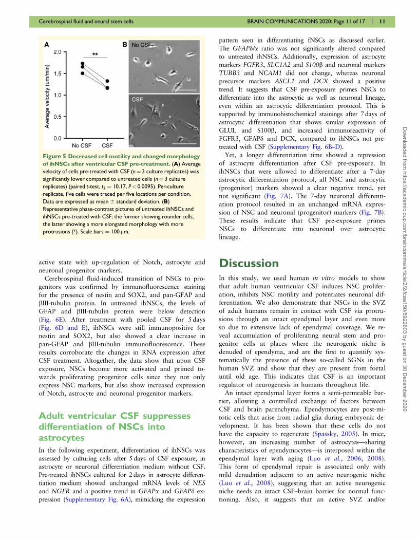
Figure 5 Decreased cell motility and changed morphology
of ihNSCs after ventricular CSF pre-treatment. (A) Average
velocity of cells pre-treated with CSF (n¼ 3 culture replicates) was
significantly lower compared to untreated cells (n¼ 3 culture
replicates) (paired t-test, t2 ¼ 10.17, P< 0.0095). Per-culture
replicate, five cells were traced per five locations per condition.
Data are expressed as mean 6 standard deviation. (B)
Representative phase-contrast pictures of untreated ihNSCs and
ihNSCs pre-treated with CSF; the former showing rounder cells,
the latter showing a more elongated morphology with more
protrusions (*). Scale bars ¼ 100 lm.
Cerebrospinal fluid and neural stem cells BRAIN COMMUNICATIONS 2020: Page 11 of 17 | 11
Dow
nloaded from https://academ
ic.oup.com/braincom
ms/article/2/2/fcaa150/5922603 by guest on 30 D
ecember 2020

Figure 6 Upon treatment with ventricular CSF, ihNSCs transition to a more active state with up-regulation of astrocyte and
neuronal progenitor markers. (A) Schematic overview of markers used to characterize the effect of CSF on lineage progression (for
references, see Supplementary material Table 5). qNSC ¼ quiescent NSC; aNSC ¼ activated NSC; NPC ¼ neural progenitor cell. (B,C) After
5 days of CSF treatment, cells were collected and used for mRNA expression analysis (n¼ 6 untreated wells, n¼ 11 wells treated with CSF of
individual donors); AluS, elongation factor 1-alpha (EF1a) and actin were used as reference genes. GFAPa (Mann–Whitney U test, U¼ 0,
P¼ 0.00016), GFAPd (Mann–Whitney U test, U¼ 0, P¼ 0.00046) and NES (Mann–Whitney U test, U¼ 0, P¼ 0.00016) were higher, whereas the
ratio GFAPd/a (Mann–Whitney U test, U¼ 0, P¼ 0.00046) was lower expressed compared to untreated ihNSCs. NGFR (Mann–Whitney U test,
U¼ 11, P¼ 0.0273), FGFR3 (Mann–Whitney U test, U¼ 23, P¼ 0.350) and S100b (Mann–Whitney U test, U¼ 0, P¼ 0.00016) were not
12 | BRAIN COMMUNICATIONS 2020: Page 12 of 17 S. F. A. M. deSonnaville et al.
(continued)
Dow
nloaded from https://academ
ic.oup.com/braincom
ms/article/2/2/fcaa150/5922603 by guest on 30 D
ecember 2020
contact with existing ependymocytes is necessary to gen-
erate intercalating astrocytes. When greater loss of con-
tiguous ependymal cells is induced in rodents, astrocytes
are unable to take on characteristics of ependymal cells
and SGNs are formed instead (Kuo et al., 2006; Gomez-
Roldan et al., 2008; Luo et al., 2008; Jimenez et al.,
2009; Roales-Bujan et al., 2012; Muthusamy et al.,2015). As SGNs do not specifically express GFAPa or
HLA-DR, a local immune reaction or reactive gliosis
seems unlikely. Instead, our data indicate that SGNs are
composed of accumulated proliferating neural stem and
progenitor cells, and hence corroborating our in vitro
findings that CSF stimulates proliferation and differenti-
ation of NSCs and inhibits motility.
The effect we describe about human CSF on NSC pro-
liferation is in agreement with the results of Zhu et al.(2015), showing that human ventricular CSF from hydro-
cephalic patients promotes proliferation of human foetal
neural progenitor cells (NPCs), and the data of
Buddensiek et al. (2010) indicating that human lumbarCSF from hydrocephalic patients reduces adult human
NSC proliferation in vitro. We ascribe these seemingly
opposite findings to differences in origin, and therefore
composition, of the CSF as gradual changes in CSF com-
position along the flow pathway between the brain and
lumbar sac have been described, forming a rostral–caudal
gradient of factors (Sommer et al., 2002; Brandner et al.,
2014). In agreement with the proliferation/migration di-
chotomy stating that proliferation and migration tempor-
ally are mutually exclusive behaviours (Giese et al.,
1996), we observed a decreased motility of NSCs after
pre-treatment with CSF in vitro. These results seem in
contrast with the findings of Zhu et al. (2015)—who
found an increase in speed, travel distance and migration
capacity of NPCs upon CSF treatment—possibly due to
differences in their experimental set-up—i.e. measuring
motility on polyurethane acrylate–nanopatterns with poly-
L-ornithine and laminin-coating after CSF pre-treatment
differentially expressed, whereas GLUL (Mann–Whitney U test, U¼ 0, P¼ 0.00016) and SLC1A2 (Mann–Whitney U test, U¼ 0, P¼ 0.00016) were
higher expressed. ASCL1 (Mann–Whitney U test, U¼ 0, P¼ 0.00016), DCX (Mann–Whitney U test, U¼ 2, P¼ 0.000646) and NCAM1 (Mann–
Whitney U test, U¼ 2, P¼ 0.000646) as well as TUBB3 (Mann–Whitney U test, U¼ 0, P¼ 0.00016) were highly expressed compared to
untreated ihNSCs. Exclusion of data points belonging to the assays treated with CSF of Alzheimer patients did not change the statistical results.
Data are expressed as median fold change of relative mRNA expression of CSF-treated ihNSCs over relative expression of untreated ihNSCs
(dotted line) 6 interquartile range; Sidak corrected P value ¼ 0.0039 for multiple comparisons, *P< 0.0039, **P< 0.001, ***P< 0.0001,
****P< 0.00001. (D) ihNSCs were treated with 25% pooled CSF in ihNSC medium for 5 days and labelled for SOX2 (magenta) and nestin
(green). (E) CSF-treated ihNSCs were also stained for the astrocyte marker pan-GFAP (magenta) together with bIII-tubulin (green). Hoechst
(blue) as nuclear counter staining. Scale bars ¼ 10 lm.
Figure 7 Ventricular CSF exposure (7 days) primes ihNSCs to differentiate into neuronal over astrocytic lineage upon CSF
withdrawal. (A) CSF-treated ihNSCs were differentiated according to astrocytic differentiation protocol upon CSF withdrawal (culture
replicates, n¼ 3). Per-culture replicate, ihNSCs were untreated, respectively, pre-treated with pooled CSF of healthy controls. After 7 days,
mRNA levels of SOX2 (Mann–Whitney U test, U¼ 0, P¼ 0.1000), GFAPa (Mann–Whitney U test, U¼ 0, P¼ 0.2000), FGFR3 (Mann–Whitney U
test, U¼ 0, P¼ 0.1000), SLC1A2 (Mann–Whitney U test, U¼ 0, P¼ 0.1000), S100b (Mann–Whitney U test, U¼ 1, P¼ 0.2000) were measured.
(B) Seven days later, ihNSCs were allowed to differentiate according to neuronal differentiation protocol (culture replicates, n¼ 3), after pre-
treatment with pooled CSF, mRNA levels of SOX2 (Mann–Whitney U test, U¼ 3, P¼ 0.7000), ASCL1 (Mann–Whitney U test, U¼ 1,
P¼ 0.2000), DCX (Mann–Whitney U test, U¼ 3, P¼ 0.7000), NCAM1 (Mann–Whitney U test, U¼ 4, P> 0.9999) and TUBB3 (Mann–Whitney
U test, U¼ 4, P> 0.9999) were measured. Per-culture replicate, ihNSCs were untreated, respectively, pre-treated with pooled CSF of healthy
controls. Actin, AluS and glyceraldehyde-3-phosphate dehydrogenase (GAPDH) were used as reference genes. Data are expressed as median
fold change of relative mRNA expression of pre-treated ihNSCs over relative expression of untreated ihNSCs (dotted line); Sidak corrected P
value ¼ 0.0102.
Cerebrospinal fluid and neural stem cells BRAIN COMMUNICATIONS 2020: Page 13 of 17 | 13
Dow
nloaded from https://academ
ic.oup.com/braincom
ms/article/2/2/fcaa150/5922603 by guest on 30 D
ecember 2020

of undescribed duration, and measuring migration using
a transwell assay with glioma-conditioned medium as at-
tractant for NPCs treated with CSF during 7 and
14 days, all in differently composed media.
Based on the neurosphere assays and RNA analyses—
and in agreement with the observed formation of SGNs
where ependyma is lacking—we conclude that ventricu-
lar CSF pushes NSCs from a relative quiescent into a
more active state, being more proliferative and capable
of self-renewal as well as being less motile. Second,
when CSF is withdrawn, NSCs develop into a progeni-
tor state, still highly proliferative but less capable of
self-renewal—i.e. larger neurosphere size but lower neu-
rosphere number. The idea that NSCs exposed to CSF
become more activated is supported by our mRNA ex-
pression and immunohistochemical data showing a gen-
eral increase in RNA and protein levels in NSCs after
CSF treatment. Indeed, NSC activation is characterized
by up-regulation of protein synthesis, and subsequent
cell division (Baser et al., 2017). Also, ASCL1 expres-
sion increases upon CSF exposure, which has been
shown to promote NSC activation and proliferation
(Andersen et al., 2014). Furthermore, self-renewal and
maintenance of activated NSCs is believed to be regu-
lated by Notch1, whereas quiescent NSCs seem less reli-
ant on Notch1 signalling (Basak et al., 2012; Engler
et al., 2018); we see clear NOTCH1 up-regulation and
activation of the Notch pathway after CSF treatment.
Thus, our data indicate that CSF of adult humans acti-
vates NSCs, and hence promoting proliferation and pri-
ming NSCs to differentiate. Additionally, we found an
indication that CSF promotes neuronal over astrocytic
differentiation. Interestingly, Schwarz et al. (2017) have
shown that human lumbar CSF improves survival and
enhances network function of neurons in vitro. Also,
Veening and Barendregt (2010) describe that in vivo
neurons in the SVZ are in contact with CSF via proc-
esses that terminate in the ventricle and actively release
neuromodulators in the CSF that can exert downstream
effects on target neurons. These observations hint that
CSF might not only potentiate neuronal differentiation,
but also impact mature neurons.
The use of post-mortem material is a strength as well
as a weakness of our study. We were able to use brain
tissue slides of a total of 40 adult human donors and
13 human foetuses, and post-mortem CSF of 10 human
donors—these are large numbers with respect to most of
the previous literature. Limitations are related to the
characterization of the post-mortem donors. Working
with post-mortem material entails that when used direct-
ly after autopsy, only clinical information is available,
based on which donors are included as controls. Later
pathological assessment changed donor status of two
CSF donors into controls having early stages Alzheimer
pathology (Braak 3-4)—importantly, their clinical files
underscore that there was no clinical phenotype yet. As
there was no difference found between CSF of these
particular donors and other donors in experiments in
which CSF of single donors was used, the experiments
with pooled CSF—composed of CSF of controls and
two donors with Alzheimer pathology—were included in
this article. Similarly, the donors of aNSCs were eventu-
ally classified as donors with Alzheimer’s disease,
Korsakov’s syndrome and bipolar disorder. As the effect
of CSF on aNSCs of the different donors was similar
and also showed the same pattern observed in ihNSCs,
the aNSCs of these donors were regarded as useful mod-
els. Second, as post-mortem ventricular CSF is more
readily available, obtainable in larger volumes and giv-
ing the same results in experiments as ante-mortem ven-
tricular CSF, mainly post-mortem CSF was used in this
study. Since the composition of CSF may change after
death, only CSF samples with a short post-mortem delay
(<7 h) were included.
The potential of CSF to stimulate neurogenesis is clinic-
ally relevant in two ways. The most common neurodege-
nerative diseases in elderly—Alzheimer’s and Parkinson’s
disease—are characterized by extensive neuronal degener-
ation. Our previous studies have shown that NSCs are
still present in the SVZ of these patients, and are still
able to proliferate and differentiate into neurons and glial
cells in vitro (van den Berge et al., 2011; van Strien
et al., 2014). Stimulation of endogenous NSCs in SVZ of
patients with a neurodegenerative disorder could therefore
potentially lead to neuronal replacement. The fact that
we show that CSF promotes neuronal over astrocytic lin-
eage differentiation is interesting in this respect. Second,
it has been hypothesized that mutated NSCs are the ini-
tiating cells in glioma, i.e. the most common primary ma-
lignant brain tumour (Qui~nones-Hinojosa and Chaichana,
2007; Jackson and Alvarez-Buylla, 2008). These glioma
stem cells functionally resemble NSCs (Ignatova et al.,2002; Galli et al., 2004; Singh et al., 2004; Jackson
et al., 2006), and gliomas primarily seem to arise in the
SVZ (Lantos and Pilkington, 1979; Zhu et al., 2005; Lim
et al., 2007; Chaichana et al., 2008; Lee et al., 2018).
There are indications that gliomas arising in closer prox-
imity to the SVZ are more likely to be of a proneural or
neural subtype (Steed et al., 2016); noteworthy in the
light of our finding that CSF promotes neuronal lineage
differentiation. Thus, understanding how CSF stimulates
NSCs will also help to elucidate the contribution of CSF
to gliomagenesis.
ConclusionIn conclusion, we show that ventricular CSF continues to
influence NSC proliferation, motility and differentiation
in adult humans, despite age-related changes in CSF com-
position that have been described by others. Factors in
the CSF and related mechanisms responsible for these
effects are yet unknown, and should be focus of future
research.
14 | BRAIN COMMUNICATIONS 2020: Page 14 of 17 S. F. A. M. deSonnaville et al.
Dow
nloaded from https://academ
ic.oup.com/braincom
ms/article/2/2/fcaa150/5922603 by guest on 30 D
ecember 2020
Supplementary materialSupplementary material is available at Brain
Communications online.
AcknowledgementsPost-mortem human brain material and CSF were obtained
from The Netherlands Brain Bank, Netherlands Institute for
Neuroscience (Amsterdam, The Netherlands; open access:
www.brainbank.nl). Ante-mortem CSF was obtained from
UMC Utrecht (Utrecht, The Netherlands), the Ludwig
Maximilian University of Munich (Munich, Germany) and
Amsterdam UMC (Amsterdam, The Netherlands). The
authors acknowledge the HIS Mouse facility of the AMC,
Amsterdam, and the Bloemenhove clinic (Heemstede, The
Netherlands) for providing foetal tissues.
FundingThis study was funded by Stichting ParkinsonFonds (EMH),
Nederlandse Organisatie voor Wetenschappelijk Onderzoek
NWO-talentprogramma Vici (NWO VICI) 865.09.003
(EMH), ZonMw Meer Kennis met Minder Dieren (ZonMW
MKMD) 11402101 (EMH) and T. & P. Bohnen fund
(PAR).
Competing interestsThe authors report no competing interests.
ReferencesAdams CW, Abdulla YH, Torres EM, Poston RN. Periventricular
lesions in multiple sclerosis: their perivenous origin and relationshipto granular ependymitis. Neuropathol Appl Neurobiol 1987; 13:
141–52.Alonso MI, Lamus F, Carnicero E, Moro JA, de la Mano A,
Fernandez JMF, et al. Embryonic cerebrospinal fluid increasesneurogenic activity in the brain ventricular-subventricular zone ofadult mice. Front Neuroanat 2017; 11: 124.
Andersen J, Urban N, Achimastou A, Ito A, Simic M, Ullom K, et al. Atranscriptional mechanism integrating inputs from extracellular signals
to activate hippocampal stem cells. Neuron 2014; 83: 1085–97.Baird GS, Nelson SK, Keeney TR, Stewart A, Williams S, Kraemer S,
et al. Age-dependent changes in the cerebrospinal fluid proteome by
slow off-rate modified aptamer array. Am J Pathol 2012; 180: 446–56.Basak O, Giachino C, Fiorini E, Macdonald HR, Taylor V.
Neurogenic subventricular zone stem/progenitor cells are Notch1-de-
pendent in their active but not quiescent state. J Neurosci 2012; 32:5654–66.
Baser A, Skabkin M, Martin-Villalba A. Neural stem cell activationand the role of protein synthesis. Brain Plast 2017; 3: 1–15.
Brandner S, Thaler C, Lelental N, Buchfelder M, Kleindienst A, Maler
JM, et al. Ventricular and lumbar cerebrospinal fluid concentrationsof Alzheimer’s disease biomarkers in patients with normal pressure
hydrocephalus and posttraumatic hydrocephalus. J Alzheimers Dis2014; 41: 1–6.
Buddensiek J, Dressel A, Kowalski M, Runge U, Schroeder H,
Hermann A, et al. Cerebrospinal fluid promotes survival and astro-glial differentiation of adult human neural progenitor cells but inhib-its proliferation and neuronal differentiation. BMC Neurosci 2010;
11: 48.Buddensiek J, Dressel A, Kowalski M, Storch A, Sabolek M. Adult
cerebrospinal fluid inhibits neurogenesis but facilitates gliogenesisfrom fetal rat neural stem cells. J Neurosci Res 2009; 87: 3054–66.
Chaichana KL, McGirt MJ, Frazier J, Attenello F, Guerrero-Cazares
H, Quinones-Hinojosa A. Relationship of glioblastoma multiformeto the lateral ventricles predicts survival following tumor resection. JNeurooncol 2008; 89: 219–24.
Conover JC, Shook B. A. Aging of the subventricular zone neural stemcell niche. Aging Dis 2011; 2: 49–63.
De Filippis L, Lamorte G, Snyder EY, Malgaroli A, Vescovi AL. Anovel, immortal, and multipotent human neural stem cell line gener-ating functional neurons and oligodendrocytes. Stem Cells 2007; 25:
2312–21.Del Bigio MR. Ependymal cells: Biology and pathology. Acta
Neuropathol 2010; 119: 55–73.Doetsch F, Caille I, Lim DA, Garcıa-Verdugo JM, Alvarez-Buylla A.
Subventricular zone astrocytes are neural stem cells in the adult
mammalian brain. Cell 1999; 97: 703–16.Domınguez-Pinos MD, Paez P, Jimenez A-J, Weil B, Arraez M-A,
Perez-Fıgares J-M, et al. Ependymal denudation and alterations ofthe subventricular zone occur in human fetuses with a moderatecommunicating hydrocephalus. J Neuropathol Exp Neurol 2005;
64: 595–604.Engler A, Rolando C, Giachino C, Saotome I, Erni A, Brien C, et al.
Notch2 signaling maintains NSC quiescence in the murine ventricu-
lar-subventricular zone. Cell Rep 2018; 22: 992–1002.Galli R, Binda E, Orfanelli U, Cipelletti B, Gritti A, Vitis SD, et al.
Isolation and characterization of tumorigenic, stem-like neural pre-cursors from human glioblastoma isolation and characterization oftumorigenic, stem-like neural precursors from human glioblastoma.
Cancer Res 2004; 64: 7011–21.Gato A, Moro JA, Alonso MI, Bueno D, De La Mano A, Martın C.
Embryonic cerebrospinal fluid regulates neuroepithelial survival,proliferation, and neurogenesis in chick embryos. Anat Rec 2005;284A: 475–84.
Giese A, Loo MA, Tran N, Haskett D, Coons SW, Berens ME.Dichotomy of astrocytoma migration and proliferation. Int J Cancer
1996; 67: 275–82.Gomez-Roldan MDC, Perez-Martın M, Capilla-Gonzalez V, Cifuentes
M, Perez J, Garcıa-Verdugo JM, et al. Neuroblast proliferation on
the surface of the adult rat striatal wall after focal ependymal lossby intracerebroventricular injection of neuraminidase. J Comp
Neurol 2008; 507: 1571–87.Gray F, Lescs MC, Keohane C, Paraire F, Marc B, Durigon M, et al.
Early brain changes in HIV infection: neuropathological study of 11
HIV seropositive, non-AIDS cases. J Neuropathol Exp Neurol 1992;51: 177–85.
Honer WG, Bassett AS, Falkai P, Beach TG, Lapointe JS. A case study
of temporal lobe development in familial schizophrenia. PsycholMed 1996; 26: 191–5.
Ignatova TN, Kukekov VG, Laywell ED, Suslov ON, Vrionis FD,Steindler DA. Human cortical glial tumors contain neural stem-likecells expressing astroglial and neuronal markers in vitro. Glia 2002;
39: 193–206.Jackson EL, Alvarez-Buylla A. Characterization of adult neural stem
cells and their relation to brain tumors. Cells Tissues Organs 2008;188: 212–24.
Jackson EL, Garcia-Verdugo JM, Gil-Perotin S, Roy M, Quinones-
Hinojosa A, VandenBerg S, et al. PDGFRalpha-positive B cells areneural stem cells in the adult SVZ that form glioma-like growths in
response to increased PDGF signaling. Neuron 2006; 51: 187–99.Jimenez AJ, Domınguez-Pinos M-D, Guerra MM, Fernandez-Llebrez
P, Perez-Fıgares J-M. Structure and function of the ependymal
Cerebrospinal fluid and neural stem cells BRAIN COMMUNICATIONS 2020: Page 15 of 17 | 15
Dow
nloaded from https://academ
ic.oup.com/braincom
ms/article/2/2/fcaa150/5922603 by guest on 30 D
ecember 2020

barrier and diseases associated with ependyma disruption. Tissue
Barriers 2014; 2: e28426.Jimenez AJ, Garcıa-Verdugo JM, Gonzalez CA, Batiz LF, Rodrıguez-
Perez LM, Paez P, et al. Disruption of the neurogenic niche in the
subventricular zone of postnatal hydrocephalic hyh mice. JNeuropathol Exp Neurol 2009; 68: 1006–20.
Johansson PA, Cappello S, Gotz M. Stem cells niches during develop-ment-lessons from the cerebral cortex. Curr Opin Neurobiol 2010;20: 400–7.
Johnson KP, Johnson RT. Granular ependymitis. Occurrence in myxo-virus infected rodents and prevalence in man. Am J Pathol 1972; 67:511–26.
Kamphuis W, Orre M, Kooijman L, Dahmen M, Hol EM. Differentialcell proliferation in the cortex of the appsweps1de9 Alzheimer’s dis-
ease mouse model. Glia 2012; 60: 615–29.Kukekov VG, Laywell ED, Suslov O, Davies K, Scheffler B, Thomas
LB, et al. Multipotent stem/progenitor cells with similar properties
arise from neurogenic regions of adult human brain. Exp Neurol1999; 156: 333–44.
Kuo CT, Mirzadeh Z, Soriano-Navarro M, Rain M, Wang D, Shen J,et al. Postnatal deletion of Numb/Numblike reveals repair andremodeling capacity in the subventricular neurogenic niche. Cell
2006; 127: 1253–64.Lantos PL, Pilkington GJ. The development of experimental brain
tumours. A sequential light and electron microscope study of thesubependymal plate. I. Early lesions (abnormal cell clusters). ActaNeuropathol 1979; 45: 167–75.
Lee JH, Lee JE, Kahng JY, Kim SH, Park JS, Yoon SJ, et al. Humanglioblastoma arises from subventricular zone cells with low-leveldriver mutations. Nature 2018; 560: 243–7.
Lehtinen MK, Walsh C. A. Neurogenesis at the brain-cerebrospinalfluid interface. Annu Rev Cell Dev Biol 2011; 27: 653–79.
Lehtinen MK, Zappaterra MW, Chen X, Yang YJ, Hill AD, Lun M, etal. The cerebrospinal fluid provides a proliferative niche for neuralprogenitor cells. Neuron 2011; 69: 893–905.
Lim D. A, Cha S, Mayo MC, Chen M-H, Keles E, VandenBerg S, etal. Relationship of glioblastoma multiforme to neural stem cell
regions predicts invasive and multifocal tumor phenotype. NeuroOncol 2007; 9: 424–9.
Luo J, Daniels SB, Lennington JB, Notti RQ, Conover JC. The aging
neurogenic subventricular zone. Aging Cell 2006; 5: 139–52.Luo J, Shook BA, Daniels SB, Conover JC. Subventricular zone-medi-
ated ependyma repair in the adult mammalian brain. J Neurosci2008; 28: 3804–13.
Ma Y, Liu M, He B. Adult cerebrospinal fluid does not support
neurogenesis from fetal rat neural stem cells. Neurol Sci 2013; 34:735–9.
Middeldorp J, Boer K, Sluijs JA, De Filippis L, Encha-Razavi F, VescoviAL, et al. GFAPd in radial glia and subventricular zone progenitors inthe developing human cortex. Development 2010; 137: 313–21.
Mirzadeh Z, Merkle FT, Soriano-Navarro M, Garcia-Verdugo JM,Alvarez-Buylla A. Neural stem cells confer unique pinwheel architec-ture to the ventricular surface in neurogenic regions of the adult
brain. Cell Stem Cell 2008; 3: 265–78.Muthusamy N, Sommerville LJ, Moeser AJ, Stumpo DJ, Sannes P,
Adler K, et al. MARCKS-dependent mucin clearance and lipid me-tabolism in ependymal cells are required for maintenance of fore-brain homeostasis during aging. Aging Cell 2015; 14: 764–73.
Netherlands Brain Bank [Internet]. 2020. Amsterdam: NetherlandsInstitute for Neuroscience [cited 23 January 2020]. Available from:
https://www.brainbank.nl.Nielsen S, Arnulf Nagelhus E, Amiry-Moghaddam M, Bourque C,
Agre P, Petter Ottersen O. Specialized membrane domains for water
transport in glial cells. J Neurosci 1997; 17: 171–80.Orekovi�c D, Klarica M. The formation of cerebrospinal fluid: nearly a
hundred years of interpretations and misinterpretations. Brain ResRev 2010; 64: 241–62.
Ortega JA, Memi F, Radonjic N, Filipovic R, Bagasrawala I, Zecevic
N, et al. The subventricular zone: a key player in human neocorticaldevelopment. Neuroscientist 2018; 24: 156–70.
Qui~nones-Hinojosa A, Chaichana K. The human subventricular zone:
a source of new cells and a potential source of brain tumors. ExpNeurol 2007; 205: 313–24.
Rasband WI. [Software]. U. S. National Institutes of Health. Bethesda,Maryland, USA 2015. http://imagej.nih.gov/ij/.
Roales-Bujan R, Paez P, Guerra M, Rodrıguez S, Vıo K, Ho-Plagaro
A, et al. Astrocytes acquire morphological and functionalcharacteristics of ependymal cells following disruption ofependyma in hydrocephalus. Acta Neuropathol 2012; 124:
531–46.Roelofs RF, Fischer DF, Houtman SH, Sluijs JA, Van Haren W, Van
Leeuwen FW, et al. Adult human subventricular, subgranular, andsubpial zones contain astrocytes with a specialized intermediate fila-ment cytoskeleton. Glia 2005; 52: 289–300.
Sanai N, Alvarez-Buylla A, Berger MS. Neural stem cells and the originof gliomas. N Engl J Med 2005; 353: 811–22.
Sanai N, Nguyen T, Ihrie RA, Mirzadeh Z, Tsai H-H, Wong M, et al.Corridors of migrating neurons in the human brain and their declineduring infancy. Nature 2011; 478: 382–6.
Sanai N, Tramontin AD, Qui~nones-Hinojosa A, Barbaro NM, GuptaN, Kunwar S, et al. Unique astrocyte ribbon in adult human brain
contains neural stem cells but lacks chain migration. Nature 2004;427: 740–4.
Sarnat HB. Ependymal reactions to injury. A review. J Neuropathol
Exp Neurol 1995; 54: 1–15.Sawamoto K. New neurons follow the flow of cerebrospinal fluid in
the adult brain. Science. 2006; 311: 629–32.
Schauer R. Sialic acids as regulators of molecular and cellular interac-tions. Curr Opin Struct Biol 2009; 19: 507–14.
Schwarz N, Hedrich UBS, Schwarz H, Harshad PA, Dammeier N,Auffenberg E, et al. Human cerebrospinal fluid promotes long-termneuronal viability and network function in human neocortical orga-
notypic brain slice cultures. Sci Rep 2017; 7: 12249.Silva-Vargas V, Maldonado-Soto AR, Mizrak D, Codega P, Doetsch
F. Age-dependent niche signals from the choroid plexus regulateadult neural stem cells. Cell Stem Cell 2016; 19: 643–52.
Singh SK, Hawkins C, Clarke ID, Squire JA, Bayani J, Hide T, et al.
Identification of human brain tumour initiating cells. Nature 2004;432: 396–401.
Sommer JB, Gaul C, Heckmann J, Neundorfer B, Erbguth FJ. Doeslumbar cerebrospinal fluid reflect ventricular cerebrospinal fluid? Aprospective study in patients with external ventricular drainage. Eur
Neurol 2002; 47: 224–32.Spassky N. Adult ependymal cells are postmitotic and are derived
from radial glial cells during embryogenesis. J Neurosci 2005; 25:10–8.
Steed TC, Treiber JM, Patel K, Ramakrishnan V, Merk A, Smith AR,
et al. Differential localization of glioblastoma subtype:implications on glioblastoma pathogenesis. Oncotarget 2016; 7:24899–907.
van den Berge SA, Middeldorp J, Zhang CE, Curtis MA, Leonard BW,Mastroeni D, et al. Longterm quiescent cells in the aged human sub-
ventricular neurogenic system specifically express GFAP-d. AgingCell 2010; 9: 313–26.
van den Berge SA, van Strien ME, Korecka JA, Dijkstra AA, Sluijs JA,
Kooijman L, et al. The proliferative capacity of the subventricularzone is maintained in the parkinsonian brain. Brain 2011; 134:
3249–63.Van Dijk KD, Bidinosti M, Weiss A, Raijmakers P, Berendse HW, van
de Berg WDJ. Reduced a-synuclein levels in cerebrospinal fluid in
Parkinson’s disease are unrelated to clinical and imaging measuresof disease severity. Eur J Neurol 2014; 21: 388–94.
Van Dijk KD, Persichetti E, Chiasserini D, Eusebi P, Beccari T, CalabresiP, et al. Changes in endolysosomal enzyme activities in cerebrospinal
16 | BRAIN COMMUNICATIONS 2020: Page 16 of 17 S. F. A. M. deSonnaville et al.
Dow
nloaded from https://academ
ic.oup.com/braincom
ms/article/2/2/fcaa150/5922603 by guest on 30 D
ecember 2020
fluid of patients with Parkinson’s disease. Mov Disord 2013; 28:
747–54.van Strien ME, van den Berge SA, Hol EM. Migrating neuroblasts in
the adult human brain: a stream reduced to a trickle. Cell Res 2011;
21: 1523–5.van Strien ME, Sluijs JA, Reynolds BA, Steindler DA, Aronica E, Hol
EM. Isolation of neural progenitor cells from the human adult sub-ventricular zone based on expression of the cell surface markerCD271. Stem Cells Transl Med 2014; 3: 470–80.
Veening JG, Barendregt HP. The regulation of brain states by neuroac-tive substances distributed via the cerebrospinal fluid; a review.Cerebrospinal Fluid Res 2010; 7: 1.
Whish S, Dziegielewska KM, M~a¸llg~A¥Rd K, Noor NM, Liddelow SA,Habgood MD, et al. The inner CSF-brain barrier: developmentally
controlled access to the brain via intercellular junctions. FrontNeurosci 2015; 9: 1–15.
Zappaterra MW, Lehtinen MK. The cerebrospinal fluid: regulator of
neurogenesis, behavior, and beyond. Cell Mol Life Sci 2012; 69:2863–78.
Zhang J, Goodlett DR, Peskind ER, Quinn JF, Zhou Y, Wang Q, et
al. Quantitative proteomic analysis of age-related changes in humancerebrospinal fluid. Neurobiol Aging 2005; 26: 207–27.
Zhang J, Piontek J, Wolburg H, Piehl C, Liss M, Otten C, et al.Establishment of a neuroepithelial barrier by Claudin5a is essentialfor zebrafish brain ventricular lumen expansion. Proc Natl Acad Sci
USA 2010; 107: 1425–30.Zhu M, Feng Y, Dangelmajer S, Guerrero-Cazares H, Chaichana KL,
Smith CL, et al. Human cerebrospinal fluid regulates proliferation
and migration of stem cells through insulin-like growth factor-1.Stem Cells Dev 2015; 24: 160–71.
Zhu Y, Guignard F, Zhao D, Liu L, Burns DK, Mason RP, et al.Early inactivation of p53 tumor suppressor gene cooperating with NF1loss induces malignant astrocytoma. Cancer Cell 2005; 8: 119–30.
Cerebrospinal fluid and neural stem cells BRAIN COMMUNICATIONS 2020: Page 17 of 17 | 17
Dow
nloaded from https://academ
ic.oup.com/braincom
ms/article/2/2/fcaa150/5922603 by guest on 30 D
ecember 2020