Embed Size (px)
Citation preview
CASE /11 (S86-71 00)
ELLIS FISCHEL STATE CANCER C ENTER ORAL PATKOLOGY SEMINAR #93
O.P.S. NO. 86-1115 November 1,, 1986
CASE HISTORIES
<::ontrlbuted by Ronald W. Oxenhandler, M.D., Memorial Hospital, ~hattanooga, Tennessee.
89 year old with a two- year history of obstructive symptoms with lntermlttent bleeding. The left nasal mass has enlarged recently. A CT scan performed 7/1/86 shows a mass localized to the left naris. The doctor is unaware of any other Significant histor y.
eASE /12 (DC86-4H) (slide and x-ray) ContrJbuted by Charles A. Waldron, D.D.S., Washington Universi ty School of Dental Medicine, St. Louis, Mis.souri.
A blacK man, age ..51, presented with a modera-tely painful right mandibular swelling of some months dura1ion. Clinical examina·tlon showed a generalized enlargement of the r ight mandible- with ctlnkal evidence of-a patholqglc !racture ln the premolar area. The covering gingiva and alveolar mucosa were d lncally normal and there was no evidence of 11JUCosat ulceration or abnormal mass anywhere in the soft tissues oi the· mouth. Radiographs showed that the mandible was almost totally destroyed by an extenslve lytic process extending from the symphysis to the ascending ramus. After biopsy a mandlbular resection was performed. An x- ray of the sUrgical spechnerl ls enclos~d. The slide represents a cross section through the mandible in the canine area.
CASE 1/3 (4539) (slide and x-ray) Contributed by Y. LeGal, M.D., {nstitut O'Anatomie Pathologique, Strasbourg.
LU is a 16 year old male. An oste-olytic lesion was discovered In the maxilla in 1982. Copy of the roentgenogram from January 1985 is included.
CASE li4 (1506) Contribute9 by t.A. LovinggOQd, M.D., Southeast Missour i Hospital, Cape Girardeau, Missouri.
The patlent is a 72 year old white man who complained of a graduaJJy increasing swelling of the right facial area opposite the parotid eland. The mass occurred over a 2 month pedod. At surgical exploration the right deep lobe oi the parotid was found to contain a tumol" mass.
-.
CASE HISTORIES continued
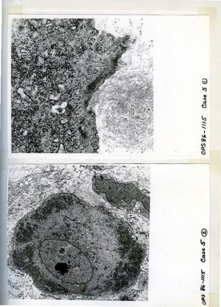
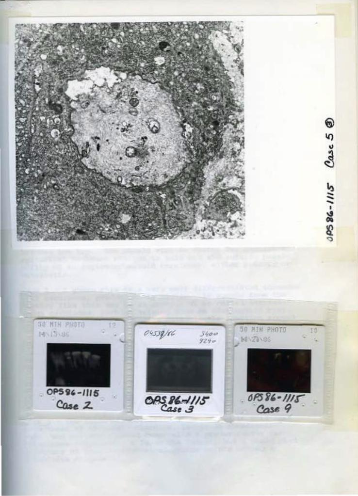
CASE //j (2 l063} (slide und 3 EM photos) Contributed by Y. LeGal, M.D., lnsti tut D'Anatomle Pathologique, Strasbourg.
cs was born in l'n~ and developed "nasal polyps of the le ft side" at the end of 19&0. Subsequently, the patient had several recurrences: In 1982 (middle turbinaire}, in 19&4 {left inteNinuso-nasal wall), in 1985 at the previous place and finally again in 1986. Despite these numerous recurr"ences there had not been bone destruction. The histoJogy o1 the tumor remains unchanged. The rnaterlal you are receiving represen ts the original biopsy performed In 1980. Included a lso are elec tro micrographs.
CASE /16 (586-2389) Contributed by J. B. Andelin, M.D., Mercy Hosital of Williston, Williston, North Dakota.
TD is a 34 year old caucasian male, user of smokeless tobacco who developed a 2 em nodule in the soft palate, mobile, nonllxed which was almost completely exclsed during biopsy. According to the patient, the lesion has been there almost 2 years.
CASE 07 (T86-4227) Contr ibuted by Charles Dunlap, D.D.S. and Bruee Barker, D.D.S., Department of Oral Pathology, University of Missour i-Kansas CJty, School of Dentist ry, Kansas Ci ty, Missouri.
A 6 J year old m~le with a 4 .. , em tumor in the soft patete.
CASE U& (86-!60& and &6-1623) (2 slides) Contributed by Charles Dunlap, D.D.S. and Bruce Barker, D.D.S., Department of Oral Pa thology, Universi ty of Missouri-Kansas City, School of Dentistry, Kansas City, Missourl.
A 7~ year old male had diffuse leukoplakia of the palate. One site had an approximately 1.2 em exophytic lesion (86-160&} and two other sites had Uatte.ned, papillary lesions; both "''ere removed and 86-1623 is representative.
CASE /19 (&6-1 466) (clinical photo only) Contributed by Charles Dunlap, D.D.S. and Bruce Barker, D.D.S., Department of Oral Pathology, University of Missouri-Kansas City, Sdlool of Dentistry, Kansas City, Missouri.
This 7:l year old had an ulcerative lesion of long duration on the buc<;al gingiva as shown. There were no other oral lesions and the medical history was unremarkable.
CASE 810 (51928} Contributed by Miguel A. Simon, M.D., Professor o( Pathology, San Juan, Argentina.
MA is a 30 year old white female who developed a onass asymptomatic located within the parotid gland area. Thi.s patient had a pigmen ted lesion re moved at an undetermined past, however, the histological na ture was no t known.
II) ... --I ~ ~ <)

Yale U niversity j NJ'I RISoU, M 0 . PNjm<fl' & Oir'l'l-.w
" ...... -. P.do4"n Dq<.- "P,W~<fr sm..I<JMd-Jifl c• Smvt P.O. S.X JJJJ ,"'J.N ~'t'll , C..W011C'W HJIH~I~
october 30, 1986
Carlos Perez-Mesa, M.D. Department of Pathology Ellis Fischel State cancer Hospital Columbia, MO 65201
Dear carlos:
Cl':ltFUJ' .d4ffA
N Yj)r:leS!rm Tdqlu= (»J) j$J•}J11J
These a.ra l'DY .impressions on -ebe c.ases that I. reee.ived from you from the Oral Pathology Seminar #93:
case l - This is a malignant vascular tumor, and my differential diagnosis is between malignant hamangiopericytoma and ""'lignant hemangioendothelioma. I slightly favor the former on the basis of the architecture, but I would like to see ~ticul~n stains and immunoperoxidase before deciding between the two. One should also do stains for keratin and epithelial membrane antigen to rule out tha outside possibility of an angiosarcomatoid carcinoma, either primary or met astatic.
case 2 - I guess this is a very well differentiated s quamous cell carcinoma, predominantly cystic. It sounds fro~ the history like this may be an example of so-celled intraosseous carcinoma. perhaps arising from an odontogenic cyst. There is a l so a very extensive osteoblastic reaction around the tooth.
case 3 - This looks like a very organoid odontogenic lesion. I suppose i~ corresponds ~o a complex or compound odontoma, but I would defer to the oral pathologist regarding the exac~ ~erminology .
Case 4 - I~ looks like a carc~osarcoma of sal~vary gland origin. A~ternatively, one may use the terminology of sarcomatoid or sarcoma-like carcinoma if one were to interpret tbe entiro neoplasm as of epithelia l derivation.
Case 5 - The three possibilities 1 considered in this case were those of chondro~d chordoma, low-grade chondrosarcoma, and malignant mdxed tumor with a predominantly cartilaginous component. I favor the former, but I would need abattory of immunocytochemical stains before making a definitive diagnosis.
case 6 - This is a carcLooma of minor salivary gland derivation, and the architectural features suggest that it belongs to the general category of adenoid cyst ic carcinoma. There is a striking hyperplasia of the overlying squamous mucosa.
Case 7 - The only name that r can think of f or this l esion is that of osteoma.
Case 8 - I would diagnose this lasion as verrucous keratosis, although I cannot rule out the possibility o f it being a early form of verrucous carcinoma.
Case 10 - This looks consistent with a malignant melanoma that has metastasized to an intraparotid lymph node .
I have been in commun~cation late1y wi th your f riend Ronald Oxenhandlcr, who tells me that he misses a lot your company and the atmosphere of Columbia, Missouri.
JR/ams
Best personal regards,
Juan Rosai, M.D. Professor of Pathology Direct or of Anatomic Pathol ogy
ELLIS fiSCHEL STATE CANCER CENTER ORAL PATHOLOGY SEMINAR NO. 93
O.P.S. 86-1115 November I ' • 1986
"OFFICIAL DIAGNOSIS''
The proceedings of this seminar were conducted in the Lodge of the Four Seasons Re5()(t, Lake Oz.ark.s, Missouri.
CASE I: ANCJOSARACOMA {S86-7100) Contdbuted by Ronald W. Oxenhandler, M.D., Memorial Hospital, Chattanooga, Tennessee.
The majority of the consultants favored the diagnosis of angiosarcoma. A few commet~ts at random:
RosaJ from Yale, "This is a •naligoant vascular tumor, and cny di!ler ential diagnosis is between malignan t hemangiopericy torna and malignant hemangioendothelioma. I sligh tly favor the former on the basis of the a rchitecture, but I would like to see reticulin stains and immunoperoxida.se before decidin& between the two. One should also do stains for keratin and epitheHal membrane antigen to ru1e out the outside possibiti ty of an angiosarcomatoid carcinoma, e i ther primary or metastatic." Meyer and Associates from St. Luke's Hospital, St. Louis, "Angiosarcoma. We had some thoughts about pyogenic granuloma, but concluded that the vascular pattern was too atypical and the sarcomatoid stroma is like that ol angiosarcomas or the head and face in older people." Glass, Young, and Rohrer from Ol<lnhoma commented, "We feel this is a malignant vascular lesion and would consider angiosarcoma and possibly Kaposi's sarcoma.'' Sprague and Associates from Nebraska, "Generically, this Is quite acceptable for a form of angiosarcoma. However, we believe it Is mqre acceptable specifically as a a Kaposi's sarcoma." Azzopa.rdi from the Unjversity of london, ''? Hamangiopericytoma-like tumor of nose but cyto logicaJiy "worse" than usual tumor , with large vesicular nuclei. Angiomatous lesions entered my di ffe rential from pertcytomatous ones, a,s some tumor cells appeared to line vascular spaces, but mostly they seemed to be around t.'\e vessds. Commoner lesions should be excluded by appropriate stains, and Teticulin might emphasize the perieytomatous pattern. Factor VIU might also be helpful, H only as a negative image.'' Toto and 1\ssociatu lrom Loyola at Chicago interpreted the lesion as maHs.nant hemagiopericytoma. Simon from Argentina interpreted as n Kaposi's sarcoma .. Abnuns from USC, "The ceUuJar atypia seems too seve.re to consider this to be beni.gn. Therefore, angiosarcoma or Kaposi's sarcoma would be Hkefy." Cardona Lopez fro1n Honduras and Eusebi from 6ologna inte r pre ted the leslon as angiosarcoma.
2
W·eidner from Wake Forest, Barker and Dunlap from the University of Missouri-Kansas City preferred pyogenic granuloma. Rowe and Stewart from Michigan also pyogenic granuloma vs. sarcoma. LeGal from Strasbourg interpreted the lesion as ''hemangioendothelioma, presumed benign." Donath from Hamburg pre(ered angiogranuloma .. OD haemangioendotheHoma.
CASE 2: RRIMARY INTRA-OSSEOUS SQUAMOUS CELL CARCINOMA PROBABLY ORIGINATING IN AN ODONTOGENIC CYST (DC86-434) Contributed by Charles A. Waldron, D.D.S., Washington University School of Dental ,\1edicine~ St. Louis, Missouri.
A few commentaries at random:
Lumerman, Fre~man, and Kerpelfrom Flushing New York, "squamous ·cell carcinoma arising in a cyst." Kru1ch~off and Eisenberg from the University oi Connecticut, uA central and well-di Uer~ntiated squamous cell carcinoma, apparently not arising (rom surface epithelium.. We question the origin of this and throw . out the possibility o[ the so-called primary intra~alveolar carcinoma." Rosai from Yale, "I guess this is a ver y.. weiJ differentiated squamous cell carcinoma, predominantly cystic. It sounds from the history Hke this may be an .~xample of so-called in tra.osscous card noma, perhaps arising from an odontogenic cyst. There is also a very extensive osteoblastic reaction around the tooth." Class, Young, and Rohf.er from Oklahoma, "We think this is a well differentiated squamous cell carcinoma which very likely might have arisen from the linfng of the odontogenic cyst which still seems to be present on the radiograph." Azzopardi from the University of London, "Burrowing squamous carcinoma, apparently as a primary intra-alveolar .Lesion. Possibly of odontogenic origin or, alternatively, from seques-tered squamous epithelium." Sprague and Associates from Nebraska, 11We are assured from the clinical history that there is no communication with this proliferating epithelium and the surface. With that in mind, we must strongly support primary interosseous cardnoma, not otherwise subclassified. We see no histopathologic evidence to support origin from odontogenic apparatus.', Hammond, Vincent, and Watson from Iowa, "Well differentiated squamous cell carcinoma, apparent!)• primary in mandible .I' Tarpley. and Corio !com Georgetown University, "Squamous cell carcinoma whh verrucous pattern with the carcinoma probably arising in a cyst.'' Azar from Tampa, ''Keratinizing squarnous cell carcinoma, presumably primary lntra-osseous .. " ScitJt>ba from SUNY at Stony Brook preferred primary intra-alveolar Carcinoma. Abrams from USC of!ercd, "The histopathology is squamous car.cinoma. In view of the history provided, it might be a primary intra-osseous squamous carcinoma."
3
CASE 3: COMPOUND ODONTOMA (4.539) Contributed by Yvon l eCal, M.D., lnstit1Jt 0 '/\natomie Pa thologique, Strasbourg.
White from Kentucky commented, HQeveloping odontoma with pc"ominent ameloblastic component which is associated with newlyformed dental hard tl$sue. Some may wish to call this the old ameloblas tic odontoma, but we believe the epithelium is act ive-ly partjcipating in matrix production and not neoplastic." Lumerman, Freedman, and Kerpel made the following commentary, "Although there is a great deal of •rename:J organ" type epithelium, we believe that most if not all of it is involved in tooth production. Therefore, although some might cooslder the diagnosis of odontoamelb!astoma, or ameloblastic tibro-odontoma, we would simply call this a complex odontoma.11
Krutchkoff and Eisenberg, "Odontoma·-plain and simple. This apparen1ly matured over the years from n lytle to an opaque process ... Tomjch !rom (ndiana also called it-complex odontoma. Az:topardl from london, "? Complex odontome, with amcloblastic epithelium one end, and tooth-bud-like malformations other end. "Osteolytic" lesion pu1;zled me. ls it not "sclerotiC'' as seen on the slide?'' Toto from Loyola. University at Chicago, ''complex odontoma. im1nature.'' Kyriakos [rom Washington University ~nd Gnepp from St. Louis University, St. Louis called It odontoma. Kahn and Sciubba from Stony Brook commented complex odontoma, adding, "The enclosed panoramic radiograph MJgge·sts a pattern unlike that for an odontoma. The single slide ex3mined would suuest a more aggressive process than an odontoma, raising the possibility of this r epresenting an ameloblastic odontoma." Abrams from USC, 11Th is Is mostly complex odontoma.. However, there is a small fragment of 11sof t tumor" present a long one side, so ameloblastlc fibro-odontoma would be an ncceptable diagnosis." Weathers from Emory, ••Actively growing odontoma {complex).t•
A lew Interpreted the Jesloo as malignant "meloblastoma.
CASE 4: CARCINOSARCOMA (U06) Contributed by T .A. Lovtnggood, M.D., Southeast Missouri Hospital, Cape Girardeau, Mis.souri.
All the consultants agreed in the malisnant nature of the lesion, however, some differences concerning histogenesis. A few random examples:
Lumerman, Freedman, and KerpeJ from FJushlng, New York, "This is a peculiar tumor demonstrating definite adenocarcinoma set jn a spindlecell sarcomatous strorna. The stroma resembles fibrosarcorna but we feel that there may be some evidence o( os1eoid formation. We're not sure if subclassifying the sarcomatous component is relevant and are inclined to call this tumor a carcinosarcoma, probably of p3rotJd gland origin."
Rosai from Yale, "It looks like a carcinosarcoma of salivary gland origin. Alte-rnatively, one may use the terminology of sarcomatoid or sarcoma- like carcinoma. if one were to interpret the entire neoplasm as of epJthelial derivation." LoCal from Strasbourg rnade the following commentary, "Carcinosarco ma or malignant mixed tumor. Bu t from what origin? How Is the PAP for S 100 pro tein? I am dreaming of a per ipheral rnallgnant nervous tumor Induced by a carcinoma of sa1ivary gland through NGF ." Tomich and Associa tes from Indiana Universi ty, "This is a remarkable case. One component appears to be similar to salivary duct carcinoma as described by Fayemi o.nd Tokcr. The stromal component appears to be neoplastic as well. Perhaps this is a carcinosarcoma. Another possibility is a malignant schwannoma with glandular dif(erenriation .... Spra.gue and Associates from Nebraska, "Since we feel comfortable that both the stroma and the epitheHum are mali&nant, we musr suppor t a diagnosis of carcinosarcom4. The besl bet would be that i t is a metastasis to this region. Because o f the stroma, we cannot support a d iagnosis of carcinoma ex pleomorphic adeno m.a.'' Hammond, Vincent and Wnt.son (rom Iowa offered, "malignant sch wannoma with glandular diCferentiation." Tarpley and Corio from Ceorgetown offered the following comment, "Carcino-sarcoma in the broad sense but with the neural and rhabdomyoblast elements present-the possibility o[ a malignant triton lumor must be considered.u Kyriakos from Washington University offered the possibility of 01Synovial sarcoma-stroma is malignant with fibrosareoma1ous areas (Cancer 50:269, 19&2)." Simon from San Juan, Argentina, "t vote for carcino$arcoma since both tumor components are neoplastic." Azar from Tampa, "Adenocarcinoma with sarcomatous stroma. This is probably a malignant pJcomorphic adenoma because of a residual focus of pleomorphic adenoma." Donath from Hamburg, "An unusual case, which I have never seen . I think i t is an adenocarclnc)lnn with a sarcoid-like stroma reac tion, 1 would not call it carcinosarcoma or malignant pleomorphic adenoma." Weidner from Wake Forest and Eusebi from Bologna call It salivary duct carcinoma. Weidner stated, '"The carcinomatous component of this tumor reminds me of so-c~lled •'saJivary .. duct carcinoma" like that described by Carland eta!. (Am J Clin Patho1 81:436·4'11, 1934). The surrounding stroma is very atypical with increased mitotic activity. Is it pseudosarcomatous desmoplastic response, or is it true sarcoma? lf the lAtter is t rue, then thi$ lesion becomes a carcinosarcoma. I also considered true malignant rnlxed turnor. In either event , th is tumor is high grade:•
Follow-up: There was no evidence of r ecurrence during the vl$it o f the pa tient to the clinic In January, 1987.
5
CASE j: CHORDOMA (23063) Contributed by Yvon LeGal, M.D., lnstltut D'Anatomie Pathologique, Strasbcurg.
The contributor, Or. LeGa!, olfered the following, "I don't know and hope to kno·;v the final answer."
Waldron and El-Mofty !rom Washington University, St. Louis called it chordoma. Lumermat'l, F reedmttn and Kerpe:_l sent thf! following commentary, "The microscopic appearance ol this lesion and the E\1 p.'lotos suggest a diag~>osis ol chordoma although the lack of bone destruction and the apparent origin of the lesion from the latera) nasal waH are perplexing. Although we favor the diag{loSis o f chordoma, we suppose one must also consider chordoid sarcoma." Rosai from Yale, "The three possiblli des I considered in thi.s case were those of chondroid dlordoma, low--grade chondrosarcoma, and malignant mixed tumor with a predominantly cartilaginous component .. I favor the former, but I would need a battery of immunocytochemical stains before making a definitive diagnosis." Meyer frorn St. Luke's Hospital in St. Louis, "1'his was a puzzler. We all considered chordoma but most of us dismissed it because of the ~pithelial growth pattern locally and the el<etron micrographs. Chondrosarcoma was the first choice of .some; others including myself favor mixed tumor (pleomorphic ndenoma)." Hori from Elkins, West Virginia fnvored choAdroid chordoma. Tomid1 from lndiana, "Chondroid chordoma is the best we could do on this c~se." Kyriakos from W;u hlng ton University, St. Louis , "Chordoma. Unusual site but chordomas not connected to clivus are reported. See Arch Laryngol 7&:163, 1963; Laryngoscope 90:612, 1980; and Laryngoscope 9~: 1063, 1984 .• Simon, Staff and Resjdents from San Juan, Argentina by u.nan1mit-y called It chordoma. Sprague from Nebraska, Azzopardi from the University of London, Hammond and Associates lrom Iowa, Tarpley and Corio from Georgetown, HM$el\ from San Francisco, Sciubba and Kahn from Stony Brook, Abrams from USC, Donath from Hamburg and Eusebi from Bologna also prefer chordoma.
Other dissenting opinions were also rendered. A few at random:
KrutchkoU and Associates from Connecticut, "&en!gr. mucoid mucosal polyp. The enclosed photocopies of electron micrographs did not have sufficient detail to be useful to us." Class, Young, and Rohrer from Oklahoma, 11We feel this represents a mucous cxlravasatlon phenomenon." Rowe and Stewart from Michigan preferred chondrosarcoma. Toto from Loyola at Chicago, "chondroidmyxoma." Dunlap and Barker from Kansa.s City, Missouri, "Benign histiocytic mucinoma:• • Azar from Tampa, "Probable, recurrent pleomorphic adenoma wlth large "myoepithelial" cell~.''
Oxenhandler from Chattanooga, "lntrarwual mixed tumor (Am J Clin Path 68:21}-21&, 1977),• Weidner from Wake Forest, "I favor a diagnosis o f mixed tumor.'' Weath«s from Emory commented, "Malignant tran$formatlon from a prevlously benign mixed tumor (carcJnoma explcomnrphic adenoma). Some of the clear cells with the strings of cytoplasm were a bit reminiscent ol the physatiferous cell s of the cordoma, but I feel that this i s adenocarcinoma." Cardona Lopez from Hondur~u preferred chondrosarco•na. welldifferentiated.
Gnepp from St. Louis University discussed the diagnosis of this case during the conference. Followjng is an excerpt of his discussion.
"The differential diagnosiS i$ between myxoid chondro5aracoma, a chordoma and benign mixed tumor. Of the three in my di!ferential, I believe mixed tumor can be eliminated on the basis of histo logy. I have never seen myoepithelial cells line up In c:h<lins, « in such variations in size and shape in mixed tumors. Also mixed tumors of the no.se behave in a fairly benign fashion and almost nevet recur. In addirion~ the majority of mixed rumors have a cellular epithelial component which is lacking ln this ca.se. The main differential diagnosis is between chordoma and myxoid chondrosarcoma. J (avor the diagnosis of chordoma on the basis o f the histology (chains of cells with vacuolated cytoplasm). To be absolu tely sure on\! can do a cytokera tin stain EMA and lysozyme. Cytokeratin and E,\111\ arc positive in chordoma while lysozyme l$ negative. The opposite is true for myxoid chondro.sarcoma.
The EM pictures were nonspedfic. Chondrosarcomas have desmosomes but no profilaments while c.hordomas have both. In addition, chordomas have a particular array (cytoplasmic inclusion) of mitochondria alternating with rough endoplasmic reticulum. Mixed tumors will have charac teristics of myoepithelial cells ond duct.al cells. The EM pictures we have are consistent with all :3, but not diagnostic of any!!
DIAGNOSiS: Chordoma ."
CASE 6: POLYMORPHOUS LOW-GRADE ADENOCARCINOMA (53~23&9) Contributed by J. 8 . Andelin, M.D., Mercy Hospital of Williston, Williston, North Dakota.
Polymorphous low-grade adenoearcinoma was the overwhelming d iagnosis including its S)'nonyms: lobular carcinoma, terminal duct carcinoma, t rabecular carcinoma, low .. grade sclerosing adeno-carc ioma . There was aiJo ~ opinion minority which include-d, "ductal adenocarcinoma; adenocarcinoma, probably adenoid cystic; monomorphic adeMma; adenoid cy!tlc t arcinoma (trabeeular type); clear cell minor salivary gland tumor suggestive of myoepithelial origin.
7
CASE 7: OSSEOUS CHOR!STOMA (SOFT TISSUE OSTEOMA) (T36-4227) Contributed by Charles Dunlap, D.D.S. and Bruce Barker, D.D.S., Department of Oral Pathology, University ol Missouri-Kansas City, School o f Dentistry, Kansas City, Missouri.
Although osse-ous dloristoma ~·as the preferred designation, others used different terminology including soft tissue osteoma; osseous melaplasia; reactlve periostal ossificatio., secondary to bone hemangioma; lipoUbromyxoma; benign mesenchymoma; peripheral ossi fying ilbroma; and hamarto1na.
Az.:topardi from London commented, "Osteomo.tosls circumscripta palatl. If there is no such condition, this is the first c.ase.'1
CASE 8: P ROU FERATIVE VERRUCOUS LEUKOPLAKIA (86-1608 and 36-16230) Contributed by Charles Dunlap, D.D.S. and Bruce Barker, D.D.S., Department of Oral Pathology, University ot Missouri-Kansas City , Sdlool of Den tistry, Kansas City, Mi"ourt.
There wa-s a range of varia tions of the diagnoses among the consultants. A random s~lection of opinions:
EI-Mofty and Waldron from Wa$hington University of St . Louis, "Both slides show atypical verrucous epithelial hyperpla.sla. 'lie strongly suspect that this patient probably has a dilfusc squamous cell carcinoma but 1 am hesitant to making unequivocal diagnosis of carcinoma on these slides. J saw many lesions of this type ln my 25 yenrs at Emory and at least around the labora tory we used to call this type of reaction "snuff dippers creeping cruds11
."
White from Kentucky ollered the fo llowing, ''86-1608-verrucous carcinoma; 86- l623-verrucous keratosis. Will not doubt progress.11
Krutchkoff and Eisenberg, "Verrucous carc inoma (with prominent suggestion or viral modification)." Tomich from Indiana, "Verrucous carcinoma . It appears as though this pat ien t has "proJiferative vern.1cous leukoplakia" as described by Hansen and his colleagues." Tarpley and C«io from Georgetown o[(er¢d, "Proliferative verrucous leukoplakia, grade 5-6.'' Cnepp from St. Louis University, "1608-squamous papilloma; 1623-verruc:ous carcinoma." Oxenhendler from Chattanooga, ''Verrucous hyperpla.la- I suspecr many pe-ople will call this a verrucous carcinoma ... Sclubba and Kahn from Stony Brook, "86-1608: A reasonably acceptable verrucous carcinoma, while the papillary lesions (86-1623) reflect an earlier stage of verrucous carcinomOL We prefer not to use the term "verrucous hyperplasia" for the latter $ince we feel the carcinoma designation more appropriate/' Donath frorn hamburg, 11Fiorid papillomatosis with severe dysplasia.n Weathers from l:.mor y, "(&6-1608) Atypical epithelial hyperplasia (verrucous .hyperplasia o[ Pindborg) or (progressive verrucous leukopla.kla of Silverman, or ••creeping crudsu of Waldron and Weathers). It Is interesting that sorne of the parakeratin, both morphologically and t inctorially, remind one of a verrucifor m xanthoma. Also, there Is abundant superimposed candidiasis. (86-1623) Verrucous carcinoma. (This Is an end stage of the process noted on the previous slide).''
8
Glass, Young, and Rohrer, m•Lou Ha.nsen's Disease'' (prolj ferative leukoplakia)." Hansen and AS:SOCiates from Universi ty of California, San Francisco, "86-1608: Proliferative ve rrucous leukoplakia, grade 7. 86- 1623: Proll I era tive verrucous leukoplakia, grade 6.'•
CASE ~ HISTOPLASMOSIS (86-1466 clinical photo only) Contributed by Charles Dunlap, o.o.S and Bruce Barker, D.D.S., Department of Oral Pathology, University of 1\>\iuouri .. Kansas City, School of De1Hisry, Kansas City, Missouri.
No microscopk preparation was distributed with this case since the contributor Celt the vlsoal charactetistics of the lesion were sufficient
bll h th d . . (?111111) to esta s e correct tagnosts. . ......
A her reviewing a ll the Ollinio.,s received, the most consistent diagnosis was "microscopic slide missing.'' Another popular diagnosis was "could be any th(n8'' although sornebody with more precision offered, "It could be from any thing, from a pyogenic granuloma a ll the way to metas'tatic malignant melanoma."
Krutchkoll and Eisenberg, "The best we can do here Is to formulate a differential diagnosi$. A) Hyperplastic granulation rusue must be considered although this is unlikely in view of yellowish nodu1arity. B) Granulomatous infection must be considered. lhis is also unlikely in view of the heaJthy status a.nd also because the lesion is apparent ly a solitary, l$olaced process. C) A maHgnant tumor appears to be most probable due to : i ) color; 2) nodularity; 3) indistinct margins." Lumerman, freedman, and Kerpel offered the following, "Our diffe rentia l includes squamous cell carcinoma, eosinophilic granuloma, pyogenic granuloma and some sort of granulomotous lesion such as a fungal lnlec tlon. We suppose !hal other more rare lesions such as metastatic carcinoma or a lymphoma should be considered as long shots." LeGal from S trasbourg, "Slide missing. 1 am g lad. No e rror on this ease." Sprague !rom Nebraska, " With no further information, we might be obliged to call this "bump on the gum." However, since this is a ver y, very erudite seminar , we must consider the possibility of a metastatic lesion versus a primary inl1ammatory lesion such as peripheral odontogenic fibroma of the WHO rype or peripheral giant ceU granuloma or similar lesions. Wea.thers from Emory, "1 would have to give a. differential on this one since we received only a ) 5 mm clinical, and 1 would suggest foreign body reaction, peripheral ossiiying fibroma, deep fungal infection, or a squamous carcinoma. l rend to prefer the latter for lhe reasons that I have seen one similar to this previously, and since this is a CPC."
During the presentation, Dr. Dunlap showed several photographs of lesions similar ro the present ono and all exhibi ted $Orne detail highly suggestive or histoplasmosis. In all instances, there was confirmatory c linical, roentgenograph ic and pathologic evide-nce.
CASE 10: METASTATIC MA!JCNANT MELANOMA (}1923) Contributed by Miguel A. Simon, M.D., Professor of Pathology, San Juan, Argentina.
The unanimous diagnosis was metastatic malignant melanoma.
Or. Simon was able to obtain additional information: the pa tien t had a malignant melanoma removed from the skin which In the opinion of Dr. Simon represenred a nodular melanoma.