Embed Size (px)
Citation preview

Page 1 of 24
Toward a Computable Model of Epithelial Differentiation in Hepatogenesis and Liver Regeneration 8/1111/30/10 12:26 PM
Toward a Computable Model of Epithelial Differentiation in Hepatogenesis and Liver Regeneration
Due to the number of proteins necessary for carrying out the many functions of mature human liver, a large number of liver-specific genes must be activated during hepatogenesis. The majority of these genes are regulated by a relatively small network of hepatocyte-enriched transcription factors: HNF-1a, 1b, 3a, 3b, 3g, 4a, 6. The sequential activation of HNF-3a, 3b, 3g indirectly regulates virtually all liver-specific genes by establishing the competency of a proliferating segment of the embryonic endoderm to differentiate into bi-potential hepatoblasts. HNF-3b then cooperates in the regulation of the competing HNF-4a directed differentiation and maintenance of the hepatocyte lineage (1), as well as the HNF-6, followed by HNF-1b, directed differentiation and maintenance of biliary epithelial cells (BEC)(2,3). In rodent partial hepatectomy (PH) models in which hepatocyte proliferation is suppressed, as well as in human massive hepatic necrosis (MHN), progeny of BEC within the Canals of Hering proliferate and transform into hepatocytes (4,5). The converse proliferation and transformation of transfused isolated hepatocytes into BEC has been observed in PH rats treated with biliary toxin DAPM and bile duct ligation (6). These observations suggest that in some conditions, damage directed against either hepatocytes or BEC may induce diffuse upregulation of the HNF regulatory network, thereby contributing to the downstream transformation of epithelial phenotypes.
HNF-MEDIATED GENE REGULATION IN THE MAMMALIAN LIVER
Transcription factors are trans-acting proteins which bind to homologous cis-acting DNA sequences within the regulatory elements of specific genes either singly or in combination (monomeric/dimeric), thereby either blocking or allowing transcription of other proteins. (7,8). Analysis of promoter and enhancer elements of genes selectively expressed in hepatocytes, primarily using transient transfection into hepatoma cell lines, led to the isolation of several distinct families of liver-enriched transcription factors, which thereby came to share the name, “Hepatocyte Nuclear Factor” (HNF) (9).
Dimeric HNF-1a and its co-expressed isoform HNF-1b (often identified as vHNF-1) are related to the homeobox proteins (7). Hepatic genes regulated by HNF-1 encode products related to diverse metabolic functions including seroprotein synthesis, carbohydrate metabolism, and detoxification. HNF-1a-/- mice studies showed that along with HNF-4a, HNF-1a regulates genes critical for glucose homeostasis (7,10).
Page 2 of 24
Toward a Computable Model of Epithelial Differentiation in Hepatogenesis and Liver Regeneration 8/1111/30/10 12:26 PM
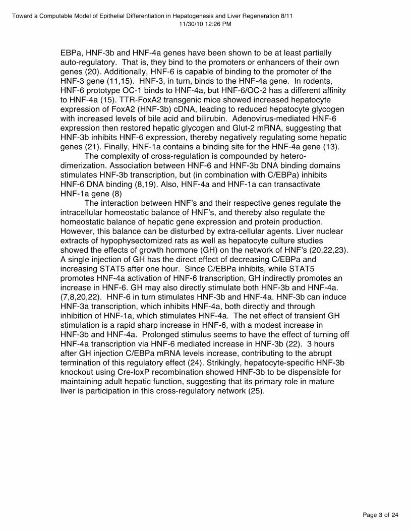
HNF-3a, -3b and -3g belong to the forkhead protein gene family (often identified as Foxa1, Foxa2, Foxa3). HNF-3 binds to DNA as a monomer using a highly-conserved winged-helix DNA binding domain (7,11). The subtypes display a 90% homology in amino acid sequence, thereby exhibiting a high degree of functional redundancy in mammalian hepatocytes (7). HNF-3b, followed by HNF-3a and HNF-3g directly promote genes involved in embryonic pattern formation and regional specification (9). Indirectly, the HNF-3 factors regulate virtually all liver-specific genes, as well as genes of the lung and pancreas (12)
HNF-4a is a member of the nuclear steroid-thyroid receptor superfamily, binding DNA strictly as a homeodimer (11,13). HNF-4a is thought to play a primary role in the initiation and maintenance of hepatocyte function (14), but not in establishing hepatic architecture. HNF-4a directly regulates numerous genes whose products include cholesterol and amino acid metabolism, glucogenesis, and blood coagulation (9). Overall, nearly half of the genes actively expressed in adult liver are regulated by HNF-4a (2,10). In adult mammals high expression is found only in liver, intestine and gut.
HNF-6 was the prototype of the novel ONECUT class of cut-homeoproteins with a single cut domain and a unique homeodomain (7,11,15,16). This unusual binding architecture makes the presence or absence of a co-activator critical to determining its activity (17). HNF-6 was originally identified, cloned and characterized as a regulator of a rat liver enzyme involved in glycolysis (PKF-2) (11). Soon after, an analogous human factor was identified as OC-2 (one-cut-2) (18). Association between HNF-6 and HNF-3b has been shown to inhibit HNF-6 DNA binding activity, thereby reducing transcription of HNF-6-dependent genes. However, the same association between HNF-3b and HNF-6 has the opposite effect on HNF-3b binding. On HNF-3b sites, HNF-6 association recruits co-activator p300/CBP to promote transcription (19) HNF-6 codes for genes regulating plasma transport, coagulation factors, and steroid metabolism (17,10)
Finally, a number of co-activators (DBP, CBP, p300/CBP) have been studied in conjunction with the HNFʼs (11). Most important is C/EBPa, a member of the family of CCAAT/enhancer-binding proteins, which share a highly conserved basic leucine zipper (bZip) domain (9). The basic activity of this factor is to bind two protein monomers into homeodimers or heterodimers (8). High levels of C/EBPa correlate with highly differentiated adult hepatocytes, suggesting a maintenance role (9). C/EBPa-/- rats show down-regulation of genes involved in hepatic glucose and lipid homeostasis. C/EBPa also inhibits cyclin-dependent kinase inhibitor p21Cip1, thereby negatively regulating hepatocyte proliferation (7).
HNF INTERACTION AND INDUCTION
Aside from cooperatively regulating the many genes involved in liver development and function, HNFs have been shown to form a complex cross-regulatory network by interacting with their own and each otherʼs genes. C/
Page 3 of 24
Toward a Computable Model of Epithelial Differentiation in Hepatogenesis and Liver Regeneration 8/1111/30/10 12:26 PM
EBPa, HNF-3b and HNF-4a genes have been shown to be at least partially auto-regulatory. That is, they bind to the promoters or enhancers of their own genes (20). Additionally, HNF-6 is capable of binding to the promoter of the HNF-3 gene (11,15). HNF-3, in turn, binds to the HNF-4a gene. In rodents, HNF-6 prototype OC-1 binds to HNF-4a, but HNF-6/OC-2 has a different affinity to HNF-4a (15). TTR-FoxA2 transgenic mice showed increased hepatocyte expression of FoxA2 (HNF-3b) cDNA, leading to reduced hepatocyte glycogen with increased levels of bile acid and bilirubin. Adenovirus-mediated HNF-6 expression then restored hepatic glycogen and Glut-2 mRNA, suggesting that HNF-3b inhibits HNF-6 expression, thereby negatively regulating some hepatic genes (21). Finally, HNF-1a contains a binding site for the HNF-4a gene (13).
The complexity of cross-regulation is compounded by hetero-dimerization. Association between HNF-6 and HNF-3b DNA binding domains stimulates HNF-3b transcription, but (in combination with C/EBPa) inhibits HNF-6 DNA binding (8,19). Also, HNF-4a and HNF-1a can transactivate HNF-1a gene (8)
The interaction between HNFʼs and their respective genes regulate the intracellular homeostatic balance of HNFʼs, and thereby also regulate the homeostatic balance of hepatic gene expression and protein production. However, this balance can be disturbed by extra-cellular agents. Liver nuclear extracts of hypophysectomized rats as well as hepatocyte culture studies showed the effects of growth hormone (GH) on the network of HNFʼs (20,22,23). A single injection of GH has the direct effect of decreasing C/EBPa and increasing STAT5 after one hour. Since C/EBPa inhibits, while STAT5 promotes HNF-4a activation of HNF-6 transcription, GH indirectly promotes an increase in HNF-6. GH may also directly stimulate both HNF-3b and HNF-4a. (7,8,20,22). HNF-6 in turn stimulates HNF-3b and HNF-4a. HNF-3b can induce HNF-3a transcription, which inhibits HNF-4a, both directly and through inhibition of HNF-1a, which stimulates HNF-4a. The net effect of transient GH stimulation is a rapid sharp increase in HNF-6, with a modest increase in HNF-3b and HNF-4a. Prolonged stimulus seems to have the effect of turning off HNF-4a transcription via HNF-6 mediated increase in HNF-3b (22). 3 hours after GH injection C/EBPa mRNA levels increase, contributing to the abrupt termination of this regulatory effect (24). Strikingly, hepatocyte-specific HNF-3b knockout using Cre-loxP recombination showed HNF-3b to be dispensible for maintaining adult hepatic function, suggesting that its primary role in mature liver is participation in this cross-regulatory network (25).

Page 4 of 24
Toward a Computable Model of Epithelial Differentiation in Hepatogenesis and Liver Regeneration 8/1111/30/10 12:26 PM
3 M. Rastegar et al. / Molecular and Cellular Endocrinology 164 (2000) 1-4
(,HNF-4
Fig. 2. Scheme of a network of GH-regulated liver transcription
factors. Thick arrows refer to effects of GH. See tell:t for details.
CfEBPll. not oppose the stimulatory effect of GH in the
first place? Indeed, C/EBPll. inhibits the hnf6 promoter
in the absence of GH. A study of the interaction of
CfEBPcx with this promoter shows that CfEBPcx bind-ing disappears quickly following GH injection, to reap-
pear at a time HNF-6 mRNA goes down again. This is
due to a GH-induced drop in the concentration of
CfEBPcx protein, the mechanism of which remains to be
investigated. Thus, the stimulation by GH of the ex-pression of the hnf6 gene brings into play at least two
stimulators, STAT5 and HNF-4, and one inhibitor,
CfEBPcx. This stimulation involves three mechanisms,
namely the binding ofSTAT5, the increased binding of
HNF-4 and the lifting of the binding of CfEBPcx, to the
hnf 6 promoter. This is followed by induction of Cf EBPll., concomitantly with the disappearance of the
effect of GH on HNF-6 (Fig. 1).
4. GH controls a network of liver transcription factors
We have mentioned that HNF-6 stimulates the ex-
pression of the hnf4 and hnf3fJ genes. This raised the
question as to whether GH increases, via induction of
HNF-6, the concentration of HNF-4 and HNF-3[3
mRNA. Following a single injection of GH, an increase
in HNF-4 and HNF-3[3 mRNA concentration is indeed
observed (Lahuna et al., 2000). The kinetics of the
increase in HNF-4 mRNA is compatible with a stimu-
lation of hnf4 gene transcription by an increased
amount of HNF-6 protein. However, the kinetics of
increase in HNF-3[3 mRNA are fast, if not faster, than
those of HNF-6 mRNA, which suggests that GH can
stimulate the hnf3fJ gene independently of HNF-6 (Fig.
1).
Three important notions emerge from the work sum-
marized above (Fig. 2). First, HNF-6 belongs to a
network of liver transcription factors that interact via
direct, feed-back, and autoregulatory loops. We have
seen that HNF-6 stimulates the transcription of the
hnf3fJ and hnf4 genes. HNF-3[3 can stimulate hnf4
(Duncan et al., 1998). HNF-4 can stimulate hnf6 (see
above). C/EBPcx stimulates hnf3fJ (Samadani et al.,
1995) and it inhibits hnf6, as discussed. On the other
hand, the positive autoregulation of hnf3fJ, hnf4 and
C/EBPa is well documented (pani et al., 1992; Legraverend et al., 1993; Spath and Weiss, 1997). The
actual operation of the network is certainly more com-
plex. For instance, the liver contains other factors of
the ONECUT class, such as OC-2. OC-2 controls some
of the genes targeted by HNF-6, e.g. hnf3fJ (Jacquemin et al., 1999). Moreover, our current study of hnf 6
knock-out mice (Jacquemin et al., 2000) suggests that HNF-6 inhibits the expression of the oc2 gene.
A second notion is that the network described above is itself regulated by GH. We have seen that HNF-6
plays here a crucial role. In fact, liver HNF-6 mRNA is
quasi undetectable in the hypophysectomized rat, i.e. in
the absence of GH. The control exerted by GH on
HNF-6 is itself complex, as it involves two other mem-
bers of the network, HNF-4 and CfEBPcx. It is note-
worthy that the increase in affinity of HNF-4 for DNA
brought about by GH treatment is specific for the hnf6
promoter, insofar as the affinity of HNF-4 for its
binding site in the hnf 1 promoter is not increased (our
unpublished data). In view of the interactions involving
HNF-6 within the network, one would expect that
other members of the network are influenced by GH.
This is the case for HNF-4 and HNF-3[3, although the
latter may be induced by mechanisms that bypass
HNF-6. On the other hand, a GH-regulated liver nu-
clear factor called GHNF plays a role in the control of
the CYPC12 gene promoter (Waxman et al., 1996), and
investigations on the transcriptional control of the ser-
ine protease inhibitors (spi) genes by GH in the rat
have shown the involvement of a GAGA box-binding
protein (Simar-Blanchet et al., 1998). Whether GHNF
and the GAGA factor, which remain to be cloned, are
part of the network is still unknown. When an extracel-
lular stimulus like GH perturbs the steady state of such
a network, and that it does so by acting both on the
proteins and on the corresponding genes, it is difficult
to predict the new steady state because of the differ-ences in half-life of the targets and of the existence of
retroacting regulatory loops.
The third issue pertains to the physiological signifi-
cance, for liver metabolism, of the control by GH of
transcription factor gene expression. This is clearly
distinct from the well-known stimulation by GH of the
early response genes c-fos and c-jun, which code for the
AP-l transcription factor. Indeed, AP-l is ubiquitous
and its induction by GH is held responsible for the
mitogenic effects of GH. Although the discovery of the
GH-IGF-I axis is four decades old, the mechanism by
which GH stimulates IGF-I gene transcription in liver
remains ill-defined. The possibility of an involvement of
HNF AND HEPATOGENESIS
The HNF network regulates key transitional events during liver development in response to a combination of pre-conditioning intra-cellular transcription factor expression patterns and extra-cellular inductive stimuli. vHNF1 has been detected when endodermal cells proliferate to form the liver primordium (9). This seems to induce HNF-3b transcription, which is followed by HNF-3a and HNF-3g. Together the HNF-3 family regulates regionalization within the early endoderm (mutations of forkhead gene result in pattern irregularities such as replacement of foregut and hindgut by ectopic head structures) (9). HNF-3 is thought to establish hepatic competency by modulating chromatin structures around enhancers of genes expressed in the developing liver (13,26). Once this competency is established, contact with the developing cardiac mesoderm establishes commitment to the hepatoblast lineage. Fibroblast growth factor (Fgf) and possibly bone morphogenic protein (BMP) seem to be necessary for this transformation (2,12). Interestingly, in the absence of mesenchymal induction, the endoderm develops pancreatic characteristics as a default (13). On the other hand, explants of mesenchymal tissue from the ventral foregut have been shown to provide the required stimulus to allow liver development to proceed (12,13,27). Once contact with mesenchymal tissue has established hepatic commitment, the endoderm is pulled away from contact with the cardiac mesenchyme. However, the auto-regulatory capacity of the HNF network seems to enable it to continue a new pattern of transcriptional activation even after the primary inductive stimulus has withdrawn.
Hepatic architecture begins to take shape as chords of primitive hepatocytes, “hepatoblasts”, extend from the foregut toward the mesenchyme of the septum transversum. These cords are arranged around an ingrowing sinusoidal vascular network arising from the viteline veins, which drains toward the cardiac primordium. Cell/cell association and/or specific signals between the endoderm anterior and fibroblastic mesenchymal tissue of the septum
Page 5 of 24
Toward a Computable Model of Epithelial Differentiation in Hepatogenesis and Liver Regeneration 8/1111/30/10 12:26 PM
transversum correlate to vHNF1 and HNF-4a induction and commitment to hepatocytes lineage. (9,12,28). HNF-4a-/- embryos lethally failed to complete gastrulation, making determination of HNF-4aʼs role in organogenesis possible. So, HNF4a-/- embryos were complimented with wild-type visceral endoderm, allowing HNF4a-/- embryos to proceed. Resulting livers were morphologically indistinguishable from wild type, but deficient in 14 genes important to hepatocytes function (29,30).
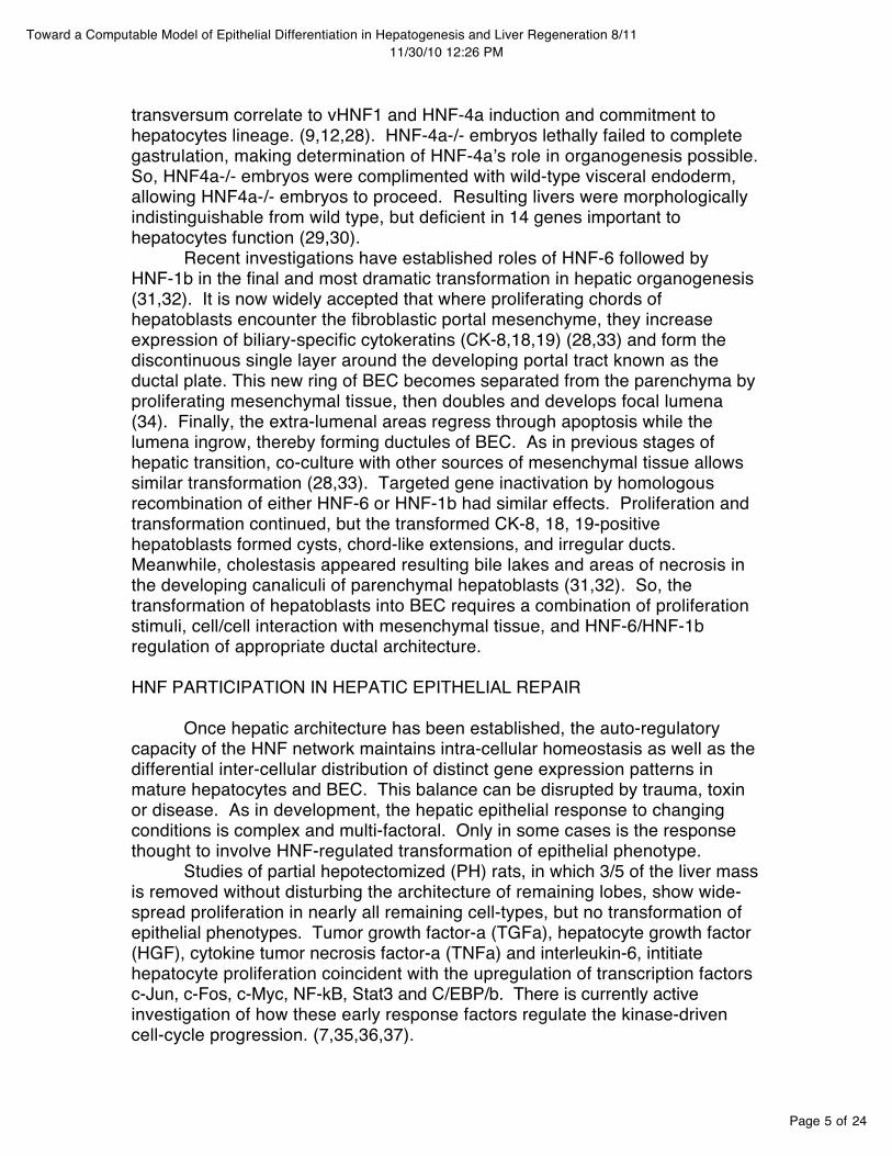
Recent investigations have established roles of HNF-6 followed by HNF-1b in the final and most dramatic transformation in hepatic organogenesis (31,32). It is now widely accepted that where proliferating chords of hepatoblasts encounter the fibroblastic portal mesenchyme, they increase expression of biliary-specific cytokeratins (CK-8,18,19) (28,33) and form the discontinuous single layer around the developing portal tract known as the ductal plate. This new ring of BEC becomes separated from the parenchyma by proliferating mesenchymal tissue, then doubles and develops focal lumena (34). Finally, the extra-lumenal areas regress through apoptosis while the lumena ingrow, thereby forming ductules of BEC. As in previous stages of hepatic transition, co-culture with other sources of mesenchymal tissue allows similar transformation (28,33). Targeted gene inactivation by homologous recombination of either HNF-6 or HNF-1b had similar effects. Proliferation and transformation continued, but the transformed CK-8, 18, 19-positive hepatoblasts formed cysts, chord-like extensions, and irregular ducts. Meanwhile, cholestasis appeared resulting bile lakes and areas of necrosis in the developing canaliculi of parenchymal hepatoblasts (31,32). So, the transformation of hepatoblasts into BEC requires a combination of proliferation stimuli, cell/cell interaction with mesenchymal tissue, and HNF-6/HNF-1b regulation of appropriate ductal architecture.
HNF PARTICIPATION IN HEPATIC EPITHELIAL REPAIR
Once hepatic architecture has been established, the auto-regulatory capacity of the HNF network maintains intra-cellular homeostasis as well as the differential inter-cellular distribution of distinct gene expression patterns in mature hepatocytes and BEC. This balance can be disrupted by trauma, toxin or disease. As in development, the hepatic epithelial response to changing conditions is complex and multi-factoral. Only in some cases is the response thought to involve HNF-regulated transformation of epithelial phenotype.
Studies of partial hepotectomized (PH) rats, in which 3/5 of the liver mass is removed without disturbing the architecture of remaining lobes, show wide-spread proliferation in nearly all remaining cell-types, but no transformation of epithelial phenotypes. Tumor growth factor-a (TGFa), hepatocyte growth factor (HGF), cytokine tumor necrosis factor-a (TNFa) and interleukin-6, intitiate hepatocyte proliferation coincident with the upregulation of transcription factors c-Jun, c-Fos, c-Myc, NF-kB, Stat3 and C/EBP/b. There is currently active investigation of how these early response factors regulate the kinase-driven cell-cycle progression. (7,35,36,37).

Page 6 of 24
Toward a Computable Model of Epithelial Differentiation in Hepatogenesis and Liver Regeneration 8/1111/30/10 12:26 PM
In other experimental systems, both the transformation of biliary-type precursors into hepatocytes as well as the converse transformation of hepatocyte-type precursors into biliary epithelial cells have been demonstrated. In rodent partial hepatectomy injury models where hepatocyte proliferation is inhibited, there is a proliferation of “oval cells” which are not observable in healthy mature tissue. More recently it has been established that these cells derive from biliary epithelial-type cells, possibly originating from facultative precursors residing in the Canals of Hering or small ductules. (38,39,40) Analogous structures can be seen in massive hepatic necrosis (MHN) of the human liver and are there termed “biliary hepatocytes”, “basophilic hepatocytes”, or “ductular hepatocytes”. It has been demonstrated that progeny of both rodent oval cells and human ductular hepatocytes are capable of transforming into architecturally appropriate BEC as well as hepatocytes, thereby restoring liver function (41,42).
More recently, the converse transformation of hepatocytes into BEC has been demonstrated. By combining partial hepatectomy with biliary toxin DAPM in DPPIV-negative rats, liver function was compromised without a disruption in hepatic architecture. DAPM inhibits biliary proliferation and thereby blocked the oval cell pathway. DPPIV-positive donor hepatocytes were then injected directly into the portal circulation and shown to proliferate, differentiate, form architecturally-appropriate DPPIV-positive ductules, and promote the restoration of normal function (6).
Several studies have attempted to establish the relationship between the course of HNF expression in hepatogenesis and that of the rodent oval cell/human ductular hepatocyte response. Prior to the discovery of HNF-6, partial hepatectomized rats treated with 2-acetyolaminofluorene to promote oval cell differentiation showed an initial upregulation of HNF1a, 1b, 3g, C/EBP and DBP in oval cells as well as in mature bile ducts and ductules. This was followed by a sudden upregulation of HNF-4 accompanied by the formation of foci of basophilic cells more strongly resembling hepatocytes (14). More recently, the examination of human livers with MHN also showed two intermediate differentiating cell types. “Ductular cells” strongly expressed biliary marker CK-7, similar morphology to biliary epithelial cells (BEC), an upregulation of HNF-1, 3, C/EBP, and DBP, but only low expression of HNF-4. The second intermediate form, “ductular hepatocytes”, showed weaker expression of CK-7, a stronger morphological similarity to hepatocytes, and prominent nuclear expression of HNF-4 (43). Both studies concluded that in the rodent oval cell response and the human ductular hepatocyte response, there seems to be a widespread activation phase involving the upregulation of HNF-1a, b, 3, C/EBP and DBP throughout most of the biliary system, which is accompanied by phenotypic changes only in its terminal branches. This is followed by a sudden upregulation of HNF-4, which is accompanied by further phenotypic changes. This suggests that HNF-4 is responsible for the final commitment of these cells to the hepatocyte lineage.
In addition to the direct evidence of HNF regulation of oval cell/ductular hepatocyte differentiation, there is indirect evidence of a similar process in other
Page 7 of 24
Toward a Computable Model of Epithelial Differentiation in Hepatogenesis and Liver Regeneration 8/1111/30/10 12:26 PM
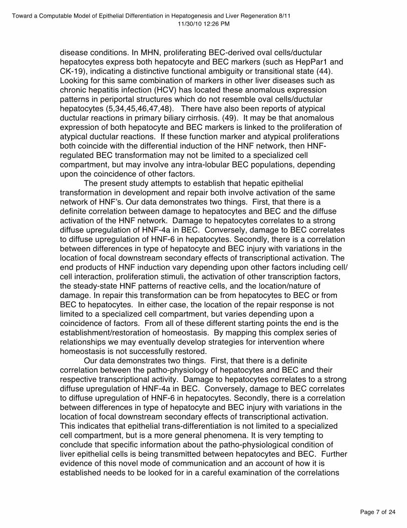
disease conditions. In MHN, proliferating BEC-derived oval cells/ductular hepatocytes express both hepatocyte and BEC markers (such as HepPar1 and CK-19), indicating a distinctive functional ambiguity or transitional state (44). Looking for this same combination of markers in other liver diseases such as chronic hepatitis infection (HCV) has located these anomalous expression patterns in periportal structures which do not resemble oval cells/ductular hepatocytes (5,34,45,46,47,48). There have also been reports of atypical ductular reactions in primary biliary cirrhosis. (49). It may be that anomalous expression of both hepatocyte and BEC markers is linked to the proliferation of atypical ductular reactions. If these function marker and atypical proliferations both coincide with the differential induction of the HNF network, then HNF-regulated BEC transformation may not be limited to a specialized cell compartment, but may involve any intra-lobular BEC populations, depending upon the coincidence of other factors.
The present study attempts to establish that hepatic epithelial transformation in development and repair both involve activation of the same network of HNFʼs. Our data demonstrates two things. First, that there is a definite correlation between damage to hepatocytes and BEC and the diffuse activation of the HNF network. Damage to hepatocytes correlates to a strong diffuse upregulation of HNF-4a in BEC. Conversely, damage to BEC correlates to diffuse upregulation of HNF-6 in hepatocytes. Secondly, there is a correlation between differences in type of hepatocyte and BEC injury with variations in the location of focal downstream secondary effects of transcriptional activation. The end products of HNF induction vary depending upon other factors including cell/cell interaction, proliferation stimuli, the activation of other transcription factors, the steady-state HNF patterns of reactive cells, and the location/nature of damage. In repair this transformation can be from hepatocytes to BEC or from BEC to hepatocytes. In either case, the location of the repair response is not limited to a specialized cell compartment, but varies depending upon a coincidence of factors. From all of these different starting points the end is the establishment/restoration of homeostasis. By mapping this complex series of relationships we may eventually develop strategies for intervention where homeostasis is not successfully restored.
Our data demonstrates two things. First, that there is a definite correlation between the patho-physiology of hepatocytes and BEC and their respective transcriptional activity. Damage to hepatocytes correlates to a strong diffuse upregulation of HNF-4a in BEC. Conversely, damage to BEC correlates to diffuse upregulation of HNF-6 in hepatocytes. Secondly, there is a correlation between differences in type of hepatocyte and BEC injury with variations in the location of focal downstream secondary effects of transcriptional activation. This indicates that epithelial trans-differentiation is not limited to a specialized cell compartment, but is a more general phenomena. It is very tempting to conclude that specific information about the patho-physiological condition of liver epithelial cells is being transmitted between hepatocytes and BEC. Further evidence of this novel mode of communication and an account of how it is established needs to be looked for in a careful examination of the correlations
Page 8 of 24
Toward a Computable Model of Epithelial Differentiation in Hepatogenesis and Liver Regeneration 8/1111/30/10 12:26 PM
between transcriptional activity and the patho-physiology of the lobule as a whole during development and repair.
MATERIALS AND METHODS
Case Selection
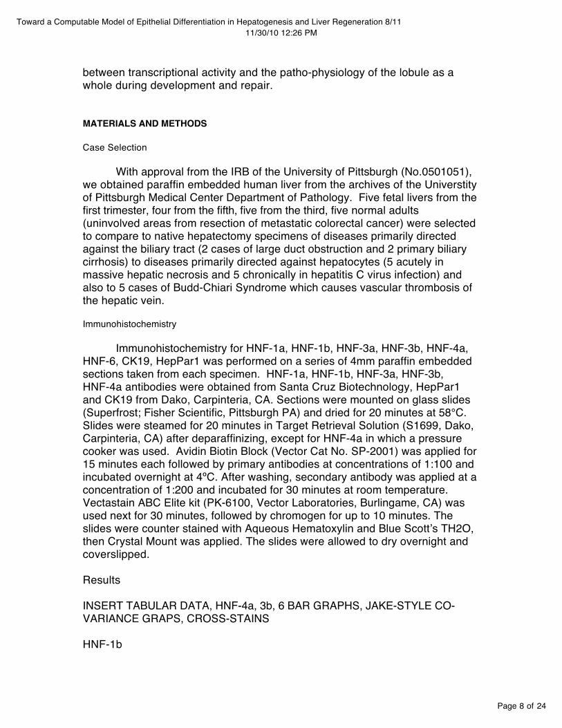
With approval from the IRB of the University of Pittsburgh (No.0501051), we obtained paraffin embedded human liver from the archives of the Universtity of Pittsburgh Medical Center Department of Pathology. Five fetal livers from the first trimester, four from the fifth, five from the third, five normal adults (uninvolved areas from resection of metastatic colorectal cancer) were selected to compare to native hepatectomy specimens of diseases primarily directed against the biliary tract (2 cases of large duct obstruction and 2 primary biliary cirrhosis) to diseases primarily directed against hepatocytes (5 acutely in massive hepatic necrosis and 5 chronically in hepatitis C virus infection) and also to 5 cases of Budd-Chiari Syndrome which causes vascular thrombosis of the hepatic vein.
Immunohistochemistry Immunohistochemistry for HNF-1a, HNF-1b, HNF-3a, HNF-3b, HNF-4a,
HNF-6, CK19, HepPar1 was performed on a series of 4mm paraffin embedded sections taken from each specimen. HNF-1a, HNF-1b, HNF-3a, HNF-3b, HNF-4a antibodies were obtained from Santa Cruz Biotechnology, HepPar1 and CK19 from Dako, Carpinteria, CA. Sections were mounted on glass slides (Superfrost; Fisher Scientific, Pittsburgh PA) and dried for 20 minutes at 58°C. Slides were steamed for 20 minutes in Target Retrieval Solution (S1699, Dako, Carpinteria, CA) after deparaffinizing, except for HNF-4a in which a pressure cooker was used. Avidin Biotin Block (Vector Cat No. SP-2001) was applied for 15 minutes each followed by primary antibodies at concentrations of 1:100 and incubated overnight at 4ºC. After washing, secondary antibody was applied at a concentration of 1:200 and incubated for 30 minutes at room temperature. Vectastain ABC Elite kit (PK-6100, Vector Laboratories, Burlingame, CA) was used next for 30 minutes, followed by chromogen for up to 10 minutes. The slides were counter stained with Aqueous Hematoxylin and Blue Scottʼs TH2O, then Crystal Mount was applied. The slides were allowed to dry overnight and coverslipped.
Results
INSERT TABULAR DATA, HNF-4a, 3b, 6 BAR GRAPHS, JAKE-STYLE CO-VARIANCE GRAPS, CROSS-STAINS
HNF-1b

Page 9 of 24
Toward a Computable Model of Epithelial Differentiation in Hepatogenesis and Liver Regeneration 8/1111/30/10 12:26 PM
HNF1b showed an even stronger specificity to biliary cells than the standard marker CK19. Hepatocytes show no HNF1b expression during any stage of development, as opposed to the early expression of CK19. Only in Budd-Chiari syndrome did we see scattered focal HNF1b staining of hepatocytes. On the other hand, the ductal plate already shows HNF1b expression in the first trimester. Biliary expression remains consistent through adulthood, with noticeable extensions from the portal region which may outline the course of the Canals of Herring.
HNF-3b
HNF-3b is consistently expressed in biliary cells from the development of the ductal plate onward, and remains consistent through all disease conditions, including expression in the ductal hepatocytes observable in massive hepatic necrosis. Hepatocytes also express HNF-3b during the first two trimesters, but not in the third or during mature normal function. Hepatocytes again became positive in biliary obstruction, primary biliary cirrhosis and Budd-Chiari syndrome, with increased periportal intensity in Budd-Chiari. There was some weak hepatocyte positivity in acute massive hepatic necrosis and all hepatocytes were negative in chronic HCV infection.
CK19 and HepPar1
CK19 and HepPar1 are standard markers of normal adult biliary and hepatocyte function, respectively. During development CK19 consistently stains the ductal plate and developing biliary ductules. Expression is maintained in all of the observed disease conditions, with noticeable staining of the ductular hepatocytes observable in acute massive hepatic necrosis. Surprisingly, during the first trimester we also observed weak CK19 expression in hepatocytes in the vicinity of the central vein, with even stronger expression in the second trimester. This decreased in the third, with no mature hepatocytes expressing CK19. In no disease conditions did hepatocytes express CK19.
We observed rare HepPar1 expression of peri-portal hepatocytes in the first trimester. By the second trimester there was greater positivity around the central vein, which increased to diffuse strong hepatocyte staining in the third trimester and continued into the mature adult hepatocytes. No biliary cells expressed HepPar1 during development. Hepatocyte HepPar1 expression was decreased in biliary obstruction, primary biliary cirrhosis, and Budd-Chiari syndrome, with hepatocyte-like cells around the portal track not expressing HepPar1 at all in PBC. Hepatoctyes showed xxx in acute massive hepatic necrosis and chronic HCV infection, with HepPar1 positive ductular hepatocytes also noticeable in massive hepatic necrosis.
Discussion
Page 10 of 24
Toward a Computable Model of Epithelial Differentiation in Hepatogenesis and Liver Regeneration 8/1111/30/10 12:26 PM
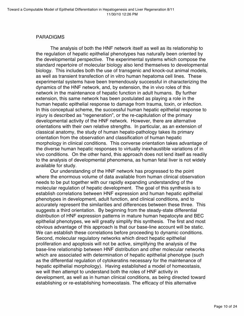
PARADIGMS
The analysis of both the HNF network itself as well as its relationship to the regulation of hepatic epithelial phenotypes has naturally been oriented by the developmental perspective. The experimental systems which compose the standard repertoire of molecular biology also lend themselves to developmental biology. This includes both the use of transgenic and knock-out animal models, as well as transient transfection of in vitro human hepatoma cell lines. These experimental systems have been tremendously successful in characterizing the dynamics of the HNF network, and, by extension, the in vivo roles of this network in the maintenance of hepatic function in adult humans. By further extension, this same network has been postulated as playing a role in the human hepatic epithelial response to damage from trauma, toxin, or infection. In this conceptual scheme, the successful human hepatic epithelial response to injury is described as “regeneration”, or the re-capitulation of the primary developmental activity of the HNF network. However, there are alternative orientations with their own relative strengths. In particular, as an extension of classical anatomy, the study of human hepato-pathology takes its primary orientation from the observation and classification of human hepatic morphology in clinical conditions. This converse orientation takes advantage of the diverse human hepatic responses to virtually inexhaustible variations of in vivo conditions. On the other hand, this approach does not lend itself as readily to the analysis of developmental phenomena, as human fetal liver is not widely available for study.
Our understanding of the HNF network has progressed to the point where the enormous volume of data available from human clinical observation needs to be put together with our rapidly expanding understanding of the molecular regulation of hepatic development. The goal of this synthesis is to establish correlations between HNF expression and human hepatic epithelial phenotypes in development, adult function, and clinical conditions, and to accurately represent the similarities and differences between these three. This suggests a third orientation. By beginning from the steady-state differential distribution of HNF expression patterns in mature human hepatocyte and BEC epithelial phenotypes, we will greatly simplify this synthesis. The first and most obvious advantage of this approach is that our base-line account will be static. We can establish these correlations before proceeding to dynamic conditions. Second, molecular regulatory networks which direct hepatic epithelial proliferation and apoptosis will not be active, simplifying the analysis of the base-line relationship between HNF distribution and other molecular networks which are associated with determination of hepatic epithelial phenotype (such as the differential regulation of cytokeratins necessary for the maintenance of hepatic epithelial morphology). Having established a model of homeostasis, we will then attempt to understand both the roles of HNF activity in development, as well as in human clinical conditions, as being directed toward establishing or re-establishing homeostasis. The efficacy of this alternative
Page 11 of 24
Toward a Computable Model of Epithelial Differentiation in Hepatogenesis and Liver Regeneration 8/1111/30/10 12:26 PM
orientation will be demonstrated if this re-formulation gives us new insights into well-attested data.
HOMEOSTASIS
The homeostatic account of the relationship between HNFʼs and human hepatic epithelial phenotypes must begin from a model of how these types are arranged in the adult human liver. This model must both depict how variations in each subtype are arranged with respect to each other in the smallest functional unit, as well as how these units are arranged in the liver as a whole (50). We then simply need to map steady-state HNF expression patterns onto this model. Once we have this mapping, we can extend the model by examining dynamic states, and eventually by a similar mapping of other regulatory networks. Surprisingly, the basic anatomic questions have not been settled.
The classical account of hepatic architecture, the hexagonal lobule, takes its orientation from the many endocrine functions of the liver, which are primarily carried out by hepatocytes (51,52,38). These include plasma glucose regulation, the synthesis of many plasma proteins including albumin and coagulation proteins, the processing of toxins, such as ammonia, for non-toxic excretion through bile or urine, and the synthesis of bile, which is necessary for dietary fat absorption (53). The arrangement of hepatocytes in the fully developed lobule is correlated to the movement of blood through each 2-mm structural unit. Each lobule has a dual blood supply. Terminal branches of the hepatic artery and portal vein attach to the portal areas of each lobule enclosed by mesenchymal connective tissue. A one-to-three ratio of oxygen-rich to nutrient-rich blood cells, representing 25% of cardiac output, mix within these lobules within innumerable fenestrated sinusoids. Red blood cells (RBCʼs) exchange metabolites with the surrounding parenchymal cells through the peri-sinusoidal Space of Disse, then exit as depleted RBCʼs through the terminal branch of the hepatic vein (THV) located at the center of each lobule. Surrounding the sinusoids, and extending microvilli into the peri-sinusoidal Space of Disse, is a large mass of parenchymal tissue organized into cribiform, anastamosing sheets of hepatocytes, which radiate from the terminal hepatic artery. The arrangement of BEC is treated secondarily in this account and is divided into an account of the intra and extra lobular segments. In our observations, the hepatic parenchyma uniformly stains for HNF-…, HepPar1, and
The converse alternative to the classical lobule is oriented by the exocrine functions of the liver, which are carried out by the branching biliary tree (49). From this perspective we see hexagonal lobules centered on the portal tract rather than the THV. In the hepatic parenchyma at the periphery of the portal lobule, minute, hexagonal bile canaliculi are formed from the specialization of adjacent surfaces of individual hepatocytes, which are sealed with tight junctions. These canaliculi connect with each other in a “chicken-wire” configuration before becoming confluent with a trough of primitive BEC in

Page 12 of 24
Toward a Computable Model of Epithelial Differentiation in Hepatogenesis and Liver Regeneration 8/1111/30/10 12:26 PM
the Canals of Herring, approximately two thirds of the distance to the portal triad (54). At this interface BEC are distinguishable by expression of intermediate filaments, cytokeratins 7 and 19 (CK-7, CK-19), as opposed to hepatocytes, which express CK-8, CK-18, and antigens related to their metabolic functions, such as HepPar1, albumin, alpha-1-antitrypsine, biliary glycoprotein-1, and others (34). The BEC at the interface can also be distinguished by a basement membrane as opposed to the canalicular membrane between hepatocytes. Before reaching the portal tract these squamous epithelial colangiocytes give rise to low-cuboidal biliary epithelial ductules, which then penetrate the mesenchymal limiting plate, and join the incoming terminal hepatic artery and portal vein in the portal triad at the center of each portal lobule. Downstream of the Canals of Hering this space becomes increasingly wider, develops a layer of mesenchymal components, and develops its own microvaculature. One liter of caustic bile is secreted each day by hepatocytes and moved out of the liver by mechanical forces in a direction opposite to the flow of blood. In our observations all BEC stained …. Interestingly, CK19 has been reported to be particularly useful for imaging the Canals of Hering (50,55).
As a third alternative, the synthesis of the former two models begins from roughly triangular functional subunits, the acini, with one apex at the THV and two portal tracts at the others (56,57,58). Not only does this approach attach equal importance to the exocrine and endocrine functions of the liver, but it is particularly useful for distinguishing the relationships between variations of hepatocytes and BEC subtypes. Within the classic acinus three zones are distinguished, graded by the depletion of oxygen and other metabolites in adjacent RBCʼs as they travel the length of the sinusoids toward the terminal hepatic vein. The intensity of expression of hepatic enzymes responsible for the biochemical transformation of toxins and metabolites varies zonally and is correlated to the changing composition of the RBCʼs as they travel the length of the sinusoids. This variation is easily correlated with variations in BEC as they transition from the Canals of Hering to the terminal bile ductules (59). The most recent refinement of the acinus concept, the cholhepaton, has the added advantage of representing a portal-portal gradient in addition to portal-central zonation (60,61,52,38). Such an approach may eventually settle the basic micro-architectural questions, and contribute toward understanding the organization and regional variations in the liver as a whole.
Our account of HNF homeostasis in mature liver leaves a number of questions. It is possible that our experimental techniques are not able to detect variations in HNF expression intensity corresponding to zonal variations within each epithelial phenotype. This is plausible given that the HNF network has been shown to have variable steady-states. Further, since transcriptional expression is far more fluid than phenotype, it may be that there are fluctuations or areas of overlap between the interface of HNF expression patterns and epithelial phenotypes in the Canals of Hering. Finally, as association with mesenchymal tissue has been shown to be significant both for the determination of epithelial phenotype and for alterations in HNF expression, closer examination of the portal interface may bear significant contrast to the

Page 13 of 24
Toward a Computable Model of Epithelial Differentiation in Hepatogenesis and Liver Regeneration 8/1111/30/10 12:26 PM
interfaces in the Canals of Hering.
DEVELOPMENT
Liver development begins within the first two weeks of embryonic development as cords of hepatoblasts extend from the foregut toward the mesenchyme of the septum transversum. These cords are arranged around an ingrowing sinusoidal vascular network arising from the viteline veins, which drains toward the cardiac primordium. In the first trimester we see significant differences between the HNF-regulated condition of hepatoblasts in proximity to and away from the mesynchymal limiting plate. Away from the mesenchyme, there is strong expression of HNF-4a and HNF-3b, with weak focal HepPar1 staining, particularly in the peri-portal region. Proximal to the mesenchyme there is a double cell layer (the ductal plate) in which HNF-1b and HNF-3b and CK19 are strongly expressed. Also, at the time of our first observations there are focal luminal spaces in the ductal plate.
Away from the mesenchyme, several transitions seem to be taking place simultaneously. Strong HNF-3b expression may be related to the opening of the cannalicular space composed of the apical surfaces of adjacent hepatoblasts. Simultaneous with this opening, which establishes one end of a new patent passageway from the parenchyma to the alimentary canal, HNF-4a is driving the activation of hepatocyte function, including HepPar1 production. We see only weak focal staining of HepPar1, particularly around the peri-portal region, suggesting that the intra-lobular opening of the biliary tree is a gradual process proceeding from the portal interface out toward the end of the Canals of Hering (38).
In the periportal region we see a different pattern of HNF expression associated with the bi-layered BEC ductal plate. Studies of proliferating cell nuclear antigen (PCNA) demonstrated that the ductal plate is not proliferative, but that these cells are derived from metaplasia, or transformation of proliferating hepatoblasts or primitive hepatocytes (34). The targeted HNF-6 and HNF-1b inactivation studies cited earlier (31, 32) showed that at least part of this transformation is independent of HNF activity. Morphological changes, termination of differentiation, and alteration in cytokeratin expression seem to be regulated by mesenchymal interaction alone. In the absence of HNF-6 and HNF-1b upregulation, metaplasia seems to continue, but results in cysts, chords and irregular configurations of transformed cells. We have also seen that prolonged stimulus of HNF-6 seems to have the effect of turning off HNF-4a transcription via increase in HNF-3b (62). So, induction of HNF-6 during ductal plate formation seems to have two effects: turning off the hepatocytes-specific gene expression pattern associated with HNF-4a, and directing appropriate ductile formation through upregulation of HNF-1b (28,38).
By the second trimester we see only scattered remnants of the ductal plate. CK-19 expression remains consistent in biliary cells, with an increase in the unexplained expression in hepatocytes around the central vein. HepPar1 expression increases in hepatocytes consistently with the further opening of the

Page 14 of 24
Toward a Computable Model of Epithelial Differentiation in Hepatogenesis and Liver Regeneration 8/1111/30/10 12:26 PM
bile ductules. This increase in hepatocyte enzymatic output inversely correlates to the decrease in HNF-4a. HNF-1b retains its specificity to biliary cells with noticeable periportal expression in small ductules or the Canals of Hering. HNF-3b remains consistent during the final remodeling of the ductal plate as well as in bile ductules, and is still present in some hepatocytes. By the third trimester the ductal plate has been entirely resorbed, CK-19 expression is now limited to the remodeled biliary system, and HepPar1 expression is strong and diffuse in hepatocytes while HNF-4a moderates to mature maintenance levels. HNF-1b remains a strong consistent indicator of biliary cells, while HNF-3b also becomes confined to biliary cells.
There are many unanswered questions about the formation and remodeling of the ductal plate. It is not clear what triggers the initial transformation of hepatoblasts to ductal plate cells nor what factors induce HNF-6 upregulation. It is not clear whether the new ductules are formed by continued proliferation and metaplasia at the portal interface, causing an ingression of older cells, or whether metaplasia subsequently takes place deeper into the lobule. It is also not clear whether the significant shift in HNF-3b expression from hepatocytes to BEC is simply related to the regulation of the HNF network, or whether it directly participates in architectural remodeling. However, one significant result seems to have come to light. The down regulation of HNF-4a and HNF-3b in hepatocytes and increase in HepPar1 production seem to be directly related to the ingression of the BEC ductules and the establishment of a patent passage from the cannaliculi to the opening biliary system. So, the final transformation from hepatoblasts to hepatocytes is dependent upon the development of BEC. In other words, the development of the two epithelial lineages are co-dependent. Induction of the hepatocytes lineage precedes that of BEC, but the architectural remodeling which depends upon appropriate HNF-regulated BEC transformation is in turn necessary for the completion of the development of mature hepatocytes
EPITHELIAL REPAIR WHICH DOES NOT INVOLVE PHENOTYPE TRANSFORMATION
Our ultimate goal is to characterize HNF regulation of epithelial transformation in hepatic injury. However, not all hepatic repair responses involve epithelial transformation. It has been noted that as a general rule, damage to hepatocytes is most efficiently repaired through replication of remaining hepatocytes, and the same holds for BEC damage (36). Interesting variations to this primary repair response have been observed in experimental conditions. The most productive model for studying hepatic repair involves partial hepatectomy (PH), which is the surgical resection of 3/5 of an adult rat liver. This procedure leaves the remaining liver mass structurally intact and functional, but not capable of meeting the metabolic demands of the host. (65). It has been observed that PH combined with inhibition of DNA synthesis through either dexamethasone (Dex) or 5-fluorouracil (FU) resulted is

Page 15 of 24
Toward a Computable Model of Epithelial Differentiation in Hepatogenesis and Liver Regeneration 8/1111/30/10 12:26 PM
restoration of liver mass and normal function through hypertrophy of periportal hepatocytes (64). In the absence of such proliferation inhibition, PH rats instead show a hyperplastic response. Immediately following surgical resection a wave of proliferation begins in the periportal region of individual lobules and spreads toward the pericentral region. The hyperplastic response involves virtually all cells in the liver. However, the overall shape of the liver does not return. Although there is widespread tissue proliferation, there is no increase in the number of functional tissue units or lobules. Instead, individual lobules increase in size and hepatocyte plates increase in thickness from an average of 1 to an average of 2 cells thickness, until function is restored. No transformation of epithelial cell types has been reported in this response (65, 6).
These studies demonstrate that hepatic epithelial proliferation and transformation are distinct and regulated by different molecular pathways. Hepatocyte proliferation has been characterized in more detail. In immediate response to injury, non-parenchymal Kupfer cells first release cytokines interleukin-6 (IL-6) and transforming growth factor-alpha (TGF-a) (63). In the second phase, non-parenchymal Stellate cells initiate proliferation through the production of growth factors transforming growth factor-b (TGF-b) and hepatocytes growth factor (HGF).
MASSIVE HEPATIC NECROSIS
Massive, hepatic necrosis, associated with a wide range of clinical conditions, can involve parenchymal loss in the entire liver, parts of the liver, in the same parts of all or most lobules, or a random distribution of hepatocyte loss. In acute massive hepatic necrosis, areas of hepatocytes are destroyed by various agents (toxins, infections, trauma).
CHRONIC HCV INFECTION
Chronic viral hepatitis C infection shows dense portal lymphoid aggregates, nonsupportive lymphocytic cholangitis and mild macrovesicular fatty changes. In chronic HCV, damage is also directed primarily to hepatocytes, but we see areas of IEC in other areas of the lobule than in MHN, and their appearance is less regular.
CIRRHOSIS
PBC is a variation of the above scenario. PBC shows fibrosis and nodular regeneration, often surrounded by ductular, fibrous or piecemeal necrosis and ductopenia.
Cirrhosis is a scarring of the epithelium which distorts areas of the model and blocks repair. There is a proliferation of ductules (cholangioles). we observed a decrease in the HepPar1 expression of hepatocytes, with some peri-portal hepatocytes failing to express HepPar1 at all in PBC. This suggests that biliary damage correlates to decrease hepatocyte function. All biliary cells,

Page 16 of 24
Toward a Computable Model of Epithelial Differentiation in Hepatogenesis and Liver Regeneration 8/1111/30/10 12:26 PM
including proliferating ductules showed normal CK19, HNF-1b and HNF-3b expression. Interestingly, in both disease conditions hepatocytes also showed expression of HNF-3b, similarly to the first and second trimester, but not third or healthy adult.
BILE DUCT OBSTRUCTION
Large duct obstruction is characterized by portal inflammatory infiltrate, portal edema and the proliferation of ducts and ductules. Duct obstruction establishes a similar set of response forces, but if the obstruction cannot be removed, we should expect to see a proliferation of abnormal ductular structures as the steady sustained forces follow the path of least resistance to restoring homeostasis.
BUDD-CHIARI SYDROME
Budd-Chiari syndrome, usually caused by the thrombotic occlusion of the hepatic veins, result in sinusoidal dilation, particularly in zone 3, followed by centrilobular and midzonal loss of parenchyma to blood lakes. This is the only case in which we see what may be periportal hepatocyte expression of HNF-1b as well as periportal expression of HNF-3b.
STEM-CELLS
Finally, we should address the issue of stem cells. Characterizing the specific sequences of transcriptional activation involved in various phases of development is absolutely essential to understanding the whole. However, once our regulatory network is established, those same sequences of events will have very different effects. Therefore, it is not possible to isolate “the liver program” responsible for development and regeneration. Similarly, although isolating the specific cells which transform in a particular clinical case, or isolating pluripotent cells which can integrate with the epithelium is extremely important, these investigations still need to be put into the context of the organic whole.
THE NEW MODEL
We now need to examine how the system of correlations which we have described determines the epithelial response to various sorts of damage. We have established that there are two mature epithelial types, each with a range of subtypes. These types interface in organogenesis when the lumena formed of the apical surfaces of adjacent hepatocytes are connected with the lumena formed of the basal surfaces of adjacent BEC. In mature tissue this interface lies in the Canals of Herring. We have also established that the difference between the two epithelial subtypes, and gradations in each subtype, are driven by alterations in activation patterns of the HNF-4a, 3b, 6 transcription factor

Page 17 of 24
Toward a Computable Model of Epithelial Differentiation in Hepatogenesis and Liver Regeneration 8/1111/30/10 12:26 PM
loop. Transcriptional loop induction in one direction drives hepatocytes differentiation. Induction in the other direction drives BEC differentiation. Finally, our data has demonstrated that hepatocytes and BEC cross-regulate each othersʼ transcriptional activity. That is, damage to hepatocytes has the effect of changing the direction of the regulatory loop at the BEC end, and visa versa. In mature tissue, the steady-state patterns of transcriptional activity interface correspond to the hepatocytes/BEC interface.
The easiest way to understand the system of correlations which we have just described is to consider its geometry in abstraction. We will first describe a construction and then relate it to the regulatory system of the lobule. We will avoid unnecessary formalization in order to make the resulting proofs intelligible to the non-mathematician.
To begin, consider two parallel discs, E and M. M has a hole in the center. We will label the sides of the discs, a,b,c,d. The discs are limitlessly deformable, elastic, and when necessary, sticky. Extend six parallel finger-like deformations from E to M (this construction has an affinity to the number six which will be explained later). Invert the ends of each finger back along the axis of each interior. In cross-section we now have six pairs of concentric circles. Note that the interior of each finger is continuous with the exterior. Now, insert the ends of all six fingers through the hole in M, adhere the sides of the portions of adjacent fingers which project through M. Take a circle around the tips of the fused fingers which contains each inversion. Extend this circle, forming a tube. If we only consider the portion of b inside of M, we have separated b into non-continuous regions. We will call these regions “red” and “blue”. Pulling the fused section of the fingers further through the hole in M exposes part of red. Divide this red area into two by drawing a circle containing each and extending them. Fuse the sides of red, blue and the hole in M. Now, bring the circumfrence of M around the fingers and form a circle similar and parallel to the hole in M. We will designate the holes “left” and “right”. We now have a globe with two holes through which surface b projects. No portion of blue in accessible from any area of E which is outside of left hole. Join the areas of red outside of left hole into a cylinder and extend. Grade decreasing intensities of red and blue from left to right.
Finally, consider the portion of blue which is now inside the globe. We are going to put a series of rings of cross-caps along blue and join them on the other side with a hexagonal juncture to the surface a, which can be seen as the underside of red. A cross-cap is a four-dimenional incision which allows us to relate the cylinder formed by the top of blue with the cylinder formed by the bottom of red. This is similar to the familiar Klein bottle which has some unusual properties. We can see that each point of fusion is associated with a unique red-to-blue value, establishing a co-ordinate system for our interfaces.
We are now ready to state the theorem which relates the inductive hypothesis of the empirical experiments to our construction:
Ochoaʼs theorem: If we designate the above constuction “Ochoaʼs knot”, then Ochoaʼs knot is isomorphic to the regulatory system which relates hepatic

Page 18 of 24
Toward a Computable Model of Epithelial Differentiation in Hepatogenesis and Liver Regeneration 8/1111/30/10 12:26 PM
epithelial phenotype to HNF expression patterns.
We will now demonstrate the theorem. The attentive reader will have noted that neither a Klein bottle nor our construction can be realized in three dimensions without passing through itself. If we allowed such punctures we would have bile leaking into our blood supply. So, to demonstrate our theorem, we will make two three dimensional constructions and then relate them.
Begin again from our two-dimensional discs. Change E into a three-dimensional disc composed of deformable polyhedra with six-sided faces. M can be an elastic three-dimensional membrane. Instead of stretching a surface, we will now be building out by adhering new polyhedra. So, build out six fingers formed by rings of units separated from each other by one unit and with a one-unit core. Continue the construction as in Ochoaʼs knot until reaching the cross-cap construction. In the underside of red we are going to make rings of indentations by exposing a ring of red sides and deforming them into a disc so that interior red bottoms form hexagonal tubes. Instead of cross-caps, we are going to delete blue units, deform exposed sides into dics, and adhere to the hexagonal red tubes. Also adhere all adjacent surfaces of red and blue bottoms. As with the cross-cap construction, each interface is associated with a unique red to blue value. We can see that this construction resembles the general structure of the lobule.
Our final construction is a bit more complex. Begin with a very, long coil which tapers in one direction. Attach one end to itself, forming a circle, like a slinky. When the coil is tightened at one end, the circle contracts into a hex. When the coil is loosened, it expands into a loose circle. Let the coil taper drastically from one end to the other. The face is on the cellular scale, the other end is on the molecular scale. Take two of these constructions and join their small ends. Now tightening the coil will simultaneously deform both faces. We will call the two faces “top” and “bottom”. Build a polyhedra with side faces (the shape of the polyhedra as a whole is unknown since no platonic regular polyhedra has hexagonal faces). Regions of the coil are orientable to one another relative to top and bottom. Identify three sections of the middle of the coil which taper relative to one another, a, b, c. Bend each section into an arc. Let turning a and b clockwise relative to each other tighten the screw. Let turning b, c counterclockwise loosen the screw.
Make a series of three slits in one side face of the polyhedra and selectively insert a and b or b and c, or just b into the slits. We now have orientable, selectively expressible external gears which can alter the shape of the polygon. Now, instead of inserting actual sections of the interior of the coil into the slits, selectively copy sections to be externally expressed. Relative to the outside, a copy section cannot be distinguished from an actual coil section. By using this trick, we can relate coils to other combinations of pairs of polygon faces, and can associate gears in variable combinations and orientations, driving polygon phenotypic changes.
Finally we need to associate our two three-dimensional constructions. Make slits in upstream and down stream sides of each red unit. Selectively

Page 19 of 24
Toward a Computable Model of Epithelial Differentiation in Hepatogenesis and Liver Regeneration 8/1111/30/10 12:26 PM
express a and b, but invert top and bottom relative to each unit up to right hole. As we add each additional unit in the construction, loosen the coil and connect external gears. When we invert the ends of our growing tubes at right hole, change a/b expression to progressive degrees of b/c expression. At each pseudo-cross-cap, a unique a/b value is associated with a unique b/c value. A tube formed by the bottom of red is associated with a tube formed by the top of blue, and transcriptional top and bottom are inverted relative to each macro unit. Note that although we have only two transcriptional patterns corresponding to red and blue formation, we have also established gradations of phenotypes. Once this system if relations is engaged, red and blue coil tensions are balance against one another and the system is in homeostasis.
Now we need to see how this system of relations responds to various stimuli.
1. GH induction in development and PH.2. Field communication across gaps and around obstructions.
If we follow the set of correlations which we have established, we arrive at a surprisingly complex structure with properties similar to the four-dimensional topological space known as the “Klein bottle”. A Klein bottle, is a closed non-orientable surface of Euler characteristic 0 that has no inside or outside (66,67). It can be physically realized only in four dimensions, since it must pass through itself without the presence of a hole. Its topology is equivalent to a pair of cross-caps with coinciding boundaries (68). Except in the present case there is a cross-cap established at each interface of the ingrowing biliary convolution and each cannalicus. If we consider a section of the three-dimensional injection of this structure, we get a genuine cylinder with “pores” or holes connecting the “inside” and “outside”. By coloring each of these with a gradient of two different colors, we can see that each ring of cross-caps corresponds to a unique combination of gradients, establishing a coordinate system which can then be extended to the whole structure. We can now use this coordinate system and the counter-intuitive properties of this structure to convey specific information between hepatocytes and BEC.
CONCLUSION
So, there are not two types of mature hepatic epithelial cells: biliary epithelial cells and hepatocytes. Hepatic epithelial repair does not depend upon bone-marrow derived pluripotent stem cells. There are no dedicated or facultative bi-potential hepatic precursor cells. There is, however, one underlying process by which hepatic epithelial cells proliferate and differentiate in response to the gradually changing sets of forces active during hepatogenesis, which fluctuate only slightly in the mature healthy state. The same epithelial response is responsible for repair following trauma. If functional architecture is not disturbed by the trauma, as in partial hepatectomy, then the two sets of forces are not imbalanced, but increase in intensity, resulting in a
Page 20 of 24
Toward a Computable Model of Epithelial Differentiation in Hepatogenesis and Liver Regeneration 8/1111/30/10 12:26 PM
repair response which resembles a continuation of the process of hepatogenesis. If functional architecture is disrupted then the two sets of forces will be imbalanced, requiring both proliferation and differentiation to reach a new state of equilibrium.
HNF alteration differs relative to the nature of the damage. In no case is there a simple recapitulation of the sequence of HNF expression seen in hepatogenesis. These observations suggest that there is not a dedicated compartment of hepatic precursors which, when activated, initiate a pre-programmed HNF-driven differentiation sequence. Rather, there is a single HNF regulatory network which can be activated in a different sequences. Further the effect of each sequence also depends upon the developmental state, particularly before and after the transformation of a largely homogeneous mass of hepatoblasts into an organic whole which inter-connects gradations of a symmetrical pair epithelial types. The result is that a range of hepatic epithelial cells can respond to damage by proliferating and differentiating in a selective HNF-driven response which varies depending upon the location and nature of the damage.
So, given that we have established our thesis in principle, the next step is to render the model digitally with additional data taken from carefully staged developmental and clinical observations. As the model develops, we can incorporate more transcription factors as well as enzymatic production, secondary morphological characteristics, etc.. Looking back, the model has already done explanatory work. Looking forward, its potential uses are numerous. A scaled-up model may be useful in making clinical diagnoses, in modeling therapeutic strategies, and for integrating the vast information contained in the genome with gross physiological phenomena. Once successful, it is a small step to applying the same techniques to other organs. It appears, then, that our great patron, Prometheus, father of the arts, has just given up his last and greatest secret.
1. Ryffel GU. Mutations in the human genes encoding the transcription factors of the hepatocyte nuclear factor (HNF)1 and HNF4 families: functional and pathological consequences. J Mol Endocrinol 2001;27:11-29.2. Lemaigre F, Zaret KS. Liver development update: new embryo models, cell lineage control, and morphogenesis. Curr Opin Genet Dev 2004;14:582-590.3. Plumb-Rudewiez N, Clotman F, Strick-Marchand H, Pierreux CE, Weiss MC, Rousseau GG, Lemaigre FP. Transcription factor HNF-6/OC-1 inhibits the stimulation of the HNF-3alpha/Foxa1 gene by TGF-beta in mouse liver. Hepatology 2004;40:1266-1274.4. Craig CE, Quaglia A, Selden C, Lowdell M, Hodgson H, Dhillon AP. The histopathology of regeneration in massive hepatic necrosis. Semin Liver Dis 2004;24:49-64.5. Demetris AJ, Seaberg EC, Wennerberg A, Ionellie J, Michalopoulos G. Ductular reaction after submassive necrosis in humans. Special emphasis on analysis

Page 21 of 24
Toward a Computable Model of Epithelial Differentiation in Hepatogenesis and Liver Regeneration 8/1111/30/10 12:26 PM
of ductular hepatocytes. Am J Pathol 1996;149:439-448.6. Michalopoulos GK, Barua L, Bowen WC. Transdifferentiation of rat hepatocytes into biliary cells after bile duct ligation and toxic biliary injury. Hepatology 2005;41:535-544.7. Costa RH, Kalinichenko VV, Holterman AX, Wang X. Transcription factors in liver development, differentiation, and regeneration. Hepatology 2003;38:1331-1347.8. Schrem H, Klempnauer J, Borlak J. Liver-enriched transcription factors in liver function and development. Part I: the hepatocyte nuclear factor network and liver-specific gene expression. Pharmacol Rev 2002;54:129-158.9. Cereghini S. Liver-enriched transcription factors and hepatocyte differentiation. Faseb J 1996;10:267-282.10. Odom DT, Zizlsperger N, Gordon DB, Bell GW, Rinaldi NJ, Murray HL, Volkert TL, et al. Control of pancreas and liver gene expression by HNF transcription factors. Science 2004;303:1378-1381.11. Lemaigre FP, Durviaux SM, Truong O, Lannoy VJ, Hsuan JJ, Rousseau GG. Hepatocyte nuclear factor 6, a transcription factor that contains a novel type of homeodomain and a single cut domain. Proc Natl Acad Sci U S A 1996;93:9460-9464.12. Zaret KS. Regulatory phases of early liver development: paradigms of organogenesis. Nat Rev Genet 2002;3:499-512.13. Duncan SA. Transcriptional regulation of liver development. Dev Dyn 2000;219:131-142.14. Nagy P, Bisgaard HC, Thorgeirsson SS. Expression of hepatic transcription factors during liver development and oval cell differentiation. J Cell Biol 1994;126:223-233.15. Jacquemin P, Lannoy VJ, Rousseau GG, Lemaigre FP. OC-2, a novel mammalian member of the ONECUT class of homeodomain transcription factors whose function in liver partially overlaps with that of hepatocyte nuclear factor-6. J Biol Chem 1999;274:2665-2671.16. Samadani U, Costa RH. The transcriptional activator hepatocyte nuclear factor 6 regulates liver gene expression. Mol Cell Biol 1996;16:6273-6284.17. Lannoy VJ, Rodolosse A, Pierreux CE, Rousseau GG, Lemaigre FP. Transcriptional stimulation by hepatocyte nuclear factor-6. Target-specific recruitment of either CREB-binding protein (CBP) or p300/CBP-associated factor (p/CAF). J Biol Chem 2000;275:22098-22103.18. Lemaigre F. [Structure, function and physiopathological implications of HNF-6, a prototype of a new family of gene expression regulators]. Bull Mem Acad R Med Belg 1999;154:295-301.19. Rausa FM, Tan Y, Costa RH. Association between hepatocyte nuclear factor 6 (HNF-6) and FoxA2 DNA binding domains stimulates FoxA2 transcriptional activity but inhibits HNF-6 DNA binding. Mol Cell Biol 2003;23:437-449.20. Rastegar M, Rousseau GG, Lemaigre FP. CCAAT/enhancer-binding protein-alpha is a component of the growth hormone-regulated network of liver transcription factors. Endocrinology 2000;141:1686-1692.

Page 22 of 24
Toward a Computable Model of Epithelial Differentiation in Hepatogenesis and Liver Regeneration 8/1111/30/10 12:26 PM
21. Tan Y, Adami G, Costa RH. Maintaining HNF6 expression prevents AdHNF3beta-mediated decrease in hepatic levels of Glut-2 and glycogen. Hepatology 2002;35:790-798.22. Lahuna O, Rastegar M, Maiter D, Thissen JP, Lemaigre FP, Rousseau GG. Involvement of STAT5 (signal transducer and activator of transcription 5) and HNF-4 (hepatocyte nuclear factor 4) in the transcriptional control of the hnf6 gene by growth hormone. Mol Endocrinol 2000;14:285-294.23. Rastegar M, Szpirer C, Rousseau GG, Lemaigre FP. Hepatocyte nuclear factor 6: organization and chromosomal assignment of the rat gene and characterization of its promoter. Biochem J 1998;334 (Pt 3):565-569.24. Rastegar M, Lemaigre FP, Rousseau GG. Control of gene expression by growth hormone in liver: key role of a network of transcription factors. Mol Cell Endocrinol 2000;164:1-4.25. Sund NJ, Ang SL, Sackett SD, Shen W, Daigle N, Magnuson MA, Kaestner KH. Hepatocyte nuclear factor 3beta (Foxa2) is dispensable for maintaining the differentiated state of the adult hepatocyte. Mol Cell Biol 2000;20:5175-5183.26. Zaret K. Early liver differentiation: genetic potentiation and multilevel growth control. Curr Opin Genet Dev 1998;8:526-531.27. Deutsch G, Jung J, Zheng M, Lora J, Zaret KS. A bipotential precursor population for pancreas and liver within the embryonic endoderm. Development 2001;128:871-881.28. Lemaigre FP. Development of the biliary tract. Mech Dev 2003;120:81-87.29. Duncan SA, Navas MA, Dufort D, Rossant J, Stoffel M. Regulation of a transcription factor network required for differentiation and metabolism. Science 1998;281:692-695.30. Li J, Ning G, Duncan SA. Mammalian hepatocyte differentiation requires the transcription factor HNF-4alpha. Genes Dev 2000;14:464-474.31. Clotman F, Lannoy VJ, Reber M, Cereghini S, Cassiman D, Jacquemin P, Roskams T, et al. The onecut transcription factor HNF6 is required for normal development of the biliary tract. Development 2002;129:1819-1828.32. Coffinier C, Gresh L, Fiette L, Tronche F, Schutz G, Babinet C, Pontoglio M, et al. Bile system morphogenesis defects and liver dysfunction upon targeted deletion of HNF1beta. Development 2002;129:1829-1838.33. Shiojiri N, Koike T. Differentiation of biliary epithelial cells from the mouse hepatic endodermal cells cultured in vitro. Tohoku J Exp Med 1997;181:1-8.34. Cocjin J, Rosenthal P, Buslon V, Luk L, Jr., Barajas L, Geller SA, Ruebner B, et al. Bile ductule formation in fetal, neonatal, and infant livers compared with extrahepatic biliary atresia. Hepatology 1996;24:568-574.35. Diehl AM. Cytokine regulation of liver injury and repair. Immunol Rev 2000;174:160-171.36. Fausto N. Liver regeneration. J Hepatol 2000;32:19-31.37. Michalopoulos GK, DeFrances MC. Liver regeneration. Science 1997;276:60-66.
Page 23 of 24
Toward a Computable Model of Epithelial Differentiation in Hepatogenesis and Liver Regeneration 8/1111/30/10 12:26 PM
38. Crawford JM. Development of the intrahepatic biliary tree. Semin Liver Dis 2002;22:213-226.39. Nussler AK, Vergani G, Gollin SM, Dorko K, Morris SM, Jr., Demetris AJ, Nomoto M, et al. Isolation and characterization of a human hepatic epithelial-like cell line (AKN-1) from a normal liver. In Vitro Cell Dev Biol Anim 1999;35:190-197.40. Paku S, Schnur J, Nagy P, Thorgeirsson SS. Origin and structural evolution of the early proliferating oval cells in rat liver. Am J Pathol 2001;158:1313-1323.41. Alison M, Golding M, Lalani EN, Nagy P, Thorgeirsson S, Sarraf C. Wholesale hepatocytic differentiation in the rat from ductular oval cells, the progeny of biliary stem cells. J Hepatol 1997;26:343-352.42. Lazaro CA, Rhim JA, Yamada Y, Fausto N. Generation of hepatocytes from oval cell precursors in culture. Cancer Res 1998;58:5514-5522.43. Hakoda T, Yamamoto K, Terada R, Okano N, Shimada N, Suzuki T, Mizuno M, et al. A crucial role of hepatocyte nuclear factor-4 expression in the differentiation of human ductular hepatocytes. Lab Invest 2003;83:1395-1402.44. Rubin EM, Martin AA, Thung SN, Gerber MA. Morphometric and immunohistochemical characterization of human liver regeneration. Am J Pathol 1995;147:397-404.45. Harada K, Kono N, Tsuneyama K, Nakanuma Y. Cell-kinetic study of proliferating bile ductules in various hepatobiliary diseases. Liver 1998;18:277-284.46. Nakanuma Y, Ohta G. Immunohistochemical study on bile ductular proliferation in various hepatobiliary diseases. Liver 1986;6:205-211.47. Slott PA, Liu MH, Tavoloni N. Origin, pattern, and mechanism of bile duct proliferation following biliary obstruction in the rat. Gastroenterology 1990;99:466-477.48. van Eyken P, Sciot R, Callea F, Desmet VJ. A cytokeratin-immunohistochemical study of focal nodular hyperplasia of the liver: further evidence that ductular metaplasia of hepatocytes contributes to ductular "proliferation". Liver 1989;9:372-377.49. Desmet VJ. Intrahepatic bile ducts under the lens. J hepatol 1985;1:545-559.50. Saxena R, Theise ND, Crawford JM. Microanatomy of the human liver-exploring the hidden interfaces. Hepatology 1999;30:1339-1346.51. Kiernan F. The Anatomy and Physiology of the Liver. Philosophical
Transactions of the Royal Society of London, Vol 123 (1833), 711-770.52. Ekataksin W, Zou AA, Wake K, Chunhabundit P, Somana R, Nishida J, McDonnell D, et al. the hepatic microciculatory subunits: an over-three-century-long search for the missing link between an exocrine unit and an endocrine unit in mammalian liver lobules. In: Motta PM ed. Recent Advances in Microscopy of cells, tissues and organs. Rome:U of Rome La Sapienza Press, 1997;375-380.53. Michalopoulos GK, DeFrances M. Liver regeneration. Adv Biochem Eng Biotechnol 2005;93:101-134.54. Saxena R, Theise N. Canals of Hering: Recent Insights and Current Knowledge. Seminars in Liver Disease 2004;24(1):43-48.

Page 24 of 24
Toward a Computable Model of Epithelial Differentiation in Hepatogenesis and Liver Regeneration 8/1111/30/10 12:26 PM
55. Haruna Y, Saito K, Spaulding S, Nalesnik MA, Gerber MA. Identification of bipotential progenitor cells in human liver development. Hepatology 1996;23:476-481.56. Elias H. A re-examination of the structure of the mammalian liver II: the hepatic lobule and its relation to the vascular and biliary systems. Am J Anat 1949;85:379-456.57. Rappaport AM. The structural and functional unit in the human liver (liver acinus). Anat Rec 1958;130:673-689.58. Rappaport AM, Borowy ZJ, Lougheed WM, Lotto WN. Subdivision of hexagonal liver lobules into a structural and functional unit; role in hepatic physiology and pathology. Anat Rec 1954;119:11-33.59. Lindros KO, Oinonen T, Issakainen J, Nagy P, Thorgeirsson SS. Zonal distribution of transcripts of four hepatic transcription factors in the mature rat liver. Cell Biol Toxicol 1997;13:257-262.60. Matsumoto T, Komori R, Magara T, Ui T, Kawakami M, Tokuda T, Takasaki S, et al. A study on the normal structure of the human liver, with special reference to its angioarchitecture. Jikeikai Med J 1979;26:1-40.61. Nakanuma Y, Hoso M, Sanzen T, Sasaki M. Microstructure and development of the normal and pathologic biliary tract in humans, including blood supply. Microsc Res Tech 1997;38:552-570.62. Lahuna O, Rastegar M, Maiter D, Thissen J-P, Lemaigre FP, Rousseau GG. Involvement of STAT5 (signal transducer and activator of transcription 5) and HNF-4 (hepatocytes nuclear factor 4) in the transcriptional control of the HNF6 gene by growth hormone. Mol Endoc 2000;14:285-294.63. Zimmerman A. Regulation of liver regeneration. Nephrol Dial Transplant (2004);19:iv4-iv10.64. Nagy P, Teramoto T, Factor VM, Sanchez A, Schnur J, Paku S, Thorgeirsson SS. Reconstitution of liver mass via cellular hypertrophy in the rat. Hepatology 2001;33:339-345.65. Taub R. Liver regeneration: from myth to mechanism. Nat Rev Mol Cell Biol 2004;5:836-847.66. Dodson, C. T. J. and Parker, P. E. A User's Guide to Algebraic Topology. Dordrecht, Netherlands: Kluwer, 1997. 67. Hilbert, D. and Cohn-Vossen, S. Geometry and the Imagination. New York: Chelsea, pp. 308-311, 1999.68. Francis, G. K. and Weeks, J. R. "Conway's ZIP Proof." Amer. Math. Monthly 106, 393-399, 1999.