Embed Size (px)
Citation preview
A Short Review on Dermatoglyphics
Lakshmi Prabha.J, Thenmozhi.R Saveetha Dental College
Abstract: Dermatoglyphics refers to the formation of naturally occurring ridges on certain body parts namely palms, fingers, soles and toes as a consequence of continuous friction which occurs in these areas. These are areas where hair usually doesn’t grow and this area enhances contact while preventing slippage. Most dermatoglyphics are correlated with genetic abnormalities and are useful in biomedical studies. They are used in the diagnosis of congenital malformations. The uniqueness of a person’s finger prints led to the analyses of one’s potential, personality and preferences by analyzing dermatoglyphics. The uniqueness is because of the reason that Dermatoglyphics is the reflection of DNA and hence does not change. Finger prints of both hands are not the same and they don’t increase in size except in cases of serious injuries. Finger prints persists lifelong unless when there is damage to dermis. During development various creases develop on the brain and are reflected on the fingerprints representing the various regions of brain. This review article deals with dermatoglyphic studies mainly based on ridge patterns of palms and fingers and the pathologies related to it. Key Words: Dermatoglyphics, finger prints, dermis, ridge patterns.
INTRODUCTION:
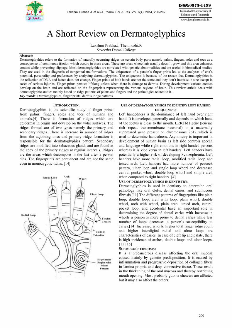
Dermatoglyphics is the scientific study of finger prints from palms, fingers, soles and toes of humans and animals.[4] There is formation of ridges which are epidermal in origin and develop on the volar surfaces. The ridges formed are of two types namely the primary and secondary ridges. There is increase in number of ridges from the adjoining ones and primary ridge formation is responsible for the dermatoglyphics pattern. Secondary ridges are modified into sebaceous glands and are found at the apex of the primary ridges at regular intervals. Ridges are the areas which decompose in the last after a person dies. The fingerprints are permanent and are not the same even in monozygotic twins. [14]
USE OF DERMATOGLYPHICS TO IDENTIFY LEFT HANDED
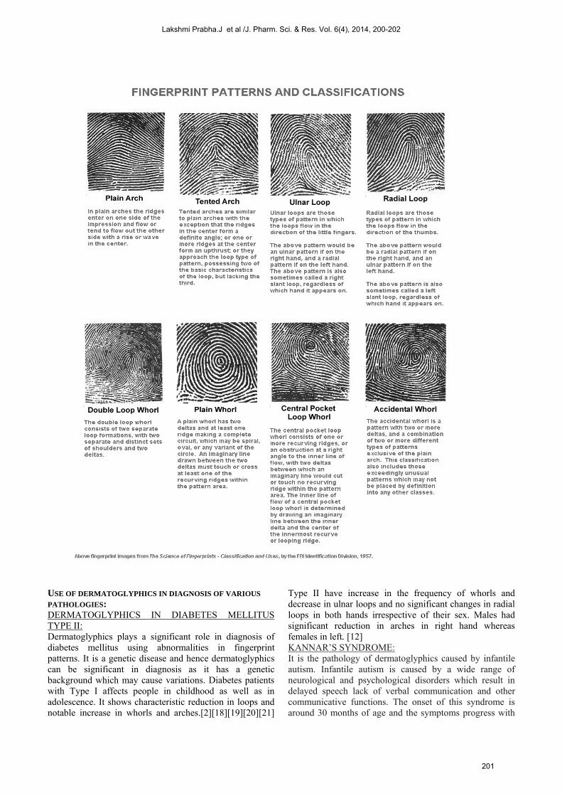
UNIQUENESS: Left handedness is the dominance of left hand over right hand. It is developed paternally and depends on which hand of the foetus is close to the mouth. LRRTM1 gene (leucine rich repeat transmembrane neuronal) is a maternally suppressed gene present on chromosome 2p12 which is used to determine handedness. Asymmetry is important in development of human brain as left side controls speech and language while right emotions in right handed persons whereas it is vice versa in left handers. Left handers have potentially a higher risk of developing Schizophrenia. Left handers have more radial loop, modified radial loop and tented arch. Left handers had more number of peacock pattern, ulnar loop and single loop whorl and decreased central pocket whorl, double loop whorl and simple arch when compared to right handers. [4] USE OF DERMATOGLYPHICS IN DENTISTRY: Dermatoglyphics is used in dentistry to determine oral pathology like oral clefts, dental caries, and submucous fibrosis.[11] The different patterns of fingerprints like plain loop, double loop, arch with loop, plain whorl, double whorl, arch with whorl, plain arch, tented arch, central pocket loop, and accidental have an important role in determining the degree of dental caries with increase in whorls a person is more prone to dental caries while less number of loops decreases a person’s susceptibility to caries.[14] Increased whorls, higher total finger ridge count and higher interdigital radial and ulnar loops are characteristics of caries. In case of cleft lip and palate, there is high incidence of arches, double loops and ulnar loops. [11][15] SUBMUCOUS FIBROSIS: It is a precancerous disease affecting the oral mucosa caused mainly by genetic predisposition. It is caused by inflammation and progressive deposition of collagen fibers in lamina propria and deep connective tissue. These result in the thickening of the oral mucosa and thereby restricting mouth opening. Most probably gutkha chewers are affected but it may also affect the others.
Lakshmi Prabha.J et al /J. Pharm. Sci. & Res. Vol. 6(4), 2014, 200-202
200
USE OF DERMATOGLYPHICS IN DIAGNOSIS OF VARIOUS
PATHOLOGIES: DERMATOGLYPHICS IN DIABETES MELLITUS TYPE II: Dermatoglyphics plays a significant role in diagnosis of diabetes mellitus using abnormalities in fingerprint patterns. It is a genetic disease and hence dermatoglyphics can be significant in diagnosis as it has a genetic background which may cause variations. Diabetes patients with Type I affects people in childhood as well as in adolescence. It shows characteristic reduction in loops and notable increase in whorls and arches.[2][18][19][20][21]
Type II have increase in the frequency of whorls and decrease in ulnar loops and no significant changes in radial loops in both hands irrespective of their sex. Males had significant reduction in arches in right hand whereas females in left. [12] KANNAR’S SYNDROME: It is the pathology of dermatoglyphics caused by infantile autism. Infantile autism is caused by a wide range of neurological and psychological disorders which result in delayed speech lack of verbal communication and other communicative functions. The onset of this syndrome is around 30 months of age and the symptoms progress with
Lakshmi Prabha.J et al /J. Pharm. Sci. & Res. Vol. 6(4), 2014, 200-202
201
the onset of Asperger syndrome and Ret syndrome. Severe forms of manifestations include hearing loss, mental retardation with epilepsy, dislexy, Martin bell’s syndrome and rare cases of tuberous sclerosis. In Digital dermatoglyphics, patients suffering from this syndrome have a high frequency of arches and lower loops. Arches of fourth and fifth fingers and first finger of left hand shows higher frequency than the others. Palmer dermatoglyphic distortions were common in left palm. These patients show prominent increase in ulnar loops. [1][3][9][10][23]
OTHER PATHOLOGICAL CONDITIONS: DOWN’S SYNDROME: Down’s syndrome is a genetic disorder caused by presence of third copy of chromosome 21. It is a common disease characterized by facial abnormalities and decreased intellectual capacities. It results in severe infections of upper respiratory tract due to partial defects in immune systems.[6] Manifestations of down’s syndrome include high frequency of creases, bilateral, radial loops on digits 4 and 5 and ulnar loops.[11][15] HYPOPARATHYROIDISM: Hypoparathyroidism is a decreased function of parathyroid hormone due to surgery of thyroid and parathyroid glands. It causes cramping and twitching of muscles leading to tetany and several other symptoms. Hypoparathyroidism is caused by reduction in circulating parathyroid hormones, hypocalcaemia and hyperphosphatemia.[5] It is characterized by short broad bands and increased arch patterns. [11][15] RUBINSTEIN-TAYBI SYNDROME: It is also known as Rubinstein syndrome or broad thumb hallux syndrome characterized by shot stature, mental disability, broad thumbs and first toes and facial anomalies. They show four or more arches in finger tips and are bilateral.[11][15] PALMAR DERMATOGLYPHICS IN MALE CATIONIC SCHIZOPHRENIA: Schizophrenia is a neural disorder in which the patient has impairment in perception and thought processing.[4] In this case subjects were found to have more number of arches and loops and less whorls. [7]
CONCLUSION: This review brings out the importance of dermatoglyphic studies in various fields. The dermatoglyphics are important in forensic sciences due to their important feature that fingerprints are unchanged in due course even after death. Different patterns of fingerprints represent various pathologies. Dentists are benefitted as they analyzers are able to determine clefts, caries and submucous fibrosis by observing the various patterns of the patients palms. By analyzing these patterns, the analyses were able to find significant variations which represent those pathologies.
REFERENCES: 1. Ana Ţarcă, C. Barabolski, PATHOLOGY OF
DERMATOGLYPHICS IN INFANTILE AUTISM, THE JOURNAL OF PREVENTIVE MEDICINE 2003; 11 (1): 11-17
2. Ana Ţarcă, Elena Tuluc, DERMATOGLYPHICS IN INSULIN – DEPENDENT DIABETES OR DIABETES MELLITUS TYPE 1 (T1DM), THE JOURNAL OF PREVENTIVE MEDICINE 2005; 13 (1-2): 43-53
3. Bowman EP: Asperger’s Syndrome and Autism. British Journal of Psychiatry, 1983, 143: 261-265.
4. Chandan Kumar Sinha, Monika Meel and Bituparna Bayan, Using Dermatoglyphics Pattern to Identify the Left Handed Unique Pattern and its Biological Significance-If Any, World Applied Sciences Journal 20 (8): 1107-1113, 2012
5. Changlin Ding, Bruce Buckingham, and Michael A. Levine, Familial isolated hypoparathyroidism caused by a mutation in the gene for the transcription factor GCMB
6. G. Ram and J. Chinen, Allergy and Immunology Section, Department of Pediatrics, Baylor College of Medicine, Texas Children’s Hospital. Houston, Houston, TX, USA, The journal of translational immunology
7. H. P. JHINGAN, G. C. MUNJAL, DERMATOGLYPHICS IN MALE CATATONIC SCHIZOPHRENICS, Indian J. Psychiat. (1990), 32(2), 188-192
8. Jian XC, Liu SF, Shen ZH, Yang YH. Histomrphology of oral submucous Fibrosis, Report of 24 cases, Chinese Med J. 1988, 101; 505-9
9. Langenbeck W, Varga I, Hausman I: The Predictive Value of Dermatoglyphics in the Diagnosis of FRA (X)- Positive Martin Bell Syndrome (MBS), American Journal of Med. Genet., 1988, 169-175.
10. . Meilă P, Milea Şt: Textbook of pediatrics, Med. Publ. House Bucureşti, 1988, 6: 340-346 (in Romanian).
11. NS Priya, P Sharada, N Chaitanya Babu, HC Girish, DERMATOGLYPHICS IN DENTISTRY: AN INSIGHT, 10.5005/jp-journals-10015-1221
12. Pathan Ferozkhan J. & Gosavi Anjali G., Dermatoglyphics in Type II Diabetes Mellitus
13. Pindborg JJ. Barmes D, Roed-Peterson B. Epidemiology and histology of oral leukoplakia and leukoedema among Papuans and new Guineans, Cancer 1968; 22: 379-84.
14. PR Abilash, R Divyashree, Shankar Gouda Pat Mohlt Gupta, T Chandrasekar, R Karthikeyan, Dermatoglyphics in Patients with Dental Caries: A Study on 1250
15. Preus M, Fraser F. Dermatoglyphics and syndrome, Amer J Dis child 1972; 24:933-42
16. Schauman Blanka Milton A: , Dermatoglyphics in Medical Disorders., Springer Verlag, New York-Heidelberg Berlin. 1976Individuals, 10.5005/jp-journals-10024-1235
17. Tamgire Dw , Fulzele Rr , Chimurkar Vk , Rawlani Ss , Sherke Ar, Qualitative Dermatoglyphic Analysis of Finger Tip Patterns In Patients Of Oral Sub Mucous Fibrosis, IOSR Journal of Dental and Medical Sciences (IOSR-JDMS) e-ISSN: 2279-0853, p-ISSN: 2279-0861. Volume 6, Issue 5 (May.- Jun. 2013), PP 24-27
18. Ţarcă Ana: Structura dermatoglifică, a populaţiei din trei provincii istorice româneşti (Moldova, Maramureş şi Bucovina). Teză de doctorat, Ed.Univ. ,, Al.I.Cuza” Iaşi, 1995, 122-139.
19. Ţarcă Ana: Patologia dermatoglifelor în afecţiuni oculare. Revista Medico- Chirurgicală, Iaşi. 2000, 104 (3): 113-118.
20. Ţarcă Ana: The Pathological Aspects of Dermatoglyphics in Cardio-Vascular Diseases. J. Med. Prev., Iaşi. 2000, 8 (3): 31-38.
21. Ţarcă Ana, Barabolski C: Contributions to the Dermatoglyphic Diagnosis in Epilepsy. J. Med. Prev., Iaşi. 2002, 10 (2): 28-34.
22. Ţurai C, Leonida CI: Amprente papilare. Ed. Medicală, Bucureşti. 1979, 211-264
23. Uta F: Autism and Asperger Syndrome. Cambridge Univ. Press, 1997, 37-183.
24. Veena HS, Cross sectional study of Palmer dermatoglyphics among gutkha chewers with and without oral submucous fibrosis, Karnataka, Bengaluru: Rajiv Gandhi University of Health Sciences Mar 2006.
25. WHO Meeting Report. Control of oral cancer in developing countries. WHO Bull 1984;62:617
Lakshmi Prabha.J et al /J. Pharm. Sci. & Res. Vol. 6(4), 2014, 200-202
202