Embed Size (px)
Citation preview

University of CyprusBiomedical Imaging and Applied OpticsBiomedical Imaging and Applied Optics
Reflectance and Diffuse S tSpectroscopy

Spectroscopy
• What is it ? from the Greek:t l l k t b• spectro = color + scope = look at or observe
= measuring/recording the colors of light• What can we learn from it?What can we learn from it?
• Energy levels of atoms and molecules• Fundamental processes• The molecular constituents in tissues• The molecular constituents in tissues
• When you measure how much of each color there is, you’re measuring a “spectrum.”
dispersion l t
spectrum to be analyzed
element
2
Wavelengths separated by angle

Newton’s prism
3

Dispersion with a Prism
• n=n(λ)n n(λ)
monochromatic light
white light
4

Dispersion with a Prism
• Resolving Power prismdnR td
λδλ λ
= =
• Spectral Resolution βφα
dδλ λ
Rλδλ =
• Useful where only low resolving power is required
φα
D1
L
D2
R
p q• Advantages:
• simple (but glass must be of high quality)
t
Prism, index nq y)• multiple-order overlap not a problem
- only one order!
• Disadvantages:g• high resolving power not possible• resolving power/resolution can vary
strongly with λ
5

Rainbows
• What causes rainbows?ff• Diffraction by rain drops
• Angle between incident and diffracted rays
• 42 degrees for red• 42 degrees for red • 40 degrees for violet.
• Form a circular rim of color in the sky a rainbow! y
• Secondary rainbows • Double reflection of sunlight inside
the raindropsthe raindrops• Appear at an angle of 50–53
degrees• The droplets have to be the rightThe droplets have to be the right
size to get two reflections to work
• Higher order rainbows are possible
6
possible

Dispersion with a Grating
• Diffraction grating g g• An optical unit that
separates polychromatic light into constantlight into constant monochromatic composition.
7

Dispersion with a Grating
• Multi-slit arrangementg• Uses diffraction to separate
light wavelengths with high a
θ
resolution and high intensity.
• The resolving power is
Constructive interference whena·sinθ = mλ, where m is an integer
a
• The resolving power is achieved by interference of light.
8
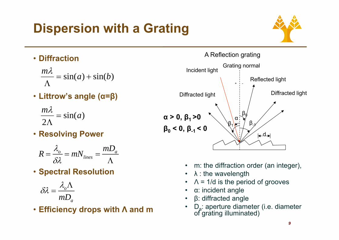
Dispersion with a Grating
• DiffractionGrating normal
A Reflection grating
Diffracted light
Reflected light
Grating normal
+ -
Incident light
Diff t d li ht
)sin()sin( bam+=
Λλ
• Littrow’s angle (α=β)
αβ0
Diffracted lightDiffracted light
α > 0, β1 >0)sin(2
am=
Λλ
• Resolving Powerβ-1β1
d
, β1
β0 < 0, β-1 < 02Λ
mDλ
• Spectral Resolution• m: the diffraction order (an integer), • λ : the wavelength
o alines
mDR mNλδλ
= = =Λ
p λ : the wavelength• Λ = 1/d is the period of grooves• α: incident angle• β: diffracted angle
o
amDλδλ Λ
=
9
• Efficiency drops with Λ and m β g
• Da: aperture diameter (i.e. diameter of grating illuminated)
a

Anatomy of a grating spectrometer
• Spectrometer• Device to obtain
spectrum of light• Usually gratingUsually grating
• Width of slit determines:
• Resolving power (w ↓, R↑)
• Throughput SNR (w ↓Throughput SNR (w ↓, I ↓)
• Hence there is always ya tradeoff between throughput and spectral information
10
spectral information

Reflectance Spectroscopy
• MeasuresC• Changes to source spectrum
• Effects of• Elastic ScatteringElastic Scattering
• Mie and Rayleigh• Absorption
• Many absorbers in tissuey
• Instrumentation• Single point
Imaging• Imaging• Many collection fibers imaging
spectrograph• Limited spatial resolution
• Camera with variable filter• Limited spectral resolution
• Applications
11
• E.g. pulse oximeter

Reflectance Spectroscopy
fiber profiber pro
d
tissue ρ
d
(D=1/μt’) and A relates to internal reflection.)
12Mourant, etc., and Bigio, Appl. Optics 36, No.4, p.949 (1997)

The Pulse Oximeter
• Function: MeasureFunction: Measure arterial blood saturation
Ad t• Advantages:• Non-invasive• Highly portable• Continuous monitoringContinuous monitoring• Cheap
R li bl• Reliable
13

The Pulse Oximeter
• How: Illuminate tissue at 2 wavelengths
100
• Illuminate tissue at 2 wavelengths • straddling isosbestic point (eg. 650 • and 805 nm)
• Isosbestic point: wavelength where Hb1
10
Isosbestic point: wavelength where Hband HbO2 spectra cross.
• Detect signal transmitted through finger
0 01
0.1
• Isolate varying signal due to pulsatileflow (arterial blood)
• Assume detected signal is proportional t b ti ffi i t
0.01
300 400 500 600 700 800 900 1000 1100 1200 1300
[ ] [ ]{ }HbHbO bbO111
2*10ln λλλ εεμ +=to absorption coefficient • Get the two μa• Calculate the concentrations (Two
measurements two unknowns)
[ ] [ ]{ }HbHbO HbHbOa 2 210ln εεμ +=
[ ] [ ]{ }HbHbO HbHbOa22
2
22*10ln λλλ εεμ +=
measurements, two unknowns)• Calibrate instrument by correlating
detected signal to arterial saturation measurements from blood samples
[ ][ ] %100*saturation O Arterial
2
22 HbHbO
HbO+
=
14
easu e e ts o b ood sa p esε is the extinction coefficient

The Pulse Oximeter
• Limitations:Limitations:• Reliable when O2
saturation above 70%• Not very reliable when
flow slows down• Can be affected by
motion artifacts and room light variations
• Doesn’t provide tissue ti l loxygenation levels
15

Turbid Media
• How can we image bj t b dd d iobjects embedded in
turbid media?• Cheat !!Cheat !!
• Try to devise a method to detect only theto detect only the photons that have not scattered
• Generating a direct shadow• Generating a direct shadow image (like an x-ray).
• Possible methodsPossible methods• Collimated Illumination
• Use of polarizersD t t “ k ” h t
16
• Detect “snake” photons

Turbid Media
• Collimated illumination and detectionCollimated illumination and detection
Collimated laserCollimated laser
PolarizerCollimated laser PolarizerCan add polarizers:
Parallel polarizer
17

Turbid Media
• The earliest arriving h t hphotons have
traveled the straightest path Output:
• Can we select only the earliest photons?
• Time gating methods
Input: very short li ht
Output: scattered photons
tissue
• Time gating methods• Streak camera• Time-to amplitude
converters
light pulse
converters• Coherence gates• Nonlinear optical gates
Kerr gate
t• Kerr gate• Second harmonic
generation• Raman amplification
18
p

Turbid Media
• But, are there enough unscatteredphotons??
• Beer’s law can also tell Output:• Beer s law can also tell us how many photons remain unscattered after a given distance in
Input: very short li ht
Output: scattered photons
tissue
a given distance in tissue:
IU = I0e− ′ μ s L
light pulse
• A typical value for many tissues: μs’ ~ 10 cm-1
IU I0et
• ⇒ for optical mammography there are very few
19
unscattered photons

Turbid Media
How can we image with diffuse light?• The “forward” problem
• Asks: if we know the light going in, and we know what the hidd bj t i l l t h t h thhidden object is, can we calculate what reaches the surface in different locations?
• The “inverse” problem• The inverse problem• Asks: if we know the light going in, and we measure the
light coming out at various locations, what can we say b t th hidd bj t?about the hidden object?
• MethodsMonte Carlo• Monte Carlo
• Diffusion Approxiamtion• Time Domain vs Frequency domain
20
Time Domain vs. Frequency domain
Turbid Media
How do we solve an inverse problem?p• Sometimes done as an iteration of forward calculations:calculations:1. Must make assumptions about the optical properties
of the surrounding tissueg2. Make an initial “guess” about the location, size and
optical properties of the lesion.3. Do a forward propagation calculation and see how
those results compare with the measurements.4. Re-estimate the properties of the lesion based on
the difference, and recalculate the forward problem.5 R t ti !!!
21
5. Repeat many times!!!

Diffuse Optical tomography (DOT)
• Year discovered: ~1988
• Form of radiation: Near-infrared light (non-ionizing)
• Energy/wavelength of radiation: ~1 eV / 600–1000 nm
• Imaging principle: Interaction• Imaging principle: Interaction (absorption, elastic scattering) of light w/ tissue
• Imaging volume: ~103 cm3
• Resolution: Low (~1cm)Resolution: Low ( 1cm)
• Applications: Perfusion, functional imaging
22

Diffuse Optical tomography (DOT)
DOT and CTSource
DOT and CT
• Superficial Similarities • Generation: x-ray tube• Detection: Detector
(i h barrays (ion.-chambers, scint. + photodiode)Computer reconstruction
Object
• Computer reconstruction of 2D slices/ 3D volumetric imagesvolumetric images
• Essential DifferencesDetector
23
• No scattering!

Diffuse Optical tomography (DOT)
• Principles of DOT• Scattering dominated• Limited penetration depth (~cm), low res. (mm-cm)• Economic, functional (hemodynamics)Economic, functional (hemodynamics)
dete
ctor
/ det
ecto
r
sour
ce
sour
ce
light source detector
S Dsc
reen
/
scre
en /
light
s
obstacle (absorber) obstacle (absorber)
light
s
Clear medium Scattering medium SDD
105
106
cm -1
M-1
]
Hb
HbO2103
104
105
extin
ctio
n co
eff.
[c
24
DDHbO2
400 500 600 700 800 900 1000
Wavelength [nm]
102Mol
ar
Many sources / many detectors

Diffuse Optical tomography (DOT)
• DOT Applications - BrainSource / Detector 1
DOT Applications Brain2-3 cm
Detector 2
Detector 3
Scalp
Bone
CSF
Cortex
25

Diffuse Optical tomography (DOT)
• DOT Applications - BreastDOT Applications Breast
20 20 20lob loblob
18
16
14
)
215lob
14
16
18
20
18
16
14
12m)
18
16
14
m)
Arm12
10
8
6
x (c
m
2
4
6
8
10
12
x (
cm
)12
10
8
6
x (c
m
Cancer
12
10
8
6x
(cm
Cancer
0 2 4 6 8 10
6
4
2
0
Cancer0 2 4 6 8 10
y (cm)
0
2
0 2 4 6 8 10
6
4
2
0
Cancer
0 2 4 6 8 10
6
4
2
0
Cancer
0 2 4 6 8 10y (cm)
low high
0 2 4 6 8 10y (cm)
0 2 4 6 8 10y (cm)
26

Atomic spectroscopy
• Atomic spectroscopy • Not very relevant to medical
applications.• Flames, electrical discharges
(especially in gases)(especially in gases)• However, one method may prove
useful Laser-induced breakdown spectroscopy (LIBS)spect oscopy ( S)
• Laser-induced breakdown spectroscopy (LIBS)
• Atomic emission spectroscopy• Highly energetic laser pulse focused
to form a plasma• Emit light of characteristic
frequencies• LIBS at surface of skin could be used
t h t l t i ti
27
to sense heavy metal contamination