Embed Size (px)
Citation preview
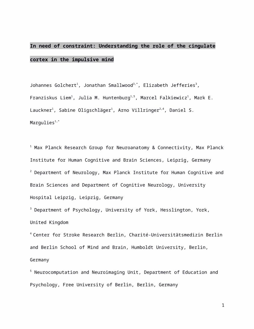
In need of constraint: Understanding the role of the cingulate cortex in the
impulsive mind
Johannes Golchert1, Jonathan Smallwood3,*, Elizabeth Jefferies3, Franziskus Liem1, Julia M.
Huntenburg1,5, Marcel Falkiewicz1, Mark E. Lauckner1, Sabine Oligschläger1, Arno Villringer2,4,
Daniel S. Margulies1,*
1 Max Planck Research Group for Neuroanatomy & Connectivity, Max Planck Institute for
Human Cognitive and Brain Sciences, Leipzig, Germany
2 Department of Neurology, Max Planck Institute for Human Cognitive and Brain Sciences and
Department of Cognitive Neurology, University Hospital Leipzig, Leipzig, Germany
3 Department of Psychology, University of York, Hesslington, York, United Kingdom
4 Center for Stroke Research Berlin, Charité-Universitätsmedizin Berlin and Berlin School of
Mind and Brain, Humboldt University, Berlin, Germany
5 Neurocomputation and Neuroimaging Unit, Department of Education and Psychology, Free
University of Berlin, Berlin, Germany
* Correspondence:
[email protected] or [email protected]
1

Highlights
Functional connectivity of the ACC relates to impulsivity in 2 independent samples
Positive urgency predicts connectivity from subgenual cingulate to bilateral parietal
Perseverance predicts connectivity from rostral cingulate to dorsolateral prefrontal
Premeditation predicts connectivity from rostral cingulate to occipital/parietal
Regions from perseverance and premeditation overlapped with multiple demand network
2

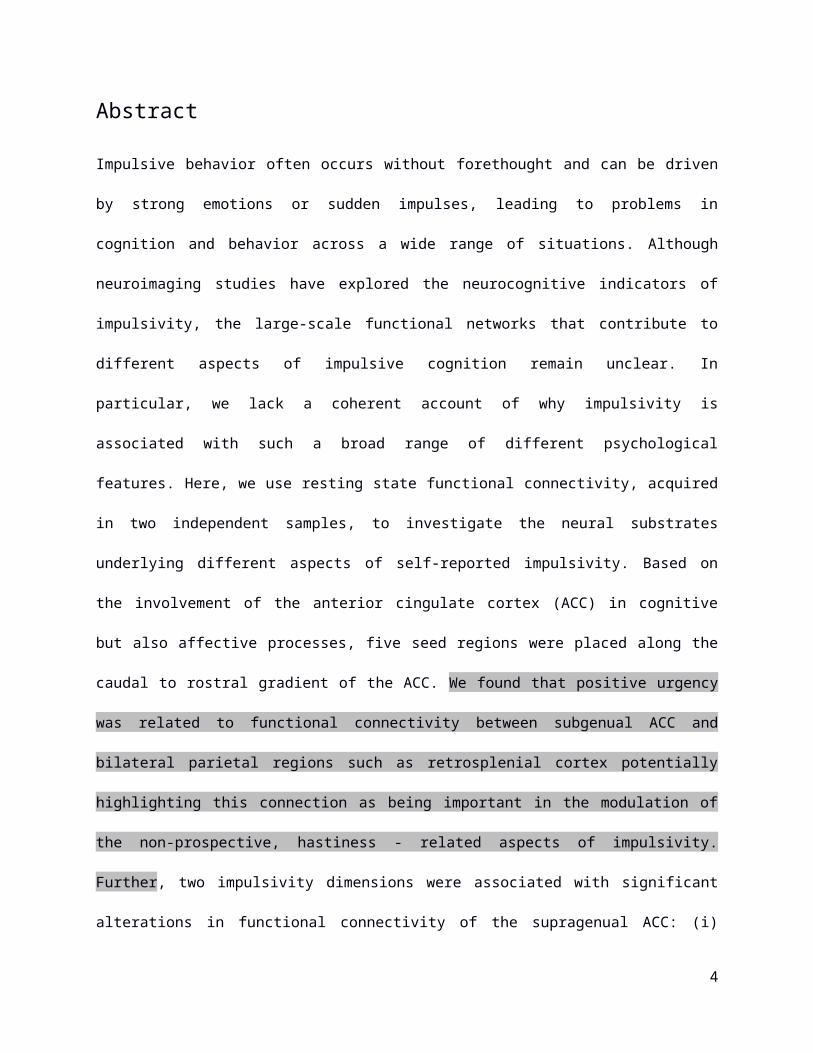
Abstract
Impulsive behavior often occurs without forethought and can be driven by strong emotions or
sudden impulses, leading to problems in cognition and behavior across a wide range of
situations. Although neuroimaging studies have explored the neurocognitive indicators of
impulsivity, the large-scale functional networks that contribute to different aspects of impulsive
cognition remain unclear. In particular, we lack a coherent account of why impulsivity is
associated with such a broad range of different psychological features. Here, we use resting
state functional connectivity, acquired in two independent samples, to investigate the neural
substrates underlying different aspects of self-reported impulsivity. Based on the involvement of
the anterior cingulate cortex (ACC) in cognitive but also affective processes, five seed regions
were placed along the caudal to rostral gradient of the ACC. We found that positive urgency
was related to functional connectivity between subgenual ACC and bilateral parietal regions
such as retrosplenial cortex potentially highlighting this connection as being important in the
modulation of the non-prospective, hastiness - related aspects of impulsivity. Further, two
impulsivity dimensions were associated with significant alterations in functional connectivity of
the supragenual ACC: (i) lack of perseverance was positively correlated to connectivity with the
bilateral dorsolateral prefrontal cortex and right inferior frontal gyrus and (ii) lack of
premeditation was inversely associated with functional connectivity with clusters within bilateral
occipital cortex. Further analysis revealed that these connectivity patterns overlapped with
bilateral dorsolateral prefrontal and bilateral occipital regions of the multiple demand network, a
large-scale neural system implicated in the general control of thought and action. Together
these results demonstrate that different forms of impulsivity have different neural correlates,
which are linked to the functional connectivity of a region of anterior cingulate cortex. This
suggests that poor perseveration and premeditation might be linked to dysfunctions in how the
3
rostral zone of the ACC interacts with the multiple demand network that allows cognition to
proceed in a controlled way.
Keywords: impulsivity, UPPS-P, functional connectivity, anterior cingulate cortex, multiple
demand network
4
1. Introduction
Impulsive behavior occurs when actions are “poorly conceived, prematurely expressed, unduly
risky or inappropriate to the situation and often result in undesirable consequences” (Daruna
and Barnes, 1993, p. 23; cf. Arce and Santisteban, 2006, Dalley et al., 2011). It is a broad
construct that captures the influence of motor, cognition and emotion on behavior (see
Evenden, 1999) with a wide range of functional outcomes (for reviews see: Dalley et al., 2011;
Moeller et al., 2001). Different aspects of impulsivity can be assessed by questionnaires such as
the UPPS impulsive behavior scale (Whiteside and Lynam, 2001) that uses a factor analytic
approach to identify 4 components: (i) negative urgency, which refers to acting rashly out of a
negative mood; (ii) lack of perseverance that describes circumstances of having difficulties in
staying on task; (iii) lack of premeditation - reflecting the hampered ability to anticipate future
consequences of an action; and (iv) sensation seeking, a tendency to seek and engage in
exciting, novel and dangerous situations. Cyders and Smith (2007) added a fifth component —
positive urgency — to differentiate between rash actions that occur under conditions of either
negative or positive mood.
Although the construct of impulsivity as a wide ranging trait with a heterogeneous mixture of
affective and cognitive factors is well established, it is unclear why such a variety of different
cognitive and behavioral attributes tend to co-occur in individuals. It is possible that
understanding the neurocognitive architecture that supports different types of impulsive
behavior could help explain what causes this clustering of diverse symptoms under the rubric of
the same broad psychological construct. To date, neurocognitive evidence has implicated
alterations in how the cortex interacts with limbic and striatal systems (e.g. Brewer and Potenza,
2008; Brown et al., 2006; Dalley et al., 2011; Robbins, 2007) as well as differences in the global
level of brain networks organization (Davis et al., 2013). One cortical structure that may mediate
5

impulsive behavior is the anterior cingulate cortex (ACC). Recently, Casetellanos-Ryan and
Seguin (2015) argued that, along with other regions of the prefrontal cortex, the ACC is
important in impulsivity because of its role in many cognitive functions that seem to be altered in
impulsivity. The authors argue that many of the cognitive processes that contribute to impulsive
decision making and behavior involve different aspects of executive functions (such as planning,
response/motor inhibition, problems in perceiving/attending environmental information, selective
attention and working memory). These processes are partly mediated by the interactions
between the ACC and other aspects of the PFC (for a detailed description see their review).
Studies have implicated the ACC in a wide range of different cognitive processes that could
have relevance to impulsivity. These include conflict monitoring (Botvinick et al., 1999, 2001,
2004), attentional (e.g. Aarts and Roelofs, 2011; Crottaz-Herbette and Menon, 2006) and motor
control (e.g. Asemi et al., 2015; Chouinard and Paus, 2006), working memory (e.g. Lenartowicz
and McIntosh, 2005), task switching (e.g. Woodward et al., 2008), adaptive choices
(Economides et al., 2014) and response inhibition (Braver et al., 2001). It has also been shown
to be involved in more complex social processes (Lavin et al., 2013), as well as in the regulation
of emotional states (for a review see: Etkin et al., 2011) and task disengagement (Bernhardt et
al., 2014). Consistent with a role in many different processes, the functional architecture of the
ACC suggests it is important for integrating and processing information from different sources to
guide behavior (cf. Kelly et al., 2009). It is a “cortical hub” with projections to several different
brain areas both in non-humans (Devinsky et al., 1995) and humans (e.g. Hagmann et al.,
2008) allowing it to integrate both bottom-up and top-down signals from different regions of
cortex (e.g. Comte et al., 2014). Accordingly, various lines of research have demonstrated the
existence of functionally distinct subdivisions within the ACC (e.g. Bush et al., 2000). Functional
connectivity studies have shown that connectivity patterns are organized along a rostral-caudal
gradient with regions in the midcingulate cortex, located caudally to the ACC, involved in
6
motoric processing, more anterior regions implicated in general process of control and
subgenual aspects implicated in the limbic system and default mode network (Margulies et al.,
2007). It is important to note that these patterns of connectivity do not mean that subregions of
the ACC serve discrete functions in all circumstances. For example, there are a number of fMRI
studies showing activation associated with emotion regulation in rostral and dorsal regions of
the ACC, as well as in the subgenual areas (e.g., Domes et al., 2010; Goldstein et al., 2007;
Gröne et al., 2015; Ochsner and Gross, 2005; see also Vogt, 2005 for a review on ACC peak
activation sites during processing of simple emotions).
The current study explores whether different aspects of impulsivity can be understood in terms
of functional variation in dissociable networks anchored in the ACC. There are several different
ways in which the functional architecture of the anterior cingulate could account for different
aspects of impulsive behavior. One possibility is that because the anterior cingulate is itself a
functionally heterogeneous region of cortex (Bush et al., 2000, Margulies et al., 2007) different
cognitive features of impulsivity could depend on dysregulation in different regions of the
anterior cingulate. According to this modular account, population level variation in different
aspects of impulsivity would be associated with variation in the functional connectivity of
different subregions of the ACC. A second possibility is that a single region of the ACC could be
linked to different aspects of impulsivity by exhibiting different patterns of connectivity, each
associated with a different type of impulsive behavior. Consistent with this hub view, evidence
has implicated the mid-anterior cingulate in the multiple demand network (MDN), a system that
plays an important role in the production of controlled cognition across a wide range of different
circumstances (Duncan, 2010). According to this hub account, cross-sectional variation in
different elements of impulsivity could be related to unique patterns of functional connectivity
from the same region within the anterior cingulate cortex, particularly regions involved in the
MDN.
7
To understand how different aspects of impulsivity are related to the functional connectivity of
the ACC we analyzed resting state functional connectivity data from more than 200 individuals,
acquired at two different sites. In both samples, impulsivity was assessed using the UPPS-P
impulsive behavior scale (Cyders and Smith, 2007) that captures different dimensions of
impulsivity. We selected five caudal to rostral regions of interest reflecting its spectrum of
connectivity patterns across cognitive and affective domains (e.g. Margulies et al., 2007). We
performed a seed-based functional connectivity analysis to examine if its connectivity profiles
vary with respect to differences in cognitive or affective components of impulsivity. We were
interested in whether different aspects of impulsivity are associated with dissociable patterns of
functional connectivity within subregions of the anterior cingulate.
2. Methods
2.1 Participants
Two independent datasets were used in this study: (i) the Max Planck Institute sample (MPI-S),
Max Planck Institute of Human Cognitive and Brain Sciences in Leipzig, Germany, and (ii) the
enhanced Nathan Kline Institute-Rockland Sample (NKI-RS, Nooner et al., 2012; made publicly
available as a prospective dataset through the International Neuroimaging Data-sharing
Initiative). Both research projects were approved by an Ethics committee (MPI-S: medical
department of University of Leipzig; NKI-RS: Nathan Kline Institute and Montclair State
University).
The MPI-S dataset contained brain imaging and behavioral data from a sample of 112 healthy
participants (63 female subjects) with a mean age of 26.48 (SD = 4.73, range: 21 - 44) years.
The data from the NKI-RS set comprised a sample of 92 (53 female) healthy subjects (age: M =
8

26.89 (SD = 8.7), range: 18 - 44). The groups did not differ in terms of their respective gender
distributions (Chi-square: X2(1) = 0.0028, p = 0.96) nor with respect to age (Welch Test:
t(134,07) = 0.4, p = 0.69). All participants were recruited through advertisement, gave written
informed consent, have been screened about past and present neurological or psychiatric
disorders, and were monetarily reimbursed at the end of the study. In both samples, all
participants were part of larger research projects and other measurements have been
administered during data collection. For the present study, only the UPPS-P impulsive behavior
scale (Cyders and Smith, 2007) and magnetic resonance imaging (MRI) data were analyzed.
2.2 Measurement of trait impulsivity
In order to assess trait impulsivity, participants of both samples filled out the UPPS-P impulsive
behavior scale (Cyders and Smith, 2007; for the MPI-S cohort the questionnaire was translated
into German by a qualified translator). The UPPS-P is a 59-item self report inventory in its
revised version (originally UPPS; Whiteside and Lynam, 2001) that quantifies five different
aspects of impulsive behavior: (i) negative urgency, which refers to the tendency to experience
strong impulses under conditions of negative affect; (ii) lack of perseverance that reflects the
experience of having problems with remaining focused on a task that might be boring or too
difficult; (iii) lack of premeditation that describes the tendency to engage in an act without
reflecting the consequences of that act beforehand; (iv) sensation seeking that further
comprises two aspects: (a) the propensity to enjoy and chase exciting activities; (b) an
openness to engage in new experiences that might be dangerous; and (v) positive urgency that
involves the tendency towards rash actions in response to very positive mood. Each item can be
scored on a four-point Likert scale, ranging from 1 (strongly agree) to 4 (strongly disagree).
Reversed items are recoded afterwards so that higher scores indicate a more pronounced level
of self-reported trait impulsivity. In both samples, reliability analyses revealed an adequate level
of internal consistency (Cronbach’s alpha) for each of the five subscales (see Table 1).
9

2.3 MRI scan acquisition
MPI-S data were acquired on a 3T Siemens Magnetom Verio Scanner and included four resting
state functional magnetic resonance imaging (rs-fMRI) sequences as well as an anatomical T1-
weighted scan. The rs-fMRI data were recorded using a BOLD-weighted multiband EPI
sequence (Feinberg et al., 2010; Moeller et al., 2010; TR = 1400 ms, TE = 39.4 ms, flip angle =
69°, multiband acceleration factor = 4, voxel size = 2.3 mm isotropic, 64 slices, 657 volumes,
duration = 15.30 min). A high-resolution anatomical image was additionally acquired using a
MP2RAGE sequence (Marques et al. 2010; TR = 5000 ms, TE = 2.92 ms, TI1 = 700 ms, TI2 =
2500 ms, flip angle 1 = 4°, flip angle 2 = 5°, voxel size = 1.0 mm isotropic, duration = 8.22 min).
NKI-RS data were acquired on a 3T Siemens Trio Scanner, and comprised a rs-fMRI scan
acquired with a BOLD-weighted multiband EPI sequence (TR = 645 ms, TE = 30 ms, flip angle
= 60°, multiband acceleration factor = 4, voxel size = 3mm isotropic, 40 slices, 900 volumes,
duration = 10 min.) and a high resolution anatomical T1-weighted image acquired with a
MPRAGE sequence (TR = 1900 ms, TE = 2.52 ms, flip angle = 9°, voxel size = 1.0 mm
isotropic). In both rs-fMRI assessments subjects were instructed to keep their eyes open and to
focus on a fixation cross (Nooner et al., 2012)
2.4 Preprocessing
All preprocessing steps were streamlined using Nipype (Gorgolewski et al., 2011), incorporating
tools from FreeSurfer (Dale et al., 1999; Fischl et al., 1999), FSL (Jenkinson et al., 2012), AFNI
(Cox, 1996), ANTs (Avants et al., 2011), CBS Tools (Bazin et al., 2014) and Nitime (Rokem et
al., 2009). Reproducible workflows containing all implementation details for both the MPI-S1 and
1 https://github.com/NeuroanatomyAndConnectivity/pipelines/blob/master/src/lsd_lemon/lsd_resting.py
10
NKI-RS2 data are available online. In brief, structural preprocessing was implemented by
masking the background of the T1-weighted images using CBS Tools before volumetric
segmentation and surface reconstruction with FreeSurfer’s recon-all pipeline. A spatial
transformation between the individual structural space and MNI152 standard space was
calculated using diffeomorphic nonlinear registration implemented in ANTs SyN.
Resting state preprocessing of the MPI-S data included the following steps: (i) removing the first
five EPI volumes from each resting-state scan to allow for signal equilibration; (ii) motion
correction; (iii) distortion correction; (iv) coregistration to the individual T1-weighted image; (v)
denoising via two steps: a) the 6 motion regressors and first derivatives, motion and signal-
intensity outliers (based on Nipype rapidart interface) as well as linear and quadratic trends
were removed in a general linear model (GLM); b) the residual time series was used to derive 6
principal components most representative of physiological noise using the aCompCor method
within white matter and CSF (Behzadi et al., 2007). These are removed along with the initial
regressors in a second GLM; (vi) band pass temporal filtering (0.01 – 0.1 Hz) and mean
centering as well as variance normalization of the denoised timeseries using Nitime; and (vii)
spatial normalization to MNI152 standard space (2 mm isotropic) using the transformation
derived during structural preprocessing. The NKI-RS data were preprocessed in a similar way
with the following exceptions: (1) distortion correction was not performed as no field maps were
acquired; (2) denoising was carried out in one step using the Friston 24-parameter-model
(Friston et al., 1996) as implemented in C-PAC: 5 principal components (aCompCor), motion
and signal-intensity outliers as well as linear and quadratic trends were regressed out; and (3)
spatial normalization towards MNI152 standard space was performed with a resulting resolution
of 3 mm isotropic.
2 https://github.com/NeuroanatomyAndConnectivity/nki_nilearn/blob/master/preprocessing_pipeline.py
11
2.5 First level analysis
Five seed regions were selected along the dorsal to caudal gradient of the ACC (Kelly et al.,
2009; Margulies et al., 2007). Spherical regions of interest (ROIs) with a radius of 4mm were
bilaterally placed at the following coordinates (MNI): seed s1 (±5, -10, 47), seed s3 (±5, 14, 42),
seed s5 (±5, 34, 28), seed s7 (±5, 47, 11), seed i9 (±5, 25, -10) (see Figure 1; coordinates and
nomenclature taken from Margulies et al., 2007). The time series of all voxels within these
regions were then extracted and averaged. For each dataset, the subject specific correlation
between the averaged seed time series and every other voxel in the brain was calculated. The
correlation maps were calculated using AFNI’s 3dfim+ command. The individual correlation
maps were then Fisher r-to-z transformed and smoothed with a Gaussian kernel of 6 mm.
Because the MPI-S dataset comprises four resting state scans, we ran this individual seed-
based functional connectivity analysis for each scan separately and then averaged the four
smoothed correlation maps. Correlation maps of the NKI-RS data were up-sampled into 2 mm
voxel space to allow for comparison between both samples. As before, all steps were
implemented using Nipype (Gorgolewski et al., 2011). In order to allow for comparison between
datasets, correlation maps were normalized (z-scored) separately for each individual prior to the
group-level analysis. That is, the correlation maps were standardized voxelwise across
participants, a step that allows the connectivity from different datasets to be compared.
2.6 Group level analysis
Higher-level ordinary least squares multiple regression analysis was performed using FMRIB's
Local Analysis of Mixed Effects (FLAME OLS, as implemented in FSL FEAT). In order to
increase the statistical power of our model, both datasets were simultaneously entered into the
analyses. The five impulsivity subscales were used as covariates of interest, while controlling for
age, sex, in-scanner motion (mean framewise displacement (Power et al., 2012)) and dataset
12
membership. All covariates were z-scored (except for sex, which was demeaned) across the
two datasets. The concatenated connectivity maps for each region of the ACC were the
dependent variables. One model was run per seed region. For each impulsivity subscale, a
positive and a negative contrast has been computed, were the positive contrast highlights
regions whose connectivity with the seed region are positively associated with the measure of
impulsivity, and the negative contrasts highlight regions showing lower connectivity with the
seed region with increasing impulsivity. In addition, we also tested for interactions of the slopes
between groups (F-test). Following Eklund et al. (2016), spatial maps were thresholded at z =
3.1 and we selected an alpha value that corrected for the number of voxels in the brain, the
number of the seed regions and the two tailed nature of the comparisons (p < 0.05 FWE / 10 = p
< 0.005). Thresholded z-stat maps of each seed’s functional connectivity pattern as modulated
by each respective impulsivity dimension were derived by this analysis. The unthresholded
maps for all the analyses reported in this paper are available at Neurovault at the following URL:
neurovault.org/collections/MXBTLTPY/
3. Results3.1 Self-reported impulsivity
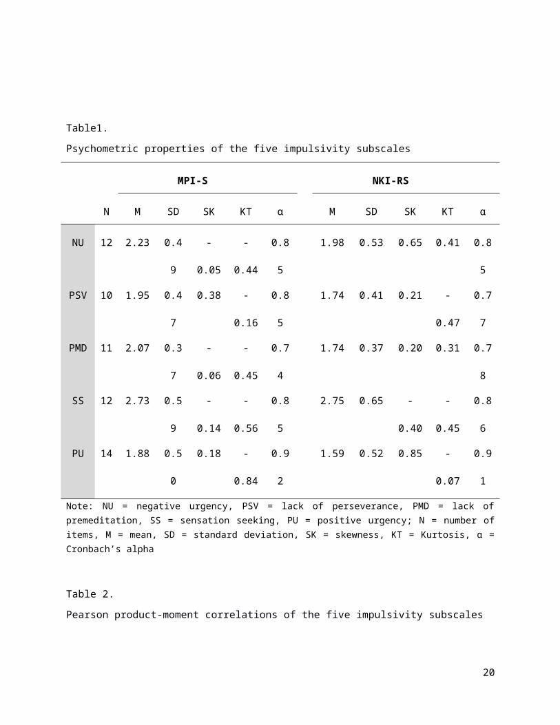
For both datasets internal consistency for the five impulsivity subscales are presented in Table
1. The subscales had acceptable psychometric properties across both samples (all alphas >
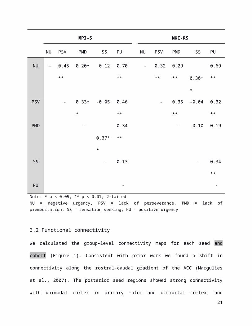
0.74). The correlations between each of the components of impulsivity are presented in Table 2.
Consistent with previous research, especially positive and negative urgency were highly
correlated, whereas sensation seeking was not correlated with a lack of perseverance in both
samples (e.g. Cyders and Smith, 2007). In addition, a lack of premeditation was correlated with
both sensation seeking and positive urgency in the MPI-S dataset and sensation seeking and
negative urgency were correlated in the NKI-RS data.
13

Table1.
Psychometric properties of the five impulsivity subscales
MPI-S NKI-RS
N M SD SK KT α M SD SK KT α
NU 12 2.23 0.49 -0.05 -0.44 0.85 1.98 0.53 0.65 0.41 0.85
PSV 10 1.95 0.47 0.38 -0.16 0.85 1.74 0.41 0.21 -0.47 0.77
PMD 11 2.07 0.37 -0.06 -0.45 0.74 1.74 0.37 0.20 0.31 0.78
SS 12 2.73 0.59 -0.14 -0.56 0.85 2.75 0.65 -0.40 -0.45 0.86
PU 14 1.88 0.50 0.18 -0.84 0.92 1.59 0.52 0.85 -0.07 0.91
Note: NU = negative urgency, PSV = lack of perseverance, PMD = lack of premeditation, SS = sensation seeking, PU = positive urgency; N = number of items, M = mean, SD = standard deviation, SK = skewness, KT = Kurtosis, α = Cronbach’s alpha
Table 2.
Pearson product-moment correlations of the five impulsivity subscales
MPI-S NKI-RS
NU PSV PMD SS PU NU PSV PMD SS PU
NU - 0.45** 0.20* 0.12 0.70** - 0.32** 0.29** 0.30** 0.69**
PSV - 0.33** -0.05 0.46** - 0.35** -0.04 0.32**
PMD - 0.37** 0.34** - 0.10 0.19
SS - 0.13 - 0.34**
PU - -
Note: * p < 0.05, ** p < 0.01, 2-tailedNU = negative urgency, PSV = lack of perseverance, PMD = lack of premeditation, SS = sensation seeking, PU = positive urgency
14
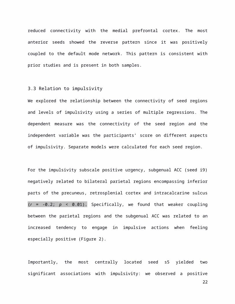
3.2 Functional connectivity
We calculated the group-level connectivity maps for each seed and cohort (Figure 1).
Consistent with prior work we found a shift in connectivity along the rostral-caudal gradient of
the ACC (Margulies et al., 2007). The posterior seed regions showed strong connectivity with
unimodal cortex in primary motor and occipital cortex, and reduced connectivity with the medial
prefrontal cortex. The most anterior seeds showed the reverse pattern since it was positively
coupled to the default mode network. This pattern is consistent with prior studies and is present
in both samples.
3.3 Relation to impulsivity
We explored the relationship between the connectivity of seed regions and levels of impulsivity
using a series of multiple regressions. The dependent measure was the connectivity of the seed
region and the independent variable was the participants’ score on different aspects of
impulsivity. Separate models were calculated for each seed region.
For the impulsivity subscale positive urgency, subgenual ACC (seed i9) negatively related to
bilateral parietal regions encompassing inferior parts of the precuneus, retrosplenial cortex and
intracalcarine sulcus (r = -0.2; p < 0.01). Specifically, we found that weaker coupling between
the parietal regions and the subgenual ACC was related to an increased tendency to engage in
impulsive actions when feeling especially positive (Figure 2).
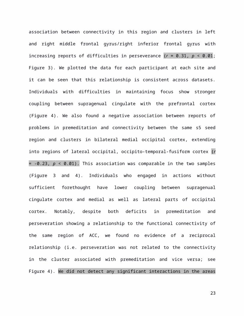
Importantly, the most centrally located seed s5 yielded two significant associations with
impulsivity: we observed a positive association between connectivity in this region and clusters
in left and right middle frontal gyrus/right inferior frontal gyrus with increasing reports of
difficulties in perseverance (r = 0.31, p < 0.01; Figure 3). We plotted the data for each
15
participant at each site and it can be seen that this relationship is consistent across datasets.
Individuals with difficulties in maintaining focus show stronger coupling between supragenual
cingulate with the prefrontal cortex (Figure 4). We also found a negative association between
reports of problems in premeditation and connectivity between the same s5 seed region and
clusters in bilateral medial occipital cortex, extending into regions of lateral occipital, occipito-
temporal-fusiform cortex (r = -0.23, p < 0.01). This association was comparable in the two
samples (Figure 3 and 4). Individuals who engaged in actions without sufficient forethought
have lower coupling between supragenual cingulate cortex and medial as well as lateral parts of
occipital cortex. Notably, despite both deficits in premeditation and perseveration showing a
relationship to the functional connectivity of the same region of ACC, we found no evidence of a
reciprocal relationship (i.e. perseveration was not related to the connectivity in the cluster
associated with premeditation and vice versa; see Figure 4). We did not detect any significant
interactions in the areas reported. We observed no significant association between the
impulsivity dimensions and the other seeds.
3.4 Similarities to the Multiple Demand Network
The s5 seed revealed a pattern of connectivity that linked impulsivity to a network that
encompassed lateral frontal, occipital and parietal regions. These regions are members of the
multiple demand network (MDN). Therefore, we explored whether the observed patterns of
functional connectivity associated with perseveration and premeditation captured regions of this
network. We compared the spatial distribution of seed s5 connectivity associated with poor
premeditation and perseverance with a spatial map of the MDN (from Fedorenko et al., 2013;
see Figure 5) and found evidence of substantial overlap. The connectivity patterns from seed s5
associated with poor perseverance show overlap in the inferior frontal sulcus in the left
hemisphere and in the inferior frontal junction in both hemispheres. The weaker connections to
occipital and parietal regions related to difficulties in premeditation overlapped with the MDN
16
bilaterally in regions of lateral occipital cortex as well as in dorsal regions of the parietal cortex.
Also, we observed that the seed region fell at the ventral anterior boundary of this network (see
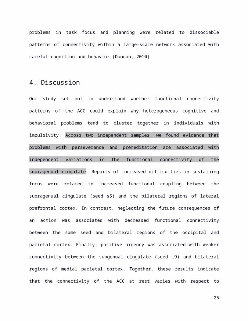
grey panel). Thus, different patterns of connectivity associated with problems in task focus and
planning were related to dissociable patterns of connectivity within a large-scale network
associated with careful cognition and behavior (Duncan, 2010).
4. Discussion
Our study set out to understand whether functional connectivity patterns of the ACC could
explain why heterogeneous cognitive and behavioral problems tend to cluster together in
individuals with impulsivity. Across two independent samples, we found evidence that problems
with perseverance and premeditation are associated with independent variations in the
functional connectivity of the supragenual cingulate. Reports of increased difficulties in
sustaining focus were related to increased functional coupling between the supragenual
cingulate (seed s5) and the bilateral regions of lateral prefrontal cortex. In contrast, neglecting
the future consequences of an action was associated with decreased functional connectivity
between the same seed and bilateral regions of the occipital and parietal cortex. Finally, positive
urgency was associated with weaker connectivity between the subgenual cingulate (seed i9)
and bilateral regions of medial parietal cortex. Together, these results indicate that the
connectivity of the ACC at rest varies with respect to different aspects of impulsivity, a pattern
that suggests that this region may be important in explaining why different constructs often co-
occur together in impulsive individuals.
Our data suggest that dysfunctions in a central region of the cingulate cortex are associated
with a variety of different aspects of the psychological features that are associated with
impulsivity. We found that a lack of perseverance relates to increased functional communication
17
between the rostral supragenual ACC and regions of the dorsolateral prefrontal cortex. Lateral
prefrontal regions have been implicated in impulsive behavior previously (Aron et al., 2003; Ding
et al., 2014; Weafer et al., 2015; Winstanley et al., 2006) and more generally in goal
maintenance and pursuit (Berkman et al., 2012). Since the supragenual ACC is implicated in
evaluative processes such as conflict monitoring (e.g. Botvinick et al., 2004), the observed
elevated connectivity between that region and dorsolateral prefrontal cortex might reflect an
overly excessive coupling between conflict monitoring and goal maintenance systems. This in
turn could result in a failure to sustain attention as individuals may switch between different goal
states. Problems in premeditation were associated with reduced connectivity between the same
rostral supragenual region of ACC and medial/lateral regions of the visual cortex, including
fusiform, medial and lateral occipito-temporal cortex. These regions not only play important
roles in extracting higher order information from sensory input (for a review see e.g. Grill-
Spector and Malach, 2004), but are also modulated by attention (e.g. Woldorff et al., 2004). For
instance, calculating effective connectivity, Crottaz-Herbette & Menon (2006) demonstrated
increased effects of the ACC on striate regions during a visual oddball task supporting the ACCs
role in attentional modulation during sensory processing. Since attentional control underlies the
capacity to follow up on goals despite distractions (cf. Aarts & Roelofs, 2011), it is possible that
the observed reductions in connectivity reflects a situation in which distracting environmental
information are insufficiently suppressed at the moment when an action is initiated. This would
support claims that impulsive behavior is initiated under conditions when sensory evidence has
been inadequately sampled (“reflection impulsivity”, Kagan, 1966; cf. Dalley et al., 2011;
Evenden, 1999), and could explain why impulsive individuals are often unduly influenced by
distractor processes (Yantis et al., 2012) making it difficult to keep track of specific goals.
Finally, we also found that positive urgency related to decreased functional connectivity
between the subgenual ACC and bilateral parietal regions such as retrosplenial cortex. These
regions fall within the so-called default mode network (DMN), a system that is known to be
18
important in envisioning the future (Schacter and Addis, 2007). Studies have suggested that
imagining future outcomes helps participants to make more effective long term decisions
(Peters and Büchel, 2010). One possible interpretation of the data associating the connectivity
of the subgenual ACC with retrosplenial cortex is that it describes an absence of effective
prospection that leads to the rash behavior that can characterize the trait of positive urgency.
Regardless of the precise psychological mechanisms through which the observed patterns of
connectivity contribute to different types of impulsivity, the fact that dissociable patterns of
functional connectivity associated with distinct aspects of impulsivity emerge from the same
region of cingulate cortex indicates that the rostral supragenual ACC is an important hub
regions whose functional connectivity provides important insights into the cognitive processes
that underpin impulsivity.
The observed patterns of connectivity also provide a possible explanation why impulsivity is
associated with problems in cognition and behavior across a wide range of situations. We found
that two types of impulsivity emerged from differences in cingulate connectivity with a distributed
set of regions across the cortex that are known to be important across a range of different task
conditions — the multiple demand network (MDN; Duncan, 2010). Since the MDN network is
defined through its importance in a wide range of different situations (Fedorenko et al., 2013),
our demonstration that its communication with a core hub in the cingulate cortex becomes
deregulated in impulsivity might explain why problems in cognition and behavior emerge across
a range of situations. Problems linked to impulsivity, such as the lacking ability to think ahead,
or to focus on a task, have a pervasive influence on behavior since they reflect dysfunctions in
communication between an important cortical hub and a neurocognitive system that support
thought, cognition and action in a domain general manner. Although our study highlights the
visual similarity between the MDN and the functional patterns associated with impulsivity, this
does not constitute a formal statistical link. Building on our observation, future work could
19
profitably explore the resting state functional connectivity patterns associated with impulsivity
and the neural activation that is common to many different difficult tasks, either in a large scale
study or through a formal meta-analysis using tools such as Neurosynth (Yarkoni et al., 2011).
Indeed, a recent meta-analysis has highlighted the viability of understanding complex traits like
impulsivity through their shared links to self-report and tasks (Sharma et al., 2014).
Limitations
The main focus of our study was to explore whether several personality facets related to
impulsive behavior can be explained by dissociable functional networks anchored along the
caudal-rostral gradient of the ACC. Although our results support this hypothesis the correlational
nature of the analyses performed limits the interpretational framework by making it impossible to
infer any causal or hierarchical relationship. From a mechanistic point of view, this leaves open
questions about the origin of inter-individual differences in those traits with respect to functional
connectivity. Along similar lines, we restricted our analyses to connectivity profiles of the
anterior cingulate cortex by using an a priori analytical approach, which, although appropriate
with respect to our circumscribed hypotheses, prevents the identification of any association
“outside” the ACC. It is important to note that while the ACC is important for impulsivity
(Castellanos-Ryan & Seguin, 2015) there is evidence that other cortical regions such as
ventromedial prefrontal cortex, orbitofrontal cortex, pre-supplementary motor area or
dorsolateral prefrontal cortex, but also midbrain (limbic, striatal) regions are similarly important
for understanding the various aspects of impulse control (see e.g., Li et al., 2013; Robbins et al.,
2012; Shannon et al., 2011). Indeed, studies suggest that the interaction between these two
systems is key to understanding the manifoldness of the construct (e.g. Dalley et al., 2011, cf.
Castellanos-Ryan & Seguin, 2015). Although this study might be the first examining different
impulsiveness-related personality traits in terms of their underlying intrinsic functional brain
organization using rs-fMRI in healthy adults, we encourage future studies to investigate those
20
traits by using more fine grained and data-driven methods that target the whole brain instead of
specific regions of interest. Lastly, although our analysis highlights the ACC as important in the
genesis of impulsive behavior, our analysis does not support a detailed mapping between the
cognitive processes that are characterized by our functional connectivity evidence. For example,
it is possible that a single cognitive process is represented in the s5 region of the ACC and that
the manner that this function expresses itself in its interactions with other regions of cortex
determines whether behavior is characterized as lacking perseverance or premeditation.
Alternatively, s5 may reflect the convergence of different types of cognitive and emotional
processing that are themselves represented in other cortical regions. In contrast to our lack of
detail at the psychological level, our data does allow us to make precise statements at the level
of functional connectivity because the s5 seed of the anterior cingulate cortex is able to
discriminate between different facets of impulsive behavior. Building on our observations that
the anterior cingulate, and in particular the s5 region, are important in different aspects of
impulsivity, we hope that future studies will be able to specify the psychological functions that
this region supports, and its role in impulsivity, with greater detail.
Conclusions and future directions
Using a large sample of participants recruited from two different countries, we found consistent
evidence that different types of impulsivity are reflected by different patterns of functional
coupling between regions in ACC with lateral and medial parietal cortex, as well as lateral
frontal cortex. The observed connectivity was topographically consistent with the distribution of
the MDN (Duncan, 2010) - a system important for the general control of thought and action. The
observed association with the MDN helps to explain why impulsive behavior tends to occur in
many different situations. Interestingly, we could differentiate forms of impulsivity based on the
functional coupling of the supragenual seed s5, a region on the boundary of the MDN, rather
than seed s3, a region within the network itself. This suggests that problems may be in the
21
integration between the MDN and other large scale systems. Both task disengagement
(Christoff et al., 2009; Mason et al., 2007) and future planning (Schacter and Addis, 2007) have
been linked to the DMN, and so it is possible that our findings reveal that the cognitive problems
associated with impulsivity emerge from dysfunctions in interactions between the MDN and
DMN. As seed s5 marks the transition between regions in the cingulate cortex coupled to the
DMN and those that are not, this could explain why this region of cortex seems especially
important in impulsivity. Based on these findings, we hypothesize that certain aspects of
impulsivity emerge from dysfunctions in the way the cingulate cortex controls interactions
between the DMN and MDN. This hypothesis could further be explored by assessing how the
integrity of the white and grey matter of these regions is linked both to traits of impulsivity and
their cognitive manifestations (poor planning and task disengagement). More generally, our
study provides evidence that several important aspects of impulsivity can be related to the
behavior of the anterior cingulate cortex at rest, an observation that means in the future
researchers may be able to use measures of functional connectivity from the anterior cingulate
as an additional outcome measure for evaluating drug and psychological interventions for
individuals with problems in impulsivity.
Acknowlegements. EJ was supported by grants from BBSRC (BB/J006963/1) and the European
Research Council (SEMBIND - 283530). FL was supported by the Swiss National Science
Foundation (P2ZHP1_155200). JS was supported by the European Research Council
(WANDERINGMINDS – 646927). This publication was also made possible through the support
of a grant from the John Templeton Foundation, “Prospective Psychology Stage 2: A Research
Competition” to Martin Seligman. The opinions expressed in this publication are those of the
author(s) and do not necessarily reflect the views of the John Templeton Foundation.
22
23

Figure Captions
Figure 1 Group functional connectivity maps for each of the five regions of the anterior cingulate
cortex (ACC)
The right panel illustrates the seed locations within the ACC as well as the exact coordinates
(below; Figure adapted from Margulies, 2007). The left panel presents a tabular overview of the
group functional connectivity maps for each seed separately for the two samples (MPI-S
dataset, NKI-RS dataset) used in this study.
Acronyms: MPI-S – Max Planck Institute Sample, NKI-RS – Nathan Kline Institute Rockland
Sample
Figure 2 Positive Urgency varies with functional connectivity of the subgenual region of the
anterior cingulate cortex (ACC)
The figure represents the cluster corrected maps visualizing the characteristics of the functional
connectivity profile of the seed region i9 as altered by positive urgency. Across two independent
datasets, functional connectivity between the ACC seed and regions within the bilateral parietal
cortex were negatively predicted by positive urgency. The scatterplot displays the averaged
functional connectivity values extracted from that cluster and plotted against the individual self-
report scores separately for each dataset. Spatial maps were thresholded using FSL’s
easythresh: Z = 3.1, FWE p < 0.005).
Figure 3 Lack of perseverance and lack of premeditation differently modulate functional
connectivity patterns of the most rostral supragenual region of the anterior cingulate cortex
The upper panel of the figure depicts the cluster corrected maps of the functional connectivity
profile of the supragenual seed region s5 that covaries with lack of perseverance: two significant
clusters within the bilateral dorsolateral prefrontal cortex (red) were derived by the analysis
24
across two independent datasets. The lower panel shows the corresponding results for lack of
premeditation: Two significant clusters within the bilateral occipital cortex (blue) were obtained
by this analysis. Spatial maps were thresholded using FSL’s easythresh: Z = 3.1, FWE p <
0.005).
Figure 4 Scatterplots demonstrating the linear relation between extracted functional connectivity
values and behavioral self-report scores.
The left panel depicts the two different connectivity profiles of supragenual seed region s5 as
modulated by lack of perseverance and lack of premeditation (cf. Figure 2). The scatterplots
show the averaged functional connectivity values extracted from these clusters plotted against
the self-report scores for each participant, separately for each dataset. In order to demonstrate
the independence of the results, we also plotted the opposite behavioral scores against the
cluster values (e.g. lack of premeditation scores against clusters derived by lack of
perseverance and vice versa).
Figure 5 Clusters associated with a lack of perseverance and premeditation overlap with the
multiple demand network
This figure illustrates the overlap of the the seed-based functional connectivity results for lack of
perseverance (red) and lack of premeditation (blue) and the pattern of the multiple demand
network (green; from Fedorenko et al., 2013) proposed by Duncan (2010). Overlaps in bilateral
dorsolateral prefrontal cortex as well as in bilateral occipital cortex are indicated in yellow and
light blue, respectively. These maps are fully saturated to maximize the visibility of spatial
overlap.
25
References
Aarts, E., Roelofs, A., 2011. Attentional control in anterior cingulate cortex based on
probabilistic cueing. J. Cogn. Neurosci. 23, 716–727. doi:10.1162/jocn.2010.21435
Arce, E., Santisteban, C., 2006. Impulsivity: A review. Psicothema 18, 213–220.
Aron, A.R., Fletcher, P.C., Bullmore, E.T., Sahakian, B.J., Robbins, T.W., 2003. Stop-signal
inhibition disrupted by damage to right inferior frontal gyrus in humans. Nat. Neurosci. 6,
115–116. doi:10.1038/nn1003
Asemi, A., Ramaseshan, K., Burgess, A., Diwadkar, V. a., Bressler, S.L., 2015. Dorsal anterior
cingulate cortex modulates supplementary motor area in coordinated unimanual motor
behavior. Front. Hum. Neurosci. 9, 1–10. doi:10.3389/fnhum.2015.00309
Avants, B.B., Tustison, N.J., Song, G., Cook, P.A., Klein, A., Gee, J.C., 2011. A reproducible
evaluation of ANTs similiarity metric performance in brain image registration. Neuroimage
54, 2033–2044. doi:10.1016/j.neuroimage.2010.09.025
Bazin, P.L., Weiss, M., Dinse, J., Schäfer, A., Trampel, R., Turner, R., 2014. A computational
framework for ultra-high resolution cortical segmentation at 7Tesla. Neuroimage 93, 201–
209. doi:10.1016/j.neuroimage.2013.03.077
Behzadi, Y., Restom, K., Liau, J., Liu, T.T., 2007. A component based noise correction method
(CompCor) for BOLD and perfusion based fMRI. Neuroimage 37, 90–101.
doi:10.1016/j.neuroimage.2007.04.042
Berkman, E.T., Falk, E.B., Lieberman, M.D., 2012. Interactive effects of three core goal pursuit
processes on brain control systems: Goal maintenance, performance monitoring, and
response inhibition. PLoS One 7, e40334. doi:10.1371/journal.pone.0040334
26
Bernhardt, B.C., Smallwood, J., Tusche,. A., Ruby, F.J., Engen, H.G., Steinbeis, N., Singer, T.,
2014. Medial prefrontal and anterior cingulate cortical thickness predicts shared individual
differences in self-generated thought and temporal discounting. Neuroimage 90, 290–297.
doi:10.1016/j.neuroimage.2013.12.040
Botvinick, M.M., Braver, T.S., Barch, D.M., Carter, C.S., Cohen, J.D., 2001. Conflict monitoring
and cognitive control. Psychol. Rev. 108, 624–652. doi:10.1037//0033-295X.I08.3.624
Botvinick, M.M., Cohen, J.D., Carter, C.S., 2004. Conflict monitoring and anterior cingulate
cortex: An update. Trends Cogn. Sci. 8, 539–546. doi:10.1016/j.tics.2004.10.003
Botvinick, M.M., Nystrom, L.E., Fissell, K., Carter, C.S., Cohen, J.D., 1999. Conflict monitoring
versus selection-for-action in anterior cingulate cortex. Nature 402, 179–181.
doi:10.1038/46035
Braver, T.S., Barch, D.M., Gray, J.R., Molfese, D.L., Snyder, A., 2001. Anterior cingulate cortex
and response conflict: effects of frequency, inhibition and errors. Cereb. Cortex 11, 825–
836. doi:10.1093/cercor/11.9.825
Brewer, J.A., Potenza, M.N., 2008. The neurobiology and genetics of impulse control disorders:
Relationships to drug addictions. Biochem. Pharmacol. 75, 63–75.
doi:10.1016/j.bcp.2007.06.043
Brown, S.M., Manuck, S.B., Flory, J.D., Hariri, A.R., 2006. Neural basis of individual differences
in impulsivity: contributions of corticolimbic circuits for behavioral arousal and control.
Emotion 6, 239–245. doi:10.1037/1528-3542.6.2.239
Bush, G., Luu, P., Posner, M., 2000. Cognitive and emotional influences in anterior cingulate
cortex. Trends Cogn. Sci. 4, 215–222. doi:10.1016/S1364-6613(00)01483-2
27
Casetellanos-Ryan, N., Seguin, J.R., 2015. Prefrontal and Anterior Cingulate Cortex
Mechanisms of Impulsivity, in: Beauchaine, T.P., Hinshaw, S.P. (Eds.), The Oxford
Handbook of Externalizing Spectrum Disorders. Oxford University Press, pp. 201–219.
doi:10.1093/oxfordhb/9780199324675.013.13
Chouinard, P.A., Paus, T., 2006. The primary motor and premotor areas of the human cerebral
cortex. Neurosci. 12, 143–152. doi:10.1177/1073858405284255
Christoff, K., Gordon, A.M., Smallwood, J., Smith, R., Schooler, J.W., 2009. Experience
sampling during fMRI reveals default network and executive system contributions to mind
wandering. Proc. Natl. Acad. Sci. U. S. A. 106, 8719–8724. doi:10.1073/pnas.0900234106
Comte, M., Schön, D., Coull, J.T., Reynaud, E., Khalfa, S., Belzeaux, R., Ibrahim, el C., Guedj,
E., Blin, O., Weinberger, D.R., Fakra, E., 2014. Dissociating Bottom-Up and Top-Down
Mechanisms in the Cortico-Limbic System during Emotion Processing. Cereb. Cortex 26,
144–155. doi:10.1093/cercor/bhu185
Cox, R.W., 1996. AFNI: software for analysis and visualization of functional magnetic resonance
neuroimages. Comput. Biomed. Res. 29, 162–173. doi:10.1006/cbmr.1996.0014
Crottaz-Herbette, S., Menon, V., 2006. Where and when the anterior cingulate cortex modulates
attentional response: combined fMRI and ERP evidence. J. Cogn. Neurosci. 18, 766–80.
doi:10.1162/jocn.2006.18.5.766
Cyders, M. A., Smith, G.T., 2007. Mood-based rash action and its components: Positive and
negative urgency. Pers. Individ. Dif. 43, 839–850. doi:10.1016/j.paid.2007.02.008
Dale, A.M., Fischl, B., Sereno, M.I., 1999. Cortical surface-based analysis. I. Segmentation and
surface reconstruction. Neuroimage 9, 179–194. doi:10.1006/nimg.1998.0395
28
Dalley, J.W., Everitt, B.J., Robbins, T.W., 2011. Impulsivity, Compulsivity, and Top-Down
Cognitive Control. Neuron 69, 680–694. doi:10.1016/j.neuron.2011.01.020
Daruna, J.H., Barnes, P.A., 1993. A neurodevelopmental view of impulsivity, in: McCown, W.G.,
Johnson, J.L., Shure, M.B. (Eds.), The impulsive Client: Theory, Research and Treatment.
American Psychological Association, Washington, D.C., pp. 23–27.
doi:http://dx.doi.org/10.1037/10500-000
Davis, F.C., Knodt, A.R., Sporns, O., Lahey, B.B., Zald, D.H., Brigidi, B.D., Hariri, A.R., 2013.
Impulsivity and the modular organization of resting-state neural networks. Cereb. Cortex
23, 1444–1452. doi:10.1093/cercor/bhs126
Devinski, O., Morrell, M.J., Vogt, B.A., 1995. Contributions of the anterior cingulate cortex to
behaviour. Brain 118, 279–306. doi:10.1093/brain/118.1.279
Ding, W-N., Sun, J-H., Sun, Y-W., Chen, X., Zhou, Y., Zhuang, Z-G., Li, L., Zhang, Y., Xu, J-R.,
Du, Y-S., 2014. Trait impulsivity and impaired prefrontal impulse inhibition function in
adolescents with internet gaming addiction revealed by a Go/No-Go fMRI study. Behav.
Brain Funct. 10, 20. doi:10.1186/1744-9081-10-20
Domes, G., Schulze, L., Böttger, M., Grossmann, A., Hauenstein, K., Wirtz, P.H., Heinrichs, M.,
Herpertz, S.C., 2010. The neural correlates of sex differences in the emotional reactivity
and emotion regulation. Hum. Brain Mapp. 31, 758–769. doi:10.1002/hbm.20903
Duncan, J., 2010. The multiple-demand (MD) system of the primate brain: mental programs for
intelligent behaviour. Trends Cogn. Sci. 14, 172–179. doi:10.1016/j.tics.2010.01.004
Economides, M., Guitart-Masip, M., Kurth-Nelson, Z., Dolan, R.J., 2014. Anterior Cingulate
Cortex Instigates Adaptive Switches in Choice by Integrating Immediate and Delayed
Components of Value in Ventromedial Prefrontal Cortex. J. Neurosci. 34, 3340–3349.
doi:10.1523/JNEUROSCI.4313-13.2014
29
Eklund, A., Nichols, T.E., Knutsson, H., 2016. Cluster failure: Why fMRI inferences for spatial
extent have inflated false-positive rates. Proc. Natl. Acad. Sci. U. S. A 113, 7900–7905.
doi:10.1073/pnas.1602413113
Etkin, A., Egner, T., Kalisch, R., 2011. Emotional processing in anterior cingulate and medial
prefrontal. Trends Cogn. Sci. 15, 85–93. doi:10.1016/j.tics.2010.11.004
Evenden, J.L., 1999. Varieties of impulsivity. Psychopharmacology (Berl). 146, 348–361.
doi:10.1007/PL00005481
Fedorenko, E., Duncan, J., Kanwisher, N., 2013. Broad domain generality in focal regions of
frontal and parietal cortex. Proc. Natl. Acad. Sci. U. S. A. 110, 16616–16621.
doi:10.1073/pnas.1315235110
Feinberg, D.A., Moeller, S., Smith, S.M., Auerbach, E., Ramanna, S., Gunther, M., Glasser,
M.F., Miller, K.L., Ugurbil, K., Yacoub, E., 2010. Multiplexed echo planar imaging for sub-
second whole brain FMRI and fast diffusion imaging. PLoS One 5, e15710.
doi:10.1371/journal.pone.0015710
Fischl, B., Sereno, M.I., Dale, A.M., 1999. Cortical surface-based analysis. II: Inflation,
flattening, and a surface-based coordinate system. Neuroimage 9, 195–207.
doi:10.1006/nimg.1998.0396
Friston, K.J., Williams, S., Howard, R., Frackowiak, R.S., Turner, R., 1996. Movement-related
effects in fMRI time-series. Magn. Reson. Med. 35, 346–355.
doi:10.1002/mrm.1910350312
Goldstein, R.Z., Tomasi, D., Rajaram, S., Cottone, L.A., Zhang, L., Maloney, T., Telang, F., Alia-
Klein, N., Volkow, N.D., 2007. Role of the anterior cingulate and medial orbitofrontal cortex
in preprocessing drug cues in cocaine addiction. Neuroscience 144, 1153–1159.
doi:10.1016/j.neuroscience.2006.11.024
30

Gorgolewski, K., Burns, C.D., Madison, C., Clark, D., Halchenko, Y.O., Waskom, M.L., Ghosh,
S.S., 2011. Nipype: a flexible, lightweight and extensible neuroimaging data processing
framework in python. Front. Neuroinform. 5, 13. doi:10.3389/fninf.2011.00013
Grill-Spector, K., Malach, R., 2004. The human visual cortex. Annu. Rev. Neurosci. 27, 649–
677. doi: 10.1146/annurev.neuro.27.070203.144220
Gröne, M., Dyck, M., Koush, Y., Bergert, S., Mathiak, K.A., Alawi, E.M., Elliott, M., Mathiak, K.,
2015. Upregulation of the rostral anterior cingulate cortex can alter the perception of
emotions: fMRI-based neurofeedback at 3 and 7 T. Brain Topogr. 28, 197-207.
doi:10.1007/s10548-014-0384-4
Hagmann, P., Cammoun, L., Gigandet, X., Meuli, R., Honey, C.J., Van Wedeen, J., Sporns, O.,
2008. Mapping the structural core of human cerebral cortex. PLoS Biol. 6, 1479–1493.
doi:10.1371/journal.pbio.0060159
Jenkinson, M., Beckmann, C.F., Behrens, T.E.J., Woolrich, M.W., Smith, S.M., 2012. Fsl.
Neuroimage 62, 782–790. doi:10.1016/j.neuroimage.2011.09.015
Kagan, J., 1966. Reflection-impulsivity: the generality and dynamics of conceptual tempo. J.
Abnorm. Psychol. 71, 17–24. doi:http://dx.doi.org/10.1037/h0022886
Kelly, A.M.C., Di Martino, A., Uddin, L.Q., Shehzad, Z., Gee, D.G., Reiss, P.T., Margulies, D.S.,
Castellanos, F.X., Milham, M.P., 2009. Development of anterior cingulate functional
connectivity from late childhood to early adulthood. Cereb. Cortex 19, 640–657.
doi:10.1093/cercor/bhn117
Lavin, C., Melis, C., Mikulan, E., Gelormini, C., Huepe, D., Ibañez, A., 2013. The anterior
cingulate cortex: an integrative hub for human socially-driven interactions. Front. Neurosci.
7, 64. doi:10.3389/fnins.2013.00064
31
Lenartowicz, A., McIntosh, A.R., 2005. The role of anterior cingulate cortex in working memory
is shaped by functional connectivity. J. Cogn. Neurosci. 17, 1026–1042.
doi:10.1162/0898929054475127
Li, N., Ma, N., Liu, Y., He, X.S., Sun, D.L., Fu, X.M., Zhang, X., Han, S., Zhang, D.R., 2013.
Resting-state functional connectivity predicts impulsivity in economic decision-making. J.
Neurosci 33, 4886-4895. doi:10.1523/JNEUROSCI.1342-12.2013
Margulies, D.S., Kelly, A.M.C., Uddin, L.Q., Biswal, B.B., Castellanos, F.X., Milham, M.P., 2007.
Mapping the functional connectivity of anterior cingulate cortex. Neuroimage 37, 579–588.
doi:10.1016/j.neuroimage.2007.05.019
Marques, J.P., Kober, T., Krueger, G., van der Zwaag, W., Van de Moortele, P.F., Gruetter, R.,
2010. MP2RAGE, a self bias-field corrected sequence for improved segmentation and T1-
mapping at high field. Neuroimage 49, 1271–1281. doi:10.1016/j.neuroimage.2009.10.002
Mason, M.F., Norton, M.I., Van Horn, J.D., Wegner, D.M., Grafton, S.T., Macrae, C.N., 2007.
Wandering minds: the default network and stimulus-independent thought. Science 315,
393–395. doi:10.1126/science.1131295
Moeller, F.G., Barratt, E.S., Dougherty, D.M., Schmitz, J.M., Swann, A.C., 2001. Psychiatric
aspects of impulsivity. Am. J. Psychiatry 158, 1783–1793.
doi:10.1176/appi.ajp.158.11.1783
Moeller, S., Yacoub, E., Olman, C.A., Auerbach, E., Strupp, J., Harel, N., Ugurbil, K., 2010.
Multiband multislice GE-EPI at 7 tesla, with 16-fold acceleration using partial parallel
imaging with application to high spatial and temporal whole-brain fMRI. Magn. Reson. Med.
63, 1144–1153. doi:10.1002/mrm.22361
Nooner K.B., Colcombe S.J., Tobe R.H., Mennes M., Benedict M.M., Moreno A.L., et al., 2012.
The NKI-rockland sample: a model for accelerating the pace of discovery science in
32
psychiatry. Front. Neurosci. 6,152. doi:10.3389/fnins.2012.00152
Ochsner, K.N., Gross, J.J., 2005. The cognitive control of emotion. Trends Cogn. Sci. 9, 242–
249. doi:10.1016/j.tics.2005.03.010
Peters, J., Büchel, C., 2010. Episodic future thinking reduces reward delay discounting through
an enhancement of prefrontal-mediotemporal interactions. Neuron 66, 138–148.
doi:10.1016/j.neuron.2010.03.026.
Power, J.D., Barnes, K.A., Snyder, A.Z., Schlaggar, B.L., Petersen, S.E., 2012. Spurious but
systematic correlations in functional connectivity MRI networks arise from subject motion.
Neuroimage 59, 2142–2154. doi:10.1016/j.neuroimage.2011.10.018
Robbins, T.W., 2007. Shifting and stopping: fronto-striatal substrates, neurochemical modulation
and clinical implications. Philos. Trans. R. Soc. Lond. B. Biol. Sci. 362, 917–932.
doi:10.1098/rstb.2007.2097
Robbins, T.W., Gillian, C.M., Smith, D.G., de Wit, S., Ersche, K.D., 2012. Neurocognitive
endophenotypes of impulsivity and compulsivity: towards dimensional psychiatry. Trends
Cogn. Sci. 16, 81–91. doi:10.1016/j.tics.2011.11.009
Rokem, A., Trumpis, M., Perez, F., 2009. Nitime: time-series analysis for neuroimaging data, in:
Varoquaux, G., van der Walt, S., Millman, J. (Eds), Proceedings of the 8th Python in
Science Conference (SciPy 2009). Pasadena, pp. 68-75.
Schacter, D.L., Addis, D.R., 2007. The cognitive neuroscience of constructive memory:
Remembering the past and imagine the future. Philos. Trans. R. Soc. Lond. B. Biol. Sci.
362, 773–786. doi:10.1098/rstb.2007.2087
Shannon, B.J., Raichle, M.E., Snyder, A.Z., Fair, D.A., Mills, K.L., Zhang, D., Bache, K.,
Calhoun, V.D., Nigg, J.T., Nagel, B.J., Stevens, A.A., Kiehl, K.A., 2011. Premotor functional
33

connectivity predicts impulsivity in juvenile offenders. Proc. Natl. Acad. Sci. U. S. A. 108,
11241–11245. doi:10.1073/pnas.1108241108
Sharma, L., Markon, K.E., Clark, L.A., 2014. Toward a theory of distinct types of “impulsive”
behaviors: A meta-analysis of self-report and behavioral measures. Psychol. Bull. 140,
374–408. doi:10.1037/a0034418
Vogt, B.A., 2005. Pain and emotion interactions in subregions of the cingulate gyrus. Nat. Rev.,
Neurosci 6, 533–544. doi:10.1038/nrn1704
Weafer, J., Dzemidzic, M., Eiler, W., Oberlin, B.G., Wang, Y., Kareken, D.A., 2015. Associations
between regional brain physiology and trait impulsivity, motor inhibition, and impaired
control over drinking. Psychiatry Res. 233, 81–87. doi:10.1016/j.pscychresns.2015.04.010
Whiteside, S.P., Lynam, D.R., 2001. The five factor model and impulsivity: Using a structural
model of personality to understand impulsivity. Pers. Individ. Dif. 30, 669–689.
doi:10.1016/S0191-8869(00)00064-7
Winstanley, C.A., Eagle, D.M., Robbins, T.W., 2006. Behavioral models of impulsivity in relation
to ADHD: Translation between clinical and preclinical studies. Clin. Psychol. Rev. 26, 379–
395. doi:10.1016/j.cpr.2006.01.001
Woldorff, M.G., Hazlett, C.J., Fichtenholtz, H.M., Weissman, D.H., Dale, A.M., Song, A.W.,
2004. Functional parcellation of attentional control regions of the brain. J. Cogn. Neurosci.
16, 149-165. doi:10.1162/089892904322755638
Woodward, T.S., Metzak, P.D., Meier, B., Holroyd, C.B., 2008. Anterior cingulate cortex signals
the requirement to break inertia when switching tasks: A study of the bivalency effect.
Neuroimage 40, 1311–1318. doi:10.1016/j.neuroimage.2007.12.049
34
Yantis, S., Anderson, B.A., Wampler, E.K., Laurent, P.A., 2012. Reward and attentional control
in visual search. Nebr. Symp. Motiv. 59, 91–116.
Yarkoni, T., Poldrack, R.A., Nichols, T.E., Van Essen, D.C., Wager, T.D., 2011. Large-scale
automated synthesis of human functional neuroimaging data. Nat. Methods 8, 665–670.
doi:10.1038/nmeth.1635.
35