Embed Size (px)
Citation preview

Accepted Manuscript
Elevated translocator protein in anterior cingulate in major depression and a role forinflammation in suicidal thinking: a PET study
Sophie E. Holmes, PhD, Rainer Hinz, PhD, Silke Conen, PhD, Catherine J. Gregory,BM BCh, Julian C. Matthews, PhD, Jose M. Anton-Rodriguez, MSc, AlexanderGerhard, MD, Peter S. Talbot, MD MRCPsych
PII: S0006-3223(17)31857-7
DOI: 10.1016/j.biopsych.2017.08.005
Reference: BPS 13291
To appear in: Biological Psychiatry
Received Date: 16 February 2017
Revised Date: 4 August 2017
Accepted Date: 6 August 2017
Please cite this article as: Holmes S.E, Hinz R., Conen S., Gregory C.J, Matthews J.C, Anton-RodriguezJ.M, Gerhard A. & Talbot P.S, Elevated translocator protein in anterior cingulate in major depressionand a role for inflammation in suicidal thinking: a PET study, Biological Psychiatry (2017), doi: 10.1016/j.biopsych.2017.08.005.
This is a PDF file of an unedited manuscript that has been accepted for publication. As a service toour customers we are providing this early version of the manuscript. The manuscript will undergocopyediting, typesetting, and review of the resulting proof before it is published in its final form. Pleasenote that during the production process errors may be discovered which could affect the content, and alllegal disclaimers that apply to the journal pertain.

MANUSCRIP
T
ACCEPTED
ACCEPTED MANUSCRIPTHolmes SE et al. PET imaging of translocator protein in major depression
Page 1
Title Page
Title
Elevated translocator protein in anterior cingulate in major depression and a role for
inflammation in suicidal thinking: a PET study
Short title
PET imaging of translocator protein in major depression
Authors
Sophie E Holmes, PhD 1,2; Rainer Hinz, PhD 3; Silke Conen, PhD 1; Catherine J Gregory, BM
BCh 1; Julian C Matthews, PhD 3; Jose M Anton-Rodriguez MSc 3; Alexander Gerhard, MD
1,4,5; Peter S Talbot, MD MRCPsych 1,6.
Affiliations
1 Division of Neuroscience and Experimental Psychology, School of Biological Sciences,
Faculty of Biology, Medicine and Health, University of Manchester, Manchester Academic
Health Science Centre, Manchester M13 9PL, UK
2 Current affiliation: Department of Psychiatry, School of Medicine, Yale University, New
Haven, CT, USA
3 Division of Informatics, Imaging & Data Sciences, School of Health Sciences, Faculty of
Biology, Medicine and Health, University of Manchester, University of Manchester,
Manchester Academic Health Science Centre, Manchester M13 9PL, UK
4 Salford Royal NHS Foundation Trust, Manchester Academic Health Science Centre,
Manchester M13 9PL, UK
5 Current affiliation: Department of Nuclear Medicine and Lehrstuhl für Geriatrie,
Universitätsklinikum Essen, Germany

MANUSCRIP
T
ACCEPTED
ACCEPTED MANUSCRIPTHolmes SE et al. PET imaging of translocator protein in major depression
Page 2
6 Greater Manchester Mental Health NHS Foundation Trust, Manchester Academic Health
Science Centre, Manchester M13 9PL, UK
Word count
Abstract: 233
Article: 4959
Tables: 2
Figures: 2
Supplementary Material: 1 (separate document)
Key words
Depression; inflammation; microglia; PET; suicide; anterior cingulate
Corresponding author
Peter S. Talbot MD, MRCPsych
Senior Lecturer in Molecular Neuroimaging
The University of Manchester
Wolfson Molecular Imaging Centre
27 Palatine Road, Withington
Manchester M20 3LJ
United Kingdom
Tel: +44 (0)161 275 0015
Fax: +44 (0)161 275 0003

MANUSCRIP
T
ACCEPTED
ACCEPTED MANUSCRIPTHolmes SE et al. PET imaging of translocator protein in major depression
Page 3
Abstract
Background: Major Depressive Disorder (MDD) is associated with raised peripheral
inflammatory markers. Mounting evidence also suggests that inflammation is involved in
suicidal behavior. However, the involvement of inflammation in the brains of depressed
individuals, and its association with suicidal ideation, needs further clarification. Translocator
protein (TSPO), which is upregulated in activated glia, predominantly microglia, can be
measured as an indication of neuroinflammation in-vivo using Positron Emission
Tomography (PET) and TSPO-specific radioligands.
Methods: We used [11C](R)-PK11195 PET to compare TSPO availability in anterior
cingulate cortex (ACC), prefrontal cortex (PFC) and insula between fourteen medication-free
patients in a major depressive episode (MDE) of at least moderate severity and thirteen
matched healthy controls. In a post-hoc analysis, we also compared TSPO availability
between patients with and without suicidal thoughts.
Results: Multivariate analysis of variance indicated significantly higher TSPO in patients
compared to controls (p=0.005). The elevation was of large effect size and significant in ACC
(p=0.022; Cohen’s d=0.95), with smaller, non-significant elevations in PFC (p=0.342;
Cohen’s d=0.38) and insula (p=0.466; Cohen’s d=0.29). TSPO was not elevated in patients
without suicidal thinking, but was significantly increased in those with suicidal thoughts
compared to those without, most robustly in ACC (p=0.008) and insula (p=0.023).
Conclusions: We confirm evidence for increased TSPO availability, suggestive of
predominantly microglial activation, in the ACC during a moderate to severe MDE. Our
findings provide further incentive for evaluating anti-inflammatory therapies in MDD.

MANUSCRIP
T
ACCEPTED
ACCEPTED MANUSCRIPTHolmes SE et al. PET imaging of translocator protein in major depression
Page 4
Introduction
Major Depressive Disorder (MDD) is one of the leading causes of disability worldwide (1, 2).
However, approximately one third of patients fail to respond to conventional antidepressants
(3) and there is a pressing need to develop more effective and better tolerated treatments. A
promising avenue of research for new treatment strategies is inflammation (4-7), based on
evidence that at least a subset of individuals with MDD have higher levels of peripheral pro-
inflammatory cytokines (8-11); a high prevalence of depression in inflammatory medical
disorders (12); an association of depression and its response to treatment with polymorphisms
in inflammatory cytokine genes (13); the development of depression in patients administered
therapeutic pro-inflammatory cytokines (14-16) and healthy volunteers given a peripheral
immune challenge (17, 18); the association of inflammation with certain risk factors for
depression (19-22); and evidence that inflammation may be associated with non-
responsiveness to antidepressants (23-25).
Peripheral inflammation can lead directly to an inflammatory response in the human brain
(26). In response to inflammation, the metabolism of tryptophan is diverted from the
production of serotonin (5-HT) to kynurenine (KYN), which is subsequently converted into
the neurotoxic quinolinic acid (QUIN) by activated microglia and infiltrating macrophages
and monocytes (27, 28). That this mechanism may be involved in neuroinflammation-
associated depression is supported by observations that activation of the KYN pathway is
essential for depressive like behavior in rats (29), and that KYN and QUIN are increased in
the cerebrospinal fluid of cancer patients who had undergone interferon (IFN)-alpha therapy,
which correlated with depressive symptoms (30). Consistent with this, postmortem studies
have found increased levels of QUIN in the anterior cingulate cortex (ACC) of depressed
individuals who had committed suicide (31), and microglial and astrocytic activation in the
ACC, thalamus and frontal cortex of depressed individuals (32-35). These studies have a
number of potentially confounding factors including antemortem use of antidepressants,

MANUSCRIP
T
ACCEPTED
ACCEPTED MANUSCRIPTHolmes SE et al. PET imaging of translocator protein in major depression
Page 5
which can have significant effects on inflammatory processes (36). A crucial question is
therefore whether there is inflammation in-vivo in the brains of medication-free individuals
currently experiencing a MDE.
One index of neuroinflammation can be measured in-vivo using PET and radioligands
specific for the 18kDa translocator protein (TSPO), a mitochondrial protein that is
upregulated in activated glial cells, predominantly microglia, in a range of pathological
conditions (37). To date, there have been two published PET studies investigating TSPO in
MDD in working-age adults. The first found unaltered TSPO in a mild-to-moderate MDE
(38). A second larger study found increased TSPO in medication-free patients in a moderate-
to-severe MDE (39), most prominently in the prefrontal cortex (PFC), ACC and insula. There
was, however, considerable overlap between patients and controls, with a subset of patients
exhibiting higher levels of TSPO. This is consistent with the studies of peripheral
inflammatory markers in depression, as well as a large survey showing CRP levels ≥5mg/L in
around 30% of depressed individuals (40). This subpopulation of depressed individuals
showing heightened inflammation may benefit from anti-inflammatory treatment strategies
(23, 41, 42).
Mounting evidence also suggests that neuroinflammation may be particularly pronounced in
suicidality (43). Robust increases have been found in interleukin-1β (IL-1β) and IL-6 in blood
and postmortem samples of patients with suicidal thinking compared with patients without
suicidal thinking and controls (44). Furthermore, a higher degree of suicidal ideation has been
associated with an increased inflammatory index, independent of severity of depressive
symptoms (45). Multiple postmortem studies have found evidence for inflammation in the
brains of suicide victims (46, 47), with specific evidence for activated microglia in the ACC
and PFC of depressed individuals who had committed suicide (32, 33), and significantly less
microglial activation in the dorsal raphe nucleus in non-suicidal depressed patients who had
died of other causes compared to suicidal depressed patients and controls (48). However, no

MANUSCRIP
T
ACCEPTED
ACCEPTED MANUSCRIPTHolmes SE et al. PET imaging of translocator protein in major depression
Page 6
study to date has investigated the association between neuroinflammation and suicidality in
depression in-vivo.
The aims of the current study were therefore to investigate brain TSPO availability in MDD
and to explore factors that might be associated with heightened inflammation. We used the
prototypical TSPO radioligand [11C](R)-PK11195 to measure brain TSPO availability in
patients with moderate to severe depression who were non-smoking, medically healthy and
antidepressant- naïve or antidepressant-free for at least 8 months. We hypothesized that TSPO
availability would be higher in ACC, PFC and insula in depressed individuals compared to
matched controls. These regions were chosen as they are the three regions hypothesised a
priori in the study by Setiawan et al (39) and found to have significantly elevated TSPO; due
to their role in mood regulation (49); and based on literature implicating the ACC in
particular in the association between inflammation and depression (17, 31-33, 50, 51).
Secondary aims were to explore associations between brain TSPO levels, symptom severity,
suicidal ideation, exercise levels, childhood adversity and peripheral markers of
inflammation.

MANUSCRIP
T
ACCEPTED
ACCEPTED MANUSCRIPTHolmes SE et al. PET imaging of translocator protein in major depression
Page 7
Methods and Materials
Participants
Nineteen patients with MDD were recruited from the Manchester region of the UK by self-
referral following placement of advertisements in mental health services, voluntary
organisations, doctors’ surgeries and online. Three patients were excluded before data
collection (two for not meeting criteria, one for possibility of pregnancy). Fourteen patients (7
males; mean ± SD age 31±12 yr) completed the study.
Diagnosis was confirmed using the Structured Clinical Interview for the DSM-IV (SCID-I)
(52). All patients were in a moderate to severe MDE (mean±SD MADRS score 31±4;
mean±SD HAM-D score 20±3) and had not taken antidepressants for at least eight months.
Seven of the fourteen MDD patients had taken antidepressants in the past and seven were
antidepressant-naïve (for details of past medication use see Supplementary Table S1). Patients
were sex- and age-matched (±5 yr) with 13 healthy controls (7 males; age 33±11 yr) recruited
as part of this and another recent study (53), and scanned using the same protocol. All
participants ranged in age from 18-55, were medically healthy based on clinical history,
physical examination, routine blood tests and negative urine toxicology, and were non-
smoking. For demographic and clinical characteristics, see Table 1. Additional measures
included body mass index (BMI); childhood adversity, measured by the Childhood Adversity
Questionnaire (54); physical exercise, measured by the Godin Leisure-Time Exercise
Questionnaire (55, 56); and the following markers of inflammation in plasma: TNF-α, IFN-γ,
IL-6, IL-8, IL-1β and CRP. Exclusion criteria for all participants included substance misuse in
the previous year, lifetime history of substance dependence, anti-inflammatory medications in
the previous month, another Axis I disorder, pregnancy, and history of neurological or
autoimmune disorder. The study was approved by the Greater Manchester East Research
Ethics Committee and the United Kingdom Administration of Radioactive Substances
Advisory Committee (ARSAC). All participants provided written informed consent.

MANUSCRIP
T
ACCEPTED
ACCEPTED MANUSCRIPTHolmes SE et al. PET imaging of translocator protein in major depression
Page 8
Image acquisition and analysis
The methodology for image acquisition and analysis was recently published (57). In
summary, following intravenous injection of [11C](R)-PK11195, emission data were acquired
for 60 minutes on a high-resolution research tomograph (HRRT; Siemens/CTI, Knoxville,
Tennessee). A T1-weighted MRI brain scan was also acquired to exclude significant
abnormality, for identification of regions of interest (ROIs), and Voxel Based Morphometry
(VBM) analysis. The hypothesized regions (ACC, PFC, insula) were identified using a
maximum probability brain atlas (58, 59) in which ACC and insula are individual ROIs. Our
PFC ROI is a composite of the following atlas ROIs: middle frontal, inferior frontal and
superior frontal gyri.
There is no reference region devoid of TSPO for PET studies as TSPO expression is
ubiquitous throughout the brain. An alternative is to use a tissue with relatively low TSPO
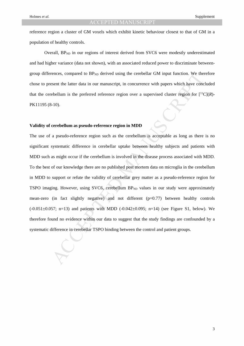
expression as a pseudo-reference region. To optimise our choice, we compared the use of two
pseudo-reference regions in our data: i) cerebellar grey matter (GM) (60); and ii) supervised
cluster reference input function (SVC6), a data-driven method which extracts a cluster of GM
voxels with kinetic behaviour closest to that of healthy GM (see Supplement for further
details). Binding potential (BPND), representing the ratio at equilibrium of specifically bound
radioligand to that of non-displaceable radioligand in tissue (61), was calculated using the
simplified reference tissue model (SRTM) (62) and the two pseudo-reference regions.
Parametric maps of BPND were generated with a basis-function implementation of the SRTM
(63) and the individualised GM brain atlases were then projected onto these parametric maps
to obtain mean BPND values for the ROIs. Overall, regional BPND values derived from SVC6
were modestly lower and had higher variance compared to using the cerebellar GM input
function (data not shown). We therefore present the latter data, in concurrence with the
superiority of the cerebellum over a data-driven approach in the literature (64) and our
previous [11C](R)-PK11195 studies on the HRRT (57, 65-68). TSPO availability in the
cerebellum (BPND derived by SVC6) did not significantly differ (p=0.77) in our data between

MANUSCRIP
T
ACCEPTED
ACCEPTED MANUSCRIPTHolmes SE et al. PET imaging of translocator protein in major depression
Page 9
healthy controls and MDD patients (see Supplementary text and Figure S1 and further
details).
Statistical analysis
Statistical analysis was performed in SPSS Statistics Version 22 (Armonk, NY: IBM Corp).
Independent-samples t-tests and univariate analysis of variance (ANOVA) were used to
assess differences between demographic, clinical and radiotracer characteristics across
groups. Sex differences were compared using Fisher's exact test (2-tailed). Group differences
in [11C](R)-PK11195 BPND were determined using a multivariate ANOVA (MANOVA), with
BPND in ACC, PFC and insula as the dependent variables, and group (MDD or healthy
controls) as the fixed independent variable. The effect size of the group differences in the
three ROIs was calculated using partial eta-squared (η2) and Cohen’s d (mean difference
divided by the pooled standard deviation). In a further exploratory analysis of potential effects
of suicidal ideation on TSPO availability, patients were stratified into those with and without
current suicidal thinking and a MANOVA performed with regional BPND (ACC, PFC and
insula) as dependent variables, and trichotomous group (healthy controls, MDD with suicidal
thoughts, MDD without suicidal thoughts) as the fixed independent variable, with Bonferroni
correction for multiple comparisons across the three groups.
The normal distribution of BPND for each combination of the variables was confirmed by
Shapiro-Wilk's test (p >0.05) and Normal Q-Q Plot. Equality of covariance was confirmed by
Box’s test. Homogeneity of variances was checked by Levine’s test. Correlations (Pearson's r,
2-tailed) were used to determine the association between TSPO availability and symptom
severity, childhood adversity, exercise and peripheral inflammatory markers in the patient
group. Comparison of these measurements with healthy controls was not performed as these
data were not collected for all the controls. Findings were considered significant at the p<0.05
level.

MANUSCRIP
T
ACCEPTED
ACCEPTED MANUSCRIPTHolmes SE et al. PET imaging of translocator protein in major depression
Page 10
Results
Patients and healthy controls were well matched for age, sex, BMI, smoking status (all non-
smoking) and injected mass of radiotracer (see Table 1). For all analyses, ANOVA
assumptions were not violated. There was no significant main effect of age on BPND
(MANOVA: F3, 22=0.85, p=0.479).
Across the hypothesized regions (ACC, PFC and insula) TSPO availability ([11C](R)-
PK11195 BPND) was higher in the MDD patients than the controls by a mean of 39%, which
was statistically significant (MANOVA, main effect of group: F3, 23=5.63, p=0.005). The
increase was highest in the ACC (67%), with smaller elevations seen in the PFC (29%) and
insula (24%). Univariate tests on the individual regions indicated that the elevation in the
ACC was of large effect size and statistically significant (F1, 25=5.99, p=0.022; partial
η2=0.193; Cohen’s d=0.95), but was of small effect size and failed to reach significance in
PFC (F1, 25=0.94, p=0.342; partial η2=0.036; Cohen’s d=0.38) or insula (F1, 25=0.549, p=0.466;
partial η2=0.021; Cohen’s d=0.29) (see Figure 1 and Table 2). The significance of these
differences was not materially altered if age was applied as a covariate (see Supplement).
In the exploratory analysis of the effects of suicidal ideation on TSPO availability, patients
were stratified into those with (n=9) and without (n=5) current suicidal thoughts. The
presence of suicidal thoughts was defined as the disclosure of suicidal thoughts during the
previous two weeks on direct enquiry and a score of 3 or higher on the ‘Suicidal Thoughts’
item of the MADRS. Their absence was defined as the denial of any suicidal thinking and a
score of zero on the MADRS item. There were no significant differences in age, sex, BMI or
injected mass of radiotracer between the two MDD subgroups. In addition, the two subgroups
were well matched for overall MDE severity on the MADRS and HAM-D, mean scores being
high in the moderate depression range for both subgroups (see Table 1). TSPO availability
differed significantly between the three groups (controls, patients with suicidal thinking, and
patients without suicidal thinking) across the three regions (MANOVA, main effect of group:

MANUSCRIP
T
ACCEPTED
ACCEPTED MANUSCRIPTHolmes SE et al. PET imaging of translocator protein in major depression
Page 11
F6, 46=4.22, p=0.002). Visual inspection (Figure 2) shows a pattern across all three regions
whereby mean BPND in patients without suicidal thinking is very similar to, or slightly lower
than, healthy controls; while BPND in patients with suicidal thinking is much higher than in
both other groups. Univariate tests on the individual regions show that these differences were
statistically significant and of large effect size in ACC (F2, 24=9.91, p=0.001; partial
η2=0.452) and insula (F2, 24=4.59, p=0.021; partial η2=0.277), and reached trend significance
in PFC (F2, 24=3.15, p=0.061; partial η2=0.208). Pairwise comparisons of BPND in each region
between the three groups, with Bonferroni adjustment for multiple comparisons, showed that
the patients with suicidal thinking had significantly higher TSPO availability than those
without suicidal thinking in ACC (+118%; p=0.008) and insula (+245%; p=0.023), and trend
higher TSPO availability in PFC (+129%; p=0.096). Patients with suicidal thinking also had
significantly higher TSPO availability than healthy controls in ACC (+107%; p=0.001) (see
Table 2 and Figure 2). Elevations compared to healthy controls in the PFC (+61%) and insula
(+66%) were not statistically significance (Table 2). Nor were there any significant
differences between healthy controls and patients without suicidal thinking in any of the
regions.
There were no significant correlations between BPND in any of the regions and symptom
severity (MADRS and HAM-D scores), duration of illness, BMI, childhood adversity, or any
of the peripheral inflammatory markers. Nor did we find any differences in concentration of
peripheral inflammatory markers between patients with and without suicidal thoughts. In the
MDD patients (n=14), a negative correlation between BPND in the ACC and their degree of
physical exercise reached trend significance (r=-0.47, p=0.07).
A post-hoc VBM analysis using SPM12 was carried out on the MRI scans to examine the
potential contribution of differences in GM volume between MDD patients and controls to
the significant between-group differences in BPND. For details of the VBM methodology, see
Supplement. No significant between-group GM volume differences were found, suggesting

MANUSCRIP
T
ACCEPTED
ACCEPTED MANUSCRIPTHolmes SE et al. PET imaging of translocator protein in major depression
Page 12
that the significant differences in BPND between groups are unlikely to be an artefact of
differences in regional tissue volumes.
A secondary between-group comparison (independent-samples t-test) on nine further ROIs is
presented in Supplementary Table S2 and Figure S2. TSPO was higher in MDD patients
compared to healthy controls in the posterior cingulate cortex (PCC; p=0.04). However, this
would not be considered significant after adjustment for multiple comparisons.
MANUSCRIP
T
ACCEPTED
ACCEPTED MANUSCRIPTHolmes SE et al. PET imaging of translocator protein in major depression
Page 13
Discussion
Our study provides the first confirmatory evidence, to the best of our knowledge, for elevated
TSPO in the ACC of drug-free, working-age adults with MDD in a moderate to severe MDE
in vivo, following the earlier report of Setiawan et al (39). It also provides the first in vivo
evidence in humans that elevated TSPO in MDD may be associated more with suicidality
than the diagnosis of MDD itself.
Under pathological conditions TSPO expression increases in microglia, infiltrating
macrophages, astrocytes, and vascular endothelial cells (69-72). However, TSPO ligand
binding appears to represent principally microglial activation in vivo (37, 73-75). We
therefore tentatively interpret our finding as support for the presence of microglial activation
in a moderate to severe MDE, while acknowledging the need for caution in interpreting
altered TSPO binding in mental disorders in the absence of more selective microglial PET
markers (72) and that we cannot exclude a contribution from other cell types.
Although our study design was independent of the Setiawan study, the patients in both studies
are comparable. All were antidepressant-free non-smokers with similar mean age, symptom
severity, and normal BMI. Our MDD group size was smaller (n=14 vs 20) while our patients
were drug-free for longer (>8 months vs >6 weeks). Given this clinical comparability, it is
interesting that our increases in TSPO across the ACC (67%), PFC (29%) and insula (24%)
(mean 39%) are similar to the increases of 32%, 26% and 33% (mean ~30%), respectively,
seen in the Setiawan study. We also found the most robust increase in the ACC. Although
TSPO was also elevated in the PFC and insula of MDD patients in our study, these group
differences were of small effect size, were not statistically significant, and we were unable to
replicate the findings of the Setiawan study in these regions. With [11C](R)-PK11195 the
signal to noise ratio is low, as reflected in the low BPND values, and this may contribute to
lack of statistical power. Post-hoc calculation based on the observed effect sizes (Cohen’s d)
of the between-group differences in our data indicates that group sizes of at least 100 would

MANUSCRIP
T
ACCEPTED
ACCEPTED MANUSCRIPTHolmes SE et al. PET imaging of translocator protein in major depression
Page 14
have been required to be fully powered to detect significant differences in PFC and insula (2-
tailed, α=0.05, power=0.8). This suggests that for these regions the combination of the size of
the biological effect, the variance in the data and the sensitivity of the methodology limit its
suitability for future studies in MDD in anything other than extremely large group sizes. For
the ACC, our sample size can detect a significant difference with a power of 0.7, suggesting
that our methodology is adequate for the ACC. However, in addition to reducing the chance
of detecting a true effect, low power also reduces the probability that a statistically significant
result reflects a true effect and increases the chance that the estimate of the effect size is
exaggerated (76). The modest group sizes and lack of statistical power in our study therefore
reduces the probability of our positive finding in the ACC and its effect size. On the other
hand, the probability that elevated TSPO in the ACC is a false positive is controlled by our
having limited our a priori hypothesis to the three regions which were themselves
hypothesised a priori in the initial study (39) based on their biological association with MDD,
and found to have significantly elevated TSPO. We would therefore have needed to be
particularly fortunate to have obtained this positive finding in the ACC. Nevertheless, further
replication studies will be important, ideally with group sizes even larger than the initial
study, to arrive at a more accurate estimation of the effect size in these regions (76).
We observed significantly greater TSPO in the ACC and insula of patients experiencing
suicidal thoughts than patients without suicidal thoughts. This is consistent with mounting
evidence for an association between inflammation and suicide (32, 33, 43, 46, 47, 77-79) and
a higher specificity of inflammation for suicide than for diagnosis (33, 47, 77). Ours is the
first study, to our knowledge, to show such an association in-vivo. However, because of the
small subgroup sizes and post-hoc nature of the analysis, our results are preliminary and
require replication. TSPO availability in the patients without suicidal thoughts was the same
as, or slightly lower than healthy controls (Figure 2). We cannot necessarily conclude that
neuroinflammation is absent in those with normal or lowered TSPO as increased levels of
inflammatory cytokines can occur with a downregulation (rather than upregulation) of TSPO

MANUSCRIP
T
ACCEPTED
ACCEPTED MANUSCRIPTHolmes SE et al. PET imaging of translocator protein in major depression
Page 15
(72). Nevertheless, the pattern of TSPO availability in our patients is consistent with recent
postmortem findings of significantly decreased microglial activation in non-suicidal
depressed patients compared to suicidal depressed patients and controls in the dorsal raphe
nucleus, which provides the major serotonergic innervation to the ACC, PFC and insula (48).
This is particularly interesting given recent evidence that abnormal 5-HT function measured
using PET predicts higher suicidal ideation and more lethal suicidal behavior (80). A
limitation for our study is that there was an overlap between patients experiencing suicidal
thoughts and those who had taken antidepressants in the past, raising the possibility that there
are other differences between the two MDD subgroups. Of the nine patients with suicidal
thinking, BPND was lower in each of the ROIs in the three patients who were antidepressant-
naïve than the six with prior medication use. The fact that these patients had been drug-free
for at least eight months makes a residual direct effect of antidepressants unlikely. However,
they reported stopping the antidepressants due to lack of efficacy, and there is some evidence
for an association between inflammation and non-responsiveness to antidepressants (23-25).
Although these are very small subgroups, we cannot exclude a potential role of treatment
resistance in the TSPO increase seen in our patients with suicidal thinking.
Our results contribute to an emerging view that glial, principally microglial, activation during
an MDE may be particularly prominent in the ACC. The ACC plays a key role in regulating
normal cognitive and emotional processing (81) and in the pathophysiology of MDD (82-87).
Postmortem studies find increased inflammatory markers in the ACC of depressed individuals
(31-33), and levels of systemic cytokines are associated with increased activation in the ACC
(17, 50, 51), suggesting that the ACC might be particularly sensitive to heightened peripheral
inflammation and be central to inflammation-induced changes in mood. The trend-significant
negative correlation between TSPO in the ACC and physical exercise in our data suggests
that a potential association between brain inflammation and exercise levels warrants further
investigation in a larger sample.

MANUSCRIP
T
ACCEPTED
ACCEPTED MANUSCRIPTHolmes SE et al. PET imaging of translocator protein in major depression
Page 16
We found no significant correlations between central TSPO and peripheral inflammatory
markers. The mechanisms of immune-to-brain communication remain to be fully elucidated.
However, this lack of correlation is consistent with previous PET studies in humans reporting
both central and peripheral measures in depression (39) and schizophrenia (57, 88), and
preclinical models involving experimental induction of both local and systemic peripheral
inflammation (89-92).
Our study has several additional limitations. Firstly, we used a pseudo-reference region
(cerebellum). Although the presence of some specific binding in the cerebellum will cause an
underestimation of the specific binding in the ROIs, this remains a reasonable approach as
long as there is no significant systematic difference in cerebellar TSPO availability between
healthy subjects and patients. To the best of our knowledge, there are no published
postmortem data on TSPO or microglia in the cerebellum in MDD so we cannot exclude the
possibility that a difference exists. Our finding of no difference in cerebellar BPND (SVC6)
between patients and controls provides some reassurance that the study findings are not
confounded by a systematic difference in cerebellar TSPO binding between controls and
patients. However, this reassurance is to a limited degree and cerebellar BPND is not as strong
as measurement of cerebellar total volume of distribution (VT) would have been using a
metabolite-corrected arterial input function. Secondly, a limitation common to all TSPO PET
studies is that microglia have a range of pro- and anti-inflammatory chemical phenotypes
including cytotoxic, repair and regeneration, and immunomodulatory (93), and at present PET
is unable to distinguish between these. However, given the postmortem studies implicating a
pro-inflammatory microglial phenotype in MDD and in suicide (31, 32), we propose that
increased TSPO binding in MDD represents a cytotoxic phenotype.
In conclusion, we have replicated the first PET findings of increased TSPO availability,
suggestive of microglial activation, in the ACC of medication-free patients in a MDE. Our
findings add support for the presence of a neuroinflammatory process in MDD and for TSPO
as a therapeutic target (71). Trials of anti-inflammatory agents in MDD have indicated that
MANUSCRIP
T
ACCEPTED
ACCEPTED MANUSCRIPTHolmes SE et al. PET imaging of translocator protein in major depression
Page 17
they might be most effective in a subset of individuals with heightened inflammation,
suggesting that a more targeted ‘personalised’ strategy might be a successful approach to
treating depression. It will therefore be important for future research to determine whether
patients with elevated TSPO would benefit from anti-inflammatory treatment. A potential
contribution of suicidality to the elevated TSPO in MDD warrants further research in
adequately powered studies.

MANUSCRIP
T
ACCEPTED
ACCEPTED MANUSCRIPTHolmes SE et al. PET imaging of translocator protein in major depression
Page 18
Acknowledgments
The authors acknowledge the contributions of operational staff at the Wolfson Molecular
Imaging Centre, including Elizabeth Barnett and Carrie-Anne Mellor for processing of blood
samples; Michael Green, Team Leader for Radiochemistry Production; PET radiographers
Mike Godfrey, Eleanor Duncan-Rouse and Gerrit Helms van der Vegte; and MR
Radiographers Amy Watkins and Barry Whitnall. Recruitment was supported by staff of the
National Institute for Health Research Clinical Research Network: Greater Manchester.
MANUSCRIP
T
ACCEPTED
ACCEPTED MANUSCRIPTHolmes SE et al. PET imaging of translocator protein in major depression
Page 19
Financial Disclosures
This work was supported by an Engineering and Physical Sciences Research Council
(EPSRC) studentship awarded to SEH. Financial support was provided by Professor Karl
Herholz and the University of Manchester’s Magnetic Resonance Imaging Facility (MRIF).
AG and RH have received funding from the European Union's Seventh Framework
Programme (FP7/2007-2013) under grant agreement number HEALTH-F2-2011-278850
(INMiND).

MANUSCRIP
T
ACCEPTED
ACCEPTED MANUSCRIPTHolmes SE et al. PET imaging of translocator protein in major depression
Page 20
Conflict of Interest
The authors report no biomedical financial interests or potential conflicts of interest.
MANUSCRIP
T
ACCEPTED
ACCEPTED MANUSCRIPTHolmes SE et al. PET imaging of translocator protein in major depression
Page 21
References
1. Vos T, Barber RM, Bell B, Bertozzi-Villa A, Biryukov S, Bolliger I, et al.
(2015): Global, regional, and national incidence, prevalence, and years
lived with disability for 301 acute and chronic diseases and injuries in
188 countries, 1990-2013: a systematic analysis for the Global Burden of
Disease Study 2013. Lancet. 386:743-800.
2. World Health Organization (2016): Depression fact Sheet. Available from:
http://www.who.int/mediacentre/factsheets/fs369/en/
3. Rush AJ, Trivedi MH, Wisniewski SR, Nierenberg AA, Stewart JW, Warden
D, et al. (2006): Acute and longer-term outcomes in depressed outpatients
requiring one or several treatment steps: a STAR D report. Am J
Psychiatry. 163:1905-1917.
4. Miller AH, Maletic V, Raison CL (2009): Inflammation and its discontents:
the role of cytokines in the pathophysiology of major depression. Biol
Psychiatry. 65:732-741.
5. Zunszain PA, Hepgul N, Pariante CM (2013): Inflammation and
depression. Curr Top Behav Neurosci. 14:135-151.
6. Pollak Y, Yirmiya R (2002): Cytokine-induced changes in mood and
behaviour: implications for 'depression due to a general medical
condition', immunotherapy and antidepressive treatment. Int J
Neuropsychopharmacol. 5:389-399.
7. Krishnadas R, Cavanagh J (2012): Depression: an inflammatory illness? J
Neurol Neurosurg Psychiatry. 83:495-502.
8. Zorrilla EP, Luborsky L, McKay JR, Rosenthal R, Houldin A, Tax A, et al.
(2001): The relationship of depression and stressors to immunological
assays: a meta-analytic review. Brain Behav Immun. 15:199-226.
9. Howren MB, Lamkin DM, Suls J (2009): Associations of depression with C-
reactive protein, IL-1, and IL-6: a meta-analysis. Psychosom Med. 71:171-
186.
10. Dowlati Y, Herrmann N, Swardfager W, Liu H, Sham L, Reim EK, et al.
(2010): A meta-analysis of cytokines in major depression. Biol Psychiatry.
67:446-457.
11. Haapakoski R, Mathieu J, Ebmeier KP, Alenius H, Kivimaki M (2015):
Cumulative meta-analysis of interleukins 6 and 1beta, tumour necrosis
factor alpha and C-reactive protein in patients with major depressive
disorder. Brain Behav Immun. 49:206-215.
12. Evans DL, Charney DS, Lewis L, Golden RN, Gorman JM, Krishnan KR, et al.
(2005): Mood disorders in the medically ill: scientific review and
recommendations. Biol Psychiatry. 58:175-189.
13. Bufalino C, Hepgul N, Aguglia E, Pariante CM (2013): The role of immune
genes in the association between depression and inflammation: a review
of recent clinical studies. Brain Behav Immun. 31:31-47.
14. Raison CL, Demetrashvili M, Capuron L, Miller AH (2005):
Neuropsychiatric adverse effects of interferon-alpha: recognition and
management. CNS Drugs. 19:105-123.
15. Capuron L, Gumnick JF, Musselman DL, Lawson DH, Reemsnyder A,
Nemeroff CB, et al. (2002): Neurobehavioral effects of interferon-alpha in

MANUSCRIP
T
ACCEPTED
ACCEPTED MANUSCRIPTHolmes SE et al. PET imaging of translocator protein in major depression
Page 22
cancer patients: phenomenology and paroxetine responsiveness of
symptom dimensions. Neuropsychopharmacology. 26:643-652.
16. Bonaccorso S, Marino V, Puzella A, Pasquini M, Biondi M, Artini M, et al.
(2002): Increased depressive ratings in patients with hepatitis C receiving
interferon-alpha-based immunotherapy are related to interferon-alpha-
induced changes in the serotonergic system. J Clin Psychopharmacol.
22:86-90.
17. Harrison NA, Brydon L, Walker C, Gray MA, Steptoe A, Critchley HD
(2009): Inflammation causes mood changes through alterations in
subgenual cingulate activity and mesolimbic connectivity. Biol Psychiatry.
66:407-414.
18. Reichenberg A, Yirmiya R, Schuld A, Kraus T, Haack M, Morag A, et al.
(2001): Cytokine-associated emotional and cognitive disturbances in
humans. Arch Gen Psychiatry. 58:445-452.
19. Pace TW, Mletzko TC, Alagbe O, Musselman DL, Nemeroff CB, Miller AH, et
al. (2006): Increased stress-induced inflammatory responses in male
patients with major depression and increased early life stress. Am J
Psychiatry. 163:1630-1633.
20. Cohen S, Janicki-Deverts D, Doyle WJ, Miller GE, Frank E, Rabin BS, et al.
(2012): Chronic stress, glucocorticoid receptor resistance, inflammation,
and disease risk. Proc Natl Acad Sci U S A. 109:5995-5999.
21. Ruiz-Nunez B, Pruimboom L, Dijck-Brouwer DA, Muskiet FA (2013):
Lifestyle and nutritional imbalances associated with Western diseases:
causes and consequences of chronic systemic low-grade inflammation in
an evolutionary context. J Nutr Biochem. 24:1183-1201.
22. Handschin C, Spiegelman BM (2008): The role of exercise and PGC1alpha
in inflammation and chronic disease. Nature. 454:463-469.
23. Raison CL, Rutherford RE, Woolwine BJ, Shuo C, Schettler P, Drake DF, et
al. (2013): A randomized controlled trial of the tumor necrosis factor
antagonist infliximab for treatment-resistant depression: the role of
baseline inflammatory biomarkers. JAMA Psychiatry. 70:31-41.
24. Eller T, Vasar V, Shlik J, Maron E (2008): Pro-inflammatory cytokines and
treatment response to escitalopram in major depressive disorder. Prog
Neuropsychopharmacol Biol Psychiatry. 32:445-450.
25. Cattaneo A, Gennarelli M, Uher R, Breen G, Farmer A, Aitchison KJ, et al.
(2013): Candidate genes expression profile associated with
antidepressants response in the GENDEP study: differentiating between
baseline 'predictors' and longitudinal 'targets'.
Neuropsychopharmacology. 38:377-385.
26. Sandiego CM, Gallezot JD, Pittman B, Nabulsi N, Lim K, Lin SF, et al.
(2015): Imaging robust microglial activation after lipopolysaccharide
administration in humans with PET. Proc Natl Acad Sci U S A. 112:12468-
12473.
27. Dantzer R, O'Connor JC, Lawson MA, Kelley KW (2011): Inflammation-
associated depression: from serotonin to kynurenine.
Psychoneuroendocrinology. 36:426-436.
28. Dantzer R (2017): Role of the kynurenine metabolism pathway in
inflammation-induced depression: preclinical approaches. Curr Top Behav
Neurosci. 31:117-138.

MANUSCRIP
T
ACCEPTED
ACCEPTED MANUSCRIPTHolmes SE et al. PET imaging of translocator protein in major depression
Page 23
29. Dantzer R (2016): Role of the kynurenine metabolism pathway in
inflammation-induced depression: preclinical approaches. Curr Top Behav
Neurosci. Epub ahead of print:DOI: 10.1007/7854_2016_1006.
30. Raison CL, Dantzer R, Kelley KW, Lawson MA, Woolwine BJ, Vogt G, et al.
(2010): CSF concentrations of brain tryptophan and kynurenines during
immune stimulation with IFN-alpha: relationship to CNS immune
responses and depression. Mol Psychiatry. 15:393-403.
31. Steiner J, Walter M, Gos T, Guillemin GJ, Bernstein HG, Sarnyai Z, et al.
(2011): Severe depression is associated with increased microglial
quinolinic acid in subregions of the anterior cingulate gyrus: evidence for
an immune-modulated glutamatergic neurotransmission? J
Neuroinflammation. 8:94.
32. Torres-Platas SG, Cruceanu C, Chen GG, Turecki G, Mechawar N (2014):
Evidence for increased microglial priming and macrophage recruitment
in the dorsal anterior cingulate white matter of depressed suicides. Brain
Behav Immun. 42:50-59.
33. Steiner J, Bielau H, Brisch R, Danos P, Ullrich O, Mawrin C, et al. (2008):
Immunological aspects in the neurobiology of suicide: elevated microglial
density in schizophrenia and depression is associated with suicide. J
Psychiatr Res. 42:151-157.
34. Rao JS, Harry GJ, Rapoport SI, Kim HW (2010): Increased excitotoxicity
and neuroinflammatory markers in postmortem frontal cortex from
bipolar disorder patients. Mol Psychiatry. 15:384-392.
35. Nagy C, Suderman M, Yang J, Szyf M, Mechawar N, Ernst C, et al. (2015):
Astrocytic abnormalities and global DNA methylation patterns in
depression and suicide. Mol Psychiatry. 20:320-328.
36. Hannestad J, DellaGioia N, Bloch M (2011): The effect of antidepressant
medication treatment on serum levels of inflammatory cytokines: a meta-
analysis. Neuropsychopharmacology. 36:2452-2459.
37. Liu GJ, Middleton RJ, Hatty CR, Kam WW, Chan R, Pham T, et al. (2014):
The 18kDa translocator protein, microglia and neuroinflammation. Brain
Pathol. 24:631-653.
38. Hannestad J, DellaGioia N, Gallezot JD, Lim K, Nabulsi N, Esterlis I, et al.
(2013): The neuroinflammation marker translocator protein is not
elevated in individuals with mild-to-moderate depression: a [11C]PBR28
PET study. Brain Behav Immun. 33:131-138.
39. Setiawan E, Wilson AA, Mizrahi R, Rusjan PM, Miler L, Rajkowska G, et al.
(2015): Role of translocator protein density, a marker of
neuroinflammation, in the brain during major depressive episodes. JAMA
Psychiatry. 72:268-275.
40. Rethorst CD, Bernstein I, Trivedi MH (2014): Inflammation, obesity, and
metabolic syndrome in depression: analysis of the 2009-2010 National
Health and Nutrition Examination Survey (NHANES). J Clin Psychiatry.
75:e1428-1432.
41. Raison CL, Miller AH (2013): Role of inflammation in depression:
implications for phenomenology, pathophysiology and treatment. Mod
Trends Pharmacopsychiatry. 28:33-48.
42. Rapaport MH, Nierenberg AA, Schettler PJ, Kinkead B, Cardoos A, Walker
R, et al. (2016): Inflammation as a predictive biomarker for response to

MANUSCRIP
T
ACCEPTED
ACCEPTED MANUSCRIPTHolmes SE et al. PET imaging of translocator protein in major depression
Page 24
omega-3 fatty acids in major depressive disorder: a proof-of-concept
study. Mol Psychiatry. 21:71-79.
43. Brundin L, Bryleva EY, Thirtamara Rajamani K (2017): Role of
Inflammation in Suicide: From Mechanisms to Treatment.
Neuropsychopharmacology. 42:271-283.
44. Black C, Miller BJ (2015): Meta-Analysis of Cytokines and Chemokines in
Suicidality: Distinguishing Suicidal Versus Nonsuicidal Patients. Biol
Psychiatry. 78:28-37.
45. O'Donovan A, Rush G, Hoatam G, Hughes BM, McCrohan A, Kelleher C, et
al. (2013): Suicidal ideation is associated with elevated inflammation in
patients with major depressive disorder. Depress Anxiety. 30:307-314.
46. Tonelli LH, Stiller J, Rujescu D, Giegling I, Schneider B, Maurer K, et al.
(2008): Elevated cytokine expression in the orbitofrontal cortex of
victims of suicide. Acta Psychiatr Scand. 117:198-206.
47. Pandey GN, Rizavi HS, Ren X, Fareed J, Hoppensteadt DA, Roberts RC, et al.
(2012): Proinflammatory cytokines in the prefrontal cortex of teenage
suicide victims. J Psychiatr Res. 46:57-63.
48. Brisch R, Steiner J, Mawrin C, Krzyżanowska M, Jankowski Z, Gos T
(2017): Microglia in the dorsal raphe nucleus plays a potential role in
both suicide facilitation and prevention in affective disorders. Eur Arch
Psychiatry Clin Neurosci. doi:10.1007/s00406-017-0774-1.
49. Goldin PR, McRae K, Ramel W, Gross JJ (2008): The neural bases of
emotion regulation: reappraisal and suppression of negative emotion. Biol
Psychiatry. 63:577-586.
50. Capuron L, Pagnoni G, Demetrashvili M, Woolwine BJ, Nemeroff CB, Berns
GS, et al. (2005): Anterior cingulate activation and error processing
during interferon-alpha treatment. Biol Psychiatry. 58:190-196.
51. Hannestad J, Subramanyam K, Dellagioia N, Planeta-Wilson B,
Weinzimmer D, Pittman B, et al. (2012): Glucose metabolism in the insula
and cingulate is affected by systemic inflammation in humans. J Nucl Med.
53:601-607.
52. First MB, Spitzer RL, Gibbon M, Williams JBW (2002): Structured Clinical
Interview for DSM-IV-TR Axis I Disorders, Research Version, Patient Edition.
(SCID-I/P). New York: Biometrics Research, New York State Psychiatric
Institute. November 2002.
53. Hunter HJ, Hinz R, Gerhard A, Talbot PS, Su Z, Holland G, et al. (2016):
Brain inflammation and psoriasis: a [11C]-(R)-PK11195 positron
emission tomography study. Br J Dermatol. 175:1082-1084.
54. Rosenman S, Rodgers B (2004): Childhood adversity in an Australian
population. Soc Psychiatry Psychiatr Epidemiol. 39:695-702.
55. Godin G, Shephard RJ (1985): A simple method to assess exercise
behavior in the community. Can J Appl Sport Sci. 10:141-146.
56. Godin G, Shephard RJ (1997): Godin leisure-time exercise questionnaire.
Med Sci Sports Exerc. 26 Suppl 6:S36-S38.
57. Holmes SE, Hinz R, Drake RJ, Gregory CJ, Conen S, Matthews JC, et al.
(2016): In vivo imaging of brain microglial activity in antipsychotic-free
and medicated schizophrenia: a [11C](R)-PK11195 positron emission
tomography study. Mol Psychiatry. 21:1672-1679.
MANUSCRIP
T
ACCEPTED
ACCEPTED MANUSCRIPTHolmes SE et al. PET imaging of translocator protein in major depression
Page 25
58. Gousias IS, Rueckert D, Heckemann RA, Dyet LE, Boardman JP, Edwards
AD, et al. (2008): Automatic segmentation of brain MRIs of 2-year-olds
into 83 regions of interest. Neuroimage. 40:672-684.
59. Hammers A, Allom R, Koepp MJ, Free SL, Myers R, Lemieux L, et al.
(2003): Three-dimensional maximum probability atlas of the human
brain, with particular reference to the temporal lobe. Hum Brain Mapp.
19:224-247.
60. Doble A, Malgouris C, Daniel M, Daniel N, Imbault F, Basbaum A, et al.
(1987): Labelling of peripheral-type benzodiazepine binding sites in
human brain with [3H]PK 11195: anatomical and subcellular distribution.
Brain Res Bull. 18:49-61.
61. Innis RB, Cunningham VJ, Delforge J, Fujita M, Gjedde A, Gunn RN, et al.
(2007): Consensus nomenclature for in vivo imaging of reversibly binding
radioligands. J Cereb Blood Flow Metab. 27:1533-1539.
62. Lammertsma AA, Hume SP (1996): Simplified reference tissue model for
PET receptor studies. Neuroimage. 4:153-158.
63. Gunn RN, Lammertsma AA, Hume SP, Cunningham VJ (1997): Parametric
imaging of ligand-receptor binding in PET using a simplified reference
region model. Neuroimage. 6:279-287.
64. Kropholler MA, Boellaard R, van Berckel BN, Schuitemaker A, Kloet RW,
Lubberink MJ, et al. (2007): Evaluation of reference regions for (R)-
[(11)C]PK11195 studies in Alzheimer's disease and mild cognitive
impairment. J Cereb Blood Flow Metab. 27:1965-1974.
65. Drake C, Boutin H, Jones MS, Denes A, McColl BW, Selvarajah JR, et al.
(2011): Brain inflammation is induced by co-morbidities and risk factors
for stroke. Brain Behav Immun. 25:1113-1122.
66. Hunter HJ, Hinz R, Gerhard A, Talbot PS, Su Z, Holland G, et al. (2016):
Brain inflammation and psoriasis: a [(11) C]-(R)-PK11195 positron
emission tomography study. Br J Dermatol. 175:1082-1084.
67. Su Z, Roncaroli F, Durrenberger PF, Coope DJ, Karabatsou K, Hinz R, et al.
(2015): The 18-kDa mitochondrial translocator protein in human
gliomas: an 11C-(R)PK11195 PET imaging and neuropathology study. J
Nucl Med. 56:512-517.
68. Su Z, Herholz K, Gerhard A, Roncaroli F, Du Plessis D, Jackson A, et al.
(2013): [11C]-(R)PK11195 tracer kinetics in the brain of glioma patients
and a comparison of two referencing approaches. Eur J Nucl Med Mol
Imaging. 40:1406-1419.
69. Chen MK, Guilarte TR (2008): Translocator protein 18 kDa (TSPO):
molecular sensor of brain injury and repair. Pharmacol Ther. 118:1-17.
70. Cosenza-Nashat M, Zhao ML, Suh HS, Morgan J, Natividad R, Morgello S, et
al. (2009): Expression of the translocator protein of 18 kDa by microglia,
macrophages and astrocytes based on immunohistochemical localization
in abnormal human brain. Neuropathol Appl Neurobiol. 35:306-328.
71. Rupprecht R, Papadopoulos V, Rammes G, Baghai TC, Fan J, Akula N, et al.
(2010): Translocator protein (18 kDa) (TSPO) as a therapeutic target for
neurological and psychiatric disorders. Nature Reviews Drug Discovery.
9:971-988.
72. Notter T, Coughlin JM, Gschwind T, Weber-Stadlbauer U, Wang Y, Kassiou
M, et al. (2017): Translational evaluation of translocator protein as a

MANUSCRIP
T
ACCEPTED
ACCEPTED MANUSCRIPTHolmes SE et al. PET imaging of translocator protein in major depression
Page 26
marker of neuroinflammation in schizophrenia. Mol Psychiatry. doi:
10.1038/mp.2016.248.
73. Venneti S, Lopresti BJ, Wiley CA (2006): The peripheral benzodiazepine
receptor (Translocator protein 18kDa) in microglia: from pathology to
imaging. Prog Neurobiol. 80:308-322.
74. Venneti S, Lopresti BJ, Wang G, Bissel SJ, Mathis CA, Meltzer CC, et al.
(2004): PET imaging of brain macrophages using the peripheral
benzodiazepine receptor in a macaque model of neuroAIDS. J Clin Invest.
113:981-989.
75. Mankowski JL, Queen SE, Tarwater PJ, Adams RJ, Guilarte TR (2003):
Elevated peripheral benzodiazepine receptor expression in simian
immunodeficiency virus encephalitis. J Neurovirol. 9:94-100.
76. Button KS, Ioannidis JP, Mokrysz C, Nosek BA, Flint J, Robinson ES, et al.
(2013): Power failure: why small sample size undermines the reliability
of neuroscience. Nat Rev Neurosci. 14:365-376.
77. Schnieder TP, Trencevska I, Rosoklija G, Stankov A, Mann JJ, Smiley J, et al.
(2014): Microglia of prefrontal white matter in suicide. J Neuropathol Exp
Neurol. 73:880-890.
78. Batty GD, Bell S, Stamatakis E, Kivimaki M (2016): Association of systemic
inflammation with risk of completed suicide in the general population.
JAMA Psychiatry. 73:993-995.
79. Lund-Sorensen H, Benros ME, Madsen T, Sorensen HJ, Eaton WW,
Postolache TT, et al. (2016): A nationwide cohort study of the association
between hospitalization with infection and risk of death by suicide. JAMA
Psychiatry. 73:912-919.
80. Oquendo MA, Galfalvy H, Sullivan GM, Miller JM, Milak MM, Sublette ME, et
al. (2016): Positron emission tomographic imaging of the serotonergic
system and prediction of risk and lethality of future suicidal behavior.
JAMA Psychiatry. 73:1048-1055.
81. Talbot PS, Cooper SJ (2006): Anterior cingulate and subgenual prefrontal
blood flow changes following tryptophan depletion in healthy males.
Neuropsychopharmacology. 31:1757-1767.
82. Bora E, Fornito A, Pantelis C, Yucel M (2012): Gray matter abnormalities
in Major Depressive Disorder: a meta-analysis of voxel based
morphometry studies. J Affect Disord. 138:9-18.
83. Anand A, Li Y, Wang Y, Wu J, Gao S, Bukhari L, et al. (2005): Activity and
connectivity of brain mood regulating circuit in depression: a functional
magnetic resonance study. Biol Psychiatry. 57:1079-1088.
84. Drevets WC (2001): Neuroimaging and neuropathological studies of
depression: implications for the cognitive-emotional features of mood
disorders. Curr Opin Neurobiol. 11:240-249.
85. Schlosser RG, Wagner G, Koch K, Dahnke R, Reichenbach JR, Sauer H
(2008): Fronto-cingulate effective connectivity in major depression: a
study with fMRI and dynamic causal modeling. Neuroimage. 43:645-655.
86. Lozano AM, Mayberg HS, Giacobbe P, Hamani C, Craddock RC, Kennedy SH
(2008): Subcallosal cingulate gyrus deep brain stimulation for treatment-
resistant depression. Biol Psychiatry. 64:461-467.
MANUSCRIP
T
ACCEPTED
ACCEPTED MANUSCRIPTHolmes SE et al. PET imaging of translocator protein in major depression
Page 27
87. Mayberg HS, Lozano AM, Voon V, McNeely HE, Seminowicz D, Hamani C,
et al. (2005): Deep brain stimulation for treatment-resistant depression.
Neuron. 45:651-660.
88. Coughlin JM, Wang Y, Ambinder EB, Ward RE, Minn I, Vranesic M, et al.
(2016): In vivo markers of inflammatory response in recent-onset
schizophrenia: a combined study using [(11)C]DPA-713 PET and analysis
of CSF and plasma. Transl Psychiatry Psychiatry. 6:e777.
89. Bay-Richter C, Janelidze S, Hallberg L, Brundin L (2011): Changes in
behaviour and cytokine expression upon a peripheral immune challenge.
Behav Brain Res. 222:193-199.
90. Qin L, Wu X, Block ML, Liu Y, Breese GR, Hong JS, et al. (2007): Systemic
LPS causes chronic neuroinflammation and progressive
neurodegeneration. Glia. 55:453-462.
91. Thomson CA, McColl A, Cavanagh J, Graham GJ (2014): Peripheral
inflammation is associated with remote global gene expression changes in
the brain. J Neuroinflammation. 11:73.
92. McColl A, Thomson CA, Nerurkar L, Graham GJ, Cavanagh J (2016): TLR7-
mediated skin inflammation remotely triggers chemokine expression and
leukocyte accumulation in the brain. J Neuroinflammation. 13:102.
93. Perry VH, Nicoll JA, Holmes C (2010): Microglia in neurodegenerative
disease. Nat Rev Neurol. 6:193-201.

MANUSCRIP
T
ACCEPTED
ACCEPTED MANUSCRIPTHolmes SE et al. PET imaging of translocator protein in major depression
Page 28
Figure legends:
Figure 1: Regional mean [11C](R)-PK11195 BPND in MDD patients and controls, showing
statistically significant elevations in ACC but not PFC or insula. BPND, binding potential;
ACC, anterior cingulate cortex; PFC, prefrontal cortex. * indicates significant at p<0.05.
Figure 2: Regional [11C](R)-PK11195 BPND in controls, MDD patients with suicidal thoughts
and MDD patients without suicidal thoughts. Horizontal bars indicate means. Open circles
represent controls (n=16), closed triangles represent MDD patients with suicidal thoughts
(n=9) and closed circles represent MDD patients without suicidal thoughts (n=6). * indicates
significant at p<0.05.
MANUSCRIP
T
ACCEPTED
ACCEPTED MANUSCRIPTHolmes SE et al. PET imaging of translocator protein in major depression
Page 29
Tables
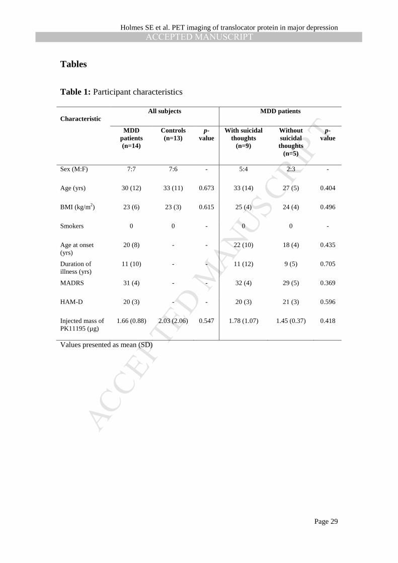
Table 1: Participant characteristics
Characteristic
All subjects MDD patients
MDD patients (n=14)
Controls (n=13)
p-value
With suicidal thoughts
(n=9)
Without suicidal thoughts
(n=5)
p-value
Sex (M:F) 7:7 7:6 - 5:4 2:3 -
Age (yrs) 30 (12) 33 (11) 0.673 33 (14) 27 (5) 0.404
BMI (kg/m2) 23 (6) 23 (3) 0.615 25 (4) 24 (4) 0.496
Smokers 0 0 - 0 0 -
Age at onset (yrs)
20 (8) - - 22 (10) 18 (4) 0.435
Duration of illness (yrs)
11 (10) - - 11 (12) 9 (5) 0.705
MADRS 31 (4) - - 32 (4) 29 (5) 0.369
HAM-D 20 (3) - - 20 (3) 21 (3) 0.596
Injected mass of PK11195 (µg)
1.66 (0.88) 2.03 (2.06) 0.547 1.78 (1.07) 1.45 (0.37) 0.418
Values presented as mean (SD)

MANUSCRIP
T
ACCEPTED
ACCEPTED MANUSCRIPT
Holmes SE et al. PET imaging of translocator protein in major depression
Page 30
Table 2: Regional TSPO availability ([11C](R)-PK11195 BPND) in MDD patients, healthy controls, and MDD patients stratified by
presence or absence of suicidal thinking
Region
MDD
patients (n=14)
Healthy controls (n=13)
MDD patients vs healthy controls
MDD without
suicidal thoughts
(n=5)
MDD with
suicidal thoughts
(n=9)
MDD with suicidal thoughts
vs healthy controls
MDD with vs without
suicidal thoughts
Difference
(%) Signif
(p)
Difference (%)
Signif (p)
Difference (%)
Signif (p)
ACC 0.162 (0.077) 0.097 (0.059) 67% 0.022* 0.092 (0.060) 0.201 (0.055) 107% 0.001* 118% 0.008*
PFC 0.116 (0.085) 0.090 (0.048) 29% 0.342 0.064 (0.074) 0.145 (0.079) 61% 0.179 129% 0.096
Insula 0.131 (0.103) 0.106 (0.068) 24% 0.466 0.051 (0.091) 0.176 (0.083) 66% 0.143 245% 0.023*
Values presented as mean (SD). ACC, anterior cingulate cortex; PFC, prefrontal cortex; BPND, binding potential; MDD, major depressive disorder. *indicates
significant at p<0.0

MANUSCRIP
T
ACCEPTED
ACCEPTED MANUSCRIPT

MANUSCRIP
T
ACCEPTED
ACCEPTED MANUSCRIPT

MANUSCRIP
T
ACCEPTED
ACCEPTED MANUSCRIPTHolmes et al. Supplement
1
Elevated Translocator Protein in Anterior Cingulate in Major Depression and a Role for Inflammation in Suicidal Thinking:
A Positron Emission Tomography Study
Supplemental Information
Past antidepressant use in MDD patients
Table S1. Details of previous antidepressants for each patient and number of months since last use, where applicable.
Patient Previous antidepressants Months without antidepressant
1 Citalopram 48 2 Citalopram 12 3 - - 4 - - 5 Sertraline, citalopram,
fluoxetine, venlafaxine 12
6 - - 7 Fluoxetine 60 8 - - 9 Fluoxetine 8 10 Amitriptyline, mirtazapine 25 11 Reboxetine, sertraline,
paroxetine 24
12 - - 13 - - 14 - -
Methodological considerations
[11C](R)-PK11195 was chosen in this study because, unlike [11C]PBR-28, [18F]DPA-714 and other
second generation TSPO radioligands, its differences in binding affinity in humans due to the
polymorphism rs6971 are negligible (1). This allowed us to include all eligible participants in this
study regardless of binding affinity, including the approximately 10% of the population who are low
affinity binders and therefore excluded from studies using second generation tracers (2).
We decided not to use an arterial input function due to the likelihood that the requirement for
arterial cannulation would further limit recruitment of an already difficult to recruit clinical population
(drug-free patients with major depression of at least moderate severity).

MANUSCRIP
T
ACCEPTED
ACCEPTED MANUSCRIPTHolmes et al. Supplement
2
Therefore the quantification of regional [11C](R)-PK11195 binding in the brain had to use a
reference tissue input function. This brought in further requirements such as that the reference input is
not affected by the disease, that its displaceable binding is insignificant relative to that in the target
area and that of homogeneity of the non-displaceable binding across the brain. However, at the same
time, results obtained from reference tissue analyses have proven to be more robust than those from
plasma input function kinetic models in cases where it had been difficult to get reliable measurements
of the fractions of unmetabolised tracer in plasma or of the plasma free fraction (3).
As TSPO expression is ubiquitous throughout the brain, there is no ideal reference region for PET
studies assessing microglial activation with TSPO radioligands. An alternative is to use a pseudo-
reference region, and our methodology used cerebellar grey matter (GM). Labelling of TSPO in post
mortem human brain with [3H]PK11195 found for the cerebellar cortex binding densities of 660 ± 85
fmol/mg protein in the granular cell layer, 191 ± 55 fmol/mg protein in the molecular cell layer and 41
± 32 fmol/mg protein in white matter (4). For comparison, the highest binding densities were found in
the dorsomedial thalamic nucleus (1912 ± 412 fmol/mg protein) and in inferior olivary nucleus of the
medulla (1655 ± 355 fmol/mg protein). This amount of specific binding in the cerebellum causes an
underestimation of the specific binding in the target regions of the brain, if a cerebellar input function
is chosen for a reference tissue model.
Therefore, data driven approaches have been developed to extract the reference tissue kinetics
from dynamic brain scans with [11C](R)-PK11195 on the voxel level (5-7). These methods do not rely
on an anatomically delineated region of interest for the definition of the reference region. Instead, they
group voxels together based on their similarity between the voxel time-activity curves.
Supervised cluster analysis (SVC6)
We therefore also analysed our data using the alternative approach of supervised cluster reference
input function (SVC6), a data-driven modelling method which segments voxels in the raw dynamic
data into six pre-defined tissue classes (normal grey and white matter, blood pool, muscle, skull and
pathological tissue with high TSPO density) based on their time activity curves, then extracts as a
MANUSCRIP
T
ACCEPTED
ACCEPTED MANUSCRIPTHolmes et al. Supplement
3
reference region a cluster of GM voxels which exhibit kinetic behaviour closest to that of GM in a
population of healthy controls.
Overall, BPND in our regions of interest derived from SVC6 were modestly underestimated
and had higher variance (data not shown), with an associated reduced power to discriminate between-
group differences, compared to BPND derived using the cerebellar GM input function. We therefore
chose to present the latter data in our manuscript, in concurrence with papers which have concluded
that the cerebellum is the preferred reference region over a supervised cluster region for [11C](R)-
PK11195 (8-10).
Validity of cerebellum as pseudo-reference region in MDD
The use of a pseudo-reference region such as the cerebellum is acceptable as long as there is no
significant systematic difference in cerebellar uptake between healthy subjects and patients with
MDD such as might occur if the cerebellum is involved in the disease process associated with MDD.
To the best of our knowledge there are no published post mortem data on microglia in the cerebellum
in MDD to support or refute the validity of cerebellar grey matter as a pseudo-reference region for
TSPO imaging. However, using SVC6, cerebellum BPND values in our study were approximately
mean-zero (in fact slightly negative) and not different (p=0.77) between healthy controls
(-0.051±0.057; n=13) and patients with MDD (-0.042±0.095; n=14) (see Figure S1, below). We
therefore found no evidence within our data to suggest that the study findings are confounded by a
systematic difference in cerebellar TSPO binding between the control and patient groups.

MANUSCRIP
T
ACCEPTED
ACCEPTED MANUSCRIPTHolmes et al. Supplement
4
Figure S1. Cerebellum BPND values in healthy controls and MDD patients.
Effect of age on group comparison
Because there was no significant main effect of age on BPND, the main analysis did not include age as
a covariate. However, for the sake of comparison we performed a secondary analysis with BPND in
ACC, PFC and insula as the dependent variables, group (MDD or healthy controls) as the fixed
independent variable, and introducing age as a covariate. This did not materially alter the statistical
significances of the main effect of group (F3, 22=5.40, p=0.006) or the between group differences: the
elevation in the ACC remained of large effect size and statistically significant (F1, 24=6.77, p=0.016;
partial h2=0.220; Cohen’s d=0.95), and of small effect size and not statistically significant in PFC (F1,
24=1.28, p=0.268; partial h2=0.051; Cohen’s d=0.38) and insula (F1, 24=0.727, p=0.402; partial
h2=0.029; Cohen’s d=0.29).
Voxel-based morphometry
A post-hoc voxel-based morphometry (VBM) analysis was carried out to examine possible
differences in grey matter volume between MDD patients and controls. Image pre-processing was
conducted using SPM12 (Statistical Parametric Mapping, Wellcome Department of Imaging
Neuroscience, Institute of Neurology, London, UK) running in MATLAB R2015a (Mathworks,
MANUSCRIP
T
ACCEPTED
ACCEPTED MANUSCRIPTHolmes et al. Supplement
5
Natick, Massachusetts). After realignment, the structural T1-weighted images were segmented into
grey matter (GM), white matter (WM) and cerebrospinal fluid (CSF). A template was created and the
deformations that best aligned the images were estimated using DARTEL (diffeomorphic
registration). Then, spatially normalised and smoothed Jacobian scaled GM images were generated
using the deformation images calculated in the previous step. For each volunteer GM tissue volumes
were calculated. Finally, a GM analysis mask (thresholded at a belonging probability >0.2) was
created in order to avoid instabilities that might occur in the analysis if the background is included.
After pre-processing, a voxel-wise two-sample t-test was run in SPM12 comparing the smoothed,
modulated, normalised, grey-matter images of our two groups. In this analysis we used the previously
created GM analysis mask for explicit masking and the previously calculated tissue volumes for
global calculation. Clusters of voxels were considered significant at a cluster-size threshold of
pFWEc<0.05 and a height-threshold of p<0.001 (uncorrected).
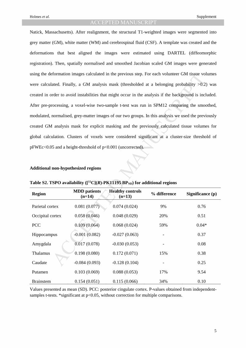
Additional non-hypothesized regions
Table S2. TSPO availability ([11C](R)-PK11195 BPND) for additional regions
Region MDD patients (n=14)
Healthy controls (n=13) % difference Significance (p)
Parietal cortex 0.081 (0.077) 0.074 (0.024) 9% 0.76
Occipital cortex 0.058 (0.046) 0.048 (0.029) 20% 0.51
PCC 0.109 (0.064) 0.068 (0.024) 59% 0.04*
Hippocampus -0.001 (0.082) -0.027 (0.063) - 0.37
Amygdala 0.017 (0.078) -0.030 (0.053) - 0.08
Thalamus 0.198 (0.080) 0.172 (0.071) 15% 0.38
Caudate -0.084 (0.093) -0.128 (0.104) - 0.25
Putamen 0.103 (0.069) 0.088 (0.053) 17% 9.54
Brainstem 0.154 (0.051) 0.115 (0.066) 34% 0.10
Values presented as mean (SD). PCC: posterior cingulate cortex. P-values obtained from independent-samples t-tests. *significant at p<0.05, without correction for multiple comparisons.

MANUSCRIP
T
ACCEPTED
ACCEPTED MANUSCRIPTHolmes et al. Supplement
6
Figure S2. TSPO availability ([11C](R)-PK11195 BPND) for additional regions

MANUSCRIP
T
ACCEPTED
ACCEPTED MANUSCRIPTHolmes et al. Supplement
7
Supplementary References
1. Owen DR, Yeo AJ, Gunn RN, Song K, Wadsworth G, Lewis A, et al. (2012): An 18-kDa translocator protein (TSPO) polymorphism explains differences in binding affinity of the PET radioligand PBR28. J Cereb Blood Flow Metab. 32:1-5.
2. Kreisl WC, Fujita M, Fujimura Y, Kimura N, Jenko KJ, Kannan P, et al. (2010): Comparison of [(11)C]-(R)-PK 11195 and [(11)C]PBR28, two radioligands for translocator protein (18 kDa) in human and monkey: Implications for positron emission tomographic imaging of this inflammation biomarker. Neuroimage. 49:2924-2932.
3. Slifstein M, Laruelle M (2001): Models and methods for derivation of in vivo neuroreceptor parameters with PET and SPECT reversible radiotracers. Nucl Med Biol. 28:595-608.
4. Doble A, Malgouris C, Daniel M, Daniel N, Imbault F, Basbaum A, et al. (1987): Labelling of peripheral-type benzodiazepine binding sites in human brain with [3H]PK 11195: anatomical and subcellular distribution. Brain Res Bull. 18:49-61.
5. Banati RB, Newcombe J, Gunn RN, Cagnin A, Turkheimer F, Heppner F, et al. (2000): The peripheral benzodiazepine binding site in the brain in multiple sclerosis: quantitative in vivo imaging of microglia as a measure of disease activity. Brain. 123:2321-2337.
6. Turkheimer FE, Edison P, Pavese N, Roncaroli F, Anderson AN, Hammers A, et al. (2007): Reference and target region modeling of [11C]-(R)-PK11195 brain studies. J Nucl Med. 48:158-167.
7. Yaqub M, van Berckel BN, Schuitemaker A, Hinz R, Turkheimer FE, Tomasi G, et al. (2012): Optimization of supervised cluster analysis for extracting reference tissue input curves in (R)-[(11)C]PK11195 brain PET studies. J Cereb Blood Flow Metab. 32:1600-1608.
8. Holmes SE, Hinz R, Drake RJ, Gregory CJ, Conen S, Matthews JC, et al. (2016): In vivo imaging of brain microglial activity in antipsychotic-free and medicated schizophrenia: a [11C](R)-PK11195 positron emission tomography study. Mol Psychiatry. 21:1672-1679.
9. Kropholler MA, Boellaard R, van Berckel BN, Schuitemaker A, Kloet RW, Lubberink MJ, et al. (2007): Evaluation of reference regions for (R)-[(11)C]PK11195 studies in Alzheimer's disease and mild cognitive impairment. J Cereb Blood Flow Metab. 27:1965-1974.
10. Su Z, Herholz K, Gerhard A, Roncaroli F, Du Plessis D, Jackson A, et al. (2013): [11C]-(R)PK11195 tracer kinetics in the brain of glioma patients and a comparison of two referencing approaches. Eur J Nucl Med Mol Imaging. 40:1406-1419.