Embed Size (px)
Citation preview

Nerve BlocksDenise Ammon T4
Anesthesia2/23/12
Brachial Plexus
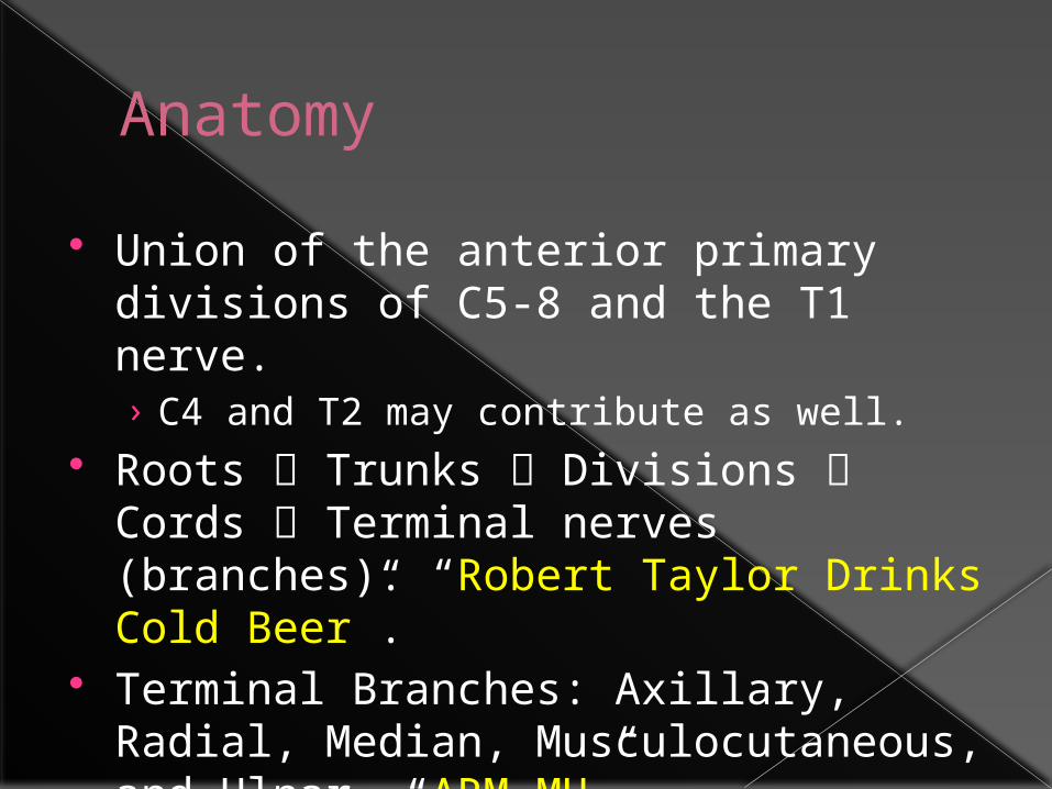
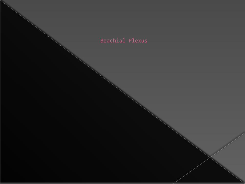
Anatomy
Union of the anterior primary divisions of C5-8 and the T1 nerve.› C4 and T2 may contribute as well.
Roots Trunks Divisions Cords Terminal nerves (branches). “Robert Taylor Drinks Cold Beer”.
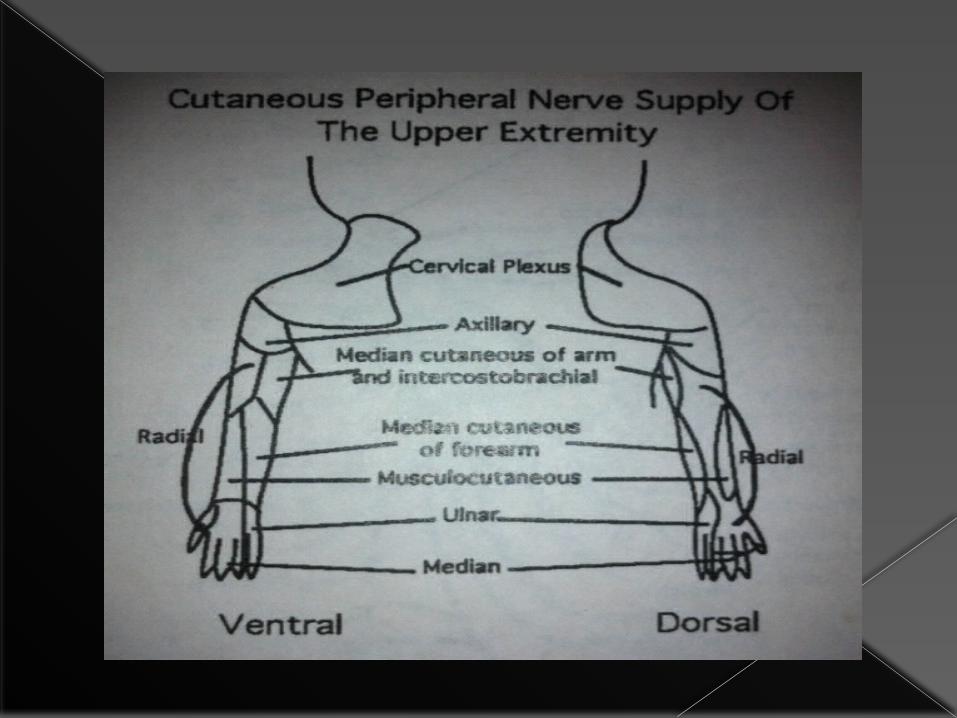
Terminal Branches: Axillary, Radial, Median, Musculocutaneous, and Ulnar. “ARM MU”.
Picture
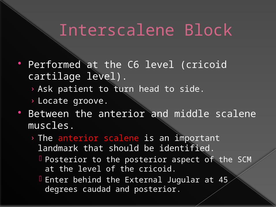
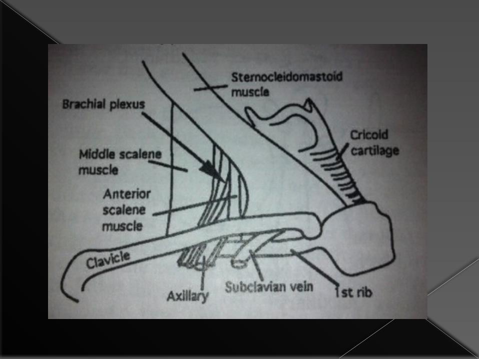
Interscalene Block
Performed at the C6 level (cricoid cartilage level).› Ask patient to turn head to side.› Locate groove.
Between the anterior and middle scalene muscles.› The anterior scalene is an important landmark
that should be identified. Posterior to the posterior aspect of the SCM at the
level of the cricoid. Enter behind the External Jugular at 45 degrees
caudad and posterior.
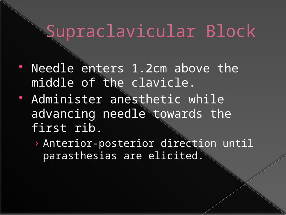
Supraclavicular Block
Needle enters 1.2cm above the middle of the clavicle.
Administer anesthetic while advancing needle towards the first rib. › Anterior-posterior direction until
parasthesias are elicited.
Sciatic Nerve
Sciatic Nerve Block
L4-5 and S1-3› Runs between the ischial spine and greater trochanter of
the femur.› Becomes superficial at the base of the gluteus maximus.
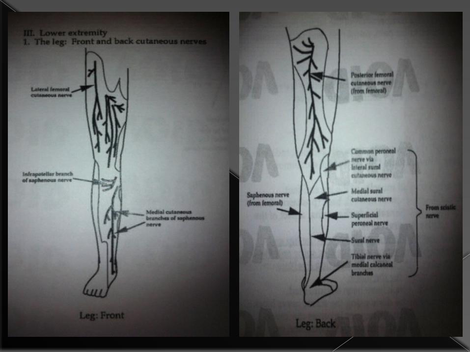
Cutaneous innervation to posterior thigh and all of the leg below the knee minus a small medial strip.
Two approaches: Posterior and Anterior. Usually block is combined with femoral, obturator,
or lateral fem cutaneous nerve blocks. Disadvantages: technically difficult, painful,
possible hematoma, nerve damage, slight drop in BP due to blood pooling.
Posterior Approach
Lateral decub position with leg to be blocked flexed at the knee with the heel resting on the opposite knee.
Connect the posterior superior iliac spine with the greater trochanter with a drawing pen. Bisect this line perpendicularly, extending caudal.
Needle entry point: 3cm downward from the perpendicular line.
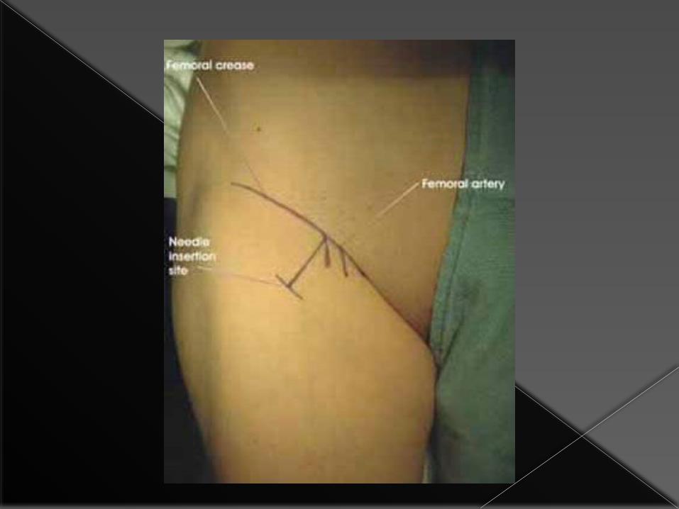
Anterior Approach
Supine position. Line from ASIS to pubic tubercle. Mark
point 2/3 of the way. Draw parallel line from greater
trochanter. From point of first line, continue down
to second line. Inject at this site until bone is hit, then direct medially.


![Trunks - Sangomaliterature.schmoozecom.com/trunks-module/userguides/trunks-module-userguide.pdfN matches any digit from 2-9. [1237-9] matches any digit in the brackets (example: 1,2,3,7,8,9)](https://img.pdfslide.us/doc/110x75/5e6974979bb9254b82492147/trunks-n-matches-any-digit-from-2-9-1237-9-matches-any-digit-in-the-brackets.jpg)