Embed Size (px)
Citation preview
1
MemoryRudolf N. Cardinal
MRCPsych 2004Tuesday 25 March
For slides, electronic handouts, and related material, see www.pobox.com/~rudolf/psychology.
Overview
Memory is a complex topic. We will discuss the various forms of memory that existfrom a theoretical and psychological perspective, discuss various influences onmemory encoding and recall, and then examine the neural structures that are respon-sible for these different forms of memory, including the medial temporal lobe anddiencephalic structures, the basal ganglia, and the prefrontal cortex. We will brieflyconsider consolidation, reconsolidation, and the role of sleep.
Types of memory
There are many forms of memory. As the process of subdividing ‘memory’ is basedon neuroscientific, as well as psychological dissociations, the number of distinctforms of memory thought to exist has changed over the years — there are somemajor controversies in this area of cognitive neuroscience. Memory is simply theability of something to retain information, thus changing its input→output function(the output it produces in response to a given stimulus). By this definition, sandpits,blackboards, and computers have memory. But there are, of course, much more so-phisticated forms of memory.
Individual versus phyletic memory; perceptual versus motor memory; activation
Before getting into the nitty-gritty, it’s worth mentioning some points made byFuster (1), who writes about memory systems from a neurobiological perspective.These are as follows. (1) Individual memories are changes in brain activity or con-nectivity that are superimposed on the pre-existing brain, but that pre-existing brainis specific to our species and shaped by evolution — these specificities can bethought of as a phyletic memory. (2) Nervous systems take in sensory input and dothings as a result; they have sensory and motor systems and complex processing inbetween; their memory systems are organized around this fundamental difference;we have perceptual and motor memories. (3) Both perceptual and motor memoriesmay be inactive — a long-term condition — or become active in the short term.
Short- versus long-term memory
Traditionally, a distinction has been made between short-term memory (STM) andlong-term memory (LTM) (2-5).
Incoming sensory information appears initially to enter a very short-term, high-capacity sensory store. Its existence was first shown by Sperling (6). He flashed a 4× 3 matrix of letters for 50 ms. If participants were asked to report all the letters(‘whole report’), they reported 4.32 letters correctly out of 12, but if they were cuedby a series of tones, presented after the visual array, to report only the top, middle,or bottom row (‘partial report’), they reported 3.04 out of 4 for each row. This im-plies that they had access to at least 9–10 out of 12 letters for a short time. It appearsthat this ‘iconic memory’ lasts about half a second: if the tone was delayed for asecond or so, participants were no better off than in the ‘whole report’ condition.The auditory version (‘echoic store’) last about 2 seconds (3, 7, 8).
From here, information appears to pass into a lower-capacity but slightly longer-lasting buffer, often known as short-term memory (STM). STM appears to have aseverely limited capacity — typically 7±2 arbitrary pieces of information (9), thoughthis can be increased by ‘chunking’ to impose structure on the stimuli; you canthereby remember seven arbitrary letters or numbers, or seven words, etc. If you arean expert in a particular domain, you may have more complex ‘chunks’ at your dis-posal and may therefore perform very well indeed; de Groot (10) showed thatgrandmasters had far superior short-term memory for valid chess positions thannovices. If subjects hear or see a long list of items and must recall them (free recall)
2
there is better recall of early and late items (primacy and recency effects); the re-cency effect can be abolished by distractors. STM has a very short duration: Peter-son & Peterson (11) found that if subjects memorized arbitrary patterns (e.g. ‘XPJ’),performed a distractor task to prevent mental rehearsal, and were then asked to recallthe pattern, they had forgotten 70% after nine seconds; obviously rehearsal can in-crease the effective duration. The digit span task is a popular test of STM.
According to early models of memory, some of the contents of STM can be passedon to LTM (12). The capacity of LTM appears effectively unlimited, and it isviewed as a permanent store. LTM can include spatial information about the world,motor skills, perceptual skills such as language perception, learned facts, etc.
Neurally, a concept of STM (or the related working memory, and primary memory)remains useful. It is likely, given its time-course and impermanence, that it reflectselectrical activity; it has been hypothesized, for example, that the 7±2 limit reflectsthe number of neuronal ensembles that can simultaneously be ‘bound’ together bysynchrony in the context of neuronal oscillations (e.g. 13). Long-term memory stor-age is probably not dependent upon reverbatory patterns of electrical activity; it in-volves synaptic plasticity, and potentially the growth of new synapses. However, theconcept of ‘LTM’ is not terribly useful, as it can be subdivided into many types ofmemory that can be dissociated neurally.
There have been theoretically important neural dissociations between STM andLTM; one such case was patient K.F. (14), who had a digit span of only 1–2 but ap-parently normal LTM, following a lesion of the perisylvian region of the left hemi-sphere (i.e. near/in auditory processing regions of cortex).
Dividing up long-term memory: declarative and nondeclarative memory
One way to begin is to divide LTM into declarative (or explicit) memory — mem-ory for events and facts — and nondeclarative (implicit) memory — the rest (see 1,chapter 2). Declarative memory includes episodic and semantic memory (15, 16),though whether these really reflect a different underlying neural process is less clear.Nondeclarative memory is a term that arose partly from the consideration of whatwas not lost from human amnesiacs (17); it includes procedural memory (18) andpriming (19). The borderlines remain controversial.
Episodic memory
‘The difference between episodic memory and semantic memory is often referred toin terms of remembering versus knowing: episodic memory is concerned with re-membering specific personal experiences, whereas semantic memory mediates whatone knows about the world. Remembering getting soaked in the London rain lastTuesday is an example of episodic memory, but knowing that it often rains in Eng-land is an example of semantic memory because it need not be acquired as a resultof a personal experience of getting wet.’ (20). Of course, semantic memory can beacquired as a result of personal experience.
An important problem in the study of episodic memory has been the lack of an ani-mal model. In the absence of this, human lesion studies have provided a large pro-portion of the evidence regarding the structures that implement episodic memory,and these lesions tend to be either precisely demarcated but large (such as the neuro-surgical lesion in H.M.) or difficult to define exactly (such as the damage caused bystroke, anoxia, or herpes simplex encephalitis). Tulving defines episodic memory asthe memory of temporally defined events in subjective time, giving the possessor theability to travel back in time to re-experience remembered events (‘autonoetic con-sciousness’, from Gr. autos self, noetikos pertaining to the mind, intellect, or processof perceiving or thinking), and has suggested that this may not be possessed by non-humans (16, 21, 22). It is difficult to know if animals ‘re-experience’ past events,but the use of simpler definitions has allowed some remarkable capabilities of ani-mals to be established. For example, Clayton & Dickinson (23) identify remember-ing what, where, and when an event occurred as key components of episodic mem-ory, and show that scrub jays encode this information.
3
Semantic memory
Semantic memory can be thought of as conceptual knowledge or memory for facts(Bogota is the capital of Colombia; i2 = –1). It does not necessarily include a repre-sentation of the episode in which the information was learned. In addition to suchabstract pieces of information, semantic knowledge is usually taken to include cate-gorical information about objects: ‘a robin is a bird’.
There is considerable controversy about the nature of semantic memories (e.g. 1,24), partly because they can be hard to define precisely. One view (25) is that se-mantic memories are episodic memories for which the detailed contextual informa-tion has disappeared, leaving only the generic features. Another (26) is that they aretwo, truly separate systems.
Consider how semantic memory can be acquired associatively. Moggy, Felix, andGarfield are all conglomerations of stimulus features (elements). ‘Moggy’ can thenbe represented as the activation of a network of ‘feature detectors’ (neuronal assem-blies) that is uniquely associated with Moggy. However, Moggy, Felix, and Garfieldhave common elements; these common features can be thought of as ‘catness’, couldbe associated with the word ‘cat’, and so on. This is one way in which semantic(categorical) information can be built up.
If this view is correct, then semantic information of this kind is intimately associatedwith perception — and indeed, action. Martin et al. (27) performed a PET study inwhich subjects identified line drawings of animals (which have to be distinguishedby differences in visual form), tools (which can be distinguished by the use to whichthey are put), and nonsense objects. In addition to common areas of activation (ani-mals and tools versus nonsense objects), there were regions that were specific to onecategory of information. Thus, animals (versus tools) activated an early visual proc-essing area of occipital cortex, while tools (versus animals) activated a premotorarea also activated by imagined hand movements. Semantic information, therefore,may be highly distributed across neocortex according to the perceptual and motornetworks that it builds upon (1). Semantic dementia (loss of knowledge aboutmeaning), agnosia, and apraxia can all follow damage to neocortical areas.
Procedural memory
Procedural memory means knowing how to do something (18). It is generallythought of as skill or habit memory. For example, patients with amnesia (see below)can have a preserved ability to learn a skill such as mirror-reading. More specifi-cally, a procedural memory is one in which the structure of the memory’s represen-tation directly reflects the use to which the knowledge will be put in controlling thesubject’s behaviour (28), as opposed to declarative knowledge, which is to some de-gree independent of the use to which it is put.
Although many tasks have been considered to be learned by habit in amnesia re-search (see below), it’s worth noting that few have proved their habits to be proce-dural in nature. What would do this? Dickinson and colleagues have developed a testfor whether actions, such as lever-pressing by rats, are governed by declarative orprocedural representations (see e.g. 29, 30) — and they can be governed by either. Ifa rat presses a lever for food, and you then devalue (e.g. poison) the food, then youcan assess the underlying representation. If the rat no longer presses the lever whenyou next test it, then it has integrated the knowledge of the food’s value with the in-formation that the lever produces the food; this requires declarative representations.If the rat presses the lever regardless, then its action is not controlled by the knowl-edge of the outcome, and is a stimulus–response habit. This level of psychologicalsophistication has yet to be applied to many of the ‘procedural’ tasks we will men-tion.
4
Priming
Priming is an increase in the speed or accuracy of a decision as a consequence ofprior exposure to some of the information involved in the decision (31). It occurs intasks where memory for the previous information is not required, and it may ad-versely affect performance, so it is assumed to be an involuntary and perhaps uncon-scious phenomenon. For example, the reaction time for ‘doctor’ is shorter if it hasbeen preceded by ‘nurse’ than if it has been preceded by ‘north’ or the non-word‘nuber’ (semantic priming). Repetition priming for visual stimuli is associated withreduced blood flow in occipital cortex (32); it is possible (but unproven) that (a)priming is a cortical effect in regions involved in processing the stimulus; (b) fol-lowing presentation of a stimulus, less neural activity is required to process the samestimulus.
Encoding, forgetting and recall
Emotional effects on memory consolidation
We saw in the Emotion/Motivation handout (q.v.) that emotionally-arousing situa-tions can improve memory consolidation, that this may be mediated by systemicadrenaline and glucocorticoids (and prevented by benzodiazepines), and that manyof these effects appear to be modulated through the amygdala (see 33, 34, 35).
Forgetting
Although the simple passage of time may be important in forgetting from STM, it iscertainly important what happens during that time — perhaps that items are dis-placed rather than (or as well as) decaying. For example, recall can be better aftersleep than after an equally long period of waking (36), though this effect might inprinciple be due to active consolidation during sleep than disruption by other activi-ties during waking. If people attempt recall of an item in the middle of a list, theirsuccess depends more on the number of items that have followed than on the timethat has elapsed (37).
Interference effects can be retroactive (new information interferes with previously-learned material) or proactive (previously-learned information interferes with newlearning) — if you have to memorize list of numbers after list of numbers, you be-come progressively worse (proactive interference) but if you then have to learn a listof letters, you become better again (release from proactive interference) (38).
Cue- or state-dependence of recall
We may be unable to remember things not just because they are not encoded in ourbrain, but because we have the information but are unable to recall or retrieve it.Providing explicit cues (clues!) can assist recall (39). These cues can be external: ifyou learn material in one room, you recall it better if you’re in the same room (40);if you learn material underwater, you recall it better underwater (41) — and thisbenefit was only for recall, not for recognition. The cues can be internal, known asstate dependence: if you learn material drunk, there is a recall benefit to being drunk(42); recall is better when you’re in the same mood as when you learned (‘mood-dependent memory’) (43) and mood affects recall in other ways — if you’re happy,you’re more likely to retrieve happy material (‘mood-congruent memory’) (44). Thismay be one reason why victims of violent crime have problems recalling details —recall occurs in a less emotionally-aroused state (45).
Schemata and memory distortion
Bartlett (46) introduced the idea that we interpret incoming information in terms ofschemata (sing. schema), stored in LTM, and that this also influences our recollec-tion. For example, Bartlett had subjects repeat an unusual short story to each other— by a process of Chinese whispers it became corrupted. These corruptions madethe story shorter, more coherent, more conventional, and more clichéd — better fit-ting with the subjects’ prior schema, perhaps. This is very important in the field of
5
eye-witness testimony, as it can lead to distortion. In 1947, Allport & Postman (47)briefly showed American subjects a picture of a white man threatening a black manwith a cutthroat razor; one subject described the scene to another, who passed it on,for 6 rounds. Half of the final participants reported that the razor was held by theblack man — subjects fitting the story to their prior schema (racial stereotype)?Later information can influence recall in a way that suggests memories are recon-structed, not simply recalled; for example, Loftus & Palmer (48) showed subjects ashort video of a car crash. If they were asked for details of what happened when thecars ‘smashed’, participants estimated the cars to be going faster than if the questionused the word ‘hit’ — and were more likely to invent the presence of broken glass.
Repression and false memories
False memories can be created, as we’ve seen, by combining actual memories withthe content of suggestions from others, but it’s very hard indeed to establish whethera given memory is false — a legal minefield. It’s also hard to establish whether re-pression has occurred; this is a major problem with Freud’s (49) repression hypothe-sis (was a memory repressed or was it never formed? If it’s retrieved, is the retrievedmemory false?). There have been some experimental attempts to demonstrate re-pression. Levinger & Clark (50) found that subjects generated free associations tonegative words (‘fear’, ‘quarrel’, etc.) slower than to neutral words, and rememberedthem more poorly immediately afterwards — had they been repressed? — but thismay be just an effect of emotional arousal: high arousal inhibits immediate recall butimproves long-term recall (51, 52).
Human organic amnesia: evidence for multiple neural memory systems
Amnesia may be retrograde (failure to retrieve previously learned material) or an-terograde (failure to learn new material). Amnesia can arise in humans from a vari-ety of causes including anoxia/ischaemia, closed head injury, encephalitis, Korsa-koff’s syndrome (deficiency of thiamine, a.k.a. vitamin B1; usually due to dietarydeficiency in alcoholics), and neurosurgery for epilepsy or tumours. It is also aprime symptom of progressive neurodegenerative disorders such as Alzheimer’sdisease.
Medial temporal lobe amnesia
Damage to the medial temporal lobes can follow surgical resection, anoxia, herpessimplex encephalitis, infarction, and sclerosis. The famous patient H.M. had his me-dial temporal lobes resected as an experimental treatment for epilepsy in 1953, whenhe was 27 (53, 54). This resulted in a severe anterograde amnesia for many forms ofmaterial from different modalities. His recall and recognition memory are severelyimpaired for lists, routes, and events. He has problems in learning about both auto-biographical episodes and new facts — i.e. in both episodic and semantic memory(to use Tulving’s distinction). He also has a mild retrograde amnesia for events fromabout 1942. The frequency of his seizures was, however, reduced!
However, H.M. has not lost all forms of memory. His digit span and visual immedi-ate memory is normal. He was able to learn new motor skills, such as mirror-writing,with practice (e.g. 55), though he was unable to remember having practised thesetasks ever before! In similar fashion, he could learn the Tower of Hanoi problem-solving puzzle. Priming is also normal in amnesiacs such as H.M. (see 56, 57, 58).His IQ is above average, and was not impaired by the surgery. Medial temporal lobeamnesia also spares eyeblink conditioning and emotional conditioning. (Famously,the Swiss psychiatrist Claparède once poked an amnesiac's hand with a pin whileshaking hands; the next day, she would not shake hands but could not rememberwhy; 59.) These findings are important, as they indicate the scope of the memorysystems that involve the medial temporal lobe — non-declarative memory systemsappear to be preserved following medial temporal lobe lesions, implying multiplememory systems in the brain.
Other patients showed similar patterns (though H.M.’s memory impairment is un-doubtedly one of the most severe); sometimes, amnesia occurred after unilateral le-
6
sions, because of pre-existing pathology on the other side. What remains unclearfrom the study of these patients is the damage necessary and sufficient to producefull-blown anterograde amnesia. H.M. certainly has considerable damage to themain structures of the limbic system which underlie the temporal lobe, the hippo-campus and amygdala; other patients have variable damage to these structures; areboth implicated? Some answer to this question was provided by the discovery of pa-tient R.B., who developed bilateral, complete, and (apparently) highly localized an-oxic damage to the CA1 field of the hippocampus after a cardiac arrest followingopen-heart surgery; histologically, he had relatively minor damage elsewhere (60).He exhibited a marked anterograde amnesia and no intellectual deterioration, butoverall his deficits were less severe than those of H.M.
Diencephalic amnesia
Patient N.A. was a 22-year-old technician in the US Air Force who was accidentallystabbed with a miniature fencing foil by a friend in 1960. The foil entered his rightnostril, penetrated the cribriform plate, and damaged his medial diencephalon, in-cluding the mediodorsal nucleus of the thalamus, the mammillary bodies, and themammillothalamic tract (61). He acquired a profound anterograde amnesia, but hadno impairments of higher cognitive function.
Patients with Korsakoff’s amnesia are frequently found on post mortem to havesustained damage to diencephalic structures including the medial thalamus, fornixand mammillary bodies. They have profound anterograde but also retrograde amne-sia as well as other cognitive deficits resembling those seen after frontal lobe lesions(see 62).
The Delay–Brion (or Papez) circuit
Thus, anterograde amnesia can result from damage to diencephalic structures, aswell as the medial temporal lobe; do these form a common circuit? Delay & Brion(63) proposed that damage to a circuit from the hippocampus → mammillary bodies→ anterior thalamic nuclei is sufficient to induce anterograde amnesia. The Delay–Brion circuit is sometimes called Papez’s circuit; Papez (64) had previouslysuggested that a wider circuit including these structures and the cingulate cortex wasinvolved in emotion. Whether diencephalic amnesia qualitatively resembles that ofmedial temporal lobe amnesia is unclear. Aggleton & Brown (56) argue that it does,in most key respects; ‘pure’ diencephalic amnesia is rare and some pathologicalprocesses affecting it (e.g. Korsakoff’s) cause widespread damage elsewhere.
Effects of selective hippocampal and/or fornix lesions
This is a matter of enduring debate.
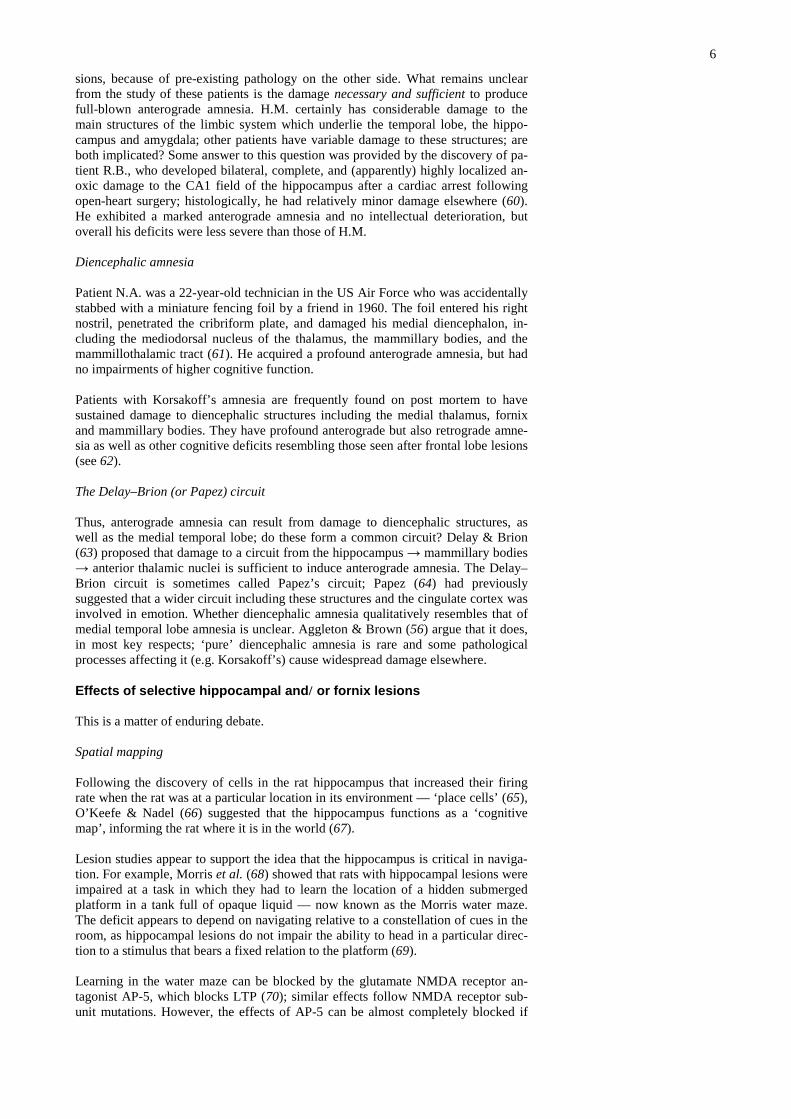
Spatial mapping
Following the discovery of cells in the rat hippocampus that increased their firingrate when the rat was at a particular location in its environment — ‘place cells’ (65),O’Keefe & Nadel (66) suggested that the hippocampus functions as a ‘cognitivemap’, informing the rat where it is in the world (67).
Lesion studies appear to support the idea that the hippocampus is critical in naviga-tion. For example, Morris et al. (68) showed that rats with hippocampal lesions wereimpaired at a task in which they had to learn the location of a hidden submergedplatform in a tank full of opaque liquid — now known as the Morris water maze.The deficit appears to depend on navigating relative to a constellation of cues in theroom, as hippocampal lesions do not impair the ability to head in a particular direc-tion to a stimulus that bears a fixed relation to the platform (69).
Learning in the water maze can be blocked by the glutamate NMDA receptor an-tagonist AP-5, which blocks LTP (70); similar effects follow NMDA receptor sub-unit mutations. However, the effects of AP-5 can be almost completely blocked if
7
the rats are trained in a different water maze beforehand (71), so the role of theNMDA receptors may not be a specifically spatial one!
Using PET imaging, Maguire et al. (72) recently found that blood flow in the (right)hippocampus was activated when London taxi drivers (expert navigators) imaginednavigating around London, compared to a control task in which they recalled fa-mous landmarks in unfamiliar cities. The posterior hippocampus is larger, and theanterior hippocampus smaller, in taxi drivers compared to controls, and this effect islarger the longer the subject has been a taxi driver (73). The hippocampus is also ac-tivated when subjects navigate around the computer game Duke Nukem (74)!
More than a map
Morris, Eichenbaum and others argue that the hippocampus doesn’t encode a map inthe sense that we’d normally use the word (see 67). Place cells tell you where youare, not where you want to go — if your place cells tell you that you’re in positionA, how do you decide to go to B and not to C? Furthermore, the arrangement ofplace cells doesn’t seem to be very consistent — they certainly don’t form a topo-graphic map of space, they lose or change their properties when the environment ex-pands, and so on. Rather, it appears that place cells encode the relationship betweensome subset of cues in the environment (independent of other cues). Furthermore,hippocampal neurons do not just encode space. Wood et al. (75) showed that hippo-campal neurons encoded a range of nonspatial features of a odour-based nonmatch-ing-to-sample task, independent of the spatial location of the stimuli.
Encoding episodes
Given the ambiguity of the AP-5 water maze experiments (see above), Morris &Frey (76) have updated their views and now see the hippocampus as vital for en-coding episodes — that it encodes rapid, one-trial episodic memory (the ‘automaticrecording of attended experience’). The ‘automatic’ property is meant to capture theidea that the animal remembers things that are not relevant to the task at hand, butthat may be recalled later. This is very much akin to human descriptions of episodicmemory. Morris & Frey attempt to go some way down this path by examining watermaze learning in a one-trial fashion; they find that the ability of rats to rememberthe most recent place they have visited in a familiar environment (one-trial delayedmatching to position in a water maze) is exquisitely sensitive to AP5 in a delay-dependent manner. Is this an episode? Well, maybe. As we said at the outset, newanimals models of episodic memory are being developed that may help the testing ofthis hypothesis. Day et al. (77) have recently shown that encoding of uniquefood/location (what/where) pairs requires hippocampal activity; this is progresstowards the what/where/when triad of Clayton & Dickinson (23).
Encoding scenes
Gaffan (78) argued that the hippocampus is required for encoding scenes — that is,complex and arbitrary stimulus patterns. Gaffan & Harrison (79) examined the ef-fects of fornix transection in the monkey. They gave the monkeys a series of objectdiscrimination problems (A versus B), in which the correct object depended uponthe position and/or visual environment of the monkey. The monkeys could learnnormally if they saw different objects in the room when A was correct than when Bwas correct. However, if the two visual environments contained the same objects,but in a different configuration, then fornix-lesioned monkeys were impaired. Gaf-fan & Harrison suggest that at least three types of memory are formed when a mon-key displaces an object and finds reward under it:
1. A simple association between the object and reward.2. A more complex association, between the background items, the object dis-
placed, and the reward. (This allows the monkey to solve problems of thekind ‘if a door handle and a coat are visible, choose object X’.)
3. An even more complex memory that encodes the identity and the spatial re-lations of the background objects, the target object, and the reward. (Thisallows the monkey to solve ‘if the radio is to the left of the tap, choose ob-ject X’.)
8
Gaffan & Harrison (79) argue that only the third type of memory — ‘snapshot’memory — is disrupted by fornix lesions. Gaffan (78) extended this finding to showthat fornix lesions impaired monkeys’ ability to learn discriminations involvingscenes from Raiders of the Lost Ark!
Representing context
Both the hypothesis that the hippocampus encodes spatial relationships, and the hy-pothesis that it encodes scenes, predict that the hippocampus might, under some cir-cumstances, be critical for contextual conditioning. For example, if a rat receivestone–shock pairings in a distinct environment, it may subsequently show ‘fearful’reactions to the tone (discrete CS conditioning) and also the environment (contextualconditioning). Indeed, hippocampal lesions often interfere with contextual condi-tioning (80). However, animals may use contextual information in a variety of waysand many of these studies do not illuminate the exact contribution made by the hip-pocampus; an excellent review is provided by Holland & Bouton (81).
Top left: the hippocampus as a cognitive map. Top right:place cells in the hippocampus. Right: encoding spatialrelationships — a special case of encoding relationships.Bottom right: transitive inference — another, more ab-stract and non-spatial case of using information about therelationships between stimuli. The rat is trained on A>B,B>C, C>D, D>E. It is tested on A>E (easy — A has al-ways been right, and E has always been wrong) and B>D(hard — the rat must infer that if B>C and C>D, thenB>D; this is called transitivity). Bottom left: fornix tran-section and perirhinal/entorhinal cortex lesions impairthe B>D probe test, but not the A>E test. Figures fromEichenbaum et al. (67)6} and Dusek & Eichenbaum(82)7).
9
Relational information
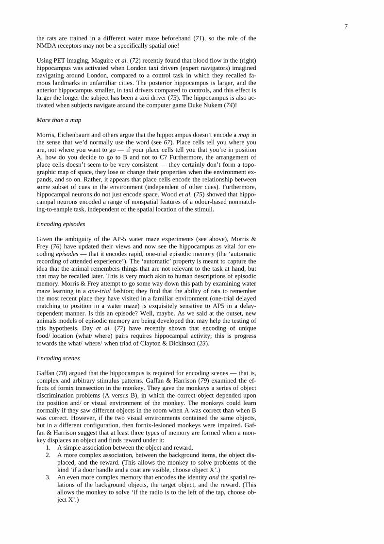
Eichenbaum et al. (67) argue that the hippocampus can encode spatial informationbecause this is a special case of encoding the relations between stimuli. They sug-gest that these relations are useful for navigation when they are spatial relations, butthat the memories encoded by the hippocampus can be used for other things. Theygive an example of a more abstract relationship: transitive inference. If a subjectlearns that B>C and C>D, then the logical property of transitivity should allow it toinfer that B>D. Dusek & Eichenbaum (82) have shown that fornix transection andperirhinal/entorhinal lesions, both of which partially disconnect the hippocampus,impair transitive inference in rats (see figure).
Contributions of rhinal cortex to memory and perception
The rhinal cortex (i.e. entorhinal + perirhinal cortex), adjacent to the hippocampus inthe medial temporal lobe, is certainly important for aspects of visual recognitionmemory (see figure). In fact, rhinal cortex is also at the end of the ventral visualprocessing ‘stream’, and is important for perception — it appears to be involved indiscriminating complex conjunctions of visual stimuli (83-85), and perhaps in asso-ciating polymodal information about objects (86). This view is right up Fuster’sstreet (1) — the idea that memory and perception are largely inseparable in cortex.
Lesions of rhinal cortex impair delayed non-matching-to-sample(DNMTS) performance (87-90): the monkey sees an object, then there’sa delay, then it sees two objects and must pick the one it hasn’t seen be-fore. Rh = rhinal cortex lesion; AH = amygdala+hippocampus lesion;CON = controls. (” means seconds, using a single object for DNMTS;LL means list length, i.e. multiple objects, and in this situation the mini-mum retention interval for each trial was 20 s × list length.)
Location of area TE (part of inferotemporalcortex) and perirhinal cortex in the rhesusmacaque monkey (91). Top: lateral view(anterior to the left). Bottom: view of theinferior surface.
Semantic memory: where? How?
There is debate not just about what semantic memories are, but how they are estab-lished. Do they begin as episodic memories but become independent of the episodicmemory system with repetition and additional association? Perhaps not. There areintriguing reports of patients who suffered perinatal hypoxia (with consequent se-vere hippocampal atrophy visible on MRI) who have severe episodic memory defi-cits. In spite of this, they showed relatively normal semantic memory for facts andwere able to attend mainstream schools (92).
Conversely, there are patients who develop semantic dementia (93), characterized byprogressive loss of conceptual knowledge about objects, facts, concepts, and wordmeanings (see 94). It has been suggested that episodic memories appear to suffer areverse temporally graded retrograde amnesia in semantic dementia — old memo-ries are remembered less well than recent ones. Structurally, this disorder is associ-ated with atrophy of the anterolateral temporal lobes (95). The pattern of semanticmemory loss is perhaps explicable in terms of random damage to a distributed corti-cal associative memory that represents associations between features (and as a con-sequence, conceptual information) according to simple statistical principles (96).
10
However, the relationship between semantic dementia and episodic memory is stillcontroversial.
A time-limited role for the hippocampus?
Retrograde amnesia
As we saw earlier, H.M. developed profound anterograde amnesia following hismedial temporal lobe resection — but also a temporally graded retrograde amnesiafor events preceding the surgery (that is, old events were recalled better than recentones). Indeed, such retrograde amnesia has been regularly noted in humans follow-ing medial temporal lobe lesions, or lesions apparently restricted to the hippocampalformation (see 97). This led to the hypothesis that the hippocampus is involved inconsolidating memories held elsewhere (54) — recent memories are vulnerable tohippocampal damage, but with time they become independent of the hippocampus.This view is highly popular, althought not the only view (98).
Prospective animal studies of retrograde amnesia
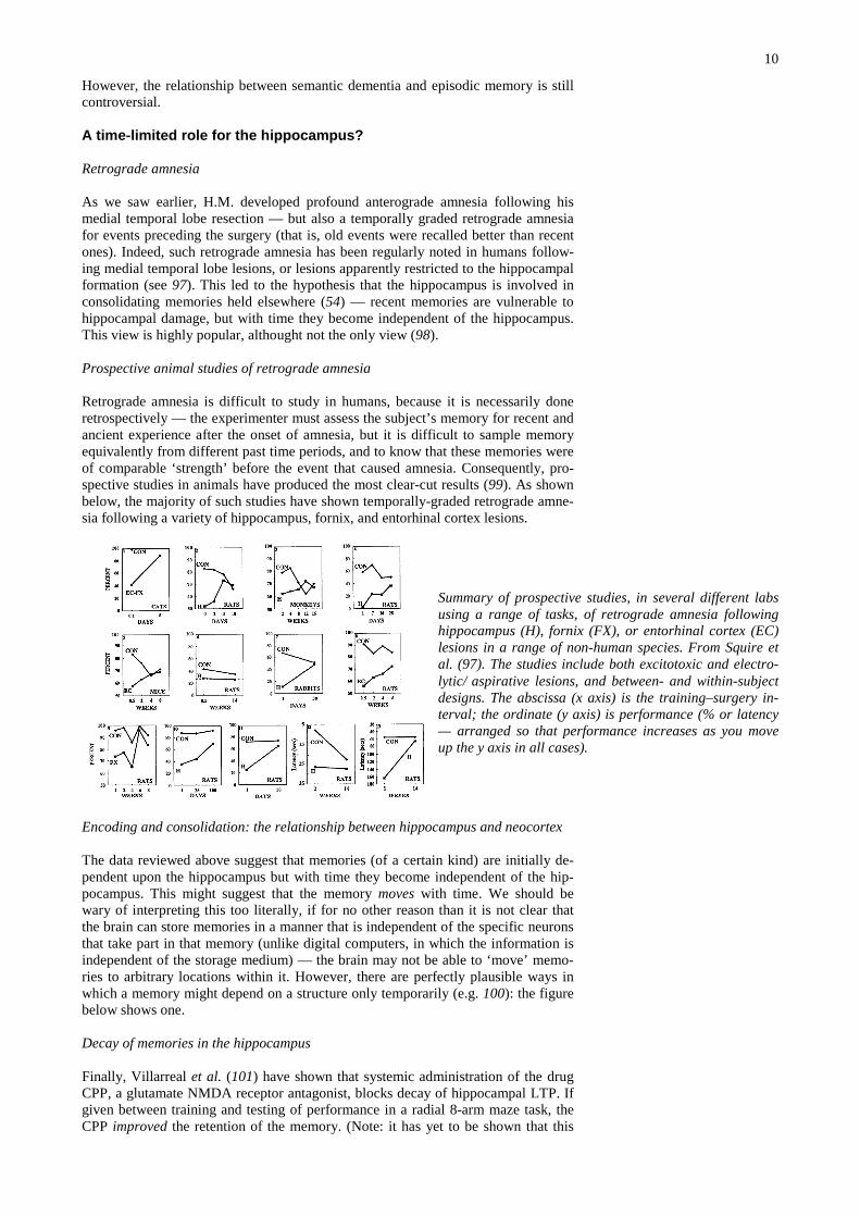
Retrograde amnesia is difficult to study in humans, because it is necessarily doneretrospectively — the experimenter must assess the subject’s memory for recent andancient experience after the onset of amnesia, but it is difficult to sample memoryequivalently from different past time periods, and to know that these memories wereof comparable ‘strength’ before the event that caused amnesia. Consequently, pro-spective studies in animals have produced the most clear-cut results (99). As shownbelow, the majority of such studies have shown temporally-graded retrograde amne-sia following a variety of hippocampus, fornix, and entorhinal cortex lesions.
Summary of prospective studies, in several different labsusing a range of tasks, of retrograde amnesia followinghippocampus (H), fornix (FX), or entorhinal cortex (EC)lesions in a range of non-human species. From Squire etal. (97). The studies include both excitotoxic and electro-lytic/aspirative lesions, and between- and within-subjectdesigns. The abscissa (x axis) is the training–surgery in-terval; the ordinate (y axis) is performance (% or latency— arranged so that performance increases as you moveup the y axis in all cases).
Encoding and consolidation: the relationship between hippocampus and neocortex
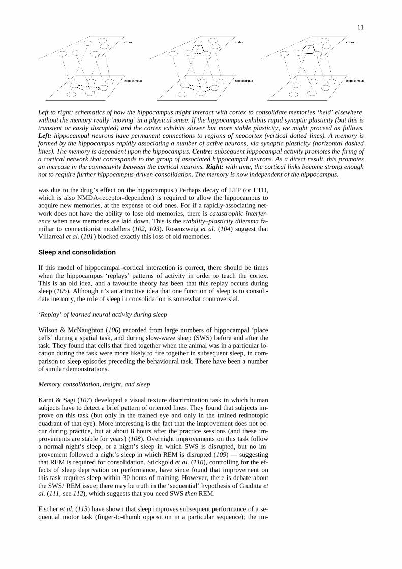
The data reviewed above suggest that memories (of a certain kind) are initially de-pendent upon the hippocampus but with time they become independent of the hip-pocampus. This might suggest that the memory moves with time. We should bewary of interpreting this too literally, if for no other reason than it is not clear thatthe brain can store memories in a manner that is independent of the specific neuronsthat take part in that memory (unlike digital computers, in which the information isindependent of the storage medium) — the brain may not be able to ‘move’ memo-ries to arbitrary locations within it. However, there are perfectly plausible ways inwhich a memory might depend on a structure only temporarily (e.g. 100): the figurebelow shows one.
Decay of memories in the hippocampus
Finally, Villarreal et al. (101) have shown that systemic administration of the drugCPP, a glutamate NMDA receptor antagonist, blocks decay of hippocampal LTP. Ifgiven between training and testing of performance in a radial 8-arm maze task, theCPP improved the retention of the memory. (Note: it has yet to be shown that this
11
was due to the drug’s effect on the hippocampus.) Perhaps decay of LTP (or LTD,which is also NMDA-receptor-dependent) is required to allow the hippocampus toacquire new memories, at the expense of old ones. For if a rapidly-associating net-work does not have the ability to lose old memories, there is catastrophic interfer-ence when new memories are laid down. This is the stability–plasticity dilemma fa-miliar to connectionist modellers (102, 103). Rosenzweig et al. (104) suggest thatVillarreal et al. (101) blocked exactly this loss of old memories.
Sleep and consolidation
If this model of hippocampal–cortical interaction is correct, there should be timeswhen the hippocampus ‘replays’ patterns of activity in order to teach the cortex.This is an old idea, and a favourite theory has been that this replay occurs duringsleep (105). Although it’s an attractive idea that one function of sleep is to consoli-date memory, the role of sleep in consolidation is somewhat controversial.
‘Replay’ of learned neural activity during sleep
Wilson & McNaughton (106) recorded from large numbers of hippocampal ‘placecells’ during a spatial task, and during slow-wave sleep (SWS) before and after thetask. They found that cells that fired together when the animal was in a particular lo-cation during the task were more likely to fire together in subsequent sleep, in com-parison to sleep episodes preceding the behavioural task. There have been a numberof similar demonstrations.
Memory consolidation, insight, and sleep
Karni & Sagi (107) developed a visual texture discrimination task in which humansubjects have to detect a brief pattern of oriented lines. They found that subjects im-prove on this task (but only in the trained eye and only in the trained retinotopicquadrant of that eye). More interesting is the fact that the improvement does not oc-cur during practice, but at about 8 hours after the practice sessions (and these im-provements are stable for years) (108). Overnight improvements on this task followa normal night’s sleep, or a night’s sleep in which SWS is disrupted, but no im-provement followed a night’s sleep in which REM is disrupted (109) — suggestingthat REM is required for consolidation. Stickgold et al. (110), controlling for the ef-fects of sleep deprivation on performance, have since found that improvement onthis task requires sleep within 30 hours of training. However, there is debate aboutthe SWS/REM issue; there may be truth in the ‘sequential’ hypothesis of Giuditta etal. (111, see 112), which suggests that you need SWS then REM.
Fischer et al. (113) have shown that sleep improves subsequent performance of a se-quential motor task (finger-to-thumb opposition in a particular sequence); the im-
Left to right: schematics of how the hippocampus might interact with cortex to consolidate memories ‘held’ elsewhere,without the memory really ‘moving’ in a physical sense. If the hippocampus exhibits rapid synaptic plasticity (but this istransient or easily disrupted) and the cortex exhibits slower but more stable plasticity, we might proceed as follows.Left: hippocampal neurons have permanent connections to regions of neocortex (vertical dotted lines). A memory isformed by the hippocampus rapidly associating a number of active neurons, via synaptic plasticity (horizontal dashedlines). The memory is dependent upon the hippocampus. Centre: subsequent hippocampal activity promotes the firing ofa cortical network that corresponds to the group of associated hippocampal neurons. As a direct result, this promotesan increase in the connectivity between the cortical neurons. Right: with time, the cortical links become strong enoughnot to require further hippocampus-driven consolidation. The memory is now independent of the hippocampus.
12
provement was specific for the practised sequence and occurred whether subjectsslept during the day or night. Sleep deprivation itself had no effect on performance.
Recently, Wagner et al. (114) have found that ‘insight learning’ — in their experi-ment, the ability to spot a hidden simplifying rule in a complex task — is hugely fa-cilitated by sleep.
Criticisms
Although many theories of sleep consolidation posited that REM sleep was criticalfor consolidation, the evidence for this is far from convincing; see Siegel (115).There is no clear evidence that REM sleep duration increases following learning; theduration of REM sleep is not obviously correlated with intellectual ability acrossspecies — dolphins, for example, have very little REM sleep — and many studies ofREM sleep disruption are subject to confounds (e.g. not controlling for stress or totalsleep deprivation). There are case reports of humans who have lost most or all REMsleep (e.g. following brainstem injury) but have no apparent memory deficits; onesubsequently went through law school and edited a puzzle section of a local news-paper (see 115). The role of SWS is perhaps better established, for certain kinds oftask (116, 117). However, a recent study showed that artificial enhancement of REMsleep improved later retention of a Y-maze task in rats (118); the debate continues.
Reconsolidation
A ‘standard’ view of consolidation would be that memories are created in a labilestate (sometimes thought of as STM), and with time, they are consolidated into astable state (LTM). For example, electroconvulsive shock (ECS, a.k.a. electrocon-vulsive therapy, ECT), which disrupts all ongoing electrical activity in the brain, in-duces amnesia if given shortly after training, but not if given a long time after train-ing (119). While the formation of new memories does not require protein synthesis,the consolidation of memories does; thus, administering the protein synthesis in-hibitor anisomycin during contextual fear conditioning does not impair the memoryof mice if they are tested one hour later, but that memory fades by 24 h as comparedto a control group (see e.g. 120, 121). Incidentally, the same is true (at a cellularlevel) of hippocampal LTP: ‘early’ LTP is not dependent upon protein synthesis, butit fades; normally, it is made long-lasting by a second phase, ‘late’ LTP, which re-quires protein synthesis (see 122).
Reconsolidation, a long-forgotten and interesting phenomenon of memory has re-cently been thrown into the limelight. As before, this hypothesis suggests thatmemories are created in a labile state and are consolidated into a stable state. How-ever, in this theory, recalling a memory returns it to the labile state. Therefore, al-though protein synthesis inhibitors don’t disrupt stable memories, they should beable to disrupt old memories that have been reactivated. Indeed, this has been ob-served (123). Recently, Nader et al. (124) found that infusions of anisomycin intothe basolateral amygdala (a critical site of plasticity for CS–US associations in-volved in conditioned freezing in the rat) disrupted memory for a CS–US associationthat had been ‘retrieved’ by presenting the CS. Appropriate controls demonstratedthat this only happened when the memory had been ‘retrieved’ in this way.
Is this important? Yes. One old case study (125) made use of the idea of reconsoli-dation. A patient had obsessive–compulsive disorder (OCD) that took the form of anobsession to kill her mother with a butcher’s knife. She had previously received 22sessions of ECS under anaesthetic (this is the normal way of doing it!). Rubin et al.made her act out her compulsion (N.B. reactivation of the memory in question) andgave here one session of ECS whilst awake. She was subsequently symptom-free forthe two years before publication of the study. This technique was effective, forvarying periods (3 months to ≥10 years), in all 28 patients tested (126). The abilityto ‘remove’ memories selectively might have enormous implications for the treat-ment of diseases including OCD, drug addiction, and so on.
13
Interference with (re)consolidation, or interference with retrieval?
It has been a matter of enduring debate whether amnesia is a result of a storage defi-cit or a retrieval deficit. For example, Warrington & Weiskrantz (58) interpreted thenormal performance of amnesiacs on memory as assessed by priming or word-completion tasks as indicating that their deficit was one of retrieval. Millin et al.(127) point out that many forms of amnesia can be reversed by reminder treatments,indicating that the memories were present all along and the deficit was one of re-trieval. Typical such studies used ECS to induce amnesia; subsequent exposure tothe CS, the US, or the ECS have all been shown to reverse the amnesia (see 127,128-130). The same question can be applied to reconsolidation (127): is it correct tosay that the reactivated memory is not stored again (reconsolidated) correctly, or cana retrieval deficit explain these results? Well, again, ‘reminder’ effects occur, im-plying a retrieval deficit (131, 132). Nader and colleagues now acknowledge this(133).
Habit learning: the dorsal striatum
The amnesia exhibited by H.M. was originally labelled ‘global anterograde amnesia’— yet a number of learning abilities were preserved in H.M. One of these was theability to learn the skill of mirror-drawing (134). The distinctions between the formsof memory that are impaired in medial temporal lobe amnesiacs and those that aren’thas been described as recognition/associative, episodic/semantic, work-ing/reference, declarative/procedural, and memories/habits (135).
Habits are the archetype of procedural memory. They are direct stimulus–response(S–R) links that are acquired as the result of reinforcement occurring when an ani-mal makes a response in the presence of a stimulus (136). Do animals have a habitsystem? Yes. We can test for it in rats using reinforcer devaluation. Rats are trainedto press a lever for food, and then they are given food and poisoned (to induce aconditioned taste aversion to that food) in the absence of the lever; after they havesampled the poisoned food, they are returned to the operant chamber and their lever-pressing is assessed (in extinction, to prevent delivery of the now-aversive foodfrom having a direct punishing effect on behaviour). Although under certain condi-tions, rats press the lever less than if the food had not been poisoned (indicating de-clarative knowledge — the effect of poisoning on lever-pressing was mediatedthrough an internal representation of the food), this is not always the case. If rats areovertrained on the lever-pressing task beforehand, reinforcer devaluation does notsuppress their lever-pressing (even though they won’t eat the food subsequently)(137). This indicates that a procedural representation has come to govern behaviour— a stimulus–response link that does not include a representation of the food (see138). It appears that S–R links develop slowly through training until (under somecircumstances) they dominate behaviour.
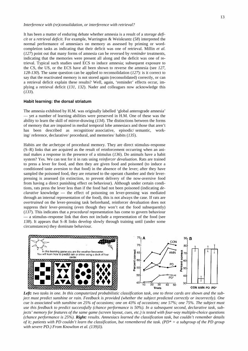
Left: two tasks in one. In this computerized probabilistic classification task, one to three cards are shown and the sub-ject must predict sunshine or rain. Feedback is provided (whether the subject predicted correctly or incorrectly). Onecue is associated with sunshine on 25% of occasions; one on 43% of occasions; one 57%; one 75%. The subject mustuse this feedback to predict successfully (chance performance is 50%). In a subsequent second, declarative task, sub-jects’ memory for features of the same game (screen layout, cues, etc.) is tested with four-way multiple-choice questions(chance performance is 25%). Right: results. Amnesiacs learned the classification task, but couldn’t remember detailsof it; patients with PD couldn’t learn the classification, but remembered the task. (PD* = a subgroup of the PD groupwith severe PD.) From Knowlton et al. (139)3).
14
So what neural structures subserve habit learning? Mishkin et al. (135) originallysuggested that a cortico-striatal system subserved habit formation. Although muchof the subsequent work on this issue has proved controversial (140-142), he wasprobably right. For example, patients with Parkinson’s disease (PD) or Huntington’sdisease (HD) are impaired on supposedly procedural tasks such as learning theTower of Hanoi puzzle (143, 144). Knowlton et al. (139) demonstrated a double dis-sociation between performance on a probabilistic classification task (impaired inPD, but not in patients amnesic secondary to hippocampal or diencephalic damage)and declarative memory for the same task (impaired in amnesiacs but not in PD pa-tients) (see figure).
This double dissociation clearly shows that the impairments in PD and hippocam-pal/diencephalic amnesia are qualitatively different. However, it does not show thatwhat the PD patients couldn’t do was learn a habit (or, for that matter, that the defi-cit was due to neostriatal dysfunction, rather than — say — prefrontal cortical do-pamine dysfunction). Unfortunately, while the learning theory definition of a habitgiven earlier is widely quoted, the learning theory methods to determine whetherbehaviour is habitual (such as reinforcer revaluation) have not adopted widely.There is no clear evidence that many of the tasks though to test ‘habits’ actually doso. Tasks have even been described as non-habit-based on the grounds that humanamnesiacs cannot learn them (145).
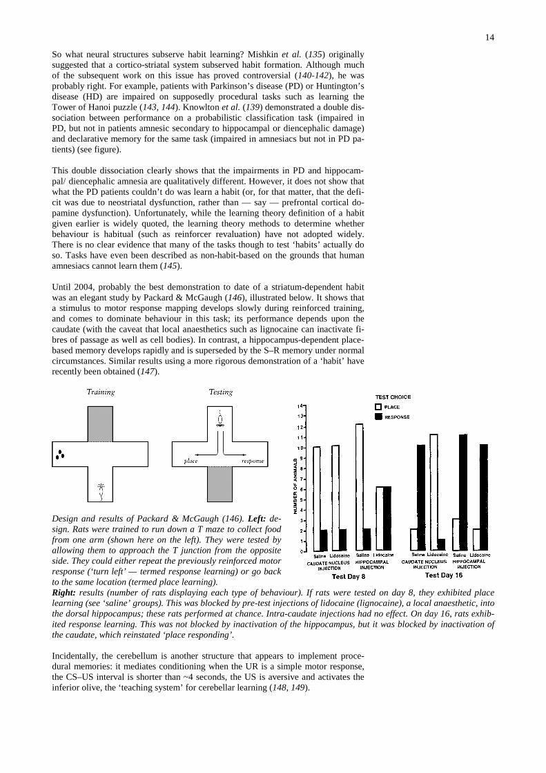
Until 2004, probably the best demonstration to date of a striatum-dependent habitwas an elegant study by Packard & McGaugh (146), illustrated below. It shows thata stimulus to motor response mapping develops slowly during reinforced training,and comes to dominate behaviour in this task; its performance depends upon thecaudate (with the caveat that local anaesthetics such as lignocaine can inactivate fi-bres of passage as well as cell bodies). In contrast, a hippocampus-dependent place-based memory develops rapidly and is superseded by the S–R memory under normalcircumstances. Similar results using a more rigorous demonstration of a ‘habit’ haverecently been obtained (147).
Design and results of Packard & McGaugh (146). Left: de-sign. Rats were trained to run down a T maze to collect foodfrom one arm (shown here on the left). They were tested byallowing them to approach the T junction from the oppositeside. They could either repeat the previously reinforced motorresponse (‘turn left’ — termed response learning) or go backto the same location (termed place learning).Right: results (number of rats displaying each type of behaviour). If rats were tested on day 8, they exhibited placelearning (see ‘saline’ groups). This was blocked by pre-test injections of lidocaine (lignocaine), a local anaesthetic, intothe dorsal hippocampus; these rats performed at chance. Intra-caudate injections had no effect. On day 16, rats exhib-ited response learning. This was not blocked by inactivation of the hippocampus, but it was blocked by inactivation ofthe caudate, which reinstated ‘place responding’.
Incidentally, the cerebellum is another structure that appears to implement proce-dural memories: it mediates conditioning when the UR is a simple motor response,the CS–US interval is shorter than ~4 seconds, the US is aversive and activates theinferior olive, the ‘teaching system’ for cerebellar learning (148, 149).
15
Encoding, retrieval, and the prefrontal cortex
The prefrontal cortex (PFC) may contribute to memory encoding and/or recall,probably via its extensive back-projections to posterior neocortex. For example, hu-mans with PFC lesions are profoundly impaired on verbal fluency tests (150) — e.g.‘please say as many words beginning with S as you can in the next minute’.
Many of the data regarding this function of the PFC come from neuroimaging stud-ies (see e.g. 151, 152). Tulving et al. (153) proposed a hemispheric encod-ing/retrieval asymmetry (HERA) model on the basis of PET studies of memorytasks; they suggested that the left PFC is more involved than the right in encodingepisodic memory (and retrieving semantic memory), whereas the right PFC is dif-ferentially more involved in episodic memory retrieval. As an example, left PFC ac-tivity at the time of processing verbal material (‘is this word abstract or concrete?’)predicts how well people subsequently remember that material (‘did you see thisword earlier?’) (154). It has been suggested that the nature of the material also de-termines the degree of left versus right activation (155).
These studies are vulnerable to a number of criticisms. One relates to whether thememory processes being observed in the scanner are episodic, semantic, both, etc. Amore serious criticism is that this imaging-based model is purely correlative; whatprocess the PFC is playing in these tasks is hard to fathom.
Dorsolateral prefrontal cortex and working memory
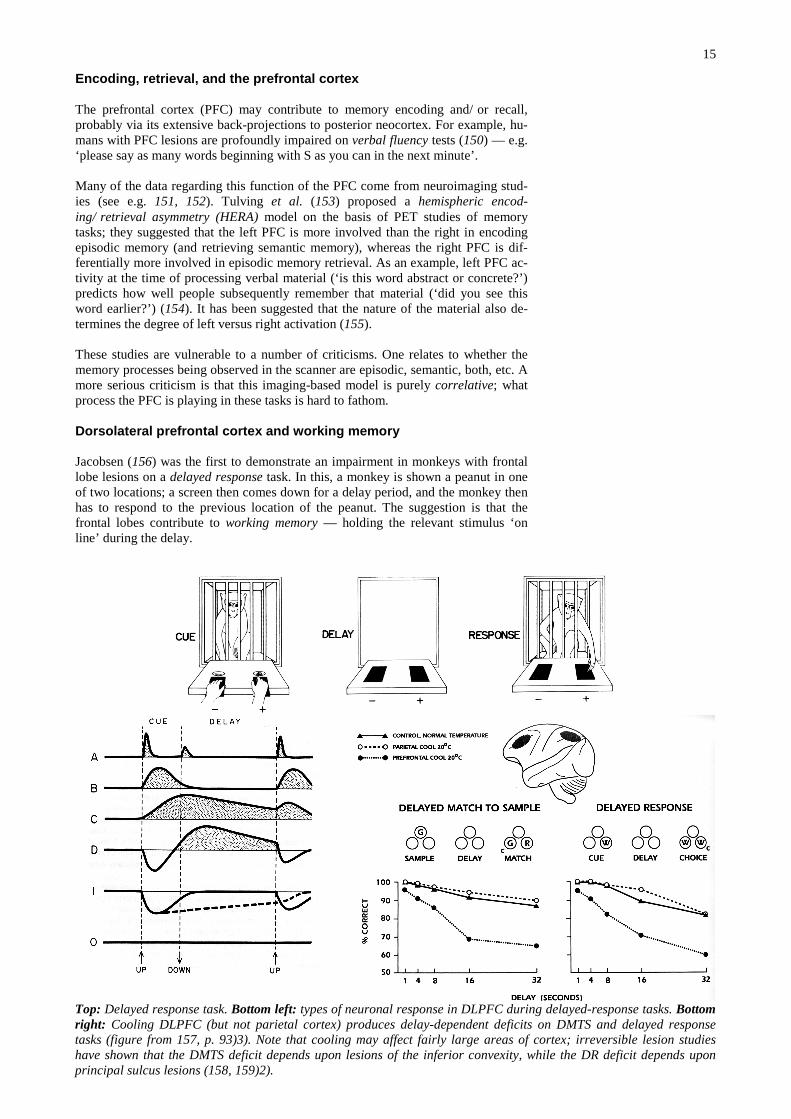
Jacobsen (156) was the first to demonstrate an impairment in monkeys with frontallobe lesions on a delayed response task. In this, a monkey is shown a peanut in oneof two locations; a screen then comes down for a delay period, and the monkey thenhas to respond to the previous location of the peanut. The suggestion is that thefrontal lobes contribute to working memory — holding the relevant stimulus ‘online’ during the delay.
Top: Delayed response task. Bottom left: types of neuronal response in DLPFC during delayed-response tasks. Bottomright: Cooling DLPFC (but not parietal cortex) produces delay-dependent deficits on DMTS and delayed responsetasks (figure from 157, p. 93)3). Note that cooling may affect fairly large areas of cortex; irreversible lesion studieshave shown that the DMTS deficit depends upon lesions of the inferior convexity, while the DR deficit depends uponprincipal sulcus lesions (158, 159)2).
16
This deficit has since been localized to the sulcus principalis (i.e. DLPFC) in mon-keys. Neurons here respond during the delay (160); cooling (161) or lesions (162) ofthe DLPFC impair delayed-response performance (see figure).
There are several lines of evidence that the PFC implements working memory via itsback projections to posterior cortex. Fuster and colleagues have shown that coolingof either DLPFC or inferotemporal cortex impair visual delayed matching-to-sample. There is a robust projection between these two regions. Furthermore, cool-ing one region affects the responses of neurons in the other (163); for example,DLPFC cooling diminished the discrimination shown by IT neurons in the delay,perhaps suggesting that the PFC is maintaining the response of the posterior corticalregion during the delay (157, 164, 165). Ruchkin et al. (166) argue along similarlines based on event-related EEG work in humans. There is a functional argument tobe made here: it is implausible that the PFC ‘contains’ the memory being held on-line, for it would have to duplicate all the perceptual capabilities of (e.g.) visualcortex in order to ‘hold’ a visual memory. Rather, by maintaining activation in vis-ual processing areas, working memory is achieved without duplicating perceptualsystems.
Bibliography
1. J. M. Fuster, Memory in the cerebral cortex: anempirical approach to neural networks in the humanand nonhuman primate (MIT Press, Cambridge,Massachusetts, 1995).2. W. James, Principles of Psychology (Holt, NewYork, 1890).3. D. E. Broadbent, Perception and Communica-tion (Pergamon, New York, 1958).4. A. Baddeley, Trends in Neurosciences 4, 176-181 (1988).5. D. A. Norman, Psychological Review 75, 522-536 (1968).6. G. Sperling, The information available in briefvisual presentation, Psychological Monographs(1960), vol. 74.7. A. M. Treisman, American Journal of Psychol-ogy 77, 206-219 (1964).8. R. D. Gross, Psychology: the Science of Mindand Behaviour (Hodder & Stoughton, London, ed.Fourth, 2001).9. G. A. Miller, Psychological Review 63, 81-97(1956).10. A. D. de Groot, Het denken van den schaker(Noord Hollandsche, Amsterdam, 1946).11. L. R. Peterson, M. J. Peterson, Journal of Ex-perimental Psychology 58, 193-198 (1959).12. R. C. Atkinson, R. M. Shiffrin, in The Psychol-ogy of Learning and Motivation, Volume 2 K. W.Spence, J. T. Spence, Eds. (Academic Press, London,1968).13. O. Jensen, J. E. Lisman, J Neurosci 18, 10688-99 (1998).14. T. Shallice, E. K. Warrington, Quarterly Jour-nal of Experimental Psychology 22, 261-273 (1970).15. E. Tulving, in Organization of Memory E.Tulving, W. Donaldson, Eds. (Academic Press,London, 1972).16. E. Tulving, H. J. Markowitsch, Hippocampus 8,198-204 (1998).17. S. Zola-Morgan, L. R. Squire, Annu Rev Neuro-sci 16, 547-63 (1993).18. N. J. Cohen, L. R. Squire, Science 210, 207-10(1980).19. E. Tulving, D. L. Schacter, Science 247, 301-6(1990).20. D. P. Griffiths, A. Dickinson, N. S. Clayton,Trends in Cognitive Sciences 3, 74-80 (1999).21. E. Tulving, American Psychologist 40, 385-398(1985).22. E. Tulving, in The Cognitive Neurosciences M.S. Gazzaniga, Ed. (MIT Press, Cambridge, MA,1995) pp. 839-847.23. N. S. Clayton, A. Dickinson, Nature 395, 272-4(1998).24. A. D. Baddeley, in Handbook of Memory Disor-ders A. D. Baddeley, M. D. Kopelman, B. A. Wilson,Eds. (John Wiley, Chichester, 2002) pp. 2-25.25. L. R. Squire, Journal of Cognitive Neuroscience4, 232–243 (1992).26. E. Tulving, European Journal of CognitivePsychology 1, 3-26 (1989).
27. A. Martin, C. L. Wiggs, L. G. Ungerleider, J. V.Haxby, Nature 379, 649-52 (1996).28. A. Dickinson, Contemporary animal learningtheory (Cambridge University Press, Cambridge,1980).29. A. Dickinson, B. Balleine, Animal Learning &Behavior 22, 1-18 (1994).30. R. N. Cardinal, J. A. Parkinson, J. Hall, B. J.Everitt, Neuroscience and Biobehavioral Reviews 26,321-352 (2002).31. D. E. Meyer, R. W. Schvaneveldt, Journal ofExperimental Psychology 90, 227-234 (1971).32. L. R. Squire, et al., Proc Natl Acad Sci U S A89, 1837-41 (1992).33. J. L. McGaugh, B. Ferry, A. Vazdarjanova, B.Roozendaal, in The amygdala: a functional analysisJ. P. Aggleton, Ed. (Oxford University Press, NewYork, 2000) pp. 391-423.34. L. Cahill, in The amygdala: a functional analy-sis J. P. Aggleton, Ed. (Oxford University Press, NewYork, 2000) pp. 425-445.35. J. L. McGaugh, Science 287, 248-51 (2000).36. J. G. Jenkins, K. M. Dallenbach, AmericanJournal of Psychology 35, 605-612 (1924).37. N. C. Waugh, D. A. Norman, PsychologicalReview 72, 89-104 (1965).38. C. D. Wickens, in Coding Processes in HumanMemory A. Melton, E. Martin, Eds. (Winston,Washington, DC, 1972).39. E. Tulving, Z. Pearlstone, Journal of VerbalLearning and Verbal Behaviour 5, 389-391 (1966).40. E. M. Abernathy, Journal of Psychology 10,293-301 (1940).41. D. Godden, A. D. Baddeley, British Journal ofPsychology 66, 325-331 (1975).42. T. Duka, R. Weissenborn, Z. Dienes, Psycho-pharmacology (Berl) 153, 295-306 (2001).43. E. Eich, J. Metcalfe, Journal of ExperimentalPsychology: Learning, Memory and Cognition 15,443-455 (1989).44. G. H. Bower, American Psychologist 36, 129-148 (1981).45. M. S. Clarke, S. Millberg, R. Erber, in Affect,Cognition and Social Behaviour K. Fiedler, J. P.Forgas, Eds. (Hogrefe, Toronto, 1987).46. F. C. Bartlett, Remembering (Cambridge Uni-versity Press, Cambridge, 1932).47. G. W. Allport, L. Postman, The Psychology ofRumour (Holt, Rinehart & Winston, New York,1947).48. E. F. Loftus, J. C. Palmer, Journal of VerbalLearning and Verbal Behaviour 13, 585-589 (1974).49. S. Freud, The Psychopathology of Everyday Life(Unwin, London, 1901).50. G. Levinger, J. Clark, Journal of Abnormal andSocial Psychology 62, 99-105 (1961).51. H. J. Eysenck, G. D. Wilson, Eds., The Experi-mental Study of Freudian Theories (Methuen, Lon-don, 1973).52. B. P. Bradley, A. D. Baddeley, PsychologicalMedicine 20, 351-355 (1990).
53. S. Corkin, D. G. Amaral, R. G. Gonzalez, K. A.Johnson, B. T. Hyman, J Neurosci 17, 3964-79(1997).54. W. B. Scoville, B. Milner, Journal of Neurol-ogy, Neurosurgery and Psychiatry 20, 11-21 (1957).55. S. Corkin, Neuropsychologia 6, 225-265 (1968).56. J. P. Aggleton, M. W. Brown, Behav Brain Sci22, 425-44; discussion 444-89 (1999).57. P. Graf, L. R. Squire, G. Mandler, J Exp PsycholLearn Mem Cogn 10, 164-78 (1984).58. E. K. Warrington, L. Weiskrantz, Nature 228,628-630 (1970).59. E. Claparède, Archives de Psychologie 11, 79-90 (1911).60. S. Zola-Morgan, L. R. Squire, D. G. Amaral, JNeurosci 6, 2950-67 (1986).61. L. R. Squire, D. G. Amaral, S. Zola-Morgan, M.Kritchevsky, G. Press, Exp Neurol 105, 23-35 (1989).62. R. P. Kessels, A. Postma, A. J. Wester, E. H. deHaan, Cortex 36, 47-57 (2000).63. J. Delay, S. Brion, Le syndrome de Korsakoff(Masson, 1969).64. J. W. Papez, Archives of Neurology and Psy-chiatry 38, 725-743 (1937).65. J. O'Keefe, J. Dostrovsky, Brain Research 34,171-175 (1971).66. J. O'Keefe, L. Nadel, The Hippocampus as aCognitive Map (Oxford, New York, 1978).67. H. Eichenbaum, P. Dudchenko, E. Wood, M.Shapiro, H. Tanila, Neuron 23, 209-26 (1999).68. R. G. Morris, P. Garrud, J. N. Rawlins, J.O'Keefe, Nature 297, 681-3 (1982).69. J. M. Pearce, A. D. Roberts, M. Good, Nature396, 75-7 (1998).70. R. G. Morris, E. Anderson, G. S. Lynch, M.Baudry, Nature 319, 774-6 (1986).71. D. M. Bannerman, M. A. Good, S. P. Butcher,M. Ramsay, R. G. Morris, Nature 378, 182-6 (1995).72. E. A. Maguire, R. S. Frackowiak, C. D. Frith, JNeurosci 17, 7103-10 (1997).73. E. A. Maguire, et al., Proc Natl Acad Sci U S A97, 4398-403 (2000).74. E. A. Maguire, et al., Science 280, 921-4 (1998).75. E. R. Wood, P. A. Dudchenko, H. Eichenbaum,Nature 397, 613-6 (1999).76. R. G. Morris, U. Frey, Philos Trans R Soc LondB Biol Sci 352, 1489-503 (1997).77. M. Day, R. Langston, R. G. Morris, Nature 424,205-9 (2003).78. D. Gaffan, European Journal of Neuroscience 4,381-388 (1992).79. D. Gaffan, S. Harrison, Experimental BrainResearch 74, 202-12 (1989).80. R. G. Phillips, J. E. LeDoux, Behavioral Neuro-science 106, 274-85 (1992).81. P. C. Holland, M. E. Bouton, Current Opinionin Neurobiology 9, 195-202 (1999).82. J. A. Dusek, H. Eichenbaum, Proc Natl AcadSci U S A 94, 7109-14 (1997).83. T. J. Bussey, L. M. Saksida, Eur J Neurosci 15,355-64 (2002).
17
84. T. J. Bussey, L. M. Saksida, E. A. Murray, Eur JNeurosci 15, 365-74 (2002).85. T. J. Bussey, L. M. Saksida, E. A. Murray, Eur JNeurosci 17, 649-60 (2003).86. E. A. Murray, B. J. Richmond, Curr Opin Neu-robiol 11, 188-93 (2001).87. M. Meunier, J. Bachevalier, M. Mishkin, E. A.Murray, J Neurosci 13, 5418-32 (1993).88. E. A. Murray, M. Mishkin, J Neurosci 18, 6568-82 (1998).89. L. Malkova, J. Bachevalier, M. Mishkin, R. C.Saunders, Neuroreport 12, 1913-7 (2001).90. M. G. Baxter, E. A. Murray, Eur J Neurosci 13,1228-38 (2001).91. E. A. Murray, T. J. Bussey, Trends in CognitiveSciences 3, 142-151 (1999).92. D. G. Gadian, et al., Brain 123 Pt 3, 499-507(2000).93. J. S. Snowden, P. J. Goulding, D. Neary, Be-havioural Neurology 2, 167-182 (1989).94. J. S. Simons, K. S. Graham, Revue de Neuro-psychologie 10, 199-215 (2000).95. J. R. Hodges, K. Patterson, S. Oxbury, E. Fun-nell, Brain 115 ( Pt 6), 1783-806 (1992).96. H. E. Moss, L. K. Tyler, J. T. Devlin, in Cate-gory specificity in brain and mind E. M. E. Forde, G.W. Humphreys, Eds. (Psychology Press, Hove, EastSussex, 2002).97. L. R. Squire, R. E. Clark, B. J. Knowlton, Hip-pocampus 11, 50-5 (2001).98. L. Nadel, M. Moscovitch, Curr Opin Neurobiol7, 217-27 (1997).99. E. A. Murray, T. J. Bussey, Hippocampus 11, 1-7 (2001).100. J. L. McClelland, B. L. McNaughton, R. C.O'Reilly, Psychol Rev 102, 419-57 (1995).101. D. M. Villarreal, V. Do, E. Haddad, B. E. Der-rick, Nat Neurosci 5, 48-52 (2002).102. S. Grossberg, Studies of Mind and Brain: Neu-ral Principles of Learning, Perception, Development,Cognition, and Motor Control (Reidel, Boston,1982).103. M. McCloskey, N. Cohen, Eds., Catastrophicinterference in connectionist networks: The sequen-tial learning problem (Academic Press, New York,1989).104. E. S. Rosenzweig, C. A. Barnes, B. L.McNaughton, Nat Neurosci 5, 6-8 (2002).105. D. Marr, Philosophical Transactions of theRoyal Society of London. Series B: Biological Sci-ences 262, 23-81 (1971).106. M. A. Wilson, B. L. McNaughton, Science 265,676-9 (1994).107. A. Karni, D. Sagi, Proc Natl Acad Sci U S A 88,4966-70 (1991).108. A. Karni, D. Sagi, Nature 365, 250-2 (1993).109. A. Karni, D. Tanne, B. S. Rubenstein, J. J.Askenasy, D. Sagi, Science 265, 679-82 (1994).110. R. Stickgold, L. James, J. A. Hobson, Nat Neu-rosci 3, 1237-8 (2000).111. A. Giuditta, et al., Behav Brain Res 69, 157-66(1995).112. M. V. Ambrosini, A. Giuditta, Sleep Med Rev 5,477-490 (2001).113. S. Fischer, M. Hallschmid, A. L. Elsner, J. Born,Proc Natl Acad Sci U S A 99, 11987-91 (2002).114. U. Wagner, S. Gais, H. Haider, R. Verleger, J.Born, Nature 427, 352-5 (2004).115. J. M. Siegel, Science 294, 1058-63 (2001).116. R. Stickgold, R. Fosse, M. P. Walker, Proc NatlAcad Sci U S A 99, 16519-21 (2002).117. R. Stickgold, J. A. Hobson, R. Fosse, M. Fosse,Science 294, 1052-7 (2001).118. W. Wetzel, T. Wagner, D. Balschun, Eur JNeurosci 18, 2611-7 (2003).119. C. P. Duncan, Journal of Comparative andPhysiological Psychology 42, 32-44 (1949).120. T. Abel, et al., Cell 88, 615-26 (1997).121. E. R. Kandel, Science 294, 1030-8 (2001).122. J. M. Beggs, et al., in Fundamental Neurosci-ence M. J. Zigmond, F. E. Bloom, S. C. Landis, J. L.Roberts, L. R. Squire, Eds. (Academic Press, Lon-don, 1999) pp. 1411-1454.123. J. R. Misanin, R. R. Miller, D. J. Lewis, Science160, 554-5 (1968).124. K. Nader, G. E. Schafe, J. E. Le Doux, Nature406, 722-6 (2000).125. R. D. Rubin, R. Fried, C. M. Franks, in Ad-vances in Behavior Therapy, 1968 R. D. Rubin, C.M. Franks, Eds. (Academic Press, New York, 1969)pp. 37-44.
126. R. D. Rubin, Can Psychiatr Assoc J 21, 87-90(1976).127. P. M. Millin, E. W. Moody, D. C. Riccio, NatRev Neurosci 2, 68-70 (2001).128. A. D. Springer, R. R. Miller, Science 177, 628-30 (1972).129. R. R. Miller, A. D. Springer, Physiology andBehavior 8, 645-651 (1972).130. R. R. Miller, C. A. Ott, A. M. Berk, A. D.Springer, J Comp Physiol Psychol 87, 717-23 (1974).131. M. E. Judge, D. Quartermain, Physiol Behav 28,585-90 (1982).132. C. F. Mactutus, J. M. Ferek, C. A. George, D. C.Riccio, Physiological Psychology 10, 79-95 (1982).133. J. Debiec, J. E. LeDoux, K. Nader, Neuron 36,527-38 (2002).134. B. Milner, in Physiologie de Hippocampe P.Passquant, Ed. (C.N.R.S., Paris, 1962) pp. 257-272.135. M. Mishkin, B. Malamut, J. Bachevalier, inNeurobiology of Learning and Memory G. Lynch, J.L. McGaugh, N. M. Weinberger, Eds. (GuildfordPress, New York, 1984) pp. 65-77.136. E. L. Thorndike, Animal intelligence: experi-mental studies (Macmillan, New York, 1911).137. C. D. Adams, Quarterly Journal of Experimen-tal Psychology, Section B - Comparative andPhysiological Psychology 34, 77-98 (1982).138. A. Dickinson, Philosophical Transactions of theRoyal Society of London, Series B - Biological Sci-ences 308, 67-78 (1985).139. B. J. Knowlton, J. A. Mangels, L. R. Squire,Science 273, 1399-402 (1996).140. S. P. Wise, Seminars in the Neurosciences 8, 39-46 (1996).141. S. P. Wise, E. A. Murray, C. R. Gerfen, CriticalReviews in Neurobiology 10, 317-356 (1996).142. N. M. White, Current Opinion in Neurobiology7, 164-169 (1997).143. N. Butters, J. Wolfe, M. Martone, E. Granholm,L. S. Cermak, Neuropsychologia 23, 729-43 (1985).144. J. A. Saint-Cyr, A. E. Taylor, A. E. Lang, Brain111 ( Pt 4), 941-59 (1988).145. K. L. Hood, B. R. Postle, S. Corkin, Neuropsy-chologia 37, 1375-86 (1999).146. M. G. Packard, J. L. McGaugh, Neurobiology ofLearning and Memory 65, 65-72 (1996).147. H. H. Yin, B. J. Knowlton, B. W. Balleine, EurJ Neurosci 19, 181-9 (2004).148. J. E. Steinmetz, Behavioural Brain Research110, 13-24 (2000).149. R. F. Thompson, R. Swain, R. Clark, P. Shink-man, Behavioural Brain Research 110, 3-11 (2000).150. B. Milner, in The frontal granular cortex andbehavior J. M. Warren, K. Akert, Eds. (McGraw-Hill, New York, 1964) pp. 313-334.151. R. L. Buckner, M. E. Wheeler, Nat Rev Neuro-sci 2, 624-34 (2001).152. R. L. Buckner, W. M. Kelley, S. E. Petersen,Nat Neurosci 2, 311-4 (1999).153. E. Tulving, S. Kapur, F. I. Craik, M. Mosco-vitch, S. Houle, Proc Natl Acad Sci U S A 91, 2016-20 (1994).154. A. D. Wagner, et al., Science 281, 1188-91(1998).155. W. M. Kelley, et al., Neuron 20, 927-36 (1998).156. C. F. Jacobsen, Comparative Psychology Mono-graphs 13, 3-60 (1936).157. J. M. Fuster, The prefrontal cortex: anatomy,physiology, and neuropsychology of the frontal lobe(Lippincott-Raven, Philadelphia, PA, ed. Third,1997).158. M. Mishkin, F. J. Manning, Brain Res 143, 313-23 (1978).159. R. E. Passingham, Brain Research 92, 89-102(1975).160. J. M. Fuster, G. E. Alexander, Science 173, 652-654 (1971).161. J. M. Fuster, G. E. Alexander, Brain Research20, 85-90 (1970).162. P. S. Goldman, H. E. Rosvold, ExperimentalNeurology 27, 291-304 (1970).163. J. M. Fuster, R. H. Bauer, J. P. Jervey, BrainRes 330, 299-307 (1985).164. M. F. S. Rushworth, A. M. Owen, Trends inCognitive Sciences 2, 46-53 (1998).165. J. D. Cohen, et al., Nature 386, 604-8 (1997).166. D. S. Ruchkin, J. Grafman, K. Cameron, R. S.Berndt, Behavioral and Brain Sciences (in press2002).


![[ COPY ] Memory and Learning - Psychology slides](https://img.pdfslide.us/doc/110x75/587c3d9a1a28ab5a1d8b58b3/-copy-memory-and-learning-psychology-slides.jpg)