Embed Size (px)
Citation preview
1
Supporting Information
Spinning micro-pipette liquid emulsion generator for single cell whole genome
amplification
Zitian Chen,a,b
Yusi Fu,a,b
Fangli Zhang,a,b
Lu Liu,a,b
Naiqing Zhang,a Dong Zhou,
a Junrui
Yang,c Yuhong Pang,
a,b and Yanyi Huang*
,a,b,d
a Biodynamic Optical Imaging Center (BIOPIC), School of Life Sciences, and College of
Engineering, Peking University, Beijing, China
b Beijing Advanced Innovation Center for Genomics (ICG), Peking University, Beijing, China
c School of Electronics Engineering and Computer Science, Peking University, Beijing, China
d Peking-Tsinghua Center for Life Sciences, Peking University, Beijing, China
* Corresponding author. E-mail: [email protected]
Electronic Supplementary Material (ESI) for Lab on a Chip.This journal is © The Royal Society of Chemistry 2016
2
Part I. Materials and methods
1. Experimental setup of SiMPLE generator and protocol of w/o emulsions generation.
A glass micropipette is attached to a load platform (Figure S1). The platform, made of
polyoxymethylene (POM), is connected to a speed-controlled servo motor (YZ-ACSD608)
through an eccentric wheel, made of copper. The eccentric distance, i.e. the rotation radius of
the glass micropipette tube, is 1.5 mm. Glass micropipettes are fabricated by a micropipette
puller (Sutter P-1000). The inner diameter of micropipette tip is around 10 µm. The surface of
the glass micropipettes is cleaned by a plasma cleaner, and then modified by
1H,1H,2H,2H-perfluorooctyl trichlorosilane (TCPFO) vapor in a vacuum desiccator for 40 min
to become hydrophobic.
The tip of micropipette was immersed into oil, and the other end of the tube is connected to a 1
ml syringe held on a syringe pump (Longer Pump TJ-2A, China) via FEP microbore tubing to
generate constant flow rate. The buffer used in dispensed phase was filtered by a 0.22μm filter
to prevent clogging at the micropipette tip.
For generating w/o emulsions, we use mineral oil (SIGMA M3516) supplemented with 4.5%
Span80 (SIGMA S6760) , 0.4% Tween80 (SIGMA P8074) and 0.05% Triton X-100 (Beyotime
ST795) in volume as continuous oil phase and MiliQ water supplemented with 1x Phi29
buffer(NEB) as dispersed phase. Density of dispersed phase and continuous phase are 1.002
kg/l and 0.784 kg/l, respectively. Interfacial tension between dispersed and continuous phase
is 6.27 dyn/cm, obtained using the pendant drop measurement. Viscocity of continuous phase
is 48.65 cP. All the physical properties above are measured under temperature of 25 �.
2. Scale analysis of forces on a drop with the specific experimental parameters.
We assume the drop as a sphere and simplify the forces acting upon it in our analysis. The
force balance of the drop can be described by the following equation.
("# + "%)' + "(' + ")' = "+' (1)
where "# is the difference between buoyancy force and drop gravity, "% is lift force, "( is
3
centrifugal force, ") is drag force, and "+ is interfacial tension.
The interfacial tension, which holds the drop on the tube, is F- = πd0γ, where γ is the
interfacial tension between the continuous phase and disperse phase, 2( is the diameter of
the neck during droplet generation. We find that 2(is in the same level with 24 (2( ≈ 24),
where24is the inner diameter of the micropipette tip. The drag force is a modification of the
Stokes formula F6 = 3πη9d(v − v6 − v9) in the situation with low Reynolds number (Re =>?@)A?
≤ 1), where d is the diameter of the drop, and D9 and EF are the dynamic viscosity and
density of the continuous phase respectively, and v is the relative velocity between the
micropipette tip and the centrifuge tube, and GF is the relative velocity between the continuous
phase and the centrifuge tube near the tip, and G) ≈HI)J
is the velocity of the expanding drop
relative to the tip. The buoyancy force, considering the gravity of the drop, is "# =KLM2NO∆ρ,
where ∆ρ = EF − E) is the density difference of the continuous and dispersed phase, and O
is gravitational acceleration. The centrifugal force is "( =KLM2N∆ER'S, where R is the angular
velocity of the tip, and S is the rotation radius of the tip. A lift force "% will act on the drop
because of the low pressure behind the micropipette tip.
With our specific experimental parameters, γ =6.27 dyn/cm, DF =48.65 cP, ∆ρ = 218kg/YN,
l = 1.5mm, 24 =15 μm, we analyze the scale of forces on a drop during its formation,
assuming ω = 400rpm (assume ω ≤ 600rpm), q = 0.5 μL/min, d =50 μm.
F- = πdeγ~3×10hiN
F6 = 3πη9d v − v6 − v9 ~3πη9dv~1×10hLN
where v9 and v6 ≈HI6J
~1mm/s are much smaller than v = ω×r~63mm/s and are
neglected.
Fl =16 πd
Ng∆ρ~1×10hKmN
F0 =16 πd
N∆ρω'l~1×10hK'N
So the buoyancy force "# , the lift force "% and the centrifugal force "( are all small in
comparison to the viscous drag force ") and interfacial tension F- and are neglected in
Equation (1).
When "# , "% and "( are all neglectable comparing to the viscous drag force ") , thus
4
Equation (1) can be simplified as balance between the interfacial tension and the stokes drag,
which leads to
))n~ +NAo@
= ( @@p)hK (2)
where 2 is drop diameter, 24 is inner diameter of the micropipette tip, q is interfacial tension
between the continuous phase and dispersed phase, D9 is dynamic viscosity of the
continuous phase, G is the relative velocity between the micropipette tip and the centrifuge
tube, and we set variable Gm = q/3D9.
3. Droplet size control, dispersity, and curve fitting.
We took bright field microscopic images using an inverted microscope (Nikon Ti-E) with a CCD
camera (Qimaging 2000R). We analyzed the pictures and calculate the size of each droplets
using MATLAB. The results are shown in Figure S2.
We used an empirical formula for predicting droplet diameters:
dde= AK(
qqm)KN(vvm)hK + A'(
qqm)KN + AN(
vvm)hK + As
with AK = 1.76, A' = 3.14, AN = 0.24, As = 0.79.
4. Experimental procedure and protocol of eWGA
We lysed single mouse ES cells in tube with volume of 2 µl, releasing genomic DNA (gDNA)
fragments. Then we dehybridize the double-strand gDNA into single strands by heat (95 � for
5 min). Prior mixed MDA reaction buffer (8 µl, containing 0.8 µl of Phi-29 polymerase (NEB), 1
µl of 50 µM random hexamer primers (Invitrogen), and 1 µl of 1 mM dNTP (NEB)) was added
to each tube at 4 �. 10 μl reaction solution was immediately tranferred into the glass
micropipette and dispersed into droplets in oil, at 4 �, within 10 min by SiMPLE generator. We
controlled the diameter of droplet (about 50 μm) by tuning the spinning speed of the
micropipette and the delivering rate of reaction buffer. As a result, 10 μl reaction solution was
separated into ~1.5×10w droplets. Isothermal amplification reaction started when we placed
the microcentrifuge tubes in thermomixer at 30 �. The whole amplification time is about 8 h.
The eWGA reaction was terminated by heat inactivation of the polymerase at 60 � for 10 min
and demulsification by votexing with 700 µl isopropanol.
5
The amplification was reproducible and validated by quantitative PCR (Figure S3). We chose
10 single-cell eWGA products to construct libraries for next-generation sequencing. Meanwhile,
two mouse ES single cell were selected to perform the MDA reaction in tube, and then
sequenced as well, for comparison. We sequenced about 0.3G bases for each library using
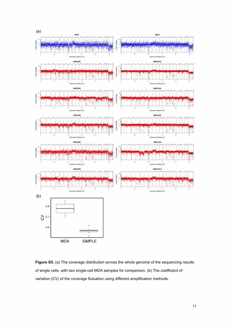
Illumina Hiseq platform. The coverage distribution across the whole genome of each sample
was listed in Figure S4.
5. A simpler SiMPLE generator combining pipette with electrical toothbrush (For Fun!)
In a very beginning of this project, we have decided to perform a 'quick and dirty' experiment to
test the idea of generation of emulsion via spinning a glass micropipette in oil. We purchased a
specific electro toothbrush (Panasonic) and replaced its brush head with a glass micropipette.
We just simply taped the micropipette to the toothbrush, and used a 20 µl conventional manual
pipette (Eppendorf) to slowly push the aqueous liquid out of the glass pipette. Although with no
precise control at all, we found this simple combination could produce a large amount of w/o
droplets within very short period of time. We noticed that the distribution of the droplets was
not monodisperse, but majority of droplets are about 50 - 100 µm in diameter, which is actually
the best size for eWGA. We also immediately realized that the motion speed of the glass
micropipette is critical since the droplet size would also be affect by the motion speed.
Interestingly this finding was verified by testing other electrical toothbrushes. Panasonic
electrical toothbrush uses circular motion to drive the brushhead, hence the linear motion
speed of micropipette is constant. While another popular brand, Philips, uses reciprocating
motion which does not provide constant linear motion speed of the brushhead, and cannot be
used in our application.
6
Part II. Supporting Figures
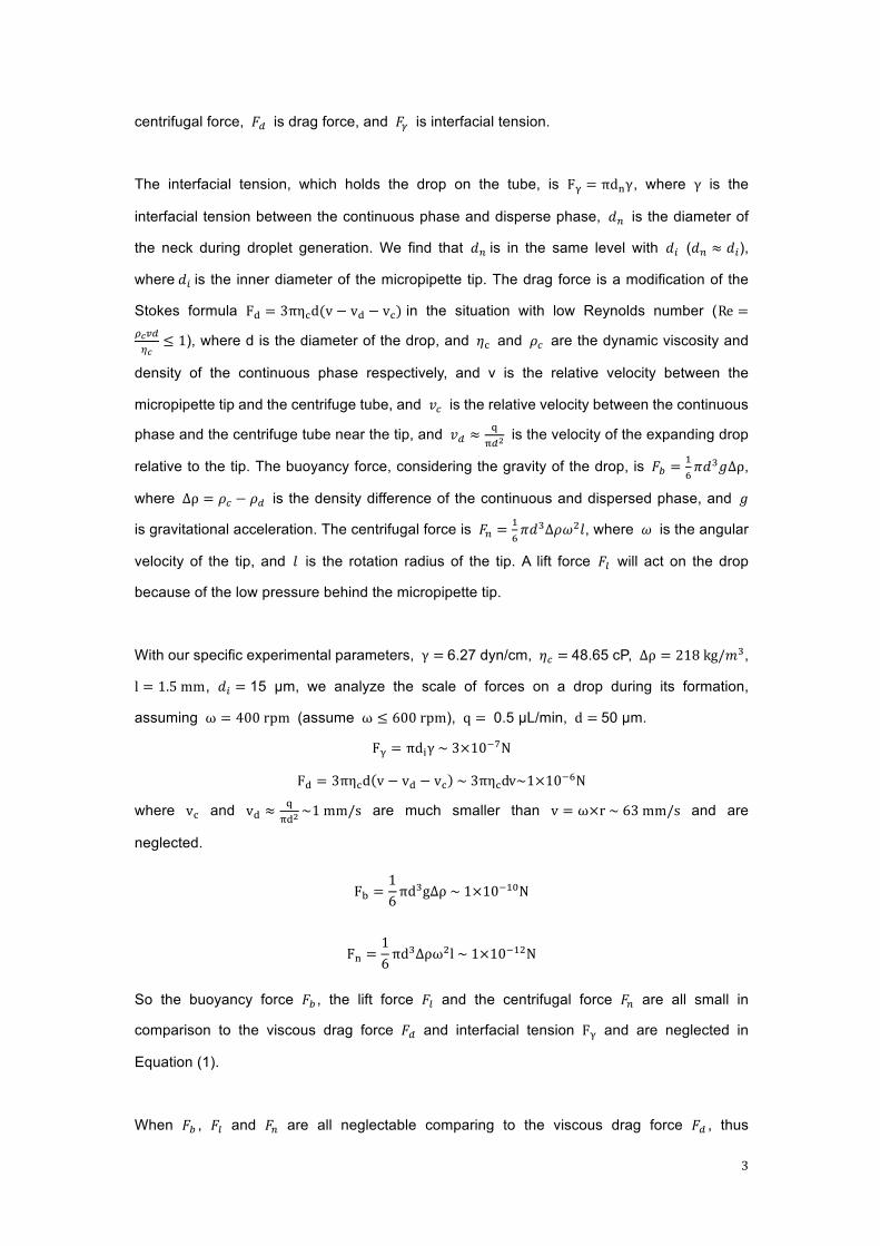
Figure S1. The design details of the loading platform (a) and the 1.5 mm off-axis eccentric
shank (b).
7
8
Cont.
Figure S2. Microscopic observation and size distribution of the w/o emulsion droplet
generated by SiMPLE generator.
9
Figure S3. Stability of w/o emulsion. The generated emulsion are placed in Nunc TopYield
strips for microscopic imaging. Microphotographs (field of view 1.5 mm x 1.5 mm) are taken at
the 1 h interval for 10 h. No noticeable fusion of fission of the droplets has been observed
during this period of time.
10
Figure S4. The quantitate PCR result of the amplified products of single cells.
11
Figure S5. (a) The coverage distribution across the whole genome of the sequencing results
of single cells, with two single-cell MDA samples for comparison. (b) The coefficient of
variation (CV) of the coverage flutuation using different amplificaiton methods.


![[XLS]sdmylife.comsdmylife.com/files/Master_Course_List_08.27.14.xlsx · Web view3. 3. 1. 1.5. 3. 3. 1.5. 1.5. 1.5. 1.5. 1.5. 1.5. 1.5. 3. 1.5. 3. 3. 3. 1.5. 1.5. 2. 3. 3. 1.5. 1.5](https://img.pdfslide.us/doc/110x75/5ac153d87f8b9a213f8cf61b/xls-view3-3-1-15-3-3-15-15-15-15-15-15-15-3-15-3-3-3.jpg)