Embed Size (px)
Citation preview
Apoptosis-101Presented on-
Date- 21-11-14
aj
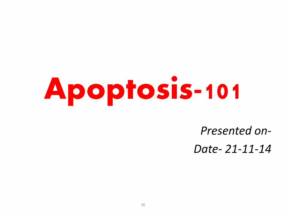
Cell response to stressful conditions and injurious stimuli
• When limits of adaptive responses are exceeded or if cells are exposed to injurious agents or stress, deprived of essential nutrients, or become compromised by mutations that affect essential cellular constituents, a sequence of events follows that is termed cell injury
• Cell death, the end result of progressive cell injury
• two principal pathways of cell death,– necrosis
– apoptosis
aj
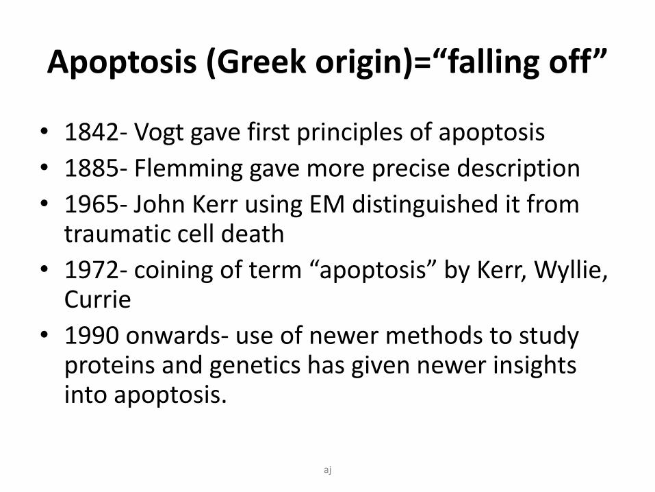
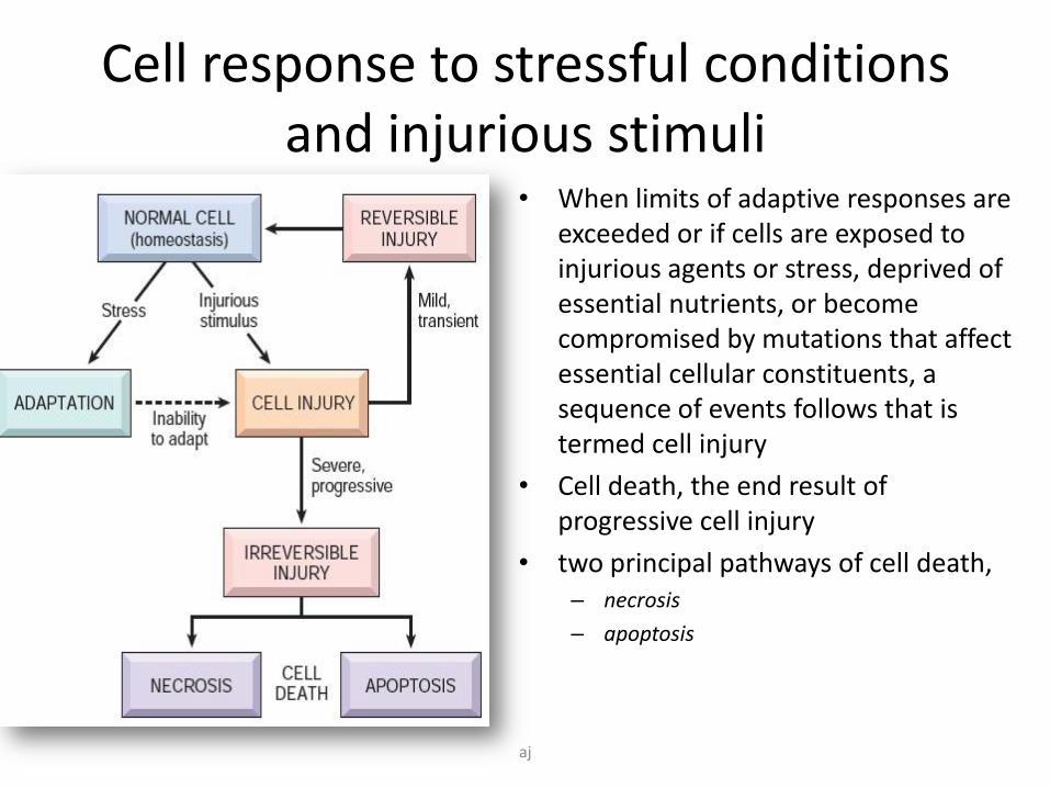
Apoptosis (Greek origin)=“falling off”
• 1842- Vogt gave first principles of apoptosis
• 1885- Flemming gave more precise description
• 1965- John Kerr using EM distinguished it from traumatic cell death
• 1972- coining of term “apoptosis” by Kerr, Wyllie, Currie
• 1990 onwards- use of newer methods to study proteins and genetics has given newer insights into apoptosis.
aj
Definition
• Apoptosis is a
– pathway of cell death that is
– induced by an internally regulated program
– in which cells destined to die activate intrinsic enzymes that degrade the cells’ own nuclear DNA and also nuclear and cytoplasmic proteins
– With minimal host reaction.
aj
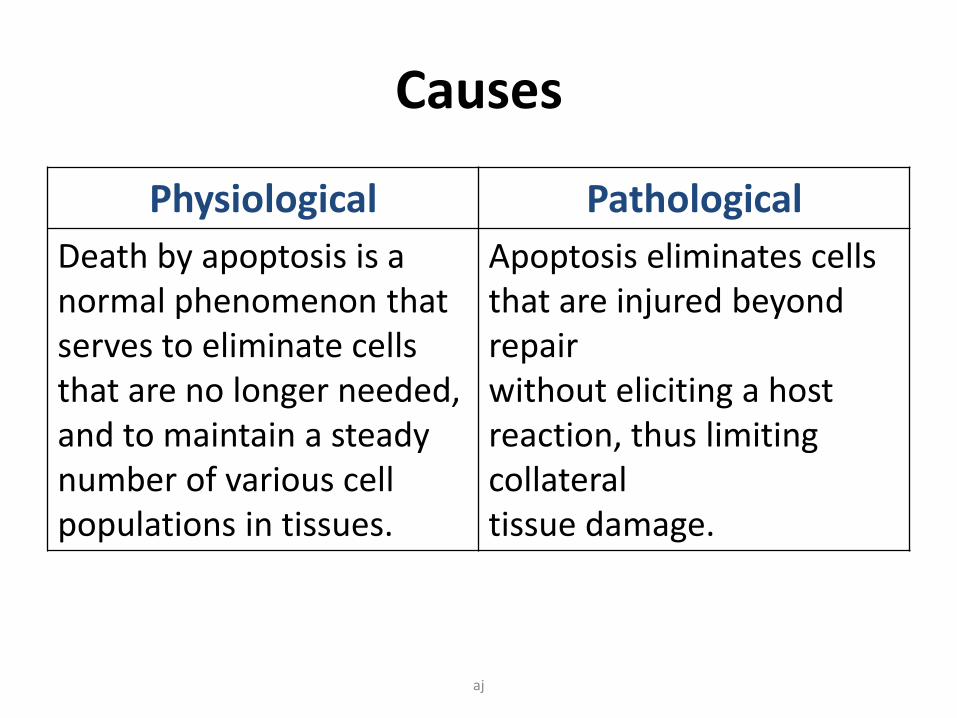
Causes
Physiological Pathological
Death by apoptosis is a normal phenomenon that serves to eliminate cells that are no longer needed, and to maintain a steady number of various cell populations in tissues.
Apoptosis eliminates cells that are injured beyond repairwithout eliciting a host reaction, thus limiting collateraltissue damage.
aj
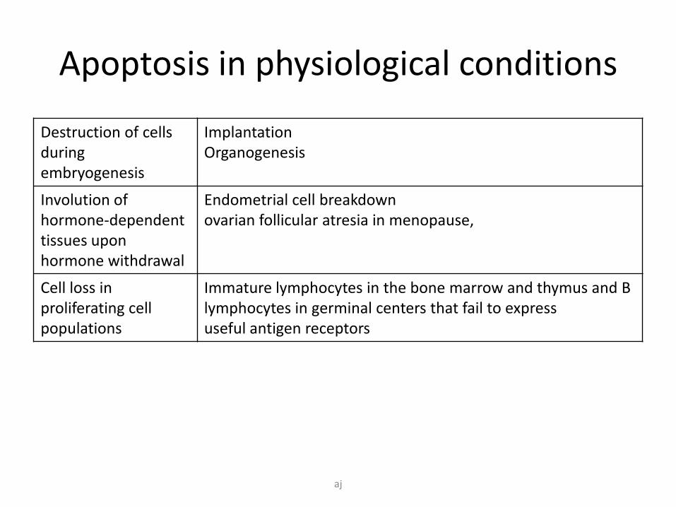
Apoptosis in physiological conditions
Destruction of cells during embryogenesis
Implantation Organogenesis
Involution of hormone-dependent tissues upon hormone withdrawal
Endometrial cell breakdownovarian follicular atresia in menopause,
Cell loss in proliferating cell populations
Immature lymphocytes in the bone marrow and thymus and B lymphocytes in germinal centers that fail to expressuseful antigen receptors
aj
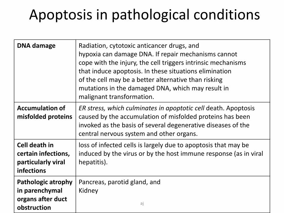
Apoptosis in pathological conditions
DNA damage Radiation, cytotoxic anticancer drugs, andhypoxia can damage DNA. If repair mechanisms cannotcope with the injury, the cell triggers intrinsic mechanismsthat induce apoptosis. In these situations eliminationof the cell may be a better alternative than riskingmutations in the damaged DNA, which may result inmalignant transformation.
Accumulation of misfolded proteins
ER stress, which culminates in apoptotic cell death. Apoptosis caused by the accumulation of misfolded proteins has been invoked as the basis of several degenerative diseases of the central nervous system and other organs.
Cell death in certain infections, particularly viral infections
loss of infected cells is largely due to apoptosis that may be induced by the virus or by the host immune response (as in viral hepatitis).
Pathologic atrophy in parenchymalorgans after duct obstruction
Pancreas, parotid gland, andKidney
aj

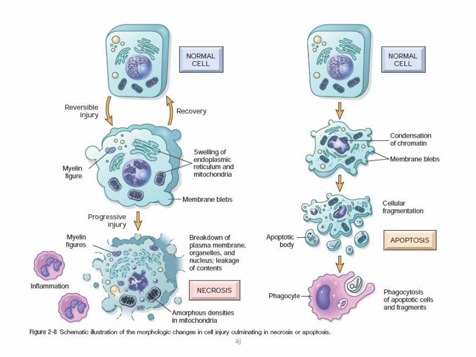
Morphological changes
aj
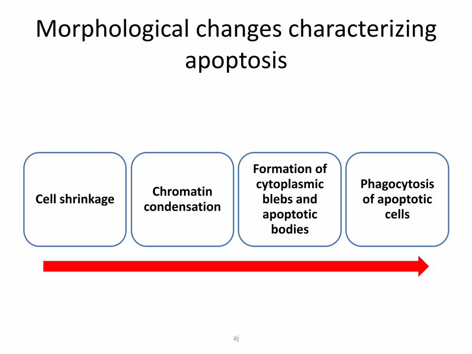
Morphological changes characterizing apoptosis
Cell shrinkageChromatin
condensation
Formation of cytoplasmic
blebs and apoptotic
bodies
Phagocytosisof apoptotic
cells
aj

Cell shrinkage
• Cell becomes smaller in size
• Cytoplasm is dense
• Organelles tightly packed
aj
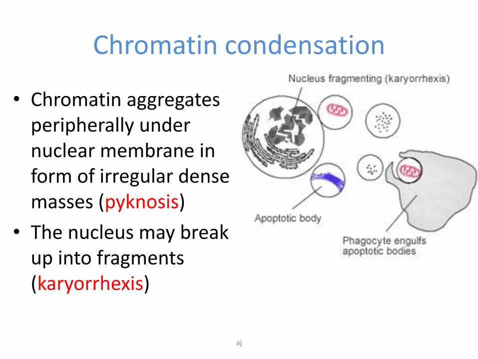
Chromatin condensation
• Chromatin aggregates peripherally under nuclear membrane in form of irregular dense masses (pyknosis)
• The nucleus may break up into fragments (karyorrhexis)
aj
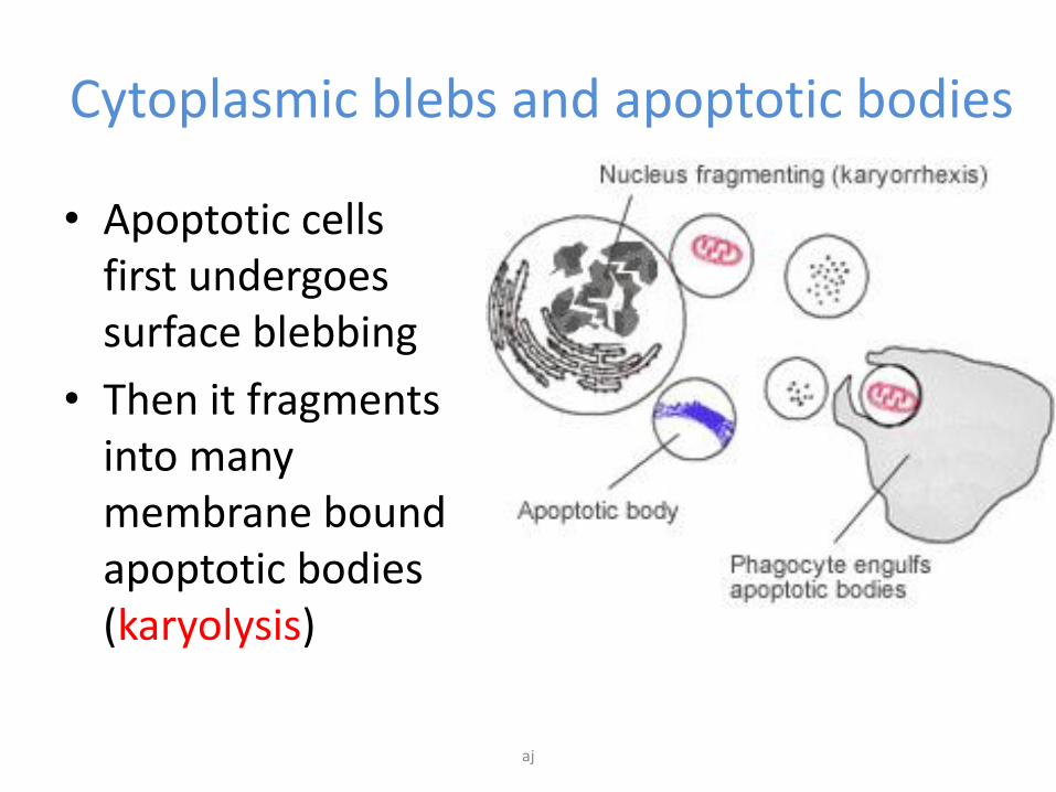
Cytoplasmic blebs and apoptotic bodies
• Apoptotic cells first undergoes surface blebbing
• Then it fragments into many membrane bound apoptotic bodies (karyolysis)
aj
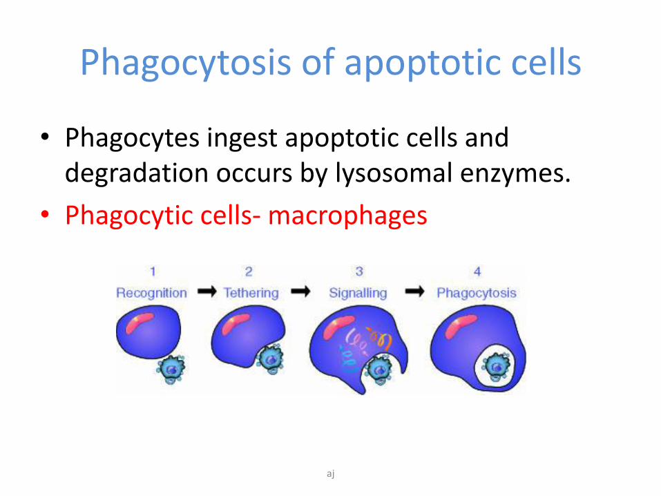
Phagocytosis of apoptotic cells
• Phagocytes ingest apoptotic cells and degradation occurs by lysosomal enzymes.
• Phagocytic cells- macrophages
aj
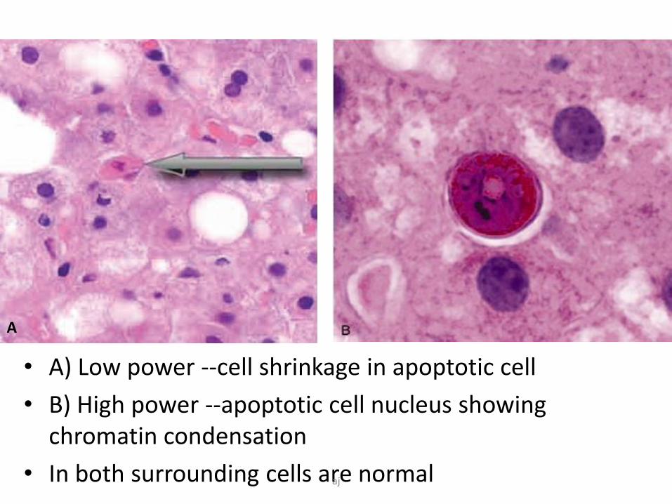
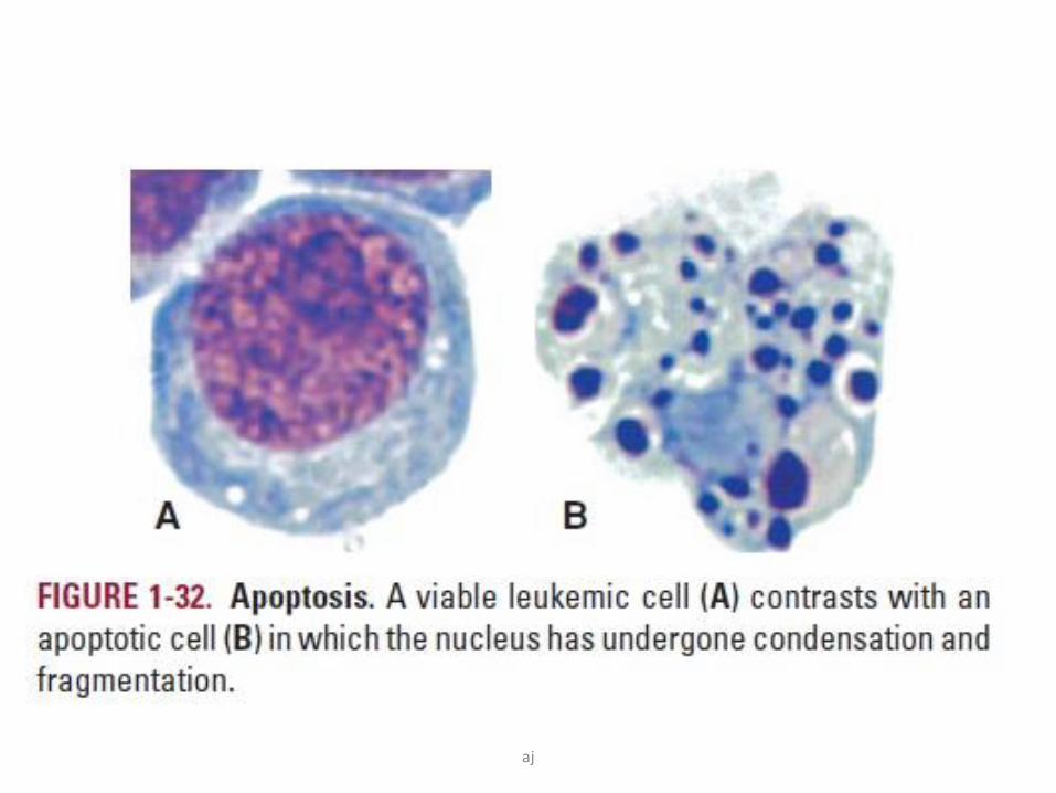
• A) Low power --cell shrinkage in apoptotic cell
• B) High power --apoptotic cell nucleus showing chromatin condensation
• In both surrounding cells are normal
A
aj
aj
Mechanism of apoptosis
aj
Apoptosis mechanism
aj
Apoptosis mechanism
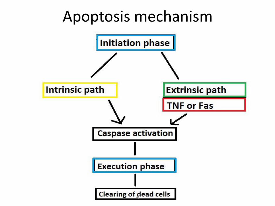
• Apoptosis results from the activation of enzymes called caspases (which exist as inactive proenzymes, or zymogens).
• Phases of apoptosis-
– Initiation- phase of activation of caspases
– Execution- activated caspases trigger cell degradation.
aj
Two Pathways of initiation of apoptosis
1. Intrinsic 2. Extrinsic
Mitochondrial Death-receptorinitiated
Initiator phase
aj
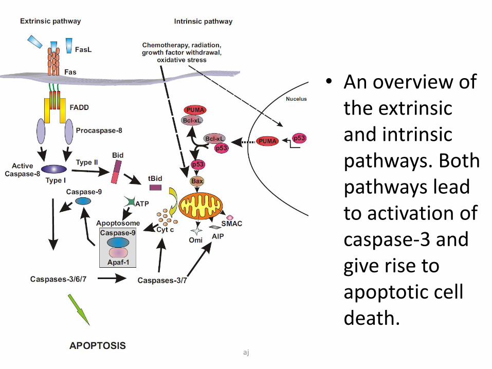
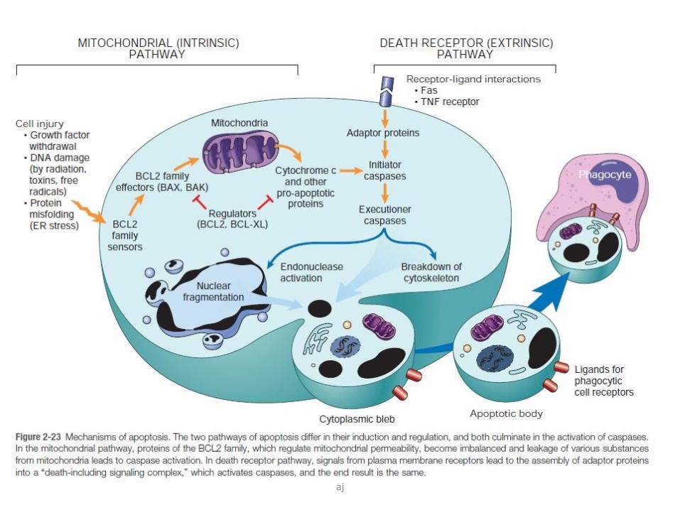
• An overview of the extrinsic and intrinsic pathways. Both pathways lead to activation of caspase-3 and give rise to apoptotic cell death.
aj
Intrinsic pathway
• Major mechanism of apoptosis in all mammalian cells.
• Increased permeability of the mitochondrial outer membrane with consequent release of death-inducing (pro-apoptotic) molecules from the mitochondrial intermembrane space into the cytoplasm.
• The release of mitochondrial pro-apoptotic proteins is tightly controlled by the – BCL2 family of proteins
aj
BCL2 family of proteins
• BCL2 stands for B-cell lymphoma 2.
aj
BCL2 family of proteins
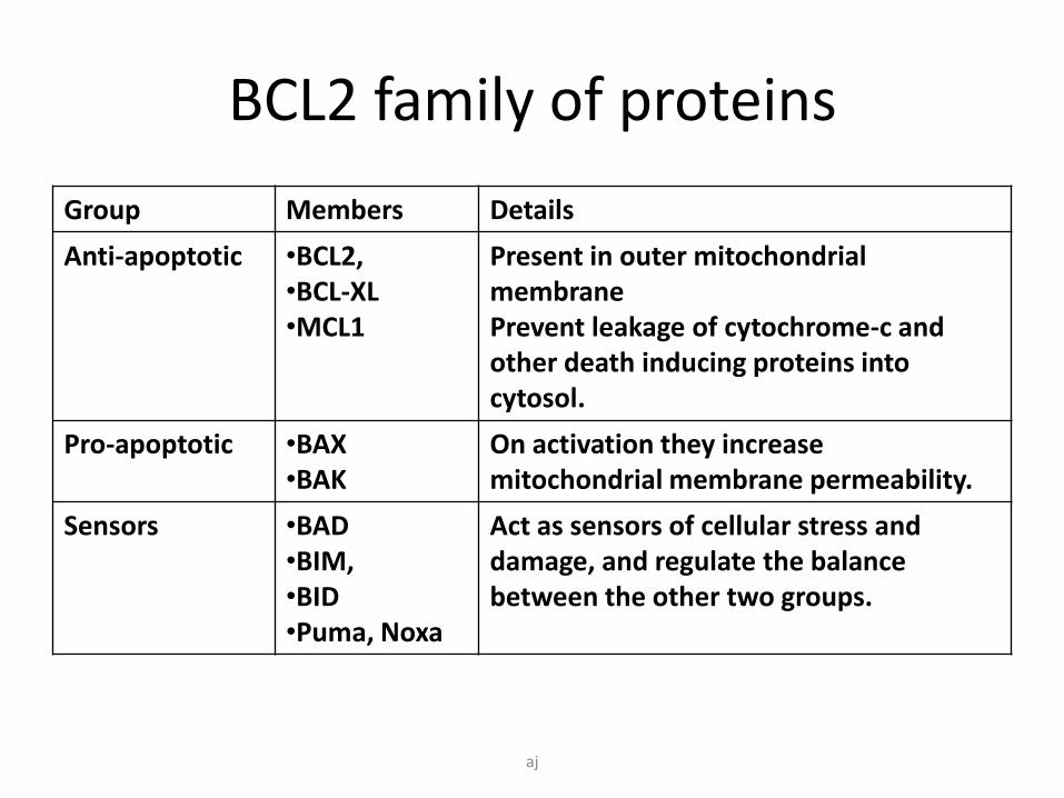
Group Members Details
Anti-apoptotic •BCL2, •BCL-XL•MCL1
Present in outer mitochondrial membranePrevent leakage of cytochrome-c and other death inducing proteins into cytosol.
Pro-apoptotic •BAX•BAK
On activation they increasemitochondrial membrane permeability.
Sensors •BAD•BIM,•BID•Puma, Noxa
Act as sensors of cellular stress and damage, and regulate the balance between the other two groups.
aj
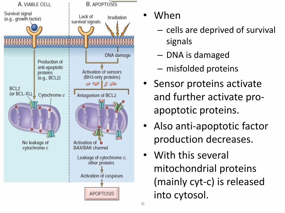
• When
– cells are deprived of survival signals
– DNA is damaged
– misfolded proteins
• Sensor proteins activate and further activate pro-apoptotic proteins.
• Also anti-apoptotic factor production decreases.
• With this several mitochondrial proteins (mainly cyt-c) is released into cytosol.
aj
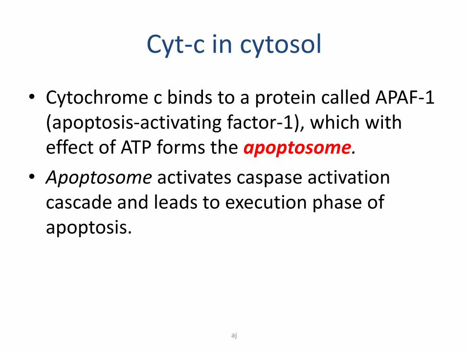
Cyt-c in cytosol
• Cytochrome c binds to a protein called APAF-1 (apoptosis-activating factor-1), which with effect of ATP forms the apoptosome.
• Apoptosome activates caspase activation cascade and leads to execution phase of apoptosis.
aj
SMAC proteins in cytosol
• Mitochondrial proteins known as SMACs (small mitochondria-derived activator of caspases) are released into the cytosol following an increase in permeability. SMAC’s neutralize inhibitor of apoptosis proteins (IAPs).
• This leads to caspase activation thus apoptosis proceeds to execution phase.
aj
Extrinsic pathways
• This pathway is initiated by engagement of plasma membrane death receptors on a variety of cells.
• These death receptors belong to TNF receptor family.
• These receptors link with a external protein to form a complex termed “death domain” which is important to activate apoptosis.
• Important death receptor activating proteins are
– TNF
– Fas ligand
aj
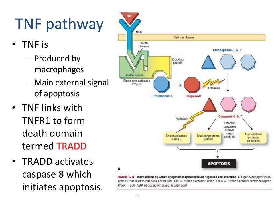
TNF pathway• TNF is
– Produced by macrophages
– Main external signal of apoptosis
• TNF links with TNFR1 to form death domain termed TRADD
• TRADD activates caspase 8 which initiates apoptosis.
aj
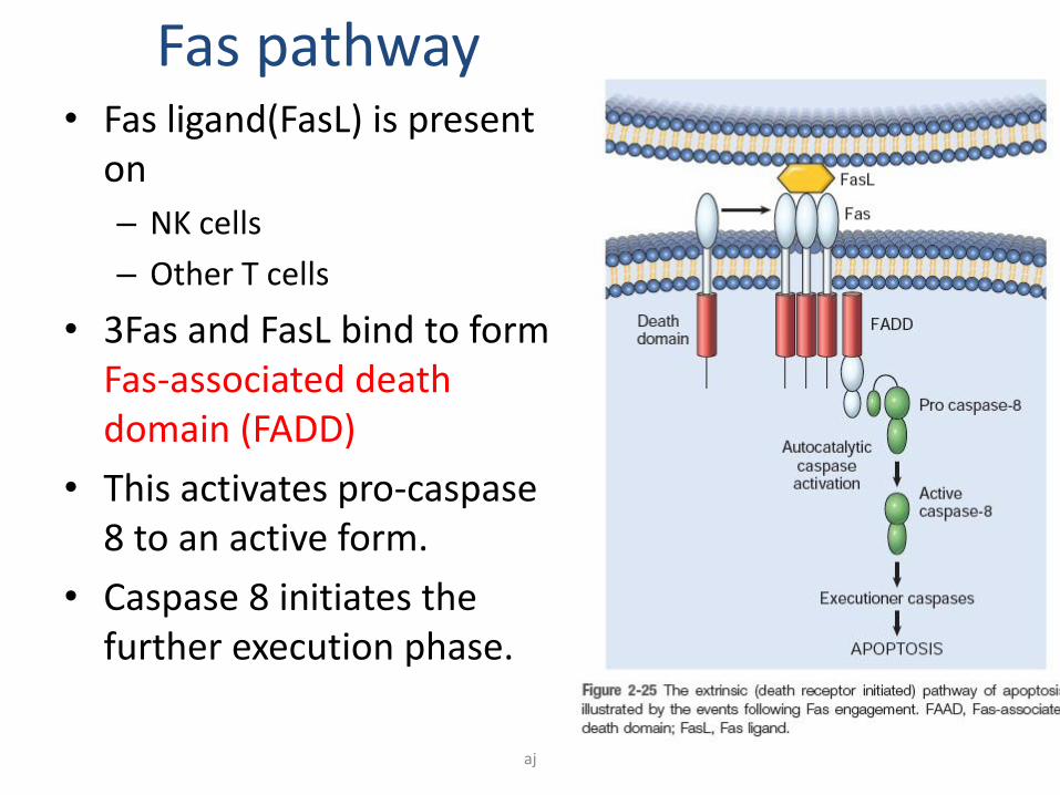
Fas pathway• Fas ligand(FasL) is present
on
– NK cells
– Other T cells
• 3Fas and FasL bind to form Fas-associated death domain (FADD)
• This activates pro-caspase8 to an active form.
• Caspase 8 initiates the further execution phase.
aj
Execution phase
• Both initiating pathways converge at level of caspase activation.
• Initiator caspases(8,9,10) activate the executioner caspases(3,6).
• These executioner caspases signal DNA cleavage.
• DNA clevage causes disintegration of nucleus(karyorrhexis) and also damages nuclear membrane.
aj
Clearing of dead cells
• Clearing of apoptotic cells is the final step of apoptosis and in healthy individuals this process is efficient and rapid enough to prevent any inflammation and necrosis.
• Phagocytic cells- macrophages.
aj
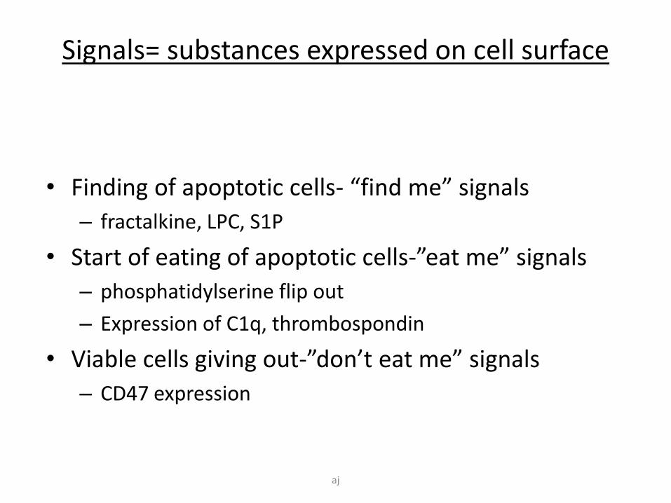
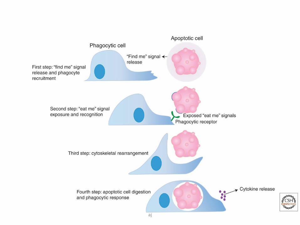
Signals= substances expressed on cell surface
• Finding of apoptotic cells- “find me” signals
– fractalkine, LPC, S1P
• Start of eating of apoptotic cells-”eat me” signals
– phosphatidylserine flip out
– Expression of C1q, thrombospondin
• Viable cells giving out-”don’t eat me” signals
– CD47 expression
aj
aj
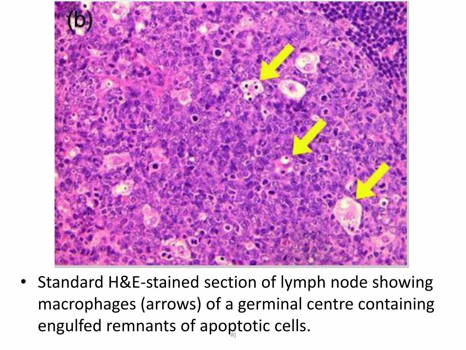
• Standard H&E-stained section of lymph node showing macrophages (arrows) of a germinal centre containing engulfed remnants of apoptotic cells.
aj
aj
• Few examples of apoptosis-
aj
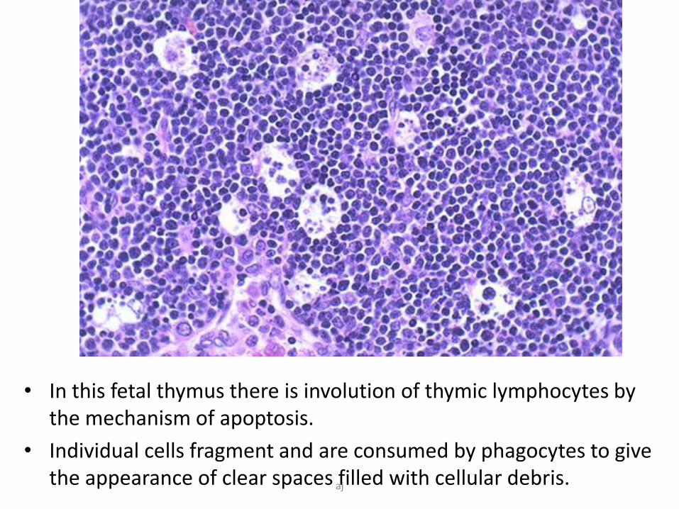
• In this fetal thymus there is involution of thymic lymphocytes by the mechanism of apoptosis.
• Individual cells fragment and are consumed by phagocytes to give the appearance of clear spaces filled with cellular debris. aj
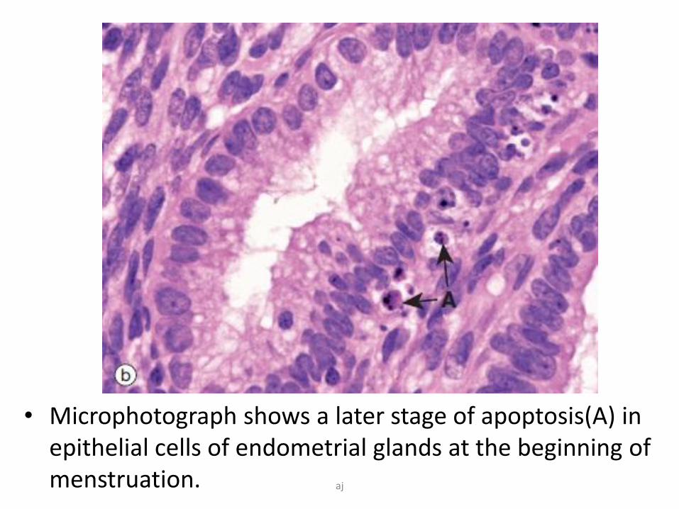
• Microphotograph shows a later stage of apoptosis(A) in epithelial cells of endometrial glands at the beginning of menstruation. aj
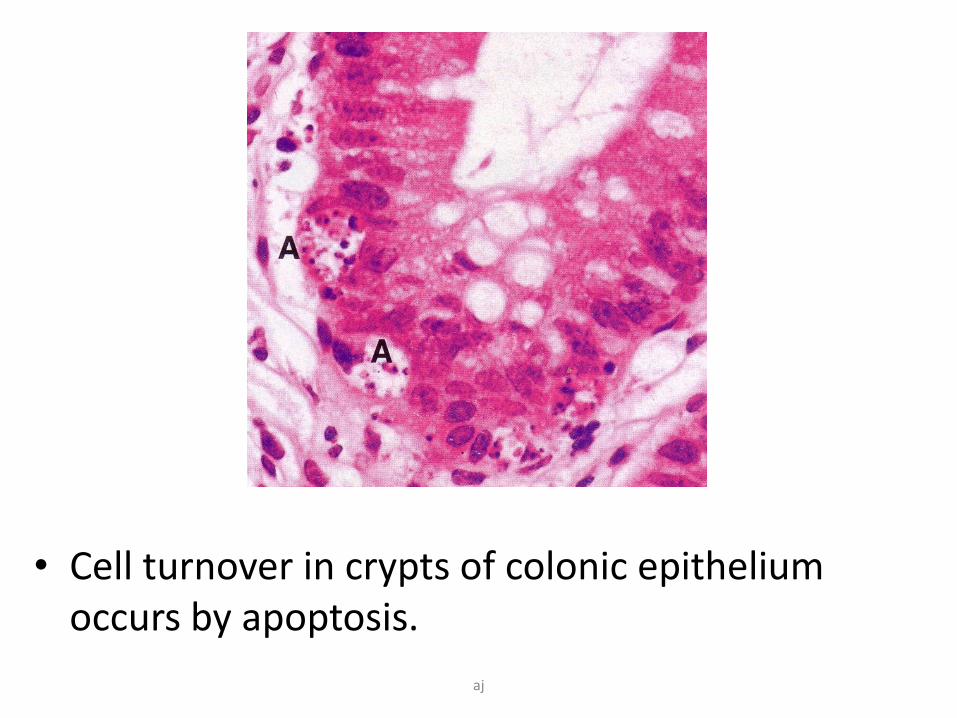
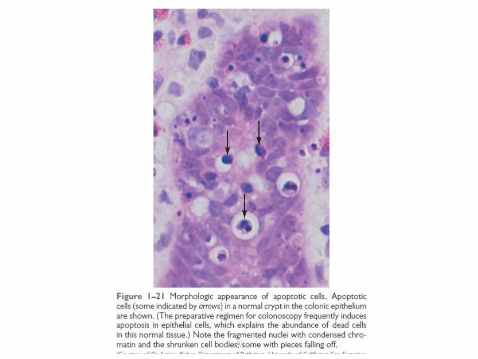
• Cell turnover in crypts of colonic epithelium occurs by apoptosis.
aj
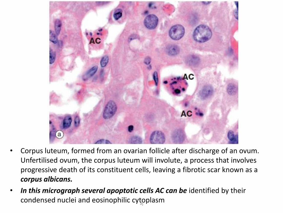
• Corpus luteum, formed from an ovarian follicle after discharge of an ovum. Unfertilised ovum, the corpus luteum will involute, a process that involves progressive death of its constituent cells, leaving a fibrotic scar known as a corpus albicans.
• In this micrograph several apoptotic cells AC can be identified by their condensed nuclei and eosinophilic cytoplasmaj
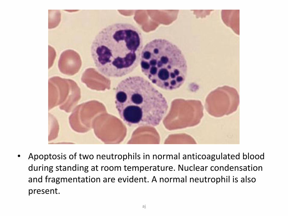
• Apoptosis of two neutrophils in normal anticoagulated blood during standing at room temperature. Nuclear condensation and fragmentation are evident. A normal neutrophil is also present.
aj
aj
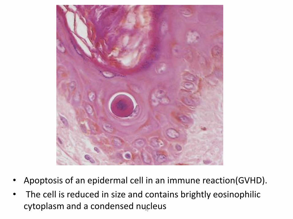
• Apoptosis of an epidermal cell in an immune reaction(GVHD).
• The cell is reduced in size and contains brightly eosinophilic cytoplasm and a condensed nucleusaj
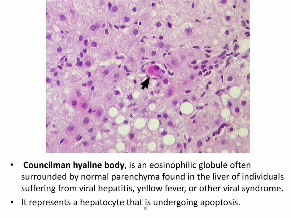
• Councilman hyaline body, is an eosinophilic globule often surrounded by normal parenchyma found in the liver of individuals suffering from viral hepatitis, yellow fever, or other viral syndrome.
• It represents a hepatocyte that is undergoing apoptosis.aj

• Erythema Multiforme (EM) is a hypersensitivity reaction usually caused by infections, mostly Herpes simplex virus.
• Keratinocytes show apoptosis.aj
Dysregulated apoptosis-too less
-too much
aj
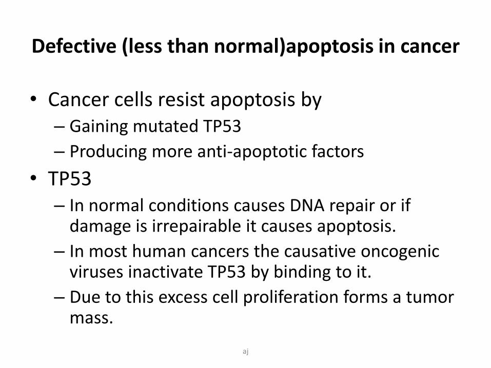
Defective (less than normal)apoptosis in cancer
• Cancer cells resist apoptosis by– Gaining mutated TP53
– Producing more anti-apoptotic factors
• TP53– In normal conditions causes DNA repair or if
damage is irrepairable it causes apoptosis.
– In most human cancers the causative oncogenicviruses inactivate TP53 by binding to it.
– Due to this excess cell proliferation forms a tumor mass.
aj
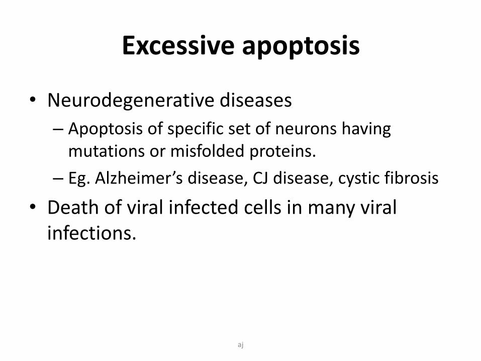
Excessive apoptosis
• Neurodegenerative diseases
– Apoptosis of specific set of neurons having mutations or misfolded proteins.
– Eg. Alzheimer’s disease, CJ disease, cystic fibrosis
• Death of viral infected cells in many viral infections.
aj
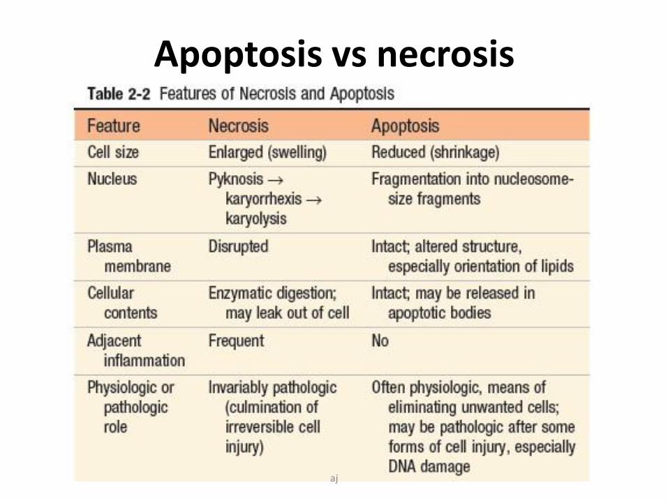
Apoptosis vs necrosis
aj
aj
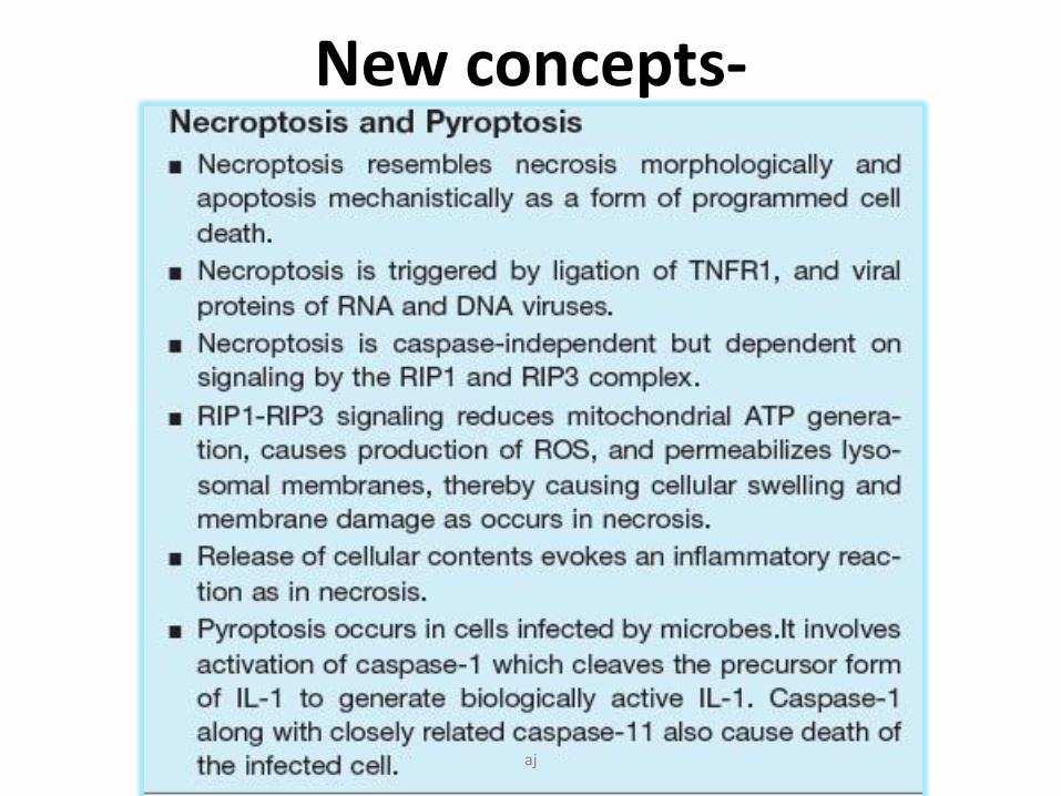
New concepts-
aj
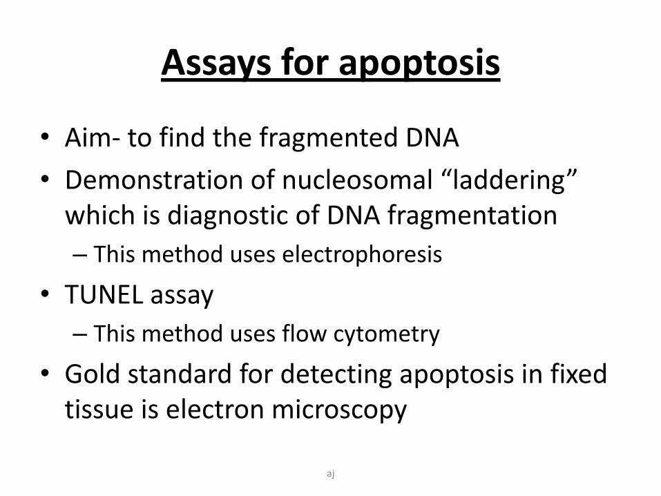
Assays for apoptosis
• Aim- to find the fragmented DNA
• Demonstration of nucleosomal “laddering” which is diagnostic of DNA fragmentation
– This method uses electrophoresis
• TUNEL assay
– This method uses flow cytometry
• Gold standard for detecting apoptosis in fixed tissue is electron microscopy
aj
S
u
m
m
a
r
yaj

Thank you
aj