Embed Size (px)
Citation preview

A modulator of the low-voltage activated T-type calcium channel that reverses HIV
glycoprotein 120-, paclitaxel-, and spinal nerve ligation-induced peripheral neuropathies
Song Cai‡,¶, Peter Tuohy†,¶, Chunlong Ma†, Naoya Kitamura†, Kimberly Gomez‡, Yuan Zhou‡,
Dongzhi Ran‡, Shreya Sai Bellampalli‡, a, Jie Yu‡, Shizhen Luo‡, Angie Dorame‡, Nancy Yen Ngan
Pham‡, Gabriella Molnar‡, John M. Streicher‡, Marcel Patek¢, Samantha Perez-Miller‡, Aubin
Moutal‡, Jun Wang†,*, and Rajesh Khanna‡, #, ¥, Δ,*
†Department of Pharmacology and Toxicology, College of Pharmacy, The University of Arizona,
Tucson, Arizona 85721, United States ‡Department of Pharmacology, College of Medicine, The University of Arizona, Tucson, Arizona,
85721, United States ¢Bright Rock Path Consulting LLC, Tucson, AZ #BIO5 Institute, 1657 East Helen Street, P.O. Box 210240, Tucson, AZ 85721, United States ¥The Center for Innovation in Brain Sciences, The University of Arizona Health Sciences, Tucson,
Arizona, United States ΔRegulonix LLC, Tucson, Arizona, United States ¶These authors contributed equally
Expanded Materials and Methods
Animals.
Pathogen-free adult male and female Sprague-Dawley rats (225-250g; Envigo, Indianapolis, IN)
were housed in temperature-controlled (23±3 ˚C) and light-controlled (12-h light/12-h dark cycle;
lights on 07:00-19:00) rooms with standard rodent chow and water available ad libitum. The
Institutional Animal Care and Use Committee of the College of Medicine at the University of
Arizona approved all experiments. All procedures were conducted in accordance with the Guide
for Care and Use of Laboratory Animals published by the National Institutes of Health and the
ethical guidelines of the International Association for the Study of Pain. Animals were randomly
assigned to treatment or control groups for the behavioral experiments. Animals were initially
housed 3 per cage but individually housed after the intrathecal cannulation. All behavioral

experiments were performed by experimenters who were blinded to the experimental groups and
treatments.
Preparation of acutely dissociated dorsal root ganglion neurons
Dorsal root ganglia from all levels were acutely dissociated using methods as described previously
[6]. Rat DRG neurons were isolated from 100g female Sprague-Dawley rats using previously
developed procedures [13]. In brief, removing dorsal skin and muscle and cutting the vertebral
bone processes parallel to the dissection stage exposed the DRGs. DRGs were then collected,
trimmed at their roots, and enzymatically digested in 3 mL bicarbonate-free, serum-free, sterile
DMEM (Cat# 11965, Thermo Fisher Scientific, Waltham, MA) solution containing neutral
protease (3.125 mg.ml-1, Cat#LS02104; Worthington, Lakewood, NJ) and collagenase type I (5
mg/mL, Cat# LS004194, Worthington, Lakewood, NJ) and incubated for 60 minutes at 37˚C under
gentle agitation. Dissociated DRG neurons (~1.5 x 10⁶) were then gently centrifuged to collect
cells and washed with DRG media DMEM containing 1% penicillin/streptomycin sulfate from
10,000 μg/mL stock, 30 ng/mL nerve growth factor, and 10% fetal bovine serum before plating
onto poly-D-lysine– and laminin-coated glass 12- or 15-mm coverslips.
Calcium imaging in acutely dissociated dorsal root ganglion neurons
Dorsal root ganglion neurons were loaded for 30 minutes at 37˚C with 3 µM Fura-2AM (Cat#
F1221, Thermo Fisher, stock solution prepared at 1mM in DMSO, 0.02% pluronic acid, (Cat#P-
3000MP, Thermo Fisher) to follow changes in intracellular calcium([Ca2+]c) in a standard bath
solution containing 139 mM NaCl, 3 mM KCl, 0.8 mM MgCl2, 1.8 mM CaCl2, 10 mM Na HEPES,
pH 7.4, 5 mM glucose exactly as previously described [1]. Fluorescence imaging was performed
with an inverted microscope, NikonEclipseTi-U (Nikon Instruments Inc., Melville, NY), using
objective Nikon Fluor 4X and a Photometrics cooled CCD camera CoolSNAPES2 (Roper
Scientific, Tucson, AZ) controlled by NIS Elements software (version 4.20, Nikon Instruments).
The excitation light was delivered by a Lambda-LS system (Sutter Instruments, Novato, CA). The
excitation filters (340 ± 5 and 380 ± 7) were controlled by a Lambda 10 to 2 optical filter change
(Sutter Instruments). Fluorescence was recorded through a 505-nm dichroic mirror at 535 ± 25 nm.
To minimize photobleaching and phototoxicity, the images were taken every ~10 seconds during
the time-course of the experiment using the minimal exposure time that provided acceptable image
quality. The changes in [Ca2+]c were monitored by following a ratio of F340/F380, calculated after
subtracting the background from both channels.
Dorsal root ganglia neuron transfection.
Collected cells were re-suspended in Nucleofector transfection reagent containing siRNA at 500
nM and 2 µg of the provided GFP plasmid as detailed previously [4]. Cells were then subjected to
electroporation protocol O-003 in an Amaxa Biosystem (Lonza, Basel, Switzerland) and plated
onto poly-D-lysine - and laminin-coated glass 12-mm coverslips. Transfection efficiencies were
routinely between 20% and 30% with ~10% cell death. Small diameter neurons were selected to
target Aδ- and c- fiber nociceptive neurons. For rat DRG culture small cells were considered to be
~ < 30 µm as determined by an eyepiece micrometer within the objective lens. Successfully
transfected cells were identified by GFP fluorescence. The siRNA sequences used were:
UAGAUAGCAAAUACUUUGGCCGGGG (for Cacna1g/CaV3.1; (Cat# RSS355855,
Thermofisher)); CAGCCAUCUUCGUGGUGGAGAUGAU (for Cacna1h/CaV3.2; (Cat#
RSS350286, Thermofisher)); CAGCAUCCUUGGGAUGCAUAUCUUU (for Cacna1i/CaV3.3;
Cat# RSS367566); and siRNA Negative Control, Med GC was used as a scrambled siRNA control
(Cat# 12935300). Cells were used 48 hrs after transfection.
Constellation Pharmacology.
These experiments were performed as described previously [13; 19], but with the following
modifications. Dorsal root ganglia neurons were loaded at 37˚C with 3µM Fura-2AM for 30
minutes in Tyrode solution (at ~310 mOsm) containing 119 mM NaCl, 2.5 mM KCl, 2 mM MgCl2,
2 mM CaCl2, 25 mM HEPES, pH 7.4, and 30 mM glucose. After a 1-minute baseline measurement,
Ca2+ influx was stimulated by the addition of the following receptor agonists: 400 nM menthol, 50
µM histamine, 10 µM adenosine triphosphate (ATP), 200 µM allyl isothiocyanate (AITC), 1 mM
acetylcholine (Ach), and 100 nM capsaicin diluted in Tyrode solution. At the end of the
constellation pharmacology protocol, cell viability was assessed by depolarization-induced Ca2+
influx using and an excitatory KCl solution comprising 32 mM NaCl, 90 mM KCl, 2 mM MgCl2,
2 mM CaCl2, 25 mM HEPES, pH 7.4, and 30 mM glucose. After the 1-minute baseline
measurement, each trigger was applied for 15 seconds in the order indicated above in 6-minute
intervals. After each trigger, bath solution was continuously perfused over the cells to wash off
excess of the trigger. This process was automated using the ValveBank II perfusion system that
controlled the perfusion of the standard bath solution and triggers (Automate Scientific, San Diego,
CA). Except for the time course experiments, 5bk was incubated overnight onto DRGs. In all cases,
5bk was also added to the Tyrode solution during the loading with Fura-2AM. Fluorescence
imaging was performed under the same conditions noted above for calcium imaging. A cell was
defined as a “responder” if its fluorescence ratio of 340 nm/380 nm was greater than 10% of the
baseline value calculated using the average fluorescence in the 30 seconds preceding application
of the trigger.
Whole-cell patch recordings of Ca2+ and Na+ currents in acutely dissociated DRG neurons.
Recordings were obtained from acutely dissociated DRG neurons as described previously [9; 14].
To isolate calcium currents, Na+ and K+ currents were blocked with 500 nM tetrodotoxin (TTX;
Alomone Laboratories) and 30 mM tetraethylammonium chloride (TEA-Cl; Sigma). Extracellular
recording solution (at ~310 mOsm) consisted of the following (in mM): 110 N-methyl-D-
glucamine (NMDG), 10 BaCl2, 30 TEA-Cl, 10 HEPES, 10 glucose, pH at 7.4, 0.001 TTX, 0.01
nifedipine. The intracellular recording solution (at ~310 mOsm) consisted of the following (in
mM): 150 CsCl2, 10 HEPES, 5 Mg-ATP, 5 BAPTA, pH at 7.4. The protocol for isolating T-type
calcium currents was previously described by Choe et al.[3] The extracellular recording solution
used to isolate T currents consisted of the following (in millimolar): 2 CaCl2, 152 TEA-Cl, 10
HEPES, pH adjusted to 7.4 with TEA-OH. The intracellular recording solution consisted of (in
millimolar): 135 tetramethylammonium hydroxide, 10 EGTA, 40 HEPES, and 2 MgCl2, pH
adjusted to 7.2 with hydrofluoric acid. Activation of ICa-T was measured by using a holding voltage
of -90 mV with voltage steps 200 ms in duration applied at 500-ms intervals in 10 mV increments
from –70 to +60 mV. Inactivation of ICa-T was determined by applying a 1500-ms conditioning
prepulse (–110 to+20 mV in 10 mV increments) after which the voltage was stepped to –30 mV
for 20 ms; a 40-ms interval with a holding voltage of -90 mV separated each acquisition. In the

deactivation tau protocol, the neuron was first held at –110 mV, then the voltage jumped to –30
mV for 10 ms followed by a 50-ms conditioning prepulse (–160 to –40 mV in 10 mV increments).
A 2-second interval with a holding voltage of –90 mV separated each acquisition. ICa-T recovery
from inactivation were obtained by using our standard double-pulse protocol with variable
interpulse duration at –90 mV after a 500-ms-long inactivating pulse (Vh=–90 mV; Vt=–30 mV).
To isolate the contributions of the HVA calcium channel subtypes, we applied all but one
of the following subunit-selective blockers (all purchased from Alomone Labs, Jerusalem):
Nifedipine (10 μM, L-type); -agatoxin GIVA (200 nM, P/Q-type) [12]; SNX-482 (200 nM, R-
type) [16]; ω-conotoxin GVIA (500 nM, N-type) [7] or TTA-P2 (1 µM, T-type)[3] to individually
isolate the subtypes.
For recording sodium currents the internal solution consisted of (in mM): 140 CsF, 10 NaCl,
1.1Cs-EGTA, and 15 HEPES (pH 7.3, mOsm/L = 290-310) and external solution contained (in
mM): 140 NaCl, 30 tetraethylammonium chloride, 10 D-glucose, 3 KCl, 1 CaCl2, 0.5 CdCl2, 1
MgCl2, and 10 HEPES (pH 7.3, mOsm/L = 310-315). DRG neurons were interrogated with
current-voltage (I-V) and activation/inactivation voltage protocols as previously described [4; 6].
The voltage protocols were as follows: (a) I-V protocol: from a −60 mV holding potential, cells
were depolarized in 150-millisecond voltage steps from −70 to +60 mV (5-mV increments) which
permitted acquisition of current density values such that we could analyze activation of sodium
channels as a function of current vs voltage and infer peak current density (normalized to cell
capacitance (in picofarads, pF)), which occurred between ~0 to 10 mV; (b) inactivation protocol:
from a −60 mV holding potential, cells were subjected to hyperpolarizing/repolarizing pulses for
1 second between −120 to 0 mV (+10 mV steps). This increment conditioned various proportions
of channels into a state of fast-inactivation – in this case 0-mV test pulse for 200 milliseconds was
able to reveal fast inactivation when normalized to maximum sodium current. Because of the
differential inactivation kinetics of TTX-resistant and TTX-sensitive channels, the fast inactivation
protocol allowed subtraction of electrically isolated TTX-R (current available after –40mV
prepulse) from total current (current available after -120mV prepulse), as previously described [4].
Pipettes with 1 to 3 MΩ resistance were used for all recordings.
The Boltzmann relation was used to determine the voltage dependence for activation of ICa
and INa wherein the conductance–voltage curve was fit by the equation G/Gmax=1/[1+exp (V0.5-

Vm)/k], where G is the conductance G=I/(Vm-ECa or ENa), Gmax is the maximal conductance
obtained from the Boltzmann fit under control conditions, V0.5 is the voltage for half-maximal
activation, Vm is the membrane potential, and k is a slope factor. ECa is the reversal potential for
ICa; ENa is the reversal potential for INa and was determined for each individual neuron. The values
of ICa and INa around the reversal potential were fit with a linear regression line to establish the
voltage at which the current was zero. The Boltzmann parameters were determined for each
individual neuron and then used to calculate the mean ± SEM.
Whole-cell recordings were obtained with a HEKA EPC-10 USB (HEKA Instruments Inc.);
data were acquired with a Patchmaster (HEKA) and analyzed with a Fitmaster (HEKA).
Capacitive artifacts were fully compensated, and series resistance was compensated by ~70%.
Recordings made from cells with greater than a 5-mV shift in series resistance compensation error
were excluded from analysis. All experiments were performed at room temperature (~23°C).
Pipettes with 1-3MΩ resistance were used for all recordings.
Calcitonin gene–related peptide release from lumbar slices.
Rats were deeply anesthetized with 5% isoflurane and then decapitated. Two vertebral incisions
(cervical and lumbar) were made in order to expose the spinal cord. Pressure was applied to a
saline-filled syringe inserted into the lumbar vertebral foramen, and the spinal cord was extracted.
Only the lumbar region of the spinal cord was used for the CGRP release assay. Baseline
treatments (#1 and #2) involved bathing the spinal cord in Tyrode’s solution. The excitatory
solution consisting of 90 mM KCl was paired with the treatment for fraction #4. These fractions
(10 minutes, 400 L each) were collected for measurement of CGRP release. Samples were
immediately flash frozen and stored in a -20 C freezer. 5bk (20 M) or vehicle (0.9% saline) was
added to the pretreatment and co-treatment fractions (#3 and 4). The concentration of CGRP
released into the buffer was measured by enzyme-linked immunosorbant assay (Cat# 589001,
Cayman Chemical, Ann Arbor, MI).
Preparation of spinal cord slices.

As described previously [22], young rats (postnatal 10-14 days) were deeply anesthetized with
diethyl ether. For spinal nerve blocking, 0.3 mL of 2% lidocaine was injected to both sides of L4
to 5 lumbar vertebrae. Laminectomy was performed from mid-thoracic to low lumbar levels, and
the spinal cord was quickly removed to cold modified artificial cerebrospinal fluid (aCSF)
oxygenated with 95% O2 and 5% CO2. The aCSF contained (in millimolar): 80 NaCl, 2.5 KCl,
1.25 NaH2PO4, 0.5 CaCl2, 3.5 MgCl2, 25 NaHCO3, 75 sucrose, 1.3 ascorbate, 3.0 sodium pyruvate,
with pH at 7.4 and osmolarity at 310 mOsm. Transverse 350-m thick slices were obtained by a
vibratome (VT1200S; Leica, Nussloch, Germany). Slices were then incubated for at least 1 hour
at RT in an oxygenated recording solution containing (in millimolar): 125 NaCl, 2.5 KCl, 2 CaCl2,
1 MgCl2, 1.25 NaH2PO4, 26 NaHCO3, 25 D-glucose, 1.3 ascorbate, 3.0 sodium pyruvate, with pH
at 7.4 and osmolarity at 320 mOsm. The slices were then positioned in a recording chamber and
continuously perfused with oxygenated recording solution at a rate of 3 to 4 mL/min before
electrophysiological recordings at RT.
Electrophysiological recording in spinal cord slices by whole-cell patch clamp.
Substantia gelatinosa neurons were visualized and identified in the slices by means of infrared
differential interference contrast video microscopy on an upright microscope (FN1; Nikon, Tokyo,
Japan) equipped with a 3 40/0.80 water-immersion objective and a charge-coupled device camera.
Patch pipettes with resistance at 6 to 10 M were made from borosilicate glass (Sutter Instruments,
Novato, CA) on a 4-steps micropipette puller (P-90; Sutter Instruments, Novato, CA). The pipette
solution contained the following (in millimolar): 120 potassium gluconate, 20 KCl, 2 MgCl 2, 2
Na 2 -ATP, 0.5 Na-GTP, 20 HEPES, 0.5 EGTA, with pH at 7.28 and osmolarity at 310 mOsm.
The membrane potential was held at -60 mV using PATCHMASTER software in combination
with a patch clamp amplifier (EPC10; HEKA Elektronik, Lambrecht, Germany).
The whole-cell configuration was obtained in voltage-clamp mode. To record spontaneous
excitatory postsynaptic currents (sEPSCs), bicuculline methiodide (10 M) and strychnine (1 M)
were added to the recording solution to block γ-aminobutyric acid-activated and glycine-activated
currents. 5bk was applied for 2-3 hours prior to the recordings. Hyperpolarizing step pulses (5 mV
in intensity, 50 milliseconds in duration) were periodically delivered to monitor the access
resistance (15-25 MΩ), and recordings were discontinued if the access resistance changed by more
than 20%. For each neuron, sEPSCs were recorded for a total duration of 2 minutes. Currents were
filtered at 3 kHz and digitized at 5 kHz. Data were further analyzed by the Mini-Analysis
(Synatosoft Inc, NJ) and Clampfit 10.7 Program. The amplitude and frequency of sEPSCs were
compared between neurons from animals in control and 5bk groups.
Implantation of intrathecal catheter.
For intrathecal (i.t.) drug administration, rats were chronically implanted with catheters as
described by Yaksh and Rudy [21]. Rats were anesthetized with ketamine/xylazine and placed in
a stereotactic head holder. The occipital muscles were separated from their occipital insertion and
retracted caudally to expose the cisternal membrane at the base of the skull. Polyethylene tubing
was passed caudally from the cisterna magna to the level of the lumbar enlargement. Animals were
allowed to recover and were examined for evidence of neurologic injury. Animals with evidence
of neuromuscular deficits were excluded.
Testing of allodynia.
The assessment of tactile allodynia (i.e., a decreased threshold to paw withdrawal after probing
with normally innocuous mechanical stimuli) consisted of testing the withdrawal threshold of the
paw in response to probing with a series of calibrated fine (von Frey) filaments. Each filament was
applied perpendicularly to the plantar surface of the paw of rats held in suspended wire mesh cages.
Withdrawal threshold was determined by sequentially increasing and decreasing the stimulus
strength (the “up and down” method), and data were analyzed with the nonparametric method of
Dixon, as described by Chaplan et al [2] and expressed as the mean withdrawal threshold.
HIV sensory neuropathy (HIV SN).
Mechanical allodynia was produced by intrathecal administration of the human immunodeficiency
virus-1 (HIV-1) envelope glycoprotein, GP120 [11]. Seven days after implantation of an
intrathecal catheter, baseline behavioral measurements were obtained and then rats were randomly
assigned to two groups. On days 10, 12 and 14, rats were injected i.t. with 300 ng of GP120
(Cat#4961, HIV-1 BaL gp120 recombinant protein, NIH-AIDS Reagent program) in a final
volume of 20 µl in 0.9% saline and 0.1% BSA. Rats were tested on day 35 (i.e., 21 days after the
last i.t. injection of GP120).
Paclitaxel-induced neuropathy model.
Rats were given paclitaxel (Cat# P-925-1, Goldbio, Olivette, MO) based on the protocol described
by Polomano et al. [18]. In brief, pharmaceutical-grade paclitaxel (Taxol) was resuspended at a
concentration of 2 mg/ml in 30% 1:1 Cremophor EL: ethanol, 70% Saline and given to the rats at
2 mg/kg intraperitoneally (i.p.) every other day for a total of 4 injections (days 0, 2, 4, and 6),
resulting in a final cumulative dose of 8 mg/kg. No abnormal spontaneous behavioral changes in
the rats were noted during or after the treatment. Animals developed mechanical hyperalgesia
within 10 days after the first paclitaxel injection.
Elevated Plus Maze (EPM).
The EPM consists of four elevated (50cm) arms (50cm long and 10cm wide) with two opposing
arms containing 30cm high opaque walls. EPM testing occurred in a quiet testing room with
ambient lighting at ~500 lux. On day of testing, rats were allowed to acclimate to the testing room
for 20 minutes. Each rat was placed in a closed arm, facing the enter platform and cage mates
started in the same closed arm. Each rat was allowed 5 minutes to explore the EPM and then
returned to its home cage. Between animals the EPM was cleaned thoroughly with Versa-Clean
(Fisher Scientific). EPM performance was recorded using an overhead video camera (MHD Sport
2.0 WiFi Action Camera, Walmart.com) for later quantification. Open and closed arm entries were
defined as the front two paws entering the arm, and open arm time began the moment the front
paws entered the open arm and ended upon exit. An anxiety index was also calculated; the index
combines EPM parameters into one unified ratio with values ranging from 0 to 1, with a higher
value indicating increased anxiety [8]. The following equation was used for calculation of the
anxiety index:
Anxiety Index = 1 – (open arm time/5 min) + (open arm entry/total entry)
Spinal nerve ligation (SNL).
Nerve ligation, performed as described earlier [10; 13], produces signs of neuropathic dysesthesias,
including tactile allodynia and thermal hypersensitivity. All nerve operations occurred 5 days after
intrathecal catheter implantation. Rats were anesthetized with 2% isoflurane in O2 anesthesia
delivered at 2 L/min. The skin over the caudal lumbar region was incised and the muscles retracted.
The L5 and L6 spinal nerves were exposed, carefully isolated, and tightly ligated with 4-0 silk distal
to the dorsal root ganglion without limiting the use of the left hind paw of the animal. All animals
were allowed 7 days to recover before any behavioral testing. Any animals exhibiting signs of
motor deficiency were euthanized.
Rotarod.
Rats were trained to walk on a rotating rod (10 rev/min; Rotamex 4/8 device) with a maximal
cutoff time of 180 seconds. Training was initiated by placing the rats on a rotating rod and allowing
them to walk until either falling off, or maximal cutoff time was reached. This process was repeated
6 times and the rats were allowed to recover for 24 hours before intrathecal compound
administration. Prior to treatment, the rats were run once on a moving rod in order to establish a
baseline value. Assessment consisted of placing the rats on the moving rod and timing until either
they fell off or reached a maximum of 180 seconds.
Competition Radioligand Binding
Details on our cell lines, culture methods, and binding methods have been reported previously [17].
All cells were Chinese Hamster Ovary (CHO-K1) cells overexpressing human opioid receptor (mu
[MOR], delta [DOR], or kappa [KOR]). Cell pellets for binding were prepared by growing cells
to confluency in 15 cm dishes, 3 per pellet. The cells were collected using 5 mM EDTA in dPBS
(no trypsin) and stored at -80°C until the assay was performed. The assay was performed by
incubating 18.5-25 μg of membrane protein with 0.97-4.76 nM of 3H-diprenorphine (PerkinElmer)
and concentration curves of 5bk or positive control (naloxone for MOR and DOR, U50,488 for
KOR) in a 200 μL volume for 1 hour at room temperature. Reactions were harvested using a 96
well format Brandel Cell Harvester, and data acquired using a PerkinElmer MicroBeta2 6-detector
96-well format scintillation counter. The data was normalized to binding in the presence of Vehicle
(100%; 0.1% DMSO and 0.1% BSA) and non-specific binding (0%; 10 μM naloxone) and reported
as the mean ± SEM. Curves were fit using a 1-site binding 3-variable nonlinear regression model
with GraphPad Prism 8.3, using the previously-measured KD values of 3H-diprenorphine in these
cells [17]. The data output was reported as the mean Ki ± SEM of N=3 independent experiments.
Statistical Analysis.
All data was first tested for a Gaussian distribution using a D’Agostino-Pearson test (Prism 8
Software, Graphpad, San Diego, CA). SNI- (day 15 post-surgery), GP120- (day 15 post last
injection) and paclitaxel- (day 15 post-injection) induced allodynia was quantified as percentage
of maximum possible allodynia using the formula: percentage allodynia = [(baseline threshold −
post-injury threshold)/baseline threshold] × 100. Reversal of allodynia by drugs (that is, anti-
allodynia) was quantified with respect to the area under the threshold-time curve (using the
trapezoidal method) over the post-injection testing period. Data are reported as percentage of the
maximum possible anti-allodynia, calculated for each rat as a ratio of its actual anti-allodynia
compared to a hypothetical situation in which the drug brought withdrawal thresholds to their
original baseline at all post-injection time points. The statistical significance of differences
between means was determined by a parametric ANOVA followed by Tukey’s post hoc or a non-
parametric Kruskal Wallis test followed by Dunn’s post-hoc test depending on if datasets achieved
normality. Behavioral data with a time course were analyzed by Two-way ANOVA with Sidak’s
post hoc test. Differences were considered significant if p≤ 0.05. Error bars in the graphs represent
mean ± SEM. See statistical analysis described in Table 1. All data were plotted in Prism 8. No
outlier data were removed.
Chemistry. Chemicals were ordered from commercial sources and were used without further
purification. Synthesis procedures for reactions described in Scheme 1 were shown below. All
final compounds were purified by flash column chromatography. 1H and 13C NMR spectra were
recorded on a Bruker-400 NMR spectrometer. Chemical shifts are reported in parts per million
referenced with respect to residual solvent (CD3OD) 3.31 ppm, (DMSO-d6) 2.50 ppm, and
(CDCl3) 7.24 ppm or from internal standard tetramethylsilane (TMS) 0.00 ppm. The following
abbreviations were used in reporting spectra: s, singlet; d, doublet; t, triplet; q, quartet; m,
multiplet; dd, doublet of doublets; ddd, doublet of doublet of doublets. All reactions were carried
out under N2 atmosphere unless otherwise stated. HPLC-grade solvents were used for all
reactions. Flash column chromatography was performed using silica gel (230–400 mesh, Merck).
Low-resolution mass spectra were obtained using an ESI technique on a 3200 Q Trap
LC/MS/MS system (Applied Biosystems). The purity was assessed by using a Shimadzu LC-MS
with a Waters XTerra MS C-18 column (part no. 186000538), 50 mm × 2.1 mm, at a flow rate of
0.3 mL/min; λ = 250 and 220 nm; mobile phase A, 0.1% formic acid in H2O, and mobile phase
B′, 0.1% formic in 60% 2-propanol, 30% CH3CN, and 9.9% H2O. All compounds submitted for
mechanistic studies were confirmed to be >95.0% purity by LC-MS traces.
Synthesis procedures.
General procedure for the synthesis of tetrazole. Aldehyde (1 mmol) and amine (1 mmol)
were added to methanol (5ml). The solution was stirred at room temperature for 10 mins. Then
TMS-N3 (1 mmol) and isocyanide (1 mmol) were added sequentially. The mixture was stirred at
room temperature overnight. Solvent was removed by rotatory evaporation and the crude product
was was purified by flash column chromatography (20-100% ethyl acetate/hexane) to give the
final product.
1-[1-(2,6-dimethylphenyl)-1H-1,2,3,4-tetrazol-5-yl](5-methylthiophen-2-yl)methyl-4-(furan-2-carbonyl)piperazine. (5aa). Yield: 90.6%. 1H NMR (400 MHz, CDCl3) δ 7.51 – 7.33 (m, 2H), 7.29 – 7.20 (m, 1H), 7.20 – 7.09 (m, 1H), 6.94 (dd, J = 3.4, 0.9 Hz, 1H), 6.66 (d, J = 3.5 Hz, 1H), 6.56 (dt, J = 3.6, 1.2 Hz, 1H), 6.44 (dd, J = 3.4, 1.8 Hz, 1H), 4.70 (s, 1H), 3.88 – 3.60 (m, 4H), 2.85 – 2.62 (m, 2H), 2.58 – 2.45 (m, 2H), 2.43 (s, 3H), 2.05 (s, 3H), 1.53 (s, 3H). 13C NMR (101 MHz, CDCl3) δ 158.92, 154.74, 147.71, 143.72, 142.24, 136.41, 135.63, 132.82, 131.48, 131.18, 129.07, 128.96, 128.92, 124.78, 116.54, 111.29, 58.95, 50.27, 17.70, 16.95, 15.36. C24H26N6O2S, EI-MS: m/z (M+H+): 463.6 (calculated), 463.6 (found). 1-(furan-2-carbonyl)-4-[(5-methylthiophen-2-yl)(1-[(1S)-1-phenylethyl]-1H-1,2,3,4-tetrazol-5-yl)methyl]piperazine. (5ab). Yield: 71.9%. 1H NMR (400 MHz, CDCl3) δ 7.46 – 7.38 (m, 1H), 7.38 – 7.27 (m, 3H), 7.24 – 7.12 (m, 2H), 6.92 (dd, J = 3.5, 0.8 Hz, 0.5H), 6.88 (dd, J = 3.5, 0.9 Hz, 0.5H), 6.64 (d, J = 3.5 Hz, 0.5H), 6.60 (dd, J = 3.2, 0.8 Hz, 1H), 6.54 – 6.49 (m, 0.5H), 6.45 – 6.36 (m, 1H), 6.08 (q, J = 7.1 Hz, 0.5H), 5.54 (q, J = 7.0 Hz, 0.5H), 5.11 (s, 0.5H), 5.06 (s,

0.5H), 3.77 – 3.44 (m, 4H), 2.67 – 2.53 (m, 1H), 2.53 – 2.40 (m, 4H), 2.40 – 2.23 (m, 2H), 2.02 – 1.89 (m, 3H). 13C NMR (101 MHz, CDCl3) δ 158.94, 158.88, 153.52, 153.32, 147.80, 147.77, 143.76, 143.67, 142.05, 141.78, 139.73, 139.23, 132.92, 132.90, 129.26, 129.16, 129.04, 128.77, 128.67, 128.61, 126.33, 126.10, 124.90, 124.79, 116.64, 116.43, 111.35, 111.27, 60.35, 59.35, 59.11, 58.84, 50.53, 49.66, 22.79, 15.42, 15.37. C24H26N6O2S, EI-MS: m/z (M+H+): 463.6 (calculated), 463.6 (found). 1-[(1-benzyl-1H-1,2,3,4-tetrazol-5-yl)(5-methylthiophen-2-yl)methyl]-4-(furan-2-carbonyl)piperazine. (5ac). Yield: 74.9%. 1H NMR (400 MHz, CDCl3) δ 7.42 (dd, J = 1.8, 0.9 Hz, 1H), 7.39 – 7.28 (m, 3H), 7.20 – 7.09 (m, 2H), 6.93 (dd, J = 3.5, 0.8 Hz, 1H), 6.64 (d, J = 3.5 Hz, 1H), 6.58 (dt, J = 3.5, 1.1 Hz, 1H), 6.43 (dd, J = 3.5, 1.7 Hz, 1H), 5.74 (d, J = 15.4 Hz, 1H), 5.48 (d, J = 15.4 Hz, 1H), 5.07 (s, 1H), 3.76 – 3.58 (m, 4H), 2.67 – 2.51 (m, 2H), 2.43 (s, 3H), 2.44 – 2.32 (m, 2H). 13C NMR (101 MHz, CDCl3) δ 158.97, 153.78, 147.82, 143.77, 142.04, 133.42, 132.58, 129.30, 129.07, 128.91, 127.52, 124.93, 116.66, 111.38, 59.72, 51.55, 50.09, 15.43. C23H24N6O2S, EI-MS: m/z (M+H+): 449.5 (calculated), 449.5 (found). 1-[(1-cyclohexyl-1H-1,2,3,4-tetrazol-5-yl)(5-methylthiophen-2-yl)methyl]-4-(furan-2carbonyl)piperazine. (5ad). Yield: 85.1%. 1H NMR (400 MHz, CDCl3) δ 7.43 (dd, J = 1.8, 0.9 Hz, 1H), 6.96 (dd, J = 3.5, 0.9 Hz, 1H), 6.77 (d, J = 3.5 Hz, 1H), 6.60 (dq, J = 3.4, 1.1 Hz, 1H), 6.44 (dd, J = 3.5, 1.8 Hz, 1H), 5.26 (s, 1H), 4.57 – 4.39 (m, 1H), 3.96 – 3.59 (m, 4H), 2.78 – 2.66 (m, 2H), 2.62 – 2.47 (m, 2H), 2.45 (s, 3H), 2.13 – 1.85 (m, 6H), 1.85 – 1.64 (m, 2H), 1.48 – 1.30 (m, 2H). 13C NMR (101 MHz, CDCl3) δ 159.08, 152.64, 147.84, 143.80, 141.89, 133.91, 128.65, 124.97, 116.75, 111.41, 60.23, 58.55, 50.63, 33.15, 33.10, 25.56, 25.50, 24.91, 15.46. C22H28N6O2S, EI-MS: m/z (M+H+): 441.6 (calculated), 441.6 (found). 1-(furan-2-carbonyl)-4-(1-[(4-methylbenzenesulfonyl)methyl]-1H-1,2,3,4-tetrazol-5-yl(5-methylthiophen-2-yl)methyl)piperazine. (5ae). Yield: 80.9%. 1H NMR (400 MHz, CDCl3) δ 7.58 – 7.46 (m, 2H), 7.43 (dd, J = 1.8, 0.9 Hz, 1H), 7.36 – 7.28 (m, 2H), 6.95 (dd, J = 3.5, 0.9 Hz, 1H), 6.84 (d, J = 3.5 Hz, 1H), 6.72 – 6.60 (m, 1H), 6.43 (dd, J = 3.5, 1.8 Hz, 1H), 6.15 (d, J = 14.4 Hz, 1H), 5.73 (s, 1H), 5.55 (d, J = 14.4 Hz, 1H), 4.00 – 3.66 (m, 4H), 2.75 – 2.55 (m, 4H), 2.47 (s, 3H), 2.44 (s, 3H). 13C NMR (101 MHz, CDCl3) δ 158.95, 154.97, 147.74, 147.05, 143.80, 142.27, 132.28, 130.56, 130.20, 129.81, 128.95, 124.99, 116.73, 111.37, 65.89, 59.41, 49.56, 21.90, 15.40. C24H26N6O4S2, EI-MS: m/z (M+H+): 527.6 (calculated), 527.4 (found). 1-(furan-2-carbonyl)-4-[(5-methylthiophen-2-yl)(1-pentyl-1H-1,2,3,4-tetrazol-5-yl)methyl]piperazine. (5af). Yield: 62.3%. 1H NMR (400 MHz, CDCl3) δ 7.43 (dd, J = 1.8, 0.9 Hz, 1H), 6.96 (dd, J = 3.5, 0.9 Hz, 1H), 6.78 (d, J = 3.5 Hz, 1H), 6.61 (dq, J = 3.4, 1.1 Hz, 1H), 6.44 (dd, J = 3.5, 1.8 Hz, 1H), 5.28 (s, 1H), 4.44 – 4.26 (m, 2H), 3.95 – 3.68 (m, 4H), 2.88 – 2.72 (m, 2H), 2.65 – 2.49 (m, 2H), 2.44 (s, 3H), 1.93 – 1.76 (m, 2H), 1.40 – 1.22 (m, 4H), 0.87 (t, J = 6.9 Hz, 3H). 13C NMR (101 MHz, CDCl3) δ 159.00, 153.19, 147.76, 143.83, 142.19, 128.97, 125.04, 116.79, 111.42, 59.74, 50.36, 48.01, 29.32, 28.67, 22.18, 15.44, 13.90. C21H28N6O2S, EI-MS: m/z (M+H+): 429.6 (calculated), 429.5 (found). 1-(furan-2-carbonyl)-4-[(5-methylthiophen-2-yl)[1-(naphthalen-2-yl)-1H-1,2,3,4-tetrazol-5-yl]methyl]piperazine. (5ag). Yield: 74.5%. 1H NMR (400 MHz, CDCl3) δ 8.03 (dt, J = 8.6, 0.7 Hz, 1H), 8.01 – 7.91 (m, 2H), 7.91 – 7.82 (m, 1H), 7.71 – 7.59 (m, 2H), 7.50 (dd, J = 8.7, 2.2 Hz,

1H), 7.43 (dd, J = 1.8, 0.9 Hz, 1H), 6.95 (dd, J = 3.4, 0.9 Hz, 1H), 6.79 (d, J = 3.5 Hz, 1H), 6.63 (dq, J = 3.3, 1.0 Hz, 1H), 6.44 (dd, J = 3.5, 1.8 Hz, 1H), 5.26 (s, 1H), 3.92 – 3.64 (m, 4H), 2.94 – 2.69 (m, 2H), 2.65 – 2.50 (m, 2H), 2.47 (s, 3H). 13C NMR (101 MHz, CDCl3) δ 159.01, 147.77, 143.79, 133.74, 132.86, 130.82, 130.37, 128.50, 128.33, 128.18, 128.02, 125.02, 124.68, 122.37, 116.69, 111.38, 58.67, 49.67, 15.48. C26H24N6O2S, EI-MS: m/z (M+H+): 485.6 (calculated), 485.6 (found). 1-[(1-tert-butyl-1H-1,2,3,4-tetrazol-5-yl)(5-methylthiophen-2-yl)methyl]-4-(furan-2-carbonyl)piperazine. (5ah). Yield: 79.3%. 1H NMR (400 MHz, CDCl3) δ 7.42 (dd, J = 1.8, 0.9 Hz, 1H), 6.94 (dd, J = 3.4, 0.9 Hz, 1H), 6.65 (d, J = 3.5 Hz, 1H), 6.57 (dq, J = 3.4, 1.1 Hz, 1H), 6.43 (dd, J = 3.5, 1.8 Hz, 1H), 5.57 (s, 1H), 3.91 – 3.63 (m, 4H), 3.01 – 2.81 (m, 2H), 2.66 – 2.52 (m, 2H), 2.44 (s, 3H), 1.73 (s, 9H). 13C NMR (101 MHz, CDCl3) δ 159.02, 153.29, 147.85, 143.74, 142.21, 129.12, 124.75, 116.57, 111.34, 61.78, 60.10, 49.89, 30.26, 15.44. C20H26N6O2S, EI-MS: m/z (M+H+): 415.5 (calculated), 415.3 (found). 1-(furan-2-carbonyl)-4-[1-(4-methoxyphenyl)-1H-1,2,3,4-tetrazol-5-yl](5-methylthiophen-2-yl)methylpiperazine. (5ai). Yield: 79.5%. 1H NMR (400 MHz, CDCl3) δ 7.42 (dd, J = 1.8, 0.9 Hz, 1H), 7.34 – 7.27 (m, 2H), 7.08 – 6.99 (m, 2H), 6.94 (dd, J = 3.5, 0.9 Hz, 1H), 6.76 – 6.68 (m, 1H), 6.63 – 6.55 (m, 1H), 6.43 (dd, J = 3.5, 1.8 Hz, 1H), 5.14 (s, 1H), 3.88 (s, 3H), 3.84 – 3.66 (m, 4H), 2.92 – 2.71 (m, 2H), 2.56 – 2.47 (m, 2H), 2.45 (s, 3H). 13C NMR (101 MHz, CDCl3) δ 161.36, 159.01, 153.45, 147.81, 143.78, 128.94, 126.95, 126.07, 124.96, 116.64, 115.10, 111.37, 58.60, 55.84, 49.73, 15.46. C23H24N6O3S, EI-MS: m/z (M+H+): 465.5 (calculated), 465.4 (found). 1-[1-(2,6-dimethylphenyl)-1H-1,2,3,4-tetrazol-5-yl](5-methylfuran-2-yl)methyl-4-(furan-2- carbonyl)piperazine. (5aj). Yield: 85.3%. 1H NMR (400 MHz, CDCl3) δ 7.43 (dd, J = 1.8, 0.9 Hz, 1H), 7.37 (t, J = 7.6 Hz, 1H), 7.25 – 7.21 (m, 1H), 7.18 – 7.13 (m, 1H), 6.93 (dd, J = 3.5, 0.9 Hz, 1H), 6.43 (dd, J = 3.5, 1.8 Hz, 1H), 6.33 (dt, J = 3.2, 0.5 Hz, 1H), 5.92 – 5.87 (m, 1H), 3.81 – 3.67 (m, 4H), 2.90 – 2.77 (m, 2H), 2.52 – 2.38 (m, 2H), 2.22 (s, 3H), 2.04 (s, 3H), 1.64 (s, 3H). 13C NMR (101 MHz, CDCl3) δ 159.02, 153.55, 153.32, 147.80, 145.02, 143.79, 136.13, 136.00, 131.69, 131.17, 128.96, 116.63, 112.98, 111.36, 106.74, 56.79, 50.28, 17.77, 17.10, 13.74. C24H26N6O3, EI-MS: m/z (M+H+): 447.5 (calculated), 447.5 (found). 1-(furan-2-carbonyl)-4-[5-(methylsulfanyl)thiophen-2-yl](1-[(1S)-1-phenylethyl]-1H-1,2,3,4-tetrazol-5-yl)methylpiperazine. (5ak). Yield: 79.6%. 1H NMR (400 MHz, CDCl3) δ 7.47 – 7.40 (m, 1H), 7.40 – 7.25 (m, 3H), 7.25 – 7.11 (m, 2H), 6.95 (dd, J = 3.5, 0.8 Hz, 0.5H), 6.93 – 6.84 (m, 1H), 6.80 (d, J = 3.7 Hz, 0.5H), 6.73 (d, J = 3.6 Hz, 0.5H), 6.62 (d, J = 3.7 Hz, 0.5H), 6.49 – 6.36 (m, 1H), 6.04 (q, J = 7.0 Hz, 0.5H), 5.64 (q, J = 7.0 Hz, 0.5H), 5.15 (s, 0.5H), 5.13 (s, 0.5H), 3.85 – 3.65 (m, 2H), 3.65 – 3.42 (m, 2H), 2.68 – 2.56 (m, 1H), 2.49 (s, 1.5H), 2.47 (s, 1.5H), 2.53 – 2.32 (m, 3H), 2.12 – 1.95 (m, 3H). 13C NMR (101 MHz, CDCl3) δ 158.93, 158.85, 152.98, 152.81, 147.69, 147.67, 143.80, 143.71, 139.94, 139.64, 139.60, 139.08, 137.03, 137.01, 129.69, 129.46, 129.26, 129.23, 129.22, 128.91, 128.78, 126.22, 126.07, 116.73, 116.53, 111.37, 111.29, 60.08, 59.28, 59.18, 58.94, 50.34, 49.56, 22.84, 22.69, 21.65, 21.50. C24H26N6O2S2, EI-MS: m/z (M+H+): 495.6 (calculated), 495.8 (found).
1-(furan-2-carbonyl)-4-(1-[(1S)-1-phenylethyl]-1H-1,2,3,4-tetrazol-5-yl(thiophen-2-yl)methyl)piperazine. (5al). Yield: 78.4%. 1H NMR (400 MHz, CDCl3) δ 7.48 – 7.39 (m, 1H), 7.39 – 7.24 (m, 4H), 7.24 – 7.12 (m, 2H), 7.02 – 6.90 (m, 1.5H), 6.90 – 6.84 (m, 1H), 6.84 – 6.77 (m, 0.5H), 6.49 – 6.35 (m, 1H), 6.10 (q, J = 7.0 Hz, 0.5H), 5.68 (q, J = 7.0 Hz, 0.5H), 5.28 (s, 1H), 3.82 – 3.63 (m, 2H), 3.63 – 3.35 (m, 2H), 2.71 – 2.52 (m, 1H), 2.52 – 2.29 (m, 3H), 2.01 – 1.89 (m, 3H). 13C NMR (101 MHz, CDCl3) δ 158.75, 158.66, 153.25, 153.04, 147.51, 147.50, 143.66, 143.57, 139.63, 138.94, 135.32, 135.16, 129.04, 128.99, 128.77, 128.59, 128.53, 127.02, 126.79, 126.61, 126.54, 126.14, 125.96, 116.40, 116.21, 111.18, 111.10, 59.56, 58.89, 58.69, 58.66, 50.19, 49.33, 22.67, 22.50. C23H24N6O2S, EI-MS: m/z (M+H+): 449.5 (calculated), 449.6 (found). 1-[1-(2,6-dimethylphenyl)-1H-1,2,3,4-tetrazol-5-yl](thiophen-2-yl)methyl-4-(furan-2 carbonyl)piperazine. (5am). Yield: 86.4%. 1H NMR (400 MHz, CDCl3) δ 7.42 (dd, J = 1.8, 0.9 Hz, 1H), 7.39 (t, J = 7.6 Hz, 1H), 7.34 – 7.28 (m, 1H), 7.28 – 7.21 (m, 1H), 7.17 – 7.09 (m, 1H), 6.98 – 6.88 (m, 3H), 6.43 (dd, J = 3.5, 1.8 Hz, 1H), 4.81 (s, 1H), 3.94 – 3.64 (m, 4H), 2.90 – 2.66 (m, 2H), 2.57 – 2.42 (m, 2H), 2.05 (s, 3H), 1.48 (s, 3H). 13C NMR (101 MHz, CDCl3) δ 158.98, 154.69, 147.82, 143.80, 136.48, 135.75, 135.37, 131.50, 131.32, 129.28, 129.08, 129.06, 127.59, 126.93, 116.70, 111.40, 58.74, 50.38, 17.83, 16.96. C23H24N6O2S, EI-MS: m/z (M+H+): 449.5 (calculated), 449.5 (found). 1-[1-(2,6-dimethylphenyl)-1H-1,2,3,4-tetrazol-5-yl](thiophen-3-yl)methyl-4-(furan-2-carbonyl)piperazine. (5an). Yield: 81.6%. 1H NMR (400 MHz, CDCl3) δ 7.42 (dd, J = 1.8, 0.9 Hz, 1H), 7.38 (t, J = 7.6 Hz, 1H), 7.30 – 7.20 (m, 2H), 7.15 – 7.02 (m, 3H), 6.95 (dd, J = 3.5, 0.9 Hz, 1H), 6.44 (dd, J = 3.5, 1.8 Hz, 1H), 4.59 (s, 1H), 3.91 – 3.66 (m, 4H), 2.81 – 2.56 (m, 2H), 2.56 – 2.36 (m, 2H), 2.03 (s, 3H), 1.36 (s, 3H). 13C NMR (101 MHz, CDCl3) δ 158.97, 147.84, 143.80, 136.62, 135.53, 131.55, 131.25, 129.06, 129.00, 128.13, 126.89, 116.71, 111.41, 59.43, 50.84, 17.80, 16.75. C23H24N6O2S, EI-MS: m/z (M+H+): 449.5 (calculated), 449.5 (found). 1-(furan-2-carbonyl)-4-[phenyl(1-[(1S)-1-phenylethyl]-1H-1,2,3,4-tetrazol-5-yl)methyl]piperazine. (5ao). Yield: 68.9%. 1H NMR (400 MHz, CDCl3) δ 7.47 – 7.38 (m, 1H), 7.38 – 7.27 (m, 6H), 7.27 – 7.18 (m, 2H), 7.17 – 7.05 (m, 2H), 6.96 – 6.84 (m, 1H), 6.47 – 6.35 (m, 1H), 5.93 (q, J = 7.0 Hz, 0.5H), 5.47 (q, J = 7.1 Hz, 0.5H), 4.92 (s, 0.5H), 4.79 (s, 0.5H), 3.82 – 3.50 (m, 4H), 2.69 – 2.55 (m, 0.5H), 2.49 – 2.26 (m, 3.5H), 1.96 – 1.81 (m, 3H). 13C NMR (101 MHz, CDCl3) δ 158.82, 158.74, 154.00, 153.62, 147.64, 143.67, 143.59, 139.47, 138.82, 133.71, 133.60, 129.16, 128.97, 128.92, 128.87, 128.66, 128.62, 128.60, 128.50, 126.30, 125.95, 116.43, 116.27, 111.22, 111.16, 64.55, 64.44, 58.59, 58.52, 53.49, 50.87, 50.37, 22.45, 22.43. C25H26N6O2, EI-MS: m/z (M+H+): 443.5 (calculated), 443.6 (found). 1-[1-(2,6-dimethylphenyl)-1H-1,2,3,4-tetrazol-5-yl](2-methylphenyl)methyl-4-(furan-2-carbonyl)piperazine. (5ap). Yield: 76.3%. 1H NMR (400 MHz, CDCl3) δ 7.42 (dd, J = 1.8, 0.9 Hz, 1H), 7.40 – 7.29 (m, 2H), 7.26 – 7.19 (m, 1H), 7.17 – 7.04 (m, 2H), 7.04 – 6.98 (m, 2H), 6.96 (dd, J = 3.4, 0.9 Hz, 1H), 6.44 (dd, J = 3.5, 1.8 Hz, 1H), 4.64 (s, 1H), 3.86 (s, 4H), 2.77 – 2.64 (m, 2H), 2.62 – 2.50 (m, 2H), 2.00 (s, 3H), 1.68 (s, 3H), 0.97 (s, 3H). 13C NMR (101 MHz, CDCl3) δ 158.96, 156.13, 147.86, 143.78, 137.37, 137.32, 134.93, 131.48, 131.26, 130.76, 130.14, 129.11, 128.83, 126.86, 116.65, 111.38, 60.73, 51.29, 18.72, 17.69, 16.19. C26H28N6O2, EI-MS: m/z (M+H+): 457.6 (calculated), 457.6 (found).
1-(furan-2-carbonyl)-4-(1-[(1S)-1-phenylethyl]-1H-1,2,3,4-tetrazol-5-yl(thian-4-yl)methyl)piperazine. (5aq). Yield: 62.8%. 1H NMR (400 MHz, CDCl3) δ 7.48 – 7.24 (m, 5H), 7.23 – 7.12 (m, 1H), 6.95 (dd, J = 3.5, 0.9 Hz, 0.5H), 6.86 (dd, J = 3.4, 0.9 Hz, 0.5H), 6.50 – 6.35 (m, 1H), 5.63 – 5.45 (m, 1H), 4.00 – 3.35 (m, 5H), 2.76 – 2.33 (m, 7H), 2.33 – 1.98 (m, 6H), 1.50 – 1.22 (m, 2H), 0.90 – 0.76 (m, 0.5H), 0.40 – 0.17 (m, 0.5H). 13C NMR (101 MHz, CDCl3) δ 159.01, 158.94, 152.38, 151.30, 147.70, 143.81, 143.67, 139.65, 139.61, 129.39, 129.36, 129.09, 128.99, 126.37, 126.18, 116.79, 116.40, 111.41, 111.27, 63.43, 63.06, 59.03, 58.75, 38.85, 37.99, 31.90, 31.41, 31.07, 28.48, 28.34, 28.27, 28.06, 23.68, 22.95. C24H30N6O2S, EI-MS: m/z (M+H+): 467.6 (calculated), 467.6 (found). 1-[1-(2,6-dimethylphenyl)-1H-1,2,3,4-tetrazol-5-yl](phenyl)methyl-4-(furan-2-carbonyl)piperazine. (5ar). Yield: 90.3%. 1H NMR (400 MHz, CDCl3) δ 7.42 (dd, J = 1.8, 0.9 Hz, 1H), 7.37 (t, J = 7.6 Hz, 1H), 7.31 – 7.18 (m, 4H), 7.17 – 7.10 (m, 2H), 7.09 – 7.02 (m, 1H), 6.95 (dd, J = 3.5, 0.9 Hz, 1H), 6.43 (dd, J = 3.5, 1.8 Hz, 1H), 4.38 – 4.24 (m, 1H), 4.01 – 3.67 (m, 4H), 2.70 – 2.45 (m, 4H), 2.02 (s, 3H), 1.09 (s, 3H). 13C NMR (101 MHz, CDCl3) δ 158.95, 147.87, 143.78, 136.94, 135.03, 131.48, 131.25, 129.54, 129.24, 129.07, 128.94, 128.90, 116.68, 111.40, 65.35, 51.52, 17.76, 16.60. C25H26N6O2, EI-MS: m/z (M+H+): 443.5 (calculated), 443.6 (found). 1-1-[1-(2,6-dimethylphenyl)-1H-1,2,3,4-tetrazol-5-yl]-2-phenylethyl-4-(furan-2-carbonyl)piperazine. (5as). Yield: 83.9%. 1H NMR (400 MHz, CDCl3) δ 7.46 (dd, J = 1.8, 0.9 Hz, 1H), 7.31 (t, J = 7.6 Hz, 1H), 7.22 – 7.06 (m, 6H), 7.06 – 6.94 (m, 2H), 6.47 (dd, J = 3.5, 1.8 Hz, 1H), 3.90 – 3.56 (m, 5H), 3.54 – 3.39 (m, 1H), 3.13 (dd, J = 12.6, 3.2 Hz, 1H), 3.07 – 2.91 (m, 2H), 2.62 – 2.45 (m, 2H), 2.07 (s, 3H), 0.97 (s, 3H). 13C NMR (101 MHz, CDCl3) δ 159.18, 154.40, 147.85, 143.82, 137.57, 136.77, 135.74, 131.59, 130.93, 129.71, 128.76, 128.62, 128.60, 126.83, 116.73, 111.43, 61.90, 48.85, 31.36, 17.91, 15.76. C26H28N6O2, EI-MS: m/z (M+H+): 457.6 (calculated), 457.6 (found). 1-(furan-2-carbonyl)-4-(2-phenyl-1-1-[(1S)-1-phenylethyl]-1H-1,2,3,4-tetrazol-5-ylethyl)piperazine. (5at). Yield: 70.9%. 1H NMR (400 MHz, CDCl3) δ 7.48 – 7.41 (m, 1H), 7.35 – 7.14 (m, 5H), 7.12 – 6.95 (m, 4H), 6.93 – 6.83 (m, 2H), 6.51 – 6.37 (m, 1H), 5.69 (q, J = 7.0 Hz, 0.5H), 4.89 (q, J = 7.0 Hz, 0.5H), 4.09 – 3.95 (m, 1H), 3.84 – 3.64 (m, 2H), 3.56 – 3.42 (m, 2H), 3.41 – 3.30 (m, 1H), 3.26 – 3.11 (m, 1H), 2.77 – 2.68 (m, 1H), 2.66 – 2.57 (m, 1H), 2.54 – 2.29 (m, 2H), 1.94 (d, J = 7.0 Hz, 1.5H), 1.65 (d, J = 7.0 Hz, 1.5H). 13C NMR (101 MHz, CDCl3) δ 159.11, 158.96, 153.62, 152.95, 147.81, 143.85, 143.73, 139.92, 139.20, 137.36, 137.28, 129.32, 129.22, 129.14, 129.07, 128.86, 128.62, 128.52, 128.49, 127.06, 126.54, 126.12, 125.86, 116.78, 116.53, 111.44, 111.34, 61.69, 61.05, 58.44, 58.31, 35.78, 33.07, 22.56, 22.17. C26H28N6O2. EI-MS: m/z (M+H+): 457.6 (calculated), 457.6 (found). 1-[1-(1-benzyl-1H-1,2,3,4-tetrazol-5-yl)-2-phenylethyl]-4-(furan-2-carbonyl)piperazine. (5au). Yield: 87.3%. 1H NMR (400 MHz, CDCl3) δ 7.50 – 7.40 (m, 1H), 7.36 – 7.21 (m, 3H), 7.21 – 7.09 (m, 3H), 7.04 – 6.88 (m, 5H), 6.51 – 6.40 (m, 1H), 5.38 (d, J = 15.5 Hz, 1H), 5.00 (d, J = 15.5 Hz, 1H), 4.03 (dd, J = 10.6, 3.9 Hz, 1H), 3.76 – 3.48 (m, 4H), 3.37 (dd, J = 13.0, 10.6 Hz, 1H), 3.20 (dd, J = 13.0, 3.9 Hz, 1H), 2.69 – 2.42 (m, 4H). 13C NMR (101 MHz, CDCl3) δ

159.06, 153.70, 147.85, 143.81, 137.30, 133.55, 129.26, 129.22, 128.86, 128.79, 127.31, 126.92, 116.71, 111.42, 61.40, 50.80, 49.20, 34.46. C25H26N6O2, EI-MS: m/z (M+H+): 443.5 (calculated), 443.6 (found). 1-(furan-2-carbonyl)-4-(1-1-[(4-methylbenzenesulfonyl)methyl]-1H-1,2,3,4-tetrazol-5-yl-2-phenylethyl)piperazine. (5av). Yield: 74.8%. 1H NMR (400 MHz, CDCl3) δ 7.46 (dd, J = 1.8, 0.9 Hz, 1H), 7.34 – 7.13 (m, 9H), 6.98 (dd, J = 3.5, 0.9 Hz, 1H), 6.46 (dd, J = 3.5, 1.8 Hz, 1H), 5.75 (d, J = 14.4 Hz, 1H), 5.36 (d, J = 14.4 Hz, 1H), 4.80 (dd, J = 9.6, 4.7 Hz, 1H), 3.93 – 3.60 (m, 4H), 3.52 – 3.38 (m, 1H), 3.38 – 3.23 (m, 1H), 2.90 – 2.64 (m, 4H), 2.39 (s, 3H). 13C NMR (101 MHz, CDCl3) δ 159.02, 155.08, 147.72, 146.81, 143.83, 137.38, 131.73, 130.39, 129.61, 128.72, 128.67, 126.89, 116.72, 111.39, 65.48, 60.58, 49.05, 32.77, 21.82. C26H28N6O4S. EI-MS: m/z (M+H+): 521.6 (calculated), 521.6 (found). 1-(furan-2-carbonyl)-4-(2-phenyl-1-1-[(1S)-1-phenylethyl]-1H-1,2,3,4-tetrazol-5-ylethyl)piperazine. (5aw). Yield: 70.8%. 1H NMR (400 MHz, CDCl3) δ 7.53 – 7.37 (m, 1H), 7.35 – 7.14 (m, 5H), 7.14 – 6.96 (m, 4H), 6.96 – 6.78 (m, 2H), 6.54 – 6.38 (m, 1H), 5.68 (q, J = 7.1 Hz, 0.5H), 4.88 (q, J = 7.0 Hz, 0.5H), 4.11 – 3.93 (m, 1H), 3.88 – 3.60 (m, 2H), 3.60 – 3.41 (m, 2H), 3.41 – 3.28 (m, 1H), 3.28 – 3.07 (m, 1H), 2.84 – 2.67 (m, 1H), 2.67 – 2.56 (m, 1H), 2.57 – 2.28 (m, 2H), 1.94 (d, J = 7.1 Hz, 1.5H), 1.64 (d, J = 7.0 Hz, 1.5H). 13C NMR (101 MHz, CDCl3) δ 159.13, 158.98, 153.63, 152.96, 147.82, 143.86, 143.75, 139.93, 139.21, 137.37, 137.29, 129.33, 129.24, 129.16, 129.08, 128.88, 128.64, 128.54, 128.51, 127.08, 126.55, 126.13, 125.87, 116.81, 116.56, 114.36, 111.46, 111.36, 61.72, 61.07, 58.46, 58.34, 49.13, 35.82, 33.08, 22.58, 22.18. C26H28N6O2, EI-MS: m/z (M+H+): 457.6 (calculated), 457.6 (found). 1-[(5-methylthiophen-2-yl)(1-[(1S)-1-phenylethyl]-1H-1,2,3,4-tetrazol-5-yl)methyl]-4-(pyridin-2-yl)piperazine. (5ax). Yield: 87.9%. 1H NMR (400 MHz, CDCl3) δ 8.21 – 8.06 (m, 1H), 7.49 – 7.37 (m, 1H), 7.37 – 7.13 (m, 5H), 6.68 (dd, J = 3.6, 1.3 Hz, 1H), 6.65 – 6.46 (m, 3H), 6.18 (q, J = 7.1 Hz, 0.5H), 5.66 (q, J = 7.0 Hz, 0.5H), 5.13 (s, 0.5H), 5.10 (s, 0.5H), 3.52 – 3.26 (m, 4H), 2.67 – 2.58 (m, 1H), 2.56 – 2.48 (m, 1H), 2.49 – 2.30 (m, 5H), 2.05 – 1.91 (m, 3H). 13C NMR (101 MHz, CDCl3) δ 159.24, 159.22, 153.70, 153.47, 147.95, 147.90, 141.79, 141.51, 139.60, 139.37, 137.53, 137.47, 133.53, 133.05, 129.15, 129.12, 128.91, 128.66, 128.61, 128.43, 126.44, 126.27, 124.81, 124.69, 113.54, 113.32, 107.12, 107.00, 60.65, 59.73, 59.06, 58.73, 50.23, 49.65, 45.08, 44.99, 22.84, 22.77, 15.40, 15.37. C24H27N7S, EI-MS: m/z (M+H+): 446.6 (calculated), 446.8 (found). 1-[(5-methylthiophen-2-yl)(1-[(1S)-1-phenylethyl]-1H-1,2,3,4-tetrazol-5-yl)methyl]-4-[3-(trifluoromethyl)pyridin-2-yl]piperazine. (5ay). Yield: 74.6%. 1H NMR (400 MHz, CDCl3) δ 8.49 – 8.32 (m, 1H), 7.90 – 7.74 (m, 1H), 7.43 – 7.22 (m, 5H), 7.04 – 6.88 (m, 1H), 6.77 – 6.67 (m, 1H), 6.64 – 6.53 (m, 1H), 6.22 (q, J = 7.1 Hz, 0.5H), 5.74 (q, J = 7.0 Hz, 0.5H), 5.14 (s, 0.5H), 5.12 (s, 0.5H), 3.33 – 3.11 (m, 4H), 2.73 – 2.54 (m, 2H), 2.54 – 2.39 (m, 5H), 2.11 – 1.94 (m, 3H). 13C NMR (101 MHz, CDCl3) δ 159.39, 159.22, 153.81, 153.53, 151.08, 150.98, 141.78, 141.50, 139.57, 139.45, 137.34, 137.29, 134.14, 133.20, 129.15, 128.87, 128.66, 128.64, 128.29, 126.51, 126.37, 124.82, 124.70, 117.08, 116.72, 116.60, 116.46, 60.68, 59.92, 59.09, 58.73, 50.48, 50.46, 50.31, 50.11, 22.89, 22.78, 15.43, 15.41. C25H26F3N7S, EI-MS: m/z (M+H+): 514.6 (calculated), 514.6 (found).

1-benzoyl-4-[(1-benzyl-1H-1,2,3,4-tetrazol-5-yl)(5-methylthiophen-2-yl)methyl]piperazine. (5az). Yield: 80.4%. 1H NMR (400 MHz, CDCl3) δ 7.47 – 7.28 (m, 8H), 7.20 – 7.03 (m, 2H), 6.69 – 6.49 (m, 2H), 5.73 (d, J = 15.4 Hz, 1H), 5.45 (d, J = 15.4 Hz, 1H), 5.07 (s, 1H), 3.87 – 3.03 (m, 4H), 2.68 – 2.48 (m, 2H), 2.45 (s, 3H), 2.43 – 2.17 (m, 2H). 13C NMR (101 MHz, CDCl3) δ 170.29, 153.71, 142.06, 135.55, 133.40, 132.80, 129.86, 129.30, 129.08, 128.83, 128.54, 127.52, 127.15, 124.94, 59.65, 51.52, 49.96, 15.45. C25H26N6OS, EI-MS: m/z (M+H+): 459.6 (calculated), 459.4 (found). 1-benzoyl-4-[(5-methylthiophen-2-yl)(1-[(1S)-1-phenylethyl]-1H-1,2,3,4-tetrazol-5-yl)methyl]piperazine. (5ba). Yield: 81.7%. 1H NMR (400 MHz, CDCl3) δ 7.48 – 7.23 (m, 8H), 7.22 – 7.11 (m, 2H), 6.67 – 6.59 (m, 1H), 6.59 – 6.56 (m, 0.5H), 6.54 – 6.48 (m, 0.5H), 6.06 (q, J = 7.0 Hz, 0.5H), 5.53 (q, J = 7.0 Hz, 0.5H), 5.13 (s, 0.5H), 5.07 (s, 0.5H), 3.80 – 3.45 (m, 2H), 3.42 – 3.05 (m, 2H), 2.66 – 2.44 (m, 2H), 2.45 (s, 1.5H), 2.42 (s, 1.5H), 2.43 – 2.17 (m, 2H), 2.03 – 1.93 (m, 3H). 13C NMR (101 MHz, CDCl3) δ 170.25, 170.16, 153.46, 153.24, 142.06, 141.77, 139.70, 139.13, 135.56, 135.43, 133.21, 132.97, 129.84, 129.73, 129.26, 129.13, 128.98, 128.76, 128.66, 128.50, 128.44, 127.08, 127.06, 126.31, 126.07, 124.89, 124.77, 60.24, 59.25, 59.06, 58.82, 50.37, 22.73, 22.70, 15.42, 15.36. C26H28N6OS, EI-MS: m/z (M+H+): 473.6 (calculated), 473.6 (found). 1-benzoyl-4-[1-(2,6-dimethylphenyl)-1H-1,2,3,4-tetrazol-5-yl](5-methylthiophen-2-yl)methylpiperazine. (5bb). Yield: 78.3%. 1H NMR (400 MHz, CDCl3) δ 7.45 – 7.30 (m, 6H), 7.26 – 7.22 (m, 1H), 7.18 – 7.08 (m, 1H), 6.74 – 6.60 (m, 1H), 6.60 – 6.50 (m, 1H), 4.70 (s, 1H), 3.88 – 3.58 (m, 2H), 3.58 – 3.26 (m, 2H), 2.83 – 2.65 (m, 2H), 2.55 – 2.31 (m, 2H), 2.42 (s, 3H), 2.04 (s, 3H), 1.53 (s, 3H). 13C NMR (101 MHz, CDCl3) δ 170.32, 154.75, 142.33, 136.56, 135.78, 135.60, 133.02, 131.61, 131.25, 129.87, 129.15, 129.06, 129.01, 128.55, 127.19, 124.88, 58.99, 17.83, 17.09, 15.47. C26H28N6OS, EI-MS: m/z (M+H+): 473.6 (calculated), 473.4 (found). 1-[1-(2,6-dimethylphenyl)-1H-1,2,3,4-tetrazol-5-yl](5-methylthiophen-2-yl)methyl-4-(2-methylphenyl)piperazine. (5bc). Yield: 85.6%. 1H NMR (400 MHz, CDCl3) δ 7.38 (t, J = 7.6 Hz, 1H), 7.30 – 7.22 (m, 1H), 7.20 – 7.08 (m, 3H), 7.03 – 6.89 (m, 2H), 6.69 (d, J = 3.5 Hz, 1H), 6.61 – 6.49 (m, 1H), 4.71 (s, 1H), 3.05 – 2.73 (m, 6H), 2.68 – 2.52 (m, 2H), 2.44 (s, 3H), 2.22 (s, 3H), 2.09 (s, 3H), 1.55 (s, 3H). 13C NMR (101 MHz, CDCl3) δ 155.15, 151.33, 142.03, 136.59, 135.97, 133.91, 132.70, 131.82, 131.14, 128.99, 128.97, 128.91, 126.67, 124.76, 123.35, 119.13, 59.37, 51.79, 50.85, 17.97, 17.94, 17.12, 15.51. C26H30N6S, EI-MS: m/z (M+H+): 459.6 (calculated), 459.6 (found). 1-[1-(2,6-dimethylphenyl)-1H-1,2,3,4-tetrazol-5-yl](5-methylthiophen-2-yl)methyl-4-(pyridin-2-yl)piperazine. (5bd). Yield: 73.1%. 1H NMR (400 MHz, CDCl3) δ 8.13 (ddd, J = 4.9, 2.0, 1.0 Hz, 1H), 7.49 – 7.33 (m, 2H), 7.25 – 7.21 (m, 1H), 7.18 – 7.12 (m, 1H), 6.69 (d, J = 3.5 Hz, 1H), 6.62 – 6.53 (m, 3H), 4.71 (s, 1H), 3.51 (t, J = 5.1 Hz, 4H), 2.86 – 2.66 (m, 2H), 2.63 – 2.46 (m, 2H), 2.42 (s, 3H), 2.05 (s, 3H), 1.56 (s, 3H). 13C NMR (101 MHz, CDCl3) δ 154.92, 147.64, 142.10, 137.80, 136.45, 135.96, 133.24, 131.71, 131.17, 129.03, 129.01, 128.96, 124.77, 113.41, 107.27, 59.19, 50.02, 45.35, 17.84, 17.12, 15.45. C24H27N7S, EI-MS: m/z (M+H+): 446.6 (calculated), 446.6 (found).

4-bromo-N-[3-(1-[(1S)-1-phenylethyl]-1H-1,2,3,4-tetrazol-5-yl[4-(pyridin-2-yl)piperazin-1-yl]methyl)phenyl]benzamide. (5be). Yield: 71.3%. 1H NMR (400 MHz, CDCl3) δ 8.23 (d, J = 6.0 Hz, 1H), 8.16 – 8.02 (m, 1H), 7.83 – 7.66 (m, 3H), 7.65 – 7.50 (m, 3H), 7.49 – 7.34 (m, 1H), 7.34 – 7.16 (m, 4H), 7.16 – 7.05 (m, 1.5H), 7.01 – 6.88 (m, 0.5H), 6.68 – 6.41 (m, 2H), 6.07 (q, J = 7.0 Hz, 0.5H), 5.70 (q, J = 6.9 Hz, 0.5H), 4.84 (s, 0.5H), 4.78 (s, 0.5H), 3.54 – 3.43 (m, 2H), 3.43 – 3.28 (m, 2H), 2.68 – 2.53 (m, 1H), 2.52 – 2.27 (m, 3H), 1.94 (d, J = 7.0 Hz, 1.5H), 1.88 (d, J = 7.0 Hz, 1.5H). 13C NMR (101 MHz, CDCl3) δ 165.12, 165.01, 159.33, 159.30, 154.34, 153.86, 148.00, 147.97, 139.61, 139.18, 138.72, 138.43, 137.64, 137.60, 135.47, 134.72, 133.68, 133.61, 132.07, 132.04, 129.75, 129.40, 129.28, 129.18, 128.93, 128.91, 128.75, 128.72, 126.84, 126.75, 126.58, 126.21, 125.28, 120.94, 120.88, 120.61, 113.68, 113.60, 107.25, 107.16, 64.97, 64.94, 59.01, 58.82, 50.79, 50.72, 45.21, 45.11, 22.82, 22.72. C32H31BrN8O, EI-MS: m/z (M+H+): 624.6 (calculated), 624.6 (found). 1-[1-(2,6-dimethylphenyl)-1H-1,2,3,4-tetrazol-5-yl](5-methylthiophen-2-yl)methyl-4-[3-(trifluoromethyl)pyridin-2-yl]piperazine. (5bf). Yield: 69.3%. 1H NMR (400 MHz, CDCl3) δ 8.40 – 8.35 (m, 1H), 7.83 – 7.76 (m, 1H), 7.38 (t, J = 7.6 Hz, 1H), 7.27 – 7.22 (m, 1H), 7.18 – 7.11 (m, 1H), 6.97 – 6.89 (m, 1H), 6.74 – 6.66 (m, 1H), 6.60 – 6.53 (m, 1H), 4.73 (s, 1H), 3.47 – 3.08 (m, 4H), 2.91 – 2.71 (m, 2H), 2.70 – 2.51 (m, 2H), 2.43 (s, 3H), 2.07 (s, 3H), 1.55 (s, 3H). 13C NMR (101 MHz, CDCl3) δ 159.31, 151.07, 137.37, 137.32, 136.50, 136.03, 131.73, 131.17, 129.03, 128.96, 125.41, 124.82, 116.81, 116.68, 116.37, 59.22, 50.49, 50.32, 17.89, 17.11, 15.49. C25H26F3N7S, EI-MS: m/z (M+H+): 514.6 (calculated), 514.6 (found). 1-(1-[1-(2,6-dimethylphenyl)-1H-1,2,3,4-tetrazol-5-yl](5-methylthiophen-2-yl)methylpiperidin-4-yl)-2,3-dihydro-1H-1,3-benzodiazol-2-one. (5bg). Yield: 72.3%. 1H NMR (400 MHz, CDCl3) δ 10.29 (s, 1H), 7.40 (t, J = 7.6 Hz, 1H), 7.32 – 7.27 (m, 1H), 7.21 – 7.13 (m, 2H), 7.12 – 7.07 (m, 1H), 7.07 – 7.00 (m, 2H), 6.77 – 6.66 (m, 1H), 6.65 – 6.54 (m, 1H), 4.77 (s, 1H), 4.44 – 4.17 (m, 1H), 3.35 – 3.16 (m, 1H), 3.09 – 2.83 (m, 1H), 2.65 – 2.32 (m, 6H), 2.32 – 2.17 (m, 1H), 2.14 (s, 3H), 1.87 – 1.70 (m, 2H), 1.59 (s, 3H). 13C NMR (101 MHz, CDCl3) δ 155.30, 155.08, 141.94, 136.44, 135.97, 133.57, 131.19, 129.02, 128.21, 124.78, 121.34, 121.11, 109.90, 109.63, 59.01, 51.48, 50.65, 48.84, 29.52, 29.25, 17.89, 17.16, 15.48. C27H29N7OS, EI-MS: m/z (M+H+): 500.6 (calculated), 500.4 (found). 1-1-[(5-bromothiophen-2-yl)(1-[(1S)-1-phenylethyl]-1H-1,2,3,4-tetrazol-5-yl)methyl]piperidin-4-yl-2,3-dihydro-1H-1,3-benzodiazol-2-one. (5bh). Yield: 70.1%. 1H NMR (400 MHz, CDCl3) δ 10.53 (s, 1H), 7.48 – 7.27 (m, 5H), 7.21 – 6.99 (m, 4H), 6.95 (d, J = 3.8 Hz, 0.5H), 6.87 (d, J = 3.8 Hz, 0.5H), 6.68 (d, J = 3.8 Hz, 0.5H), 6.56 (d, J = 3.8 Hz, 0.5H), 6.12 (q, J = 7.0 Hz, 0.5H), 5.76 (q, J = 7.0 Hz, 0.5H), 5.27 (s, 0.5H), 5.21 (s, 1H), 4.33 – 4.06 (m, 1H), 3.11 – 2.90 (m, 1H), 2.90 – 2.78 (m, 0.5H), 2.73 – 2.62 (m, 0.5H), 2.59 – 2.21 (m, 3H), 2.21 – 1.99 (m, 4H), 1.86 – 1.56 (m, 2H). 13C NMR (101 MHz, CDCl3) δ 155.33, 155.31, 152.84, 152.74, 139.43, 139.21, 137.96, 137.56, 129.43, 129.34, 129.30, 129.28, 129.12, 129.00, 128.97, 128.84, 128.82, 128.72, 128.26, 128.19, 126.69, 126.27, 121.42, 121.31, 121.06, 120.95, 114.13, 113.82, 110.01, 109.90, 109.51, 109.12, 60.01, 59.26, 58.83, 51.00, 50.51, 50.19, 49.04, 48.55, 29.47, 29.20, 29.04, 22.82, 22.70. C26H26BrN7OS, EI-MS: m/z (M+H+): 565.5 (calculated), 565.3 (found).

1-(1-[5-(methylsulfanyl)thiophen-2-yl](1-[(1S)-1-phenylethyl]-1H-1,2,3,4-tetrazol-5-yl)methylpiperidin-4-yl)-2,3-dihydro-1H-1,3-benzodiazol-2-one. (5bi). Yield: 68.2%. 1H NMR (400 MHz, CDCl3) δ 10.31 – 10.06 (2S, 1H), 7.55 – 7.22 (m, 5H), 7.22 – 6.98 (m, 4H), 6.98 – 6.63 (m, 2H), 6.17 (q, J = 7.0 Hz, 0.5H), 5.72 (q, J = 7.0 Hz, 0.5H), 5.21 (2S, 1H), 4.32 – 4.05 (m, 1H), 3.14 – 2.85 (m, 1H), 2.85 – 2.69 (m, 1H), 2.60 – 1.99 (m, 10H), 1.91 – 1.53 (m, 2H). 13C NMR (101 MHz, CDCl3) δ 155.25, 153.29, 153.17, 139.63, 139.54, 139.33, 139.20, 138.03, 137.83, 129.91, 129.68, 129.50, 129.30, 129.27, 129.19, 129.09, 128.83, 128.80, 128.73, 128.22, 128.15, 126.74, 126.38, 126.32, 121.44, 121.33, 121.11, 121.04, 109.97, 109.86, 109.63, 109.24, 60.40, 59.53, 59.23, 58.81, 51.44, 50.59, 50.48, 49.25, 48.76, 29.54, 29.26, 29.16, 29.12, 22.85, 21.87, 21.72. C27H29N7OS2, EI-MS: m/z (M+H+): 532.7 (calculated), 532.7 (found). 1-1-[(1-benzyl-1H-1,2,3,4-tetrazol-5-yl)(thiophen-2-yl)methyl]piperidin-4-yl-2,3-dihydro-1H-1,3-benzodiazol-2-one. (5bj). Yield: 86.3%. 1H NMR (400 MHz, DMSO-d6) δ 10.79 (s, 1H), 7.66 – 7.51 (m, 1H), 7.49 – 7.21 (m, 5H), 7.14 – 6.86 (m, 6H), 6.01 – 5.71 (m, 3H), 4.08 – 3.87 (m, 1H), 3.19 – 3.01 (m, 1H), 2.99 – 2.80 (m, 1H), 2.43 – 2.20 (m, 2H), 2.20 – 2.01 (m, 2H), 1.72 – 1.44 (m, 2H). 13C NMR (101 MHz, DMSO-d6) δ 153.67, 153.61, 135.98, 134.81, 129.12, 128.75, 128.70, 128.22, 128.19, 127.80, 127.11, 126.30, 120.48, 120.24, 108.74, 108.51, 56.83, 50.27, 49.81, 47.61, 28.76, 28.50. C25H25N7OS, EI-MS: m/z (M+H+): 472.6 (calculated), 472.5 (found).
1-1-[(R)-1-[(1S)-1-phenylethyl]-1H-1,2,3,4-tetrazol-5-yl(thiophen-3-yl)methyl]piperidin-4-yl-2,3-dihydro-1H-1,3-benzodiazol-2-one. (5bk). Yield: 40.3%. The characterization of this compound was reported before.[23]
1-1-[(S)-1-[(1S)-1-phenylethyl]-1H-1,2,3,4-tetrazol-5-yl(thiophen-3-yl)methyl]piperidin-4-yl-2,3-dihydro-1H-1,3-benzodiazol-2-one. (5bl). Yield: 36.9%. The characterization of this compound was reported before.[23]
1-1-[(1-cyclohexyl-1H-1,2,3,4-tetrazol-5-yl)(thiophen-3-yl)methyl]piperidin-4-yl-2,3-dihydro-1H-1,3-benzodiazol-2-one. (5bm). Yield: 74.3%. The characterization of this compound was reported before.[23]
1-1-[(1-benzyl-1H-1,2,3,4-tetrazol-5-yl)(phenyl)methyl]piperidin-4-yl-2,3-dihydro-1H-1,3-benzodiazol-2-one. (5bn). Yield: 80.5%. 1H NMR (400 MHz, DMSO-d6) δ 10.80 (s, 1H), 7.52 – 7.42 (m, 2H), 7.42 – 7.26 (m, 6H), 7.26 – 7.17 (m, 2H), 7.13 – 7.03 (m, 1H), 7.03 – 6.87 (m, 3H), 5.84 (d, J = 3.3 Hz, 2H), 5.42 (s, 1H), 4.07 – 3.92 (m, 1H), 3.02 – 2.92 (m, 1H), 2.92 – 2.76 (m, 1H), 2.36 – 2.17 (m, 3H), 2.15 – 1.96 (m, 1H), 1.67 – 1.46 (m, 2H). 13C NMR (101 MHz, DMSO-d6) δ 154.39, 153.64, 134.69, 134.64, 129.20, 129.13, 128.72, 128.26, 128.23, 128.17, 127.72, 120.48, 120.29, 108.74, 108.60, 79.16, 61.98, 50.20, 49.93, 49.88, 49.01, 28.72, 28.56. C27H27N7O, EI-MS: m/z (M+H+): 466.6 (calculated), 466.6 (found). 1-1-[(1-benzyl-1H-1,2,3,4-tetrazol-5-yl)(2-methylphenyl)methyl]piperidin-4-yl-2,3-dihydro-1H-1,3-benzodiazol-2-one. (5bo). 1H NMR (400 MHz, CDCl3) δ 10.21 (s, 1H), 7.39 – 6.98 (m, 13H), 5.78 – 5.50 (m, 1H), 5.36 – 5.04 (m, 2H), 4.41 – 4.19 (m, 1H), 2.99 – 2.77 (m, 1H), 2.77 – 2.56 (m, 2H), 2.54 – 2.29 (m, 5H), 2.29 – 2.08 (m, 1H), 1.84 – 1.58 (m, 2H). 13C NMR (101 MHz, CDCl3) δ 155.25, 154.11, 137.74, 133.33, 131.57, 129.16, 128.93, 128.68, 128.21, 127.43,
126.26, 121.36, 121.06, 109.92, 109.43, 60.92, 51.31, 51.16, 50.95, 49.06, 29.49, 19.48. C28H29N7O, EI-MS: m/z (M+H+): 480.6 (calculated), 480.6 (found). 1-1-[phenyl(1-[(1S)-1-phenylethyl]-1H-1,2,3,4-tetrazol-5-yl)methyl]piperidin-4-yl-2,3-dihydro-1H-1,3-benzodiazol-2-one. (5bp). Yield: 71.3%. 1H NMR (400 MHz, CDCl3) δ 10.27 (s, 0.5H), 10.22 (s, 0.5H), 7.48 – 6.92 (m, 1H), 6.03 (q, J = 7.0 Hz, 0.5H), 5.52 (q, J = 7.0 Hz, 0.5H), 4.99 (s, 0.5H), 4.83 (s, 0.5H), 4.36 – 4.08 (m, 1H), 3.24 – 3.06 (m, 0.5H), 2.97 – 2.75 (m, 1H), 2.71 – 2.62 (m, 0.5H), 2.60 – 2.21 (m, 3H), 2.21 – 2.01 (m, 1H), 1.99 – 1.83 (m, 3H), 1.83 – 1.60 (m, 2H). 13C NMR (101 MHz, CDCl3) δ 155.32, 154.50, 154.06, 139.55, 139.17, 134.59, 134.34, 129.29, 129.21, 129.17, 129.09, 129.00, 128.97, 128.80, 128.74, 128.67, 128.22, 128.13, 126.60, 126.22, 121.44, 121.34, 121.11, 110.00, 109.87, 109.75, 109.40, 64.86, 64.74, 58.79, 58.60, 51.54, 50.85, 50.75, 50.66, 50.50, 50.16, 29.42, 29.24, 29.05, 22.67, 22.61. C28H29N7O, EI-MS: m/z (M+H+): 480.6 (calculated), 480.4 (found). 1-1-[(2-methylphenyl)(1-[(1S)-1-phenylethyl]-1H-1,2,3,4-tetrazol-5-yl)methyl]piperidin-4-yl-2,3-dihydro-1H-1,3-benzodiazol-2-one. (5bq). Yield: 87.3%. 1H NMR (400 MHz, DMSO-d6) δ 10.81 (2S, 1H), 7.45 – 7.29 (m, 1H), 7.29 – 7.09 (m, 8H), 7.09 – 6.85 (m, 4H), 6.19 (q, J = 6.8 Hz, 0.5H), 5.91 (q, J = 6.8 Hz, 0.5H), 5.53 (s, 0.5H), 5.41 (s, 0.5H), 4.19 – 3.97 (m, 1H), 3.16 – 2.99 (m, 0.5H), 2.74 – 1.99 (m, 8.5H), 1.79 (dd, J = 6.9, 2.7 Hz, 3H), 1.72 – 1.43 (m, 2H). 13C NMR (101 MHz, DMSO-d6) δ 153.64, 153.61, 153.53, 153.28, 139.59, 139.26, 137.43, 137.13, 133.64, 133.61, 131.07, 130.84, 129.14, 129.12, 128.80, 128.62, 128.50, 128.41, 128.28, 128.24, 128.21, 128.14, 127.96, 127.86, 126.34, 126.17, 125.78, 125.58, 120.50, 120.47, 120.30, 108.77, 108.64, 108.52, 59.24, 59.18, 57.23, 50.39, 50.09, 50.02, 48.23, 48.03, 28.88, 28.70, 22.43, 22.08, 19.14, 18.98. C29H31N7O, EI-MS: m/z (M+H+): 494.6 (calculated), 494.6 (found). 1-[1-(1-[(1S)-1-phenylethyl]-1H-1,2,3,4-tetrazol-5-yl(1,2,3-thiadiazol-4-yl)methyl)piperidin-4-yl]-2,3-dihydro-1H-1,3-benzodiazol-2-one. (5br). Yield: 72.6%. 1H NMR (400 MHz, CDCl3) δ 10.28 (s, 0.7H), 10.25 (s, 0.3H), 9.28 (s, 0.7H), 9.13 (s, 0.3H), 7.56 – 7.28 (m, 5H), 7.13 – 6.95 (m, 3.4H), 6.87 – 6.77 (m, 0.6H), 6.45 (q, J = 7.0 Hz, 0.3H), 6.14 (s, 0.7H), 6.04 (q, J = 7.0 Hz, 0.7H), 5.86 (s, 0.3H), 4.18 – 3.95 (m, 1H), 3.25 – 3.06 (m, 1H), 2.87 – 2.63 (m, 1H), 2.54 – 1.65 (m, 9H). 13C NMR (101 MHz, CDCl3) δ 155.23, 155.16, 152.43, 152.38, 139.55, 139.29, 137.99, 137.84, 129.41, 129.22, 129.16, 128.92, 128.76, 128.20, 128.12, 126.57, 126.38, 121.50, 121.39, 121.12, 120.92, 110.00, 109.90, 109.50, 108.97, 59.55, 58.92, 57.82, 56.98, 52.43, 52.06, 50.47, 50.31, 47.58, 46.80, 29.46, 29.39, 29.08, 28.67, 22.99, 22.78. C24H25N9OS, EI-MS: m/z (M+H+): 488.6 (calculated), 488.5 (found). 1-1-[(2,1,3-benzoxadiazol-5-yl)(1-[(1S)-1-phenylethyl]-1H-1,2,3,4-tetrazol-5-yl)methyl]piperidin-4-yl-2,3-dihydro-1H-1,3-benzodiazol-2-one. (5bs). Yield: 69.8%. 1H NMR (400 MHz, DMSO-d6) δ 10.80 (2S, 1H), 8.22 – 8.03 (m, 1H), 8.02 – 7.82 (m, 1H), 7.70 – 7.57 (m, 0.5H), 7.52 – 7.46 (m, 0.5H), 7.47 – 7.28 (m, 2.5H), 7.27 – 7.05 (m, 3H), 7.02 – 6.91 (m, 3.5H), 6.32 (qd, J = 7.0, 1.8 Hz, 1H), 5.63 (2S, 1H), 4.22 – 4.04 (m, 0.5H), 4.03 – 3.83 (m, 0.5H), 3.17 – 3.03 (m, 0.5H), 2.98 – 2.79 (m, 1H), 2.79 – 2.66 (m, 0.5H), 2.50 – 2.31 (m, 2H), 2.31 – 2.16 (m, 1H), 2.16 – 1.80 (m, 4H), 1.73 – 1.32 (m, 2H). 13C NMR (101 MHz, DMSO-d6) δ 153.67, 153.57, 152.71, 152.67, 148.70, 148.49, 148.35, 148.27, 140.02, 139.89, 139.81, 139.53, 134.04, 133.25, 129.23, 129.03, 128.80, 128.62, 128.27, 128.20, 128.12, 127.95, 126.43,

126.18, 120.50, 120.33, 120.23, 116.11, 115.79, 115.54, 108.75, 108.60, 108.52, 61.48, 61.24, 57.34, 57.20, 49.84, 49.74, 49.63, 49.26, 48.94, 28.74, 28.49, 22.40, 22.10. C28H27N9O2, EI-MS: m/z (M+H+): 522.6 (calculated), 522.6 (found). 1-1-[(1-benzothiophen-2-yl)(1-[(1S)-1-phenylethyl]-1H-1,2,3,4-tetrazol-5-yl)methyl]piperidin-4-yl-2,3-dihydro-1H-1,3-benzodiazol-2-one. (5bt). Yield: 72.3%. 1H NMR (400 MHz, CDCl3) δ 10.03 (2S, 1H), 7.54 – 7.22 (m, 7.5H), 7.22 – 7.00 (m, 5H), 7.00 – 6.85 (m, 1.5H), 6.22 (q, J = 7.0 Hz, 0.5H), 5.67 (q, J = 7.0 Hz, 0.5H), 5.31 (s, 0.5H), 5.27 (s, 0.5H), 4.33 – 4.10 (m, 1H), 3.14 – 3.00 (m, 0.5H), 3.00 – 2.88 (m, 0.5H), 2.88 – 2.71 (m, 1H), 2.64 – 2.31 (m, 2H), 2.31 – 2.12 (m, 1H), 2.12 – 1.98 (m, 3H), 1.90 – 1.54 (m, 3H). 13C NMR (101 MHz, CDCl3) δ 155.18, 153.73, 153.54, 139.59, 139.37, 136.35, 136.13, 129.32, 129.25, 129.13, 128.84, 128.81, 128.77, 128.50, 128.19, 128.11, 127.02, 126.84, 126.75, 126.67, 126.45, 126.34, 121.44, 121.33, 121.12, 121.07, 109.94, 109.82, 109.70, 109.28, 60.19, 59.25, 59.19, 58.78, 51.59, 50.63, 50.55, 49.35, 48.77, 29.56, 29.26, 29.15, 22.87, 22.80. C30H29N7OS, EI-MS: m/z (M+H+): 536.7 (calculated), 536.5 (found).

5as
N
N
NN
NN
O
O
5be
N
N
NN
NN
N
5at
N
N
NN
NN
O
O
5au
N
N
NN
NN
O
O
5av
N
N
NN
NN
O
O
SO
O
5ab
N
N
NN
NN
O
O
S
5ac
N
N
NN
NN
O
O
S
5az
N
N
NN
NN
O
S
5ad
N
N
NN
NN
O
O
S
5aa
N
N
NN
NN
O
O
S
5ae
N
N
NN
NN
O
O
SO
OS
5ao
N
N
NN
NN
O
O
5ak
N
N
NN
NN
O
O
SS
5al
N
N
NN
NN
O
O
S
5aw
N
N
NN
NN
O
O
5aq
N
N
NN
NN
O
O
S
5ba
N
N
NN
NN
O
S
5ax
N
N
NN
NN
N
S
5ay
N
N
NN
NN
N
S
F3C
5bt
N
NN
NN
S
N
NH
O
5bp
N
NN
NN
N
NH
O
5bh
N
NN
NN
S
N
NH
O
Br
5bi
N
NN
NN
S
N
NH
O
S
5bq
N
NN
NN
N
NH
O
5bs
N
NN
NN
N
NH
O
NO
N
5br
N
NN
NN
N
NH
O
SN
N
5bk
N
NN
NNS
N
NH
O
5bg
N
NN
NN
S
N
NH
O
5bo
N
NN
NN
N
NH
O
5bj
N
NN
NN
S
N
NH
O
5bm
N
NN
NNS
N
NH
O
5bn
N
NN
NN
N
NH
O
A. General procedure for the synthesis tetrazole analogs by Ugi-Azide four-component reaction
+ + R3-NC + TMSN3MeOH
rt, 12 h
R1
X
N
R2
NN
NN
R1
X
HN
R2-CHO
1 2 3 4
5
R3n
nX = C, NN = 0, 1
B. List of compounds synthesized and tested in the Ca2+ flux assay
5af
N
N
NN
NN
O
O
S
5ag
N
N
NN
NN
O
O
S
5ah
N
N
NN
NN
O
O
S
5ai
N
N
NN
NN
O
O
S
O
5aj
N
N
NN
NN
O
O
O
5am
N
N
NN
NN
O
O
S
5ar
N
N
NN
NN
O
O
5an
N
N
NN
NN
O
O
S
5ap
N
N
NN
NN
O
O
5bb
N
N
NN
NN
O
S
5bd
N
N
NN
NN
N
S
5bf
N
N
NN
NN
N
S
F3C
5bc
N
N
NN
NN
S
5bl
N
NN
NNS
N
NH
O
NH
O
Br
Supplementary Figure. 1. Synthesis of compounds by the Ugi-Azide four-component reaction.
(A) Synthesis methodology. (B) List of compounds synthesized and tested. 5bk is also referred
to as UAWJ111 throughout this Supplementary Materials section.

B
Supplementary Figure 2. (A) List of FDA-approved oral drugs that share similar structural
features with 5bk. (B) Caculated ADME properties by Glide QikProp. 5bk is also referred to as
UAWJ111 throughout this Supplementary Materials section.
M.W. 485.6
tPSA 92.2
cLogP 4.6
Caco-2 permeability 105a
M.W. = molecular weight; tPSA = total polar surface; cLog = lipophilicity; a nm/sec

Supplementary Figure 3. 5bk does not affect hERG channels. hERG channel was expressed
in oocytes and the current was recorded with and without 100 M of the indicated compounds.
(A) Summary data of % hERG current remaining with the various compounds. E4031, a known
hERG channel blocker, was used as a positive control [20]. Amantadine was used as a negative
control. Representative traces from oocytes treated with vehicle (0.01% DMSO, black trace) or
E4031 (B, red trace), Ama (C, red trace), or 5bk (D, red trace).
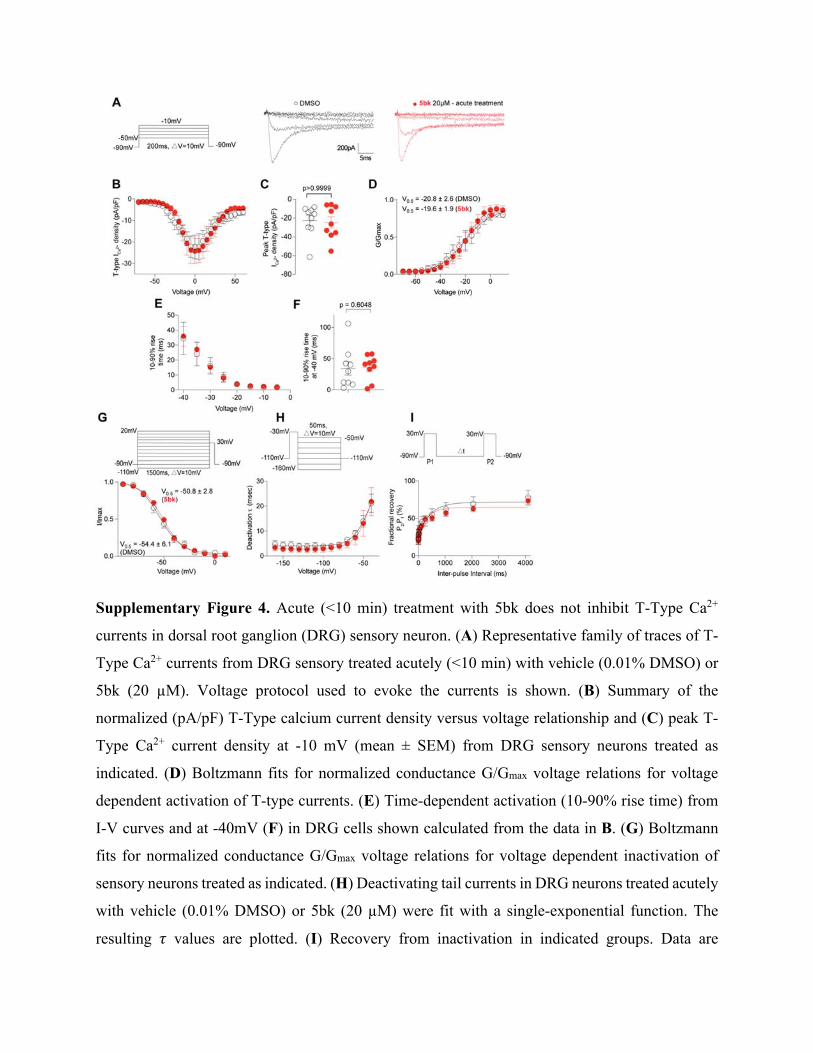
Supplementary Figure 4. Acute (<10 min) treatment with 5bk does not inhibit T-Type Ca2+
currents in dorsal root ganglion (DRG) sensory neuron. (A) Representative family of traces of T-
Type Ca2+ currents from DRG sensory treated acutely (<10 min) with vehicle (0.01% DMSO) or
5bk (20 µM). Voltage protocol used to evoke the currents is shown. (B) Summary of the
normalized (pA/pF) T-Type calcium current density versus voltage relationship and (C) peak T-
Type Ca2+ current density at -10 mV (mean ± SEM) from DRG sensory neurons treated as
indicated. (D) Boltzmann fits for normalized conductance G/Gmax voltage relations for voltage
dependent activation of T-type currents. (E) Time-dependent activation (10-90% rise time) from
I-V curves and at -40mV (F) in DRG cells shown calculated from the data in B. (G) Boltzmann
fits for normalized conductance G/Gmax voltage relations for voltage dependent inactivation of
sensory neurons treated as indicated. (H) Deactivating tail currents in DRG neurons treated acutely
with vehicle (0.01% DMSO) or 5bk (20 µM) were fit with a single-exponential function. The
resulting 𝜏 values are plotted. (I) Recovery from inactivation in indicated groups. Data are

averaged and fitted by double exponential association (p values as indicated, Mann-Whitney test).
All graphs show mean ± s.e.m. with individual data points showed when possible.
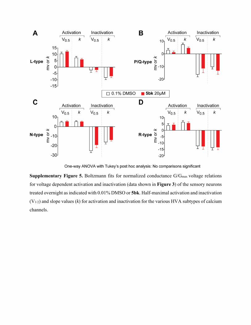
Supplementary Figure 5. Boltzmann fits for normalized conductance G/Gmax voltage relations
for voltage dependent activation and inactivation (data shown in Figure 3) of the sensory neurons
treated overnight as indicated with 0.01% DMSO or 5bk. Half-maximal activation and inactivation
(V1/2) and slope values (k) for activation and inactivation for the various HVA subtypes of calcium
channels.
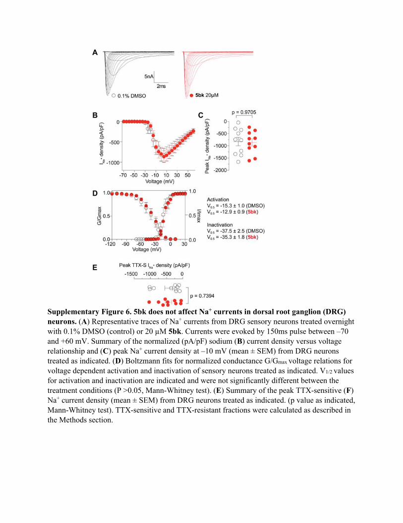
Supplementary Figure 6. 5bk does not affect Na+ currents in dorsal root ganglion (DRG) neurons. (A) Representative traces of Na+ currents from DRG sensory neurons treated overnight with 0.1% DMSO (control) or 20 µM 5bk. Currents were evoked by 150ms pulse between –70 and +60 mV. Summary of the normalized (pA/pF) sodium (B) current density versus voltage relationship and (C) peak Na+ current density at –10 mV (mean ± SEM) from DRG neurons treated as indicated. (D) Boltzmann fits for normalized conductance G/Gmax voltage relations for voltage dependent activation and inactivation of sensory neurons treated as indicated. V1/2 values for activation and inactivation are indicated and were not significantly different between the treatment conditions (P >0.05, Mann-Whitney test). (E) Summary of the peak TTX-sensitive (F) Na+ current density (mean ± SEM) from DRG neurons treated as indicated. (p value as indicated, Mann-Whitney test). TTX-sensitive and TTX-resistant fractions were calculated as described in the Methods section.

Supplementary Figure 7. Representative images of vehicle-treated DRG neurons post-
challenge with constellation pharmacology triggers. Differential interference contrast (DIC)
and pseudocolored fluorescent images of DRG neurons treated with vehicle, visualized for
Fura2-AM before and after stimulations with each of the constellation triggers: menthol (400
nM), histamine (50 µM), ATP (10 µM), AITC (200 µM), acetylcholine (1 mM), capsaicin (100
nM) and KCl (90 mM)) during Ca2+ imaging. Scale bar is 50 µm. Size heat map reports number
of DRG neurons of indicated size (measured by neuronal area) responding to constellation
triggers.
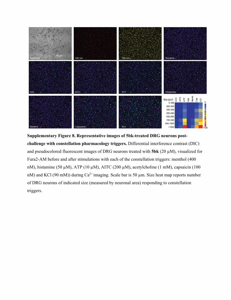
Supplementary Figure 8. Representative images of 5bk-treated DRG neurons post-
challenge with constellation pharmacology triggers. Differential interference contrast (DIC)
and pseudocolored fluorescent images of DRG neurons treated with 5bk (20 µM), visualized for
Fura2-AM before and after stimulations with each of the constellation triggers: menthol (400
nM), histamine (50 µM), ATP (10 µM), AITC (200 µM), acetylcholine (1 mM), capsaicin (100
nM) and KCl (90 mM)) during Ca2+ imaging. Scale bar is 50 µm. Size heat map reports number
of DRG neurons of indicated size (measured by neuronal area) responding to constellation
triggers.
References
[1] Brittain JM, Duarte DB, Wilson SM, Zhu W, Ballard C, Johnson PL, Liu N, Xiong W, Ripsch MS, Wang Y, Fehrenbacher JC, Fitz SD, Khanna M, Park CK, Schmutzler BS, Cheon BM, Due MR, Brustovetsky T, Ashpole NM, Hudmon A, Meroueh SO, Hingtgen CM, Brustovetsky N, Ji RR, Hurley JH, Jin X, Shekhar A, Xu XM, Oxford GS, Vasko MR, White FA, Khanna R. Suppression of inflammatory and neuropathic pain by uncoupling CRMP-2 from the presynaptic Ca(2)(+) channel complex. Nature medicine 2011;17(7):822-829.
[2] Chaplan SR, Bach FW, Pogrel JW, Chung JM, Yaksh TL. Quantitative assessment of tactile allodynia in the rat paw. Journal of neuroscience methods 1994;53(1):55-63.
[3] Choe W, Messinger RB, Leach E, Eckle VS, Obradovic A, Salajegheh R, Jevtovic-Todorovic V, Todorovic SM. TTA-P2 is a potent and selective blocker of T-type calcium channels in rat sensory neurons and a novel antinociceptive agent. MolPharmacol 2011;80(5):900-910.
[4] Dustrude ET, Moutal A, Yang X, Wang Y, Khanna M, Khanna R. Hierarchical CRMP2 posttranslational modifications control NaV1.7 function. Proceedings of the National Academy of Sciences of the United States of America 2016;113(52):E8443-E8452.
[5] Dustrude ET, Wilson SM, Ju W, Xiao Y, Khanna R. CRMP2 protein SUMOylation modulates NaV1.7 channel trafficking. The Journal of biological chemistry 2013;288(34):24316-24331.
[6] Francois-Moutal L, Wang Y, Moutal A, Cottier KE, Melemedjian OK, Yang X, Wang Y, Ju W, Largent-Milnes TM, Khanna M, Vanderah TW, Khanna R. A membrane-delimited N-myristoylated CRMP2 peptide aptamer inhibits CaV2.2 trafficking and reverses inflammatory and postoperative pain behaviors. Pain 2015;156(7):1247-1264.
[7] Gray WR, Olivera BM, Cruz LJ. Peptide toxins from venomous Conus snails. Annual review of biochemistry 1988;57:665-700.
[8] Huynh TN, Krigbaum AM, Hanna JJ, Conrad CD. Sex differences and phase of light cycle modify chronic stress effects on anxiety and depressive-like behavior. Behavioural brain research 2011;222(1):212-222.
[9] Ibrahim MM, Patwardhan A, Gilbraith KB, Moutal A, Yang X, Chew LA, Largent-Milnes T, Malan TP, Vanderah TW, Porreca F, Khanna R. Long-lasting antinociceptive effects of green light in acute and chronic pain in rats. Pain 2017;158(2):347-360.
[10] Khanna R, Yu J, Yang X, Moutal A, Chefdeville A, Gokhale V, Shuja Z, Chew LA, Bellampalli SS, Luo S, Francois-Moutal L, Serafini MJ, Ha T, Perez-Miller S, Park KD, Patwardhan A, Streicher JM, Colecraft HM, Khanna M. Targeting the CaVα-β interaction yields an antagonist of the N-type CaV2.2 channel with broad antinociceptive efficacy. Pain 2019.
[11] Milligan ED, O'Connor KA, Nguyen KT, Armstrong CB, Twining C, Gaykema RP, Holguin A, Martin D, Maier SF, Watkins LR. Intrathecal HIV-1 envelope glycoprotein gp120 induces enhanced pain states mediated by spinal cord proinflammatory cytokines. The Journal of neuroscience : the official journal of the Society for Neuroscience 2001;21(8):2808-2819.
[12] Mintz IM, Venema VJ, Swiderek KM, Lee TD, Bean BP, Adams ME. P-type calcium channels blocked by the spider toxin omega-Aga-IVA. Nature 1992;355(6363):827-829.
[13] Moutal A, Chew LA, Yang X, Wang Y, Yeon SK, Telemi E, Meroueh S, Park KD, Shrinivasan R, Gilbraith KB, Qu C, Xie JY, Patwardhan A, Vanderah TW, Khanna M, Porreca F, Khanna R. (S)-lacosamide inhibition of CRMP2 phosphorylation reduces postoperative and neuropathic pain behaviors through distinct classes of sensory neurons identified by constellation pharmacology. Pain 2016;157(7):1448-1463.
[14] Moutal A, Li W, Wang Y, Ju W, Luo S, Cai S, Francois-Moutal L, Perez-Miller S, Hu J, Dustrude ET, Vanderah TW, Gokhale V, Khanna M, Khanna R. Homology-guided mutational analysis reveals the functional requirements for antinociceptive specificity of collapsin response mediator protein 2-derived peptides. British journal of pharmacology 2017.
[15] Moutal A, Wang Y, Yang X, Ji Y, Luo S, Dorame A, Bellampalli SS, Chew LA, Cai S, Dustrude ET, Keener JE, Marty MT, Vanderah TW, Khanna R. Dissecting the role of the CRMP2-neurofibromin complex on pain behaviors. Pain 2017;158(11):2203-2221.
[16] Newcomb R, Szoke B, Palma A, Wang G, Chen X, Hopkins W, Cong R, Miller J, Urge L, Tarczy-Hornoch K, Loo JA, Dooley DJ, Nadasdi L, Tsien RW, Lemos J, Miljanich G. Selective peptide antagonist of the class E calcium channel from the venom of the tarantula Hysterocrates gigas. Biochemistry 1998;37(44):15353-15362.
[17] Olson KM, Duron DI, Womer D, Fell R, Streicher JM. Comprehensive molecular pharmacology screening reveals potential new receptor interactions for clinically relevant opioids. PloS one 2019;14(6):e0217371.
[18] Polomano RC, Mannes AJ, Clark US, Bennett GJ. A painful peripheral neuropathy in the rat produced by the chemotherapeutic drug, paclitaxel. Pain 2001;94(3):293-304.
[19] Teichert RW, Schmidt EW, Olivera BM. Constellation pharmacology: a new paradigm for drug discovery. Annual review of pharmacology and toxicology 2015;55:573-589.
[20] Vandenberg JI, Perry MD, Perrin MJ, Mann SA, Ke Y, Hill AP. hERG K(+) channels: structure, function, and clinical significance. Physiol Rev 2012;92(3):1393-1478.
[21] Yaksh TL, Rudy TA. Chronic catheterization of the spinal subarachnoid space. Physiology & behavior 1976;17(6):1031-1036.

[22] Yu J, Moutal A, Dorame A, Bellampalli SS, Chefdeville A, Kanazawa I, Pham NYN, Park KD, Weimer JM, Khanna R. Phosphorylated CRMP2 Regulates Spinal Nociceptive Neurotransmission. Molecular neurobiology 2018.
[23] Zhang J, Hu Y, Foley C, Wang Y, Musharrafieh R, Xu S, Zhang Y, Ma C, Hulme C, Wang J. Exploring Ugi-Azide Four-Component Reaction Products for Broad-Spectrum Influenza Antivirals with a High Genetic Barrier to Drug Resistance. Sci Rep 2018;8(1):4653.


![show time eu¨jatu - Welcome to Air · PDF fileCast: Salman Khan, Karisma Kapoor, Sushmita Sen dykdkj% lyeku [+kku] dfj’ek *BuNDAL BAAZ caMy ckt+ Cast: Shammi Kapoor, Rajesh Khanna,](https://img.pdfslide.us/doc/110x75/5a8f0a557f8b9a78648d6804/show-time-eujatu-welcome-to-air-salman-khan-karisma-kapoor-sushmita-sen-dykdkj.jpg)