Embed Size (px)
Citation preview
I J C T A, 9(9), 2016, pp. 3903-3909© International Science Press
Identifying new Vessels on OpticDisc using Funds PhotographM. Rajesh Khanna1, D. Anandhan2, S. Venkatesan3 and K. Nishitha4
ABSTRACT
The consequence of diabetes on the eyeball is called Diabetic Retinopathy (DR). It is notorious to damage the smallblood vessel of the retinaldehyde an this valor laed to defeat of vision. We describe a method for automaticallydetecting new-fangled vessels on the optic nerve using retinal (fundus) photography. Normal and abnoraml segmentsare first detected using a method based on Gaussian filter, Canny edge analysis and watershed transform. Parameters,associated with segment length, gradient, gradient varition, direction, grey level and vessel density are calculatedfor each image segment. Pedestal on these features, each segment is classified and trained as normal or abnormalusing a Artifical Neural Netwrok (ANN) classifier, The system is trained and tested by using 108 images withabnoraml and 98 normal images from fundus photography of one eye hospital. The discrimination act of the featureswas hardened against a clincal reference customary.
Key words: Artificial Nueral Network, Diabetic Retinopathy, Fundus camera, Optical disc
Journal of Economic Literature (JEL) Classification Number: C45
1. INTRODUCTION
In topical times, Sweden and supplementary parts of the world have been countenance with an augment inage and civilization related diseases be fond of diabetes. According to latest examination, 4% of the countrypopulation has been spotted of diabetes infirmity alone and it have been make out and acknowledged asone of the foremost cause of blindness in the nation if not correctly treated and deled with. Prematuredetection and diagnosis have been notorious as one of the way to accomplish a lessening in the cut of visualimpairment caused by diabetes with more prominence on routine medical test out with the use of uniquefacilities for detection and monitored of the whispered disease. The outcome of this on the medical personnelneed not be over stress, it has lead to increase work load on the personnel and the facilities, augment indiabetes showing activities just to mention a few. A lot of approaches have been suggested and identified asmeans of dipping the stress caused by this unvarying check up and screening related activities relatedamong which the use of medical digital picture signal is dispensation for diagnosis of diabetes relatedillness like diabetic retinopathy using imagery of retina. The effect of diabetes on the eye is called DiabeticRetinopathy (DR). It is known to damage the minute blood vessel of the retina and this strength lead to lossof vision. In the BDR phase, the arteries in the retina become damaged and leak, structured small, dot likehemorrhages. These revealed vessels often lead to engorgement or long term edema in the retina anddecreased vision.
In the PDR point, movement problems cause vicinities of the retina to happen to oxygen deprived orischemic. New-fangled fragile, vessels develop as the circulatory scheme attempts to maintain adequateoxygen levels with the retina. This phenomenon is called neo vascularisation. Blood may leak into theretinaldehyde and vitreous, causing spots or floaters, along with shrink vision. In the SDR part of theailment, here is continued anomalous vessel growth and scar tissue, which may cause solemn problems
1,2,3,4 Vel Tech Multi Tech Dr. RANGARAJAN Dr. SAKUNTHALA Engineering College, Chennai, Tamil Nadu, Emails:[email protected], [email protected], [email protected], [email protected]
3904 M. Rajesh Khanna, D. Anandhan, S. Venkatesan and K. Nishitha
such as retinal detachment and glaucoma and plodding loss of vision. This project is one of the methods ofapplying digital image dealing out to the field of medical diagnosis in regulate to lessen the moment andanxiety undergone by the ophthalmologist and other associates of the group in the screening, diagnosis andtreatment of diabetic retinopathy.
The obtainable system of working out the datasets was using support vector machines, for edge detectionit uses various detection mechanisms such as Robert’s edge detector, Prewitt edge detector and sobel edgedetector. And finally making all those segmentation and for applying rank list, it uses to ranking mechanismnamely ansari-bradely rank test and wilcoxon rank test. These two rank tests have been used to overcomethe pitfalls of each other. The inefficiency that is caused by one will overcome by other. In existingmethodology there is no complete automatic detection.
2. GENERATION OF THE DATA
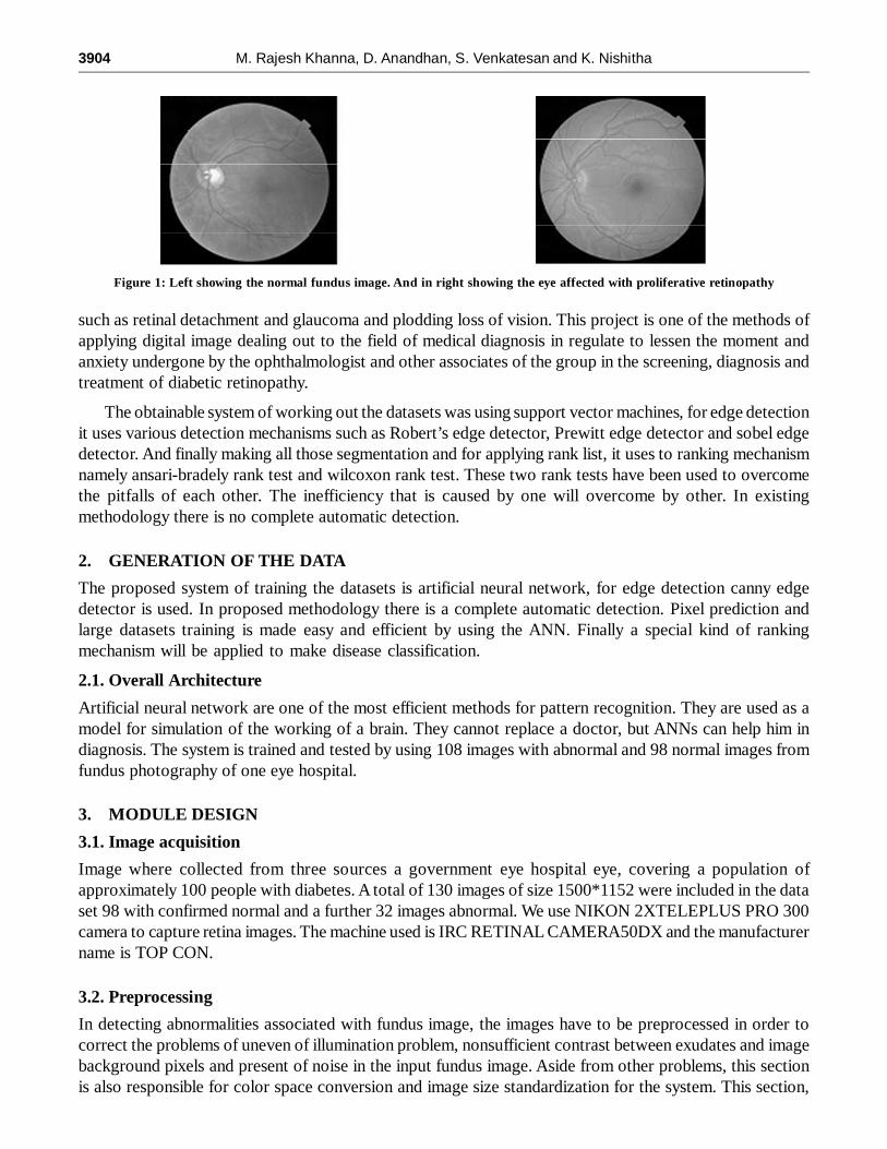
The proposed system of training the datasets is artificial neural network, for edge detection canny edgedetector is used. In proposed methodology there is a complete automatic detection. Pixel prediction andlarge datasets training is made easy and efficient by using the ANN. Finally a special kind of rankingmechanism will be applied to make disease classification.
2.1. Overall Architecture
Artificial neural network are one of the most efficient methods for pattern recognition. They are used as amodel for simulation of the working of a brain. They cannot replace a doctor, but ANNs can help him indiagnosis. The system is trained and tested by using 108 images with abnormal and 98 normal images fromfundus photography of one eye hospital.
3. MODULE DESIGN
3.1. Image acquisition
Image where collected from three sources a government eye hospital eye, covering a population ofapproximately 100 people with diabetes. A total of 130 images of size 1500*1152 were included in the dataset 98 with confirmed normal and a further 32 images abnormal. We use NIKON 2XTELEPLUS PRO 300camera to capture retina images. The machine used is IRC RETINAL CAMERA50DX and the manufacturername is TOP CON.
3.2. Preprocessing
In detecting abnormalities associated with fundus image, the images have to be preprocessed in order tocorrect the problems of uneven of illumination problem, nonsufficient contrast between exudates and imagebackground pixels and present of noise in the input fundus image. Aside from other problems, this sectionis also responsible for color space conversion and image size standardization for the system. This section,
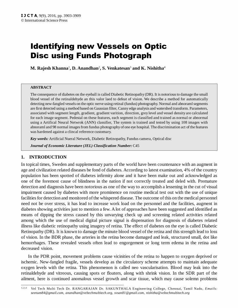
Figure 1: Left showing the normal fundus image. And in right showing the eye affected with proliferative retinopathy
Identifying new Vessels on Optic Disc using Funds Photograph 3905
which is preprocessing stage, can be regarded as the rock for this process. Preprocessing includes applyinggrey scale; normalization and Gaussian filter to get a noise free image. In electronics and signal processing,a Gaussian filter is a filter whose impulse response is a Gaussian function. Gaussian filters are designed togive no overshoot to a step function input while minimizing the rise and fall time. This behavior is closelyconnected to the ace that the Gaussian filter has the minimum possible group delay.
In Image processing, the impulse response, or impulse response function (IRF), of a dynamic system isits output when presented with a brief input signal, called an impulse. More generally, an impulse responserefers to the reaction of any dynamic system in response to some external change. In both cases, the impulseresponse describes the reaction of the system as a function of time.
3.3. Detection of new vessels
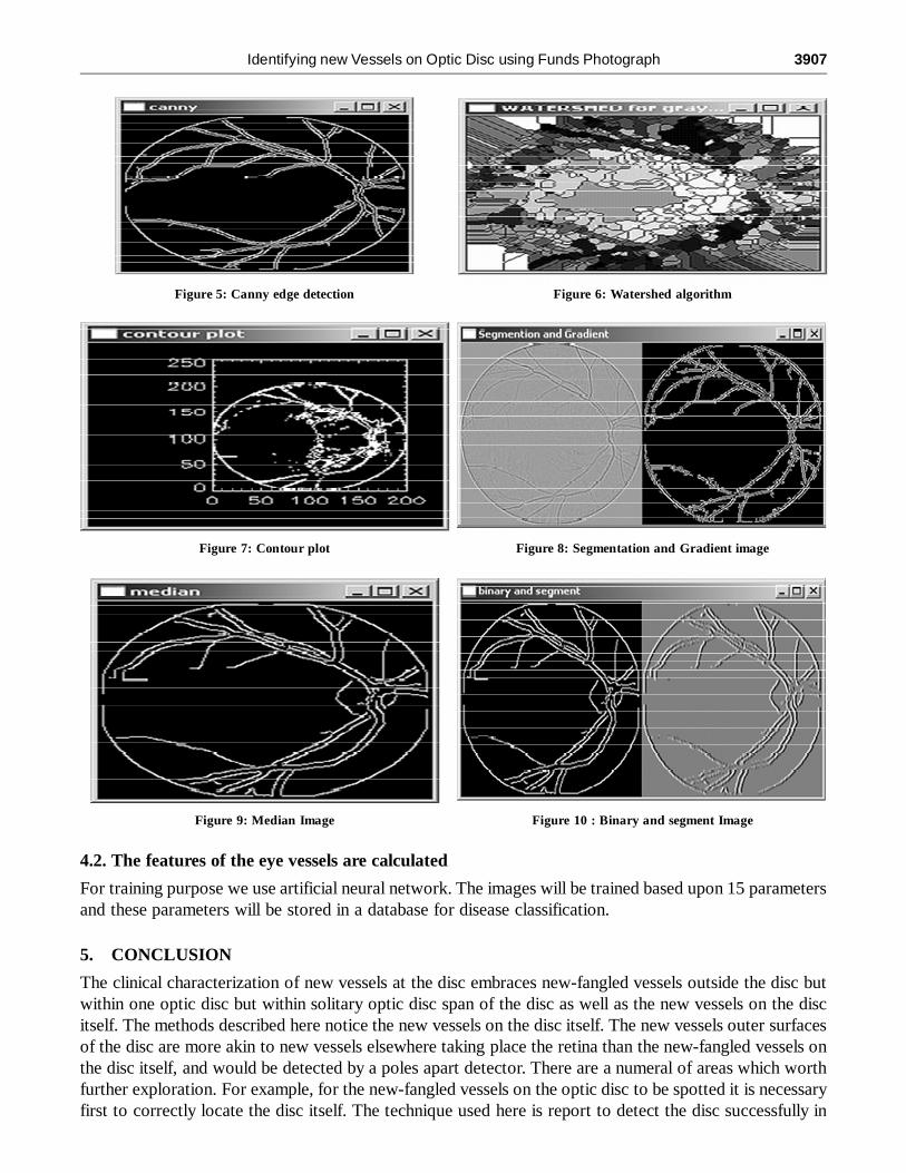
After preprocessing the detection of vessels is an important step for training and disease classification.Detection of edge’s needs the step such as canny edge detector, watershed transform, by using this cannyedge detection method we can able to make clear edges of the vessels and in order to make it more efficientwe use watershed transform which apply dam construction to give accurate edges.
3.4. Training and disease classification
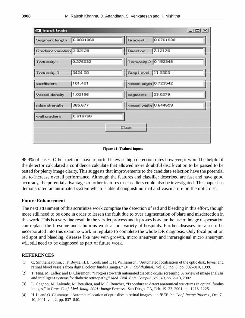
The 15 parameters will be trained using artificial neural network, forever single parameter neurons will becreated and trained features will be stored in data base for further process. Depending upon the percentageof difference the disease classification will be made and results will be displayed.
4. RESULTS
Artificial Neural Network Training and Classification
STEP 1: for each layer (except input layer)For each neuron layer;For each weight of neuronSet random weight.
STEP 2: while total network error is greater than thresholdFor each parameter (15 parameter) in training set
Figure 2: Overall Architecture
3906 M. Rajesh Khanna, D. Anandhan, S. Venkatesan and K. Nishitha
Read each parameter’s thresholdCalc values to the range between normal image and abnormal images
STEP 3: for each layer (except input layer)For each neuron in layerSum all rations of weights and last layer outputsCompute output of this layer
STEP 4: for each layer (except input layer)For each neuron in layerFor each weight of neuronCompute new eight values;
STEP 5: for each parameterFor each neuron in output layerSum output errorsCompute total networks results;
STEP 6: for each neuron in layerSum all rations of weights and last layer outputsCompute outputs of this layer;Sort and show the results;
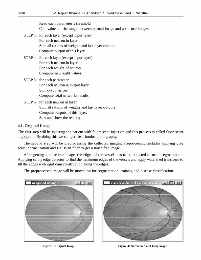
4.1. Original Image
The first step will be injecting the patient with fluorescent injection and this process is called fluorescentangiogram. By doing this we can get clear fundus photography.
The second step will be preprocessing the collected images. Preprocessing includes applying greyscale; normalization and Gaussian filter to get a noise free image.
After getting a noise free image, the edges of the vessels has to be detected to make segmentation.Applying canny edge detector to find the maximum edges of the vessels and apply watershed transform tofill the edges with rigid dam construction along the edges.
The preprocessed image will be moved on for segmentation, training and disease classification.
Figure 3: Original Image Figure 4: Normalized and Gray image
Identifying new Vessels on Optic Disc using Funds Photograph 3907
Figure 5: Canny edge detection Figure 6: Watershed algorithm
Figure 7: Contour plot Figure 8: Segmentation and Gradient image
Figure 9: Median Image Figure 10 : Binary and segment Image
4.2. The features of the eye vessels are calculated
For training purpose we use artificial neural network. The images will be trained based upon 15 parametersand these parameters will be stored in a database for disease classification.
5. CONCLUSION
The clinical characterization of new vessels at the disc embraces new-fangled vessels outside the disc butwithin one optic disc but within solitary optic disc span of the disc as well as the new vessels on the discitself. The methods described here notice the new vessels on the disc itself. The new vessels outer surfacesof the disc are more akin to new vessels elsewhere taking place the retina than the new-fangled vessels onthe disc itself, and would be detected by a poles apart detector. There are a numeral of areas which worthfurther exploration. For example, for the new-fangled vessels on the optic disc to be spotted it is necessaryfirst to correctly locate the disc itself. The technique used here is report to detect the disc successfully in
3908 M. Rajesh Khanna, D. Anandhan, S. Venkatesan and K. Nishitha
98.4% of cases. Other methods have reported likewise high detection rates however; it would be helpful ifthe detector calculated a confidence calculate that allowed more doubtful disc location to be passed to betested for plenty image clarity. This suggests that improvements to the candidate selection have the potentialare to increase overall performance. Although the features and classifier described are fast and have goodaccuracy, the potential advantages of other features or classifiers could also be investigated. This paper hasdemonstrated an automated system which is able distinguish normal and vasculature on the optic disc.
Future Enhancement
The next attainment of this scrutinize work comprise the detection of red and bleeding in this effort, thoughmore still need to be done in order to lessen the fault due to over augmentation of blare and misdetection inthis work. This is a very fine result in the verdict process and it proves how far the use of image dispensationcan replace the tiresome and laborious work at our variety of hospitals. Further diseases are also to beincorporated into this examine work in regulate to complete the whole DR diagnosis. Only focal point onred spot and bleeding, diseases like new vein growth, micro aneurysm and intraregional micro aneurysmwill still need to be diagnosed as part of future work.
REFERENCES[1] C. Sinthanayothin, J. F. Boyce, H. L. Cook, and T. H. Williamson, “Automated localisation of the optic disk, fovea, and
retinal blood vessels from digital colour fundus images,” Br. J. Ophthalmol., vol. 83, no. 8, pp. 902–910, 1999.
[2] T. Teng, M. Lefley, and D. Claremont, “Progress towards automated diabetic ocular screening: A review of image analysisand intelligent systems for diabetic retinopathy,” Med. Biol. Eng. Comput., vol. 40, pp. 2–13, 2002.
[3] L. Gagnon, M. Lalonde, M. Beaulieu, and M.C. Boucher, “Procedure to detect anatomical structures in optical fundusimages,” in Proc. Conf. Med. Imag. 2001: Image Process., San Diego, CA, Feb. 19–22, 2001, pp. 1218–1225.
[4] H. Li and O. Chutatape, “Automatic location of optic disc in retinal images,” in IEEE Int. Conf. Image Process., Oct. 7–10, 2001, vol. 2, pp. 837–840.
Figure 11: Trained Inputs
Identifying new Vessels on Optic Disc using Funds Photograph 3909
[5] J. Lowell, A. Hunter, D. Steel, A. Basu, R. Ryder, E. Fletcher, and L. Kennedy, “Optic nerve head segmentation,” IEEETrans. Med. Imag., vol. 23, no. 2, pp. 256–264, Feb. 2004.
[6] H. Li and O. Chutatape, “A model-based approach for automated feature extraction in fundus images,” in 9th IEEE Int.Conf. Computer Vision (ICCV’03), 2003, vol. 1, pp. 394–399.
[7] R. A. Abdel-Ghafar, T. Morris, T. Ritchings, and I. Wood, “Detection and characterisation of the optic disk in glaucomaand diabetic retinopathy,” presented at the Med. Image Understand. Anal. Conf., London, U.K., Sep. 23–24, 2004.
[8] A. Osareh, M. Mirmehdi, B. Thomas, and R. Markham, “Classification and localisation of diabetic-related eye disease,”in 7th Eur. Conf. Computer Vision (ECCV), May 2002, vol. 2353, LNCS, pp. 502–516.
[9] STARE ProjectWebsite Clemson Univ., Clemson, SC [Online]. Available: http://www.ces.clemson.edu~ahoover/stare
[10] M. Lalonde, M. Beaulieu, and L. Gagnon, “Fast and robust optic disk detection using pyramidal decomposition andHausdorff-based template matching,” IEEE Trans. Med. Imag., vol. 20, no. 11, pp. 1193–1200, Nov. 2001.
[11] F. ter Haar, “Automatic localization of the optic disc in digital colour images of the human retina,” M.S. thesis, UtrechtUniversity, Utrecht, The Netherlands, 2005.
[12] C. Sinthanayothin, “Image analysis for automatic diagnosis of diabetic retinopathy,” Ph.D. dissertation, University ofLondon (King’s College London), London, U.K., 1999.
[13] T.Walter and J.-C. Klein, “Segmentation of color fundus images of the human retina: Detection of the optic disc and thevascular tree using morphological techniques,” in Proc. 2nd Int. Symp. Med. Data Anal.,2001, pp. 282–287.
[14] R. Chrástek, M. Wolf, K. Donath, G. Michelson, and H. Niemann,”Optic disc segmentation in retinal images,”Bildverarbeitung für de Medizin 2002, pp. 263–266, 2002.
[15] A. Osareh, “Automated identification of diabetic retinal exudates and the optic disc,” Ph.D. dissertation, Department ofComputer Science, Faculty of Engineering, University of Bristol, Bristol, U.K., 2004.
[16] A. Osareh, M. Mirmehdi, B. Thomas, and R. Markham, “Comparison of colour spaces for optic disc localisation inretinal images,” in Proc. 16th Int. Conf. Pattern Recognition, 2002, pp. 743–746.
[17] S. F. Barrett, E. Naess, and T. Molvik, “Employing the Hough transform to locate the optic disk,” in Biomed. Sci. Instrum.,2001, vol. 37, pp. 81–86.
[18] M. Niemeijer, J. Staal, B. van Ginneken, M. Loog, and M. D. Abràmoff, J. M. Fitzpatrick and M. Sonka, Eds., “Comparativestudy of retinal vessel segmentation methods on a new publicly available database,” SPIE Med. Imag., vol. 5370, pp.648–656, 2004.
[19] A. Hoover and M. Goldbaum, “Locating the optic nerve in a retinal image using the fuzzy convergence of the bloodvessels,” IEEE Trans.Med. Imag., vol. 22, no. 8, pp. 951–958, Aug. 2003.
[20] A. Hoover and M. Goldbaum, “Fuzzy convergence,” in Proc. IEEE Computer Soc. Conf. Computer Vis. Pattern Recognit.,Santa Barbara, CA, 1998, pp. 716–721.
[21] M. Foracchia, E. Grisan, and A. Ruggeri, “Detection of optic disc in retinal images by means of a geometrical model ofvessel structure,” IEEE Trans. Med. Imag., vol. 23, no. 10, pp. 1189–1195, Oct. 2004.
[22] M. Goldbaum, S. Moezzi, A. Taylor, S. Chatterjee, J. Boyd, E. Hunter, and R. Jain, “Automated diagnosis and imageunderstanding with objectextraction, object classification, and inferencing in retinal images,” in Proc. IEEE Int. CongressImage Process., Los Alamitos, CA, 1996, vol. 3, pp. 695–698.


![[3904]Grade6 Science](https://img.pdfslide.us/doc/110x75/5695d1f51a28ab9b02989318/3904grade6-science.jpg)