Embed Size (px)
Citation preview

Description and importance of the disease: Eastern, Western and Venezuelan equine
encephalomyelitis (EEE, WEE and VEE) viruses belong to the genus Alphavirus of the family
Togaviridae. Alternate infection of birds, rodents and mosquitoes maintain these viruses in nature.
The disease occurs sporadically in horses and humans from mid-summer to late autumn. Horses and
humans are incidental end hosts for WEE. Horses are not dead-end hosts for VEE virus. The disease
in horses is characterised by fever, anorexia, and severe depression. In severe cases, it can progress
to hyperexcitability, blindness, ataxia, severe mental depression, recumbency, convulsions, and
death.
EEE virus infection in horses is often fatal, while WEE virus can cause a subclinical or mild disease
with less than 30% mortality. EEE and WEE viruses have been reported to cause disease in poultry,
game birds and ratites. Sporadic cases of EEE have been reported in cows, sheep, pigs, deer, and
dogs.
The VEE complex of viruses includes six antigenic subtypes (I–VI). Within subtype I there are five
antigenic variants (variants AB–F). Originally, subtypes I-A and I-B were considered to be distinct
variants, but they are now considered to be identical (I-AB). Antigenic variants I-AB and I-C are
associated with epizootic activity in equids and human epidemics. Historically, severe outbreaks have
involved many thousands of human and equine cases. The other three variants of subtype I (I-D, I-
E, I-F) and the other five subtypes of VEE (II–VI) circulate in natural enzootic cycles. Equidae serve
as amplifying hosts for epizootic VEE strains while enzootic VEE viruses cycle primarily in sylvatic
rodents and mosquitoes. Enzootic variants and subtypes have been considered to be nonpathogenic
for equids, but can cause clinical disease in humans. During 1993 and 1996 limited outbreaks of
encephalitis in horses in Mexico were shown to be caused by enzootic VEE viruses of subtype I-E.
More recently, sporadic outbreaks have occurred in Mexico, Central America, and northern and
western parts of South America. Human enzootic subtypes reach more broadly into northern Central
America and South America (Weaver et al., 2012).
Identification of the agent: A presumptive diagnosis of EEE, WEE or VEE can be made when
susceptible horses display the characteristic somnolence and other signs of neurological disease in
areas where haematophagous insects are active. There are no characteristic gross lesions.
Histopathological lesions can provide a presumptive diagnosis. EEE virus can usually be isolated
from the brain and sometimes other tissues of dead horses, however WEE and VEE viruses are
rarely isolated. The viruses can be isolated from field specimens by inoculating newborn mice,
embryonated chicken eggs, cell cultures, or newly hatched chickens. The virus can be identified by
reverse-transcriptase polymerase chain reaction (RT-PCR), complement fixation (CF),
immunofluorescence, or plaque reduction neutralisation (PRN) tests. Specific identification of
epizootic VEE variants can be made by the indirect fluorescent antibody test, or a differential PRN
test using subtype- or variant-specific monoclonal antibody, or by nucleic acid sequencing.
Serological tests: Antibodies can be identified by PRN test, haemagglutination inhibition (HI), CF,
or IgM capture enzyme-linked immunosorbent assay.
Requirements for vaccines: EEE and WEE vaccines are safe and immunogenic. The only
recommended vaccines against VEE are an attenuated virus vaccine, made with strain TC-83, or
inactivated virus preparations also made from this strain. Attenuated virus is immunogenic when
given by intramuscular injection, and can cause adverse reactions in the recipient.
Formalin-inactivated virulent VEE virus preparations should not be used in equids, as residual virulent
virus can remain after formalin treatment, and thereby cause severe illness in both animals and
humans. Epizootics of VEE have occurred from the use of such formalin-treated viruses.
Eastern equine encephalomyelitis (EEE), Western equine encephalomyelitis (WEE) and Venezuelan equine encephalomyelitis (VEE) viruses are members of the genus Alphavirus of the family Togaviridae. The natural ecology for virus maintenance occurs via alternate infection of birds (rodents in the case of VEE) and mosquitoes. EEE virus has also been isolated from snakes, and these may have a role as reservoir hosts (Bingham et al., 2012). Clinical disease may be observed in humans and horses, both of which are incidental dead-end hosts for WEE virus. EEE has been diagnosed in Quebec and Ontario in Canada, central and eastern regions of the United States of America (USA), the Caribbean Islands, Mexico, and Central and South America. The South American strains of EEE, previously known as lineages II, III, and IV, are now known as Madariaga virus (Arrigo, 2010) after strains causing outbreaks in Darien, Panama in 2010 (Carrera, 2013) led to the re-evaluation of viral sequencing and reclassification of the virus strains.
Disease caused by the WEE virus has been reported in the western USA and Canada, Mexico, and Central and South America (Morris, 1989; Reisen & Monath, 1989; Walton et al., 1981). Highlands J virus, antigenically related to WEE virus, has been isolated in eastern USA. Although Highlands J virus is generally believed not to cause disease in mammals, it has been isolated from the brain of a horse in Florida dying of encephalitis (Karabatsos et al., 1988).
The VEE virus complex is composed of six subtypes (I–VI). Subtype I includes five antigenic variants (AB–F), of which variants I-AB and I-C are associated with epizootic VEE in equids and concurrent epidemics in humans (Calisher et al., 1980; Monath & Trent, 1981; Pan-American Health Organization, 1972; Walton, 1981; Walton et al., 1973; Walton & Grayson, 1989). Originally, subtypes I-A and I-B were considered to be distinct variants, but they are now considered to be identical (I-AB). The epizootic variants I-AB and I-C are thought to originate from mutations of the enzootic 1-D serotype (Weaver et al., 2004); I-AB and I-C isolates have only been obtained during equine epizootics. The enzootic strains include variants I-D, I-E and I-F of subtype I, subtype II, four antigenic variants (A–D) of subtype III, and subtypes IV–VI. Normally, enzootic VEE viruses do not produce clinical encephalomyelitis in the equine species (Walton et al., 1973), but in 1993 and 1996 in Mexico, the 1-E enzootic subtype caused limited epizootics in horses. The enzootic variants and subtypes can produce clinical disease in humans (Monath & Trent, 1981; Pan-American Health Organization, 1972; Powers et al., 1997; Walton, 1981; Walton & Grayson, 1989).
Historically, epizootic VEE was limited to northern and western South America (Venezuela, Colombia, Ecuador, Peru and Trinidad) (Pan-American Health Organization, 1972). From 1969 to 1972, however, epizootic activity (variant 1-AB) occurred in Belize, Costa Rica, El Salvador, Guatemala, Honduras, Mexico, Nicaragua and the United States of America (Texas). Epizootics of VEE caused by I-AB or I-C virus have not occurred in North America and Mexico since 1972. Equine and human isolations of epizootic VEE virus were subtype I-C strains from Venezuela in 1993, 1995,1996, 1999, 2000, 2003 (Navarro et al., 2005) and Colombia in 1995. Additionally, variant 1-AB has been isolated from sentinel hamsters in Venezuela (Medina et al., 2015).
The foci of enzootic variants and subtypes are found in areas classified as tropical wet forest, i.e. those areas with a high water table or open swampy areas with meandering sunlit streams. These are the areas of the Americas where rainfall is distributed throughout the year or areas permanently supplied with water. Enzootic viruses cycle among rodents, and perhaps birds, by the feeding of mosquitoes (Monath & Trent, 1981; Pan-American Health Organization, 1972; Walton, 1981; Walton & Grayson, 1989). Enzootic VEE virus strains have been identified in the Florida Everglades (subtype II), Mexico (variant I-E), Central American countries (variant I-E), Panama (variants I-D and I-E), Venezuela (variant I-D), Colombia (variant I-D), Peru (variants 1-D, III-C, and III-D), French Guiana (variant III-B and subtype V), Ecuador (variant I-D), Bolivia (variant I-D), Suriname (variant III-A), Trinidad (variant III-A), Brazil (variants I-F and III-A and subtype IV), and Argentina (subtype VI). In an atypical ecological niche, variant III-B has been isolated in the USA (Colorado and South Dakota) in an unusual association with birds (Aguilar et al., 2009; Monath & Trent, 1981; Pan-American Health Organization, 1972; Walton, 1981; Walton & Grayson, 1989). Everglades virus is a subtype II VEE virus that infects rodents and dogs in Florida.
The clinical signs of EEE, WEE and VEE can be identical. The disease caused by either virus is also known as sleeping sickness. Following an incubation period of 5–14 days, clinical signs include fever, anorexia, and depression. A presumptive diagnosis of equine viral encephalomyelitis in unvaccinated horses can be made if the characteristic somnolence is observed during the summer in temperate climates or the wet season in tropical and subtropical climates, when the mosquito vector is plentiful. However, a number of other diseases, such as West Nile virus (Chapter 3.1.24), rabies (Chapter 3.1.17), and other infectious, parasitic, or non-infectious agents can produce similar clinical signs and the diagnosis must be confirmed by the described diagnostic test methods.
WEE virus infection in horses has been observed over a wide geographical area, e.g. sporadic cases over 2590 km2
(1000 square miles), however no disease outbreaks attributed to WEE have occurred since 1999. EEE virus infections are usually observed in limited geographical areas. Isolated events of high mortality in captive-raised game birds, primarily pheasants, chukars, aquarium penguins, and quail have been traced to WEE, EEE, or Highlands J virus infection (Morris, 1989; Reisen & Monath, 1989; Tuttle et al., 2005). Most encephalomyelitis infections in domestic fowl are caused by EEE virus and occur in the east coast states of the USA. The virus is introduced by mosquitoes, but transmission within the flocks is primarily by feather picking and cannibalism. Both EEE and WEE viruses have caused a fatal disease in ratites. Haemorrhagic enteritis has been observed in emus infected with EEE and WEE viruses, and morbidity and mortality rates may be greater than 85%. Highlands J and EEE viruses have been found to produce depression, somnolence, decreased egg production, and increased mortality in turkeys (Guy, 1997). EEE virus has been reported to cause disease in cows (McGee et al., 1992; Pursell et al., 1976), sheep (Bauer et al., 2005), pigs (Elvinger et al., 1996), white-tailed deer (Tate et al., 2005), and dogs (Farrar et al., 2005).
EEE virus causes severe disease in humans with a mortality rate of 30–70% and a high frequency of permanent sequelae in patients who survive. WEE is usually mild in adult humans, but can be a severe disease in children. The fatality rate is between 3 and 14%. Severe clinical disease and death caused by EEE and WEE viruses have been reported in laboratory workers. Laboratory manipulations should be carried out at an appropriate biosafety and containment level determined by isk analysis (see Chapter 1.1.4 Biosafety and biosecurity: Standard for managing biological risk in the veterinary laboratory and animal facilities). It is recommended that personnel be immunised against EEE and WEE viruses (United States Department of Health and Human Services, 2009). Precautions should also be taken to prevent human infection when performing post-mortem examinations on horses suspected of being infected with the equine encephalomyelitis viruses.
Human VEE virus infections have originated by aerosol transmission from the cage debris of infected laboratory rodents and from laboratory accidents. Infections with both epizootic and enzootic variants and subtypes have been acquired by laboratory workers (American Committee on Arthropod-Borne Viruses [ACAV], 1980). Severe clinical disease or death can occur in humans. Those who handle infectious VEE viruses or their antigens prepared from infected tissues or cell cultures should be vaccinated and shown to have VEE virus-specific neutralising antibody (Berge et al., 1961; Pan-American Health Organization, 1972). If vaccination is not a viable option, additional personal protective equipment to include respiratory protection is recommended for all procedures. Laboratory manipulations should be carried out at an appropriate biosafety and containment level determined by risk analysis (see chapter 1.1.4).
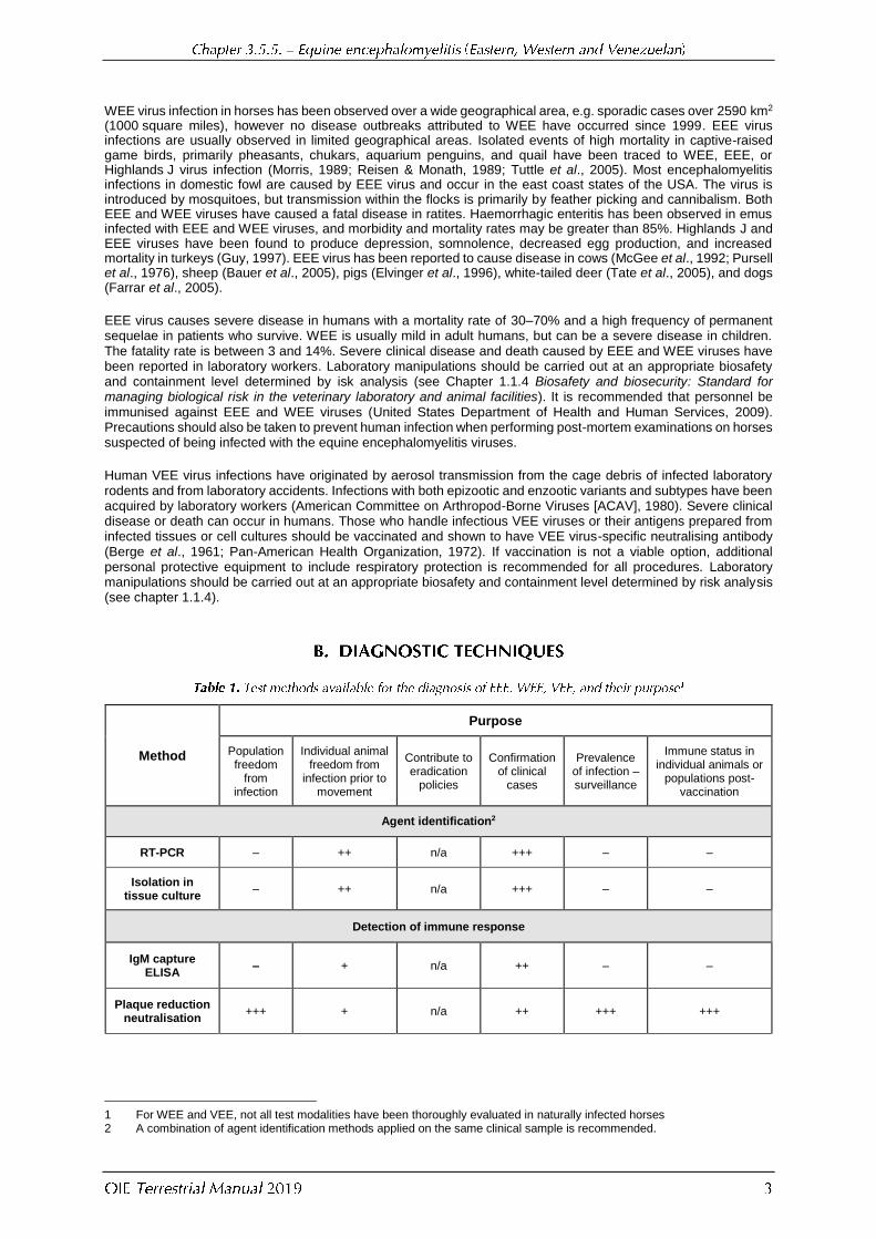
Method
Purpose
Population freedom
from infection
Individual animal freedom from
infection prior to movement
Contribute to eradication
policies
Confirmation of clinical
cases
Prevalence of infection – surveillance
Immune status in individual animals or
populations post-vaccination
Agent identification2
RT-PCR – ++ n/a +++ – –
Isolation in tissue culture
– ++ n/a +++ – –
Detection of immune response
IgM capture ELISA
– + n/a ++ – –
Plaque reduction neutralisation
+++ + n/a ++ +++ +++
1 For WEE and VEE, not all test modalities have been thoroughly evaluated in naturally infected horses 2 A combination of agent identification methods applied on the same clinical sample is recommended.
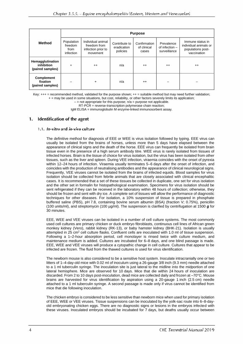
Method
Purpose
Population freedom
from infection
Individual animal freedom from
infection prior to movement
Contribute to eradication
policies
Confirmation of clinical
cases
Prevalence of infection – surveillance
Immune status in individual animals or
populations post-vaccination
Hemagglutination inhibition
(paired samples) + ++ n/a ++ ++ ++
Complement fixation
(paired samples) – + n/a ++ – –
Key: +++ = recommended method, validated for the purpose shown; ++ = suitable method but may need further validation; + = may be used in some situations, but cost, reliability, or other factors severely limits its application;
– = not appropriate for this purpose; n/a = purpose not applicable. RT-PCR = reverse-transcription polymerase chain reaction;
IgM ELISA = immunoglobulin M enzyme-linked immunosorbent assay.
The definitive method for diagnosis of EEE or WEE is virus isolation followed by typing. EEE virus can usually be isolated from the brains of horses, unless more than 5 days have elapsed between the appearance of clinical signs and the death of the horse. EEE virus can frequently be isolated from brain tissue even in the presence of a high serum antibody titre. WEE virus is rarely isolated from tissues of infected horses. Brain is the tissue of choice for virus isolation, but the virus has been isolated from other tissues, such as the liver and spleen. During VEE infection, viraemia coincides with the onset of pyrexia within 12–24 hours of infection. Viraemia usually terminates 5–6 days after the onset of infection, and coincides with the production of neutralising antibodies and the appearance of clinical neurological signs. Frequently, VEE viruses cannot be isolated from the brains of infected equids. Blood samples for virus isolation should be collected from febrile animals that are closely associated with clinical encephalitic cases. It is recommended that a set of these tissues be collected in duplicate, one set for virus isolation and the other set in formalin for histopathological examination. Specimens for virus isolation should be sent refrigerated if they can be received in the laboratory within 48 hours of collection; otherwise, they should be frozen and sent with dry ice. A complete set of tissues will allow the performance of diagnostic techniques for other diseases. For isolation, a 10% suspension of tissue is prepared in phosphate buffered saline (PBS), pH 7.8, containing bovine serum albumin (BSA) (fraction V; 0.75%), penicillin (100 units/ml), and streptomycin (100 µg/ml). The suspension is clarified by centrifugation at 1500 g for 30 minutes.
EEE, WEE and VEE viruses can be isolated in a number of cell culture systems. The most commonly used cell cultures are primary chicken or duck embryo fibroblasts, continuous cell lines of African green monkey kidney (Vero), rabbit kidney (RK-13), or baby hamster kidney (BHK-21). Isolation is usually attempted in 25 cm2 cell culture flasks. Confluent cells are inoculated with 1.0 ml of tissue suspension. Following a 1–2-hour absorption period, cell monolayer is rinsed twice with culture medium, and maintenance medium is added. Cultures are incubated for 6–8 days, and one blind passage is made. EEE, WEE and VEE viruses will produce a cytopathic change in cell culture. Cultures that appear to be infected are frozen. The fluid from the thawed cultures is used for virus identification.
The newborn mouse is also considered to be a sensitive host system. Inoculate intracranially one or two litters of 1–4-day-old mice with 0.02 ml of inoculum using a 26-gauge 3/8 inch (9.3 mm) needle attached to a 1 ml tuberculin syringe. The inoculation site is just lateral to the midline into the midportion of one lateral hemisphere. Mice are observed for 10 days. Mice that die within 24 hours of inoculation are discarded. From 2 to 10 days post-inoculation, dead mice are collected daily and frozen at –70°C. Mouse brains are harvested for virus identification by aspiration using a 20-gauge 1 inch (2.5 cm) needle attached to a 1 ml tuberculin syringe. A second passage is made only if virus cannot be identified from mice that die following inoculation.
The chicken embryo is considered to be less sensitive than newborn mice when used for primary isolation of EEE, WEE or VEE viruses. Tissue suspensions can be inoculated by the yolk-sac route into 6–8-day-old embryonating chicken eggs. There are no diagnostic signs or lesions in the embryos infected with these viruses. Inoculated embryos should be incubated for 7 days, but deaths usually occur between
2 and 4 days post-inoculation. Usually only one passage is made unless there are dead embryos from which virus cannot be isolated. Newly hatched chickens are susceptible and have been used for virus isolation. If this method is used, precautions must be taken to prevent aerosol exposure of laboratory personnel, as infected birds can shed highly infectious virus.
Viral isolates can be identified as by reverse-transcriptase polymerase chain reaction (RT-PCR), by direct or indirect fluorescent antibody test, or by plaque reduction neutralisation (PRN) tests using polyclonal or monoclonal antibodies to specific viruses obtained commercially or prepared via hyperimmunisation of animals with further collection of their serum (or ascetic fluid in case of mice) and purification of immune globulins.
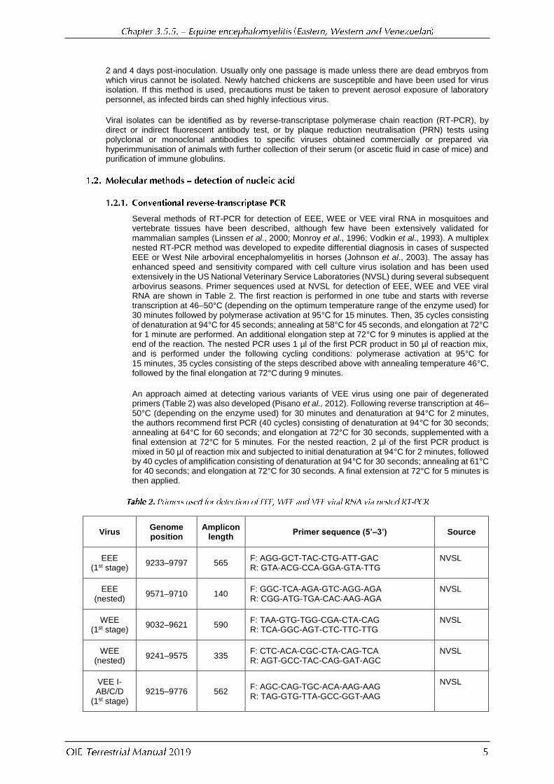
Several methods of RT-PCR for detection of EEE, WEE or VEE viral RNA in mosquitoes and vertebrate tissues have been described, although few have been extensively validated for mammalian samples (Linssen et al., 2000; Monroy et al., 1996; Vodkin et al., 1993). A multiplex nested RT-PCR method was developed to expedite differential diagnosis in cases of suspected EEE or West Nile arboviral encephalomyelitis in horses (Johnson et al., 2003). The assay has enhanced speed and sensitivity compared with cell culture virus isolation and has been used extensively in the US National Veterinary Service Laboratories (NVSL) during several subsequent arbovirus seasons. Primer sequences used at NVSL for detection of EEE, WEE and VEE viral RNA are shown in Table 2. The first reaction is performed in one tube and starts with reverse transcription at 46–50°C (depending on the optimum temperature range of the enzyme used) for 30 minutes followed by polymerase activation at 95°С for 15 minutes. Then, 35 cycles consisting of denaturation at 94°C for 45 seconds; annealing at 58°C for 45 seconds, and elongation at 72°C for 1 minute are performed. An additional elongation step at 72°C for 9 minutes is applied at the end of the reaction. The nested PCR uses 1 µl of the first PCR product in 50 µl of reaction mix, and is performed under the following cycling conditions: polymerase activation at 95°C for 15 minutes, 35 cycles consisting of the steps described above with annealing temperature 46°C, followed by the final elongation at 72°C during 9 minutes.
An approach aimed at detecting various variants of VEE virus using one pair of degenerated primers (Table 2) was also developed (Pisano et al., 2012). Following reverse transcription at 46–50°C (depending on the enzyme used) for 30 minutes and denaturation at 94°C for 2 minutes, the authors recommend first PCR (40 cycles) consisting of denaturation at 94°C for 30 seconds; annealing at 64°C for 60 seconds; and elongation at 72°C for 30 seconds, supplemented with a final extension at 72°C for 5 minutes. For the nested reaction, 2 µl of the first PCR product is mixed in 50 µl of reaction mix and subjected to initial denaturation at 94°C for 2 minutes, followed by 40 cycles of amplification consisting of denaturation at 94°C for 30 seconds; annealing at 61°C for 40 seconds; and elongation at 72°C for 30 seconds. A final extension at 72°C for 5 minutes is then applied.
Virus Genome position
Amplicon length
Primer sequence (5’–3’) Source
EEE (1st stage)
9233–9797 565 F: AGG-GCT-TAC-CTG-ATT-GAC R: GTA-ACG-CCA-GGA-GTA-TTG
NVSL
EEE (nested)
9571–9710 140 F: GGC-TCA-AGA-GTC-AGG-AGA R: CGG-ATG-TGA-CAC-AAG-AGA
NVSL
WEE (1st stage)
9032–9621 590 F: TAA-GTG-TGG-CGA-CTA-CAG R: TCA-GGC-AGT-CTC-TTC-TTG
NVSL
WEE (nested)
9241–9575 335 F: CTC-ACA-CGC-CTA-CAG-TCA R: AGT-GCC-TAC-CAG-GAT-AGC
NVSL
VEE I-AB/C/D
(1st stage) 9215–9776 562
F: AGC-CAG-TGC-ACA-AAG-AAG R: TAG-GTG-TTA-GCC-GGT-AAG
NVSL
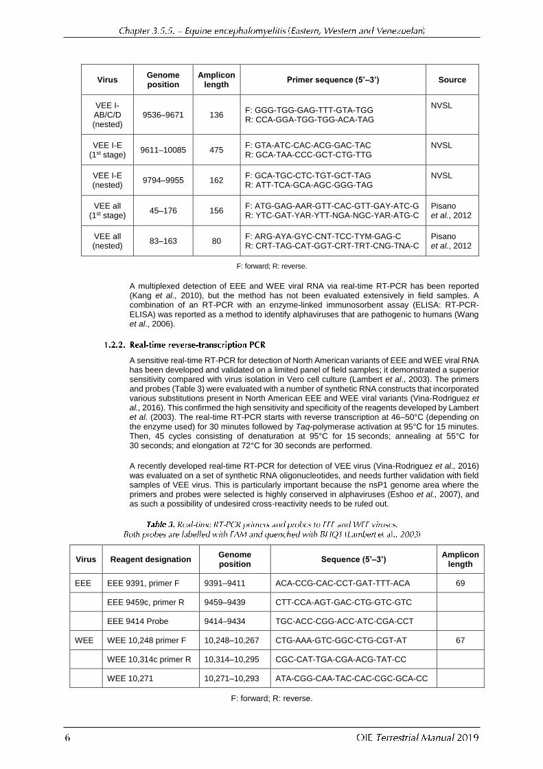
Virus Genome position
Amplicon length
Primer sequence (5’–3’) Source
VEE I-AB/C/D (nested)
9536–9671 136 F: GGG-TGG-GAG-TTT-GTA-TGG R: CCA-GGA-TGG-TGG-ACA-TAG
NVSL
VEE I-E (1st stage)
9611–10085 475 F: GTA-ATC-CAC-ACG-GAC-TAC R: GCA-TAA-CCC-GCT-CTG-TTG
NVSL
VEE I-E (nested)
9794–9955 162 F: GCA-TGC-CTC-TGT-GCT-TAG R: ATT-TCA-GCA-AGC-GGG-TAG
NVSL
VEE all (1st stage)
45–176 156 F: ATG-GAG-AAR-GTT-CAC-GTT-GAY-ATC-G R: YTC-GAT-YAR-YTT-NGA-NGC-YAR-ATG-C
Pisano et al., 2012
VEE all (nested)
83–163 80 F: ARG-AYA-GYC-CNT-TCC-TYM-GAG-C R: CRT-TAG-CAT-GGT-CRT-TRT-CNG-TNA-C
Pisano et al., 2012
F: forward; R: reverse.
A multiplexed detection of EEE and WEE viral RNA via real-time RT-PCR has been reported (Kang et al., 2010), but the method has not been evaluated extensively in field samples. A combination of an RT-PCR with an enzyme-linked immunosorbent assay (ELISA: RT-PCR-ELISA) was reported as a method to identify alphaviruses that are pathogenic to humans (Wang et al., 2006).
A sensitive real-time RT-PCR for detection of North American variants of EEE and WEE viral RNA has been developed and validated on a limited panel of field samples; it demonstrated a superior sensitivity compared with virus isolation in Vero cell culture (Lambert et al., 2003). The primers and probes (Table 3) were evaluated with a number of synthetic RNA constructs that incorporated various substitutions present in North American EEE and WEE viral variants (Vina-Rodriguez et al., 2016). This confirmed the high sensitivity and specificity of the reagents developed by Lambert et al. (2003). The real-time RT-PCR starts with reverse transcription at 46–50°C (depending on the enzyme used) for 30 minutes followed by Taq-polymerase activation at 95°С for 15 minutes. Then, 45 cycles consisting of denaturation at 95°C for 15 seconds; annealing at 55°C for 30 seconds; and elongation at 72°C for 30 seconds are performed.
A recently developed real-time RT-PCR for detection of VEE virus (Vina-Rodriguez et al., 2016) was evaluated on a set of synthetic RNA oligonucleotides, and needs further validation with field samples of VEE virus. This is particularly important because the nsP1 genome area where the primers and probes were selected is highly conserved in alphaviruses (Eshoo et al., 2007), and as such a possibility of undesired cross-reactivity needs to be ruled out.
Virus Reagent designation Genome position
Sequence (5’–3’) Amplicon
length
EEE EEE 9391, primer F 9391–9411 ACA-CCG-CAC-CCT-GAT-TTT-ACA 69
EEE 9459c, primer R 9459–9439 CTT-CCA-AGT-GAC-CTG-GTC-GTC
EEE 9414 Probe 9414–9434 TGC-ACC-CGG-ACC-ATC-CGA-CCT
WEE WEE 10,248 primer F 10,248–10,267 CTG-AAA-GTC-GGC-CTG-CGT-AT 67
WEE 10,314c primer R 10,314–10,295 CGC-CAT-TGA-CGA-ACG-TAT-CC
WEE 10,271 10,271–10,293 ATA-CGG-CAA-TAC-CAC-CGC-GCA-CC
F: forward; R: reverse.
Antigen-capture ELISA has been developed for EEE surveillance in mosquitoes. This can be used in countries that do not have facilities for virus isolation or RT-PCR (Brown et al., 2001). Immunohistochemical (IHC) procedures are very useful for diagnosis of EEE as they are carried out on fixed tissues (Pennick et al., 2012). The envelope protein of EEE virus is targeted in IHC. Necrotic and inflamed areas of the brain are examined. In EEE virus infected horses, positive staining is observed most notably in neurons and associated dendritic processes. However, failure to identify viral antigen in equine central nervous system does not rule out infection.
Serological confirmation of EEE or WEE virus infection requires a four-fold or greater increase or decrease in antibody titre in paired serum samples collected 10–14 days apart. Most horses infected with EEE or WEE virus have a high antibody titre when clinical disease is observed. Consequently, a presumptive diagnosis can be made if an unvaccinated horse with appropriate clinical signs has antibody against only EEE or WEE virus. The detection of IgM antibody by the ELISA can be suggestive for an acute infection or a recent exposure to the virus therefore interpretation of serological findings must be done in conjunction with clinical signs and epizootic situation (Sahu et al., 1994). Vaccination history must also be taken into account when interpreting results of any serological tests, particularly the PRN or virus neutralisation (VN) tests. Furthermore, there may be cross-reactions between antibody against EEE and WEE virus in such tests as complement fixation (CF) and haemagglutination inhibition (HI) tests. CF antibodies against both EEE and WEE viruses appear later and do not persist; consequently, CF test is less useful for the serological diagnosis of disease.
The CF test is frequently used for the demonstration of antibodies, although the antibodies detected by the CF test may not persist for as long as those detected by the HI or PRN tests. A sucrose/acetone mouse brain extract is commonly used as antigen. The positive antigen is inactivated by treatment with 0.1% beta-propiolactone.
In the absence of an international standard serum, the antigen should be titrated against a locally prepared positive control serum. The normal antigen, or control antigen, is mouse brain from uninoculated mice similarly extracted and diluted.
Sera are diluted 1/4 in veronal buffered saline containing 1% gelatin (VBSG), and inactivated at 56°C for 30 minutes. Titrations of positive sera may be performed using additional twofold dilutions. The CF antigens and control antigen (normal mouse brain) are diluted in VBSG to their optimal amount of fixation as determined by titration against the positive sera; guinea-pig complement is diluted in VBSG to contain 5 complement haemolytic units-50% (CH50). Sera, antigen, and complement are reacted in 96-well round-bottom microtitre plates at 4°C for 18 hours. The sheep red blood cells (SRBCs) are standardised to 2.8% concentration. Haemolysin is titrated to determine the optimal dilution for the lot of complement used. Haemolysin is used to sensitise 2.8% SRBCs and the sensitised cells are added to all wells on the microtitre plate. The test is incubated for 30 minutes at 37°C. The plates are then centrifuged (200 g), and the wells are scored for the presence of haemolysis. The following controls are used: (a) serum and control serum each with 5 CH50 and 2.5 CH50 of complement; (b) CF antigen and control antigen each with 5 CH50, and 2.5 CH50 of complement; (c) complement dilutions of 5 CH50, 2.5 CH50, and 1.25 CH50; and (d) cell control wells with only SRBCs and VBSG diluent. These controls test for anticomplementary serum and anticomplementary antigen, activity of complement used in the test, and integrity of the SRBC indicator system in the absence of complement, respectively.
To avoid anticomplementary effects, sera should be separated from the blood as soon as possible. Positive and negative control sera should be used in the test.
The antigen for the HI test is the same as described above for the CF test. The antigen is diluted so that the amount used in each haemagglutinating unit (HAU) is from four to eight times that which agglutinates 50% of the RBCs in the test system. The haemagglutination titre and optimum pH for each antigen are determined with goose RBCs diluted in pH solutions ranging from pH 5.8 to pH 6.6, at 0.2 intervals.
Sera are diluted 1:10 in borate saline, pH 9.0, and then inactivated at 56°C for 30 minutes. Kaolin treatment is used to remove nonspecific serum inhibitors. Alternatively, nonspecific inhibitors may be removed by acetone treatment of serum diluted 1:10 in PBS followed by reconstitution in borate saline.
Sera should be absorbed before use by incubation with a 0.05 ml volume of washed packed goose RBCs for 20 minutes at 4°C.
Following heat inactivation, kaolin treatment and absorption, twofold dilutions of the treated serum are prepared in borate saline, pH 9.0 with 0.4% bovalbumin. Serum dilutions (0.025 ml/well) are prepared in a 96-well round-bottom microtitre plate in twofold dilutions in borate saline, pH 9.0, with 0.4% bovalbumin. Antigen (0.025 ml/well) is added to the serum. Plates are incubated at 4°C overnight. RBCs are derived from normal white male geese3 and washed three times in dextrose/gelatin/veronal (DGV), and a 7.0% suspension is prepared in DGV. The 7.0% suspension is then diluted 1:24 in the appropriate pH solution, and 0.05 ml per well is added immediately to the plates. Plates are incubated for 30 minutes at 37°C. Positive and negative control sera are incorporated into each test. A test is considered to be valid only if the control sera give the expected results. Titres of 1:10 and 1:20 are suspect, and titres of 1:40 and above are positive.
Several kits for IgM detection from equine specimens are available commercially. The ELISA is performed by coating flat-bottomed plates with anti-equine IgM capture antibody (Sahu et al., 1994). The example below provides a generic description of the procedure, which may vary depending on the recommendations developed by manufacturer to achieve the optimum balance between sensitivity and specificity of the test.
The anti-equine IgM antibody is diluted in 0.5 M carbonate buffer, pH 9.6, and 50–100 µl of this solution is added to each well of a 96-well plate. The plates are incubated at 37°C for 1 hour, and then at 4°C overnight. Prior to use, the coated plates are washed three times with 200–300 µl/well of 0.01 M PBS containing 0.05% Tween 20. After the second wash, 200 µl/well of PBS/Tween/5% non-fat dried milk (or another blocking reagent) is added and the plates are incubated at room temperature for 1 hour. Following incubation, the plates are washed again three times with PBS/Tween. Test and control sera are diluted 1/400 in 0.01 M PBS, pH 7.2, containing 0.05% Tween 20, and 50 µl is added to each well. The plates are incubated at 37°C for 90 minutes and then washed three times. Next, 50 µl of viral antigen is added to all wells. The plates are incubated overnight at 4°C, and washed three times. Then, 50 µl of horseradish-peroxidase-conjugated monoclonal antibody (MAb) to the viral antigen used is added. The plates are incubated for 60–90 minutes at 37°C and then washed six times. Finally, 50 µl of freshly prepared ABTS (2,2’-Azino-bis-[3-ethylbenzo-thiazoline-6-sulphonic acid]) substrate and hydrogen peroxide (0.1%) is added, and the plates are incubated at room temperature for 15–40 minutes. The light absorbance is measured at 405 nm. A test sample is considered to be positive if the absorbance of the test sample in wells containing virus antigen is at least twice the absorbance of negative control serum in wells containing virus antigen and at least twice the absorbance of the sample tested in parallel in wells containing negative control antigen. To ensure specificity, each serum sample is tested for reactivity with both virus antigen and control antigen.
The PRN test is very specific and can be used to differentiate between EEE, WEE and VEE virus infections. The PRN test is performed in duck embryo fibroblast, Vero, or BHK-21 cell cultures in 25 cm2 flasks or six-well plates. Volumes listed below are for the flasks, and should be halved if test is performed in six-well plates. Prior to testing, serum is heat-inactivated at 56°C for 30 minutes. It can be screened at a 1/10 and 1/100 final dilution. Endpoints can be established using the PRN or HI tests if serial serum dilutions (e.g. 2-fold, 5-fold, 10-fold, etc.) are tested. This is particularly useful for paired samples obtained from one animal and separated by several days or weeks. Serum used in the PRN test is tested against 100 plaque-forming units (PFU) of virus (50 PFU for six-well plates). The virus/serum mixture is incubated at 37°C for 75 minutes before inoculation onto confluent cell culture monolayers in 25 cm2 flasks. The inoculum is adsorbed for 1 hour, followed by the addition of 6 ml of overlay medium. The overlay medium consists of two solutions that are prepared separately. Solution I contains 2 × Earle’s Basic Salts Solution without phenol red, 4% fetal bovine serum, 100 µg/ml gentamicin, 200 µg/ml nystatin, 0.45% solution of sodium bicarbonate, and 0.002% neutral red. When duck embryo fibroblasts are used, Solution 1 also contains 6.6% yeast extract lactalbumin hydrolysate. Solution II consists of 2% Noble agar that is sterilised and maintained at 47°C. Equal volumes of solutions I and II are adjusted to 47°C and mixed together just before use. The test is incubated for 48–72 hours, and endpoints are based
3 RBCs from adult domestic white male geese are preferred, but RBCs from other male geese can be used. If cells from
female geese are used, there may be more test variability. It has been reported that rooster RBCs cause a decrease in the sensitivity of the test.
on a 90% reduction in the number of plaques compared with the virus control flasks, which should have about 100 plaques.
Inactivated vaccines against EEE and WEE viruses are available commercially. Attenuated EEE and WEE virus vaccines have not proven satisfactory. The vaccines licensed for use in the USA are prepared using the following combinations: EEE and WEE; EEE, WEE, and VEE; and EEE and VEE. In addition, tetanus toxoid, inactivated influenza virus, inactivated herpes viruses (EHV-1 and EHV-4) and inactivated West Nile virus have been combined with EEE and WEE or EEE, WEE, and VEE. Current vaccines are prepared from virus propagated in cell culture, and inactivated with formalin (Maire et al., 1970).
The recommended vaccines against VEE infection are an attenuated virus vaccine, strain TC-83, and an inactivated virus preparation made from that strain (Monath & Trent, 1981; Pan-American Health Organization, 1972; Walton, 1981; Walton & Grayson, 1989). Directions for use provided with commercial products should be followed.
Inactivated vaccine should be administered in two doses with an interval of 2–4 weeks between doses. Annual revaccination is recommended unless otherwise is indicated on the manufacturer’s label.
Attenuated vaccine should be reconstituted with physiological saline and used immediately. Multidose vials are kept on ice while the vaccine is being used. Any vaccine not used within 4 hours of reconstitution should be safely discarded. Animals over 3 months of age are vaccinated subcutaneously in the cervical region with a single dose. Annual revaccination is recommended.
Guidelines for the production of veterinary vaccines are given in Chapter 1.1.8 Principles of veterinary vaccine production. The guidelines given below and in chapter 1.1.8 are intended to be general in nature and may be supplemented by national and regional requirements.
See chapter 1.1.8 for general requirements for Master Seeds and allowable passages for vaccine production. Suitable seed lots should be maintained at –70°C in a lyophilised state.
Standard strains of EEE and WEE viruses were isolated over 20 years ago, have been used for vaccine production and have been proven to produce a protective immunity. Strains of EEE virus that differ antigenically and in molecular structure have been identified from different geographical regions. However, the North American and Caribbean isolates appear to be similar (Weaver et al., 1994). Strains of WEE virus isolated from different countries have been found to be similar both by MAb testing and RNA oligonucleotide fingerprinting analysis (Reisen & Monath, 1989). A recent well-characterised isolate from the country where the vaccine is to be used would be advantageous. Selected viruses must be immunogenic and replicate to high titres in cell culture.
The VEE virus vaccine strain TC-83 originated from the Trinidad donkey strain (a variant of I-AB) of epizootic VEE virus isolated in 1944. This strain was derived by serial passage of the Trinidad donkey strain in fetal guinea-pig heart cells. It is safe and immunogenic at the established passage levels, and induces protective immunity in vaccinated equids, although adverse reactions can sometimes occur. The vaccine was originally developed for use in personnel involved in high-risk VEE virus research.
The MSV must be tested for purity, identity, and freedom from extraneous agents at the time before it is used in the manufacture of vaccine. The MSV must be free from bacteria, fungi and mycoplasma. The MSV is cultured on a Vero cell line and an embryonic equine cell type with confirmation by the fluorescent antibody technique to demonstrate freedom from equine herpesvirus, equine adenovirus, equine arteritis virus, bovine viral diarrhoea virus, reovirus, and rabies virus extraneous agents. The MSV must also be free from extraneous virus by cytopathic
effect (CPE) and haemadsorption on cell culture on the Vero cell line and an embryonic equine cell type.
In an immunogenicity trial, the MSV at the highest passage level intended for production must prove its efficacy (protection) in the guinea-pig vaccination/serology potency test.
In an emergency epizootic situation, there may be not enough time to fully test a new MSV for all extraneous agents; in such a situation, provisional acceptance of the new strain could be based on a risk analysis of the possibility of contamination of the antigen produced from the new MSV with extraneous agents. This risk assessment should take into account the characteristics of the process, including the nature and concentration of the inactivant for inactivated vaccines, before allowing or not the early release of the new product.
The MSV should be propagated in cell lines known to support the growth of EEE, WEE and VEE viruses. See chapter 1.1.8 for additional guidance on the preparation and testing of master cell stocks. Cell lines should be free from extraneous viruses, bacteria, fungi, and mycoplasma. Viral propagation should not exceed five passages from the MSV, unless further passages prove to provide sufficient serological titres in guinea-pigs.
The susceptible cell line is seeded into suitable vessels. Minimal essential medium, supplemented with fetal bovine serum, may be used as the medium for production. Incubation is at 37°C.
Cell cultures are inoculated directly with EEE, WEE or VEE working virus stock, which is generally 1 to 4 passages from the MSV. Inoculated cultures are incubated for 1–3 days before harvesting the culture medium. During incubation, the cultures are observed daily for CPE and bacterial contamination.
Inactivated vaccines may be chemically inactivated with formalin and mixed with a suitable adjuvant. The duration of the inactivation period is based on demonstrated inactivation kinetics.
The preservatives used are thimerosal at a 1/1000 dilution and antibiotics (neomycin, polymyxin, amphotercin B, gentamicin, and other).
All ingredients used in the manufacture of EEE, WEE and VEE vaccines should be defined in approved manufacturing protocols and consistent from batch to batch. See chapter 1.1.8 for general guidance on ingredients of animal origin. Ingredients of animal origin should be sourced from a country with negligible risk for transmissible spongiform encephalopathies (TSEs).
Production lots should be examined daily for cytopathic changes. After harvesting, the virus suspension should be tested for the presence of microbial contaminants. Production lots must be titrated in tissue culture before inactivation to standardise the product. Low-titred lots may be concentrated or blended with higher-titred lots to achieve the correct titre.
Inactivated lots must be tested for completeness of inactivation in 6- to 12-hour old chicks.
i) Sterility
Inactivated and live vaccine samples are examined for bacterial and fungal contamination. The volume of medium used in these tests should be enough to nullify any bacteriostatic or fungistatic effects of the preservatives in the product. To test for bacteria, ten vessels, each containing a minimum of 120 ml of soybean casein digest medium, are inoculated with 0.2 ml from ten final-container samples. The ten vessels are incubated at 30–35°C for
14 days and observed for bacterial growth. To test for fungi, ten vessels, each containing a minimum of 40 ml soybean casein digest medium, are inoculated with 0.2 ml from ten final-container samples. The vessels are incubated at 20–25°C for 14 days and observed for fungal growth. Individual countries may have other requirements.
ii) Identity
Separate batch tests for identity should be conducted if the batch potency test, such as tissue-culture titrations of live virus vaccines, does not sufficiently verify the identity of the agent in the vaccine. Identity tests may include fluorescent antibody or serum neutralisation assays.
iii) Safety
Batch safety testing for virus inactivation is conducted in 6- to 12-hour old chicks. Ten chicks are inoculated subcutaneously with 0.5 ml of product and observed daily for 10 days. If unfavourable reactions occur, the batch is unacceptable.
iv) Batch potency
Potency testing is performed by inoculating each of ten guinea-pigs with EEE, WEE or VEE virus vaccine stock, using one-half the horse dose on two occasions, 14–21 days apart, by the route recommended for the horse. Serum samples from each vaccinate and each control are tested 14–21 days after the second dose using the PRN test. The EEE titres should be ≥1/40, the WEE titres should be ≥1/40 and the VEE titres should be ≥1/4 (US Code of Federal Regulations, 2000), using Vero cells. If duck embryo fibroblasts are used in the PRN test, the titres will be lower. An alternative potency test is to use intracerebral challenge, 14–21 days after the second vaccination. Each guinea-pig is inoculated with 0.1 ml of virus containing 100 LD50 (50% lethal dose). Simultaneous titration is carried out. For the vaccine to be approved, 80% of the guinea-pigs must survive both viruses.
For registration of vaccine, all relevant details concerning manufacture of the vaccine and quality control testing (see Section C.2.1 and C.2.2) should be submitted to the authorities. This information shall be provided from three consecutive vaccine batches with a volume not less than 1/3 of the typical industrial batch volume.
In-process controls are part of the manufacturing process.
The final inactivated vaccine formulation should be tested in a limited number of target animals prior to a larger-scale field study. The final vaccine formulation should not cause adverse reactions.
Field safety studies should be conducted before any vaccine receives final approval. Generally, two serials should be used, in three different geographical locations under typical animal husbandry conditions, and in a minimum of 600 animals. The vaccine should be administered according to label recommendations (including booster doses) and should contain the maximum permissible amount of viral antigen. (If no maximum antigen content is specified, serials should be of anticipated typical post-marketing potency.) About one-third of the animals should be at the minimum age recommended for vaccination.
i) Precautions (hazards)
Vaccine should be identified as harmless or pathogenic for vaccinators. Manufacturers should provide adequate warnings that medical advice should be sought in the case of self-injection (including for adjuvants, oil-emulsion vaccine, preservatives, etc.) with warnings included on the product label/leaflet so that the vaccinator is aware of any danger.
To register a commercial vaccine, a batch or batches produced according to the standard method and containing the minimum amount of antigen or potency value shall prove its efficacy (protection) in the guinea-pig vaccination/serology potency test; each future commercial batch
shall be tested before release to ensure it has the same potency value demonstrated by the batch(es) used for the efficacy test(s).
Vaccinated horses may develop a serological titre which may interfere with the ability to export the horse.
Comprehensive studies on duration of immunity are not available. An annual revaccination is recommended for the inactivated vaccine. Foals vaccinated under 1 year of age should be revaccinated before the next vector season.
The inactivated vaccine is stable and immunogenic for 2 years if kept refrigerated at 2–7°C. After 2 years, vaccine should be discarded. Follow recommended expiration dates on packaging.
AGUILAR P.V., ADAMS A.P., SUÁREZ V., BEINGOLEA L., VARGAS J., MANOCK S., FREIRE J., ESPINOZA W.R., FELICES V., DIAZ A., LIANG X., ROCA Y., WEAVER S.C. & KOCHEL T.J. (2009). Genetic characterization of Venezuelan equine encephalitis virus from Bolivia, Ecuador and Peru: identification of a new subtype ID lineage. PLoS Negl. Trop. Dis., 3, e514.
AMERICAN COMMITTEE ON ARTHROPOD-BORNE VIRUSES (ACAV), SUBCOMMITTEE ON ARBOVIRUS LABORATORY SAFETY
(1980). Laboratory safety for arboviruses and certain viruses of vertebrates. Am. J. Trop. Med. Hyg., 29, 1359–1381.
ARRIGO N.C., ADAMS, A.P. & WEAVER S.C. (2010) Evolutionary patterns of eastern equine encephalitis virus in North versus South America suggest ecological differences and taxonomic revision. J. Virol., 84, 1014–1025.
BAUER R.W., GILL M.S., POSTON R.B. & KIM D. Y. (2005). Naturally occurring eastern equine encephalitis in a Hampshire wether. J. Vet. Diagn. Invest., 17, 281–285.
BERGE T.O., BANKS I.S. & TIGERTT W.D. (1961). Attenuation of Venezuelan equine encephalomyelitis virus by in vitro cultivation in guinea pig heart cells. Am. J. Hyg., 73, 209–218.
BINGHAM A.M., GRAHAM S.P., BURKETT-CADENA N.D., WHITE G.S., HASSAN H.K. & UNNASCH T.R. (2012). Detection of eastern equine encephalomyelitis virus RNA in North American snakes. Am. J. Trop. Med. Hyg., 87, 1140–1144.
BROWN T.M., MITCHELL C.J., NASCI R.S., SMITH G.C. & ROEHRIG J.T. (2001). Detection of eastern equine encephalitis virus in infected mosquitoes using a monoclonal antibody-based antigen-capture enzyme-linked immunosorbent assay. Am. J. Trop. Med. Hyg., 65, 208–213.
CALISHER C.H., SHOPE R.E., BRANDT W., CASALS J., KARABATSOS N., MURPHY F.A., TESH R.B. & WIEBE M.E. (1980). Proposed antigenic classification of registered arboviruses. I. Togavirus Alphavirus. Intervirology, 14, 229–232.
CARRERA J.-P., FORRESTER N., WANG E., VITTOR A.Y., HADDOW A.D., LÓPEZ-VERGÉS S., ABADÍA I., CASTAÑO E., SOSA
N., BÁEZ C., ESTRIPEAUT D., DÍAZ Y., BELTRÁN D., CISNEROS J., CEDEÑO H. G., TRAVASSOS DA ROSA A.P., HERNANDEZ
H., MARTÍNEZ-TORRES A.O., TESH R.B. & WEAVER S.C. (2013). Eastern Equine Encephalitis in Latin America. N. Engl. J. Med., 369, 732–744.
ELVINGER F., BALDWIN C.A., LIGGETT A.D., TANK K.N., & STALLKNECHT D.E. (1996). Prevalence of exposure to eastern equine encephalomyelitis virus in domestic and feral swine in Georgia. J. Vet. Diagn. Invest., 8, 481–484.
ESHOO M.W., WHITEHOUSE C.A., ZOLL S.T., MASSIRE C., PENNELLA T.T., BLYN L.B., SAMPATH R., HALL T.A., ECKER J.A., DESAI A., WASIELOSKI L.P., LI F., TURELL M.J., SCHINK A., RUDNICK K., OTERO G., WEAVER S.C., LUDWIG G.V., HOFSTADLER S.A. & ECKER D.J. (2007). Direct broad-range detection of alphaviruses in mosquito extracts. Virology, 368, 286–295.
FARRAR M.D., MILLER D. L., BALDWIN C. A., STIVER S. L. & HALL C. L. (2005). Eastern equine encephalitis in dogs. J. Vet. Diagn. Invest., 17, 614–617.
GUY J.S. (1997). Arbovirus Infections. In: Diseases of Poultry, Calnek B.W., Barnes H.J., Beard C.W., McDougald L.R., & Saif Y.M., ed. Iowa State University Press, Ames, Iowa, USA, 765–772.
JOHNSON D.J., OSTLUND E.N. & SCHMITT B.J. (2003). Nested multiplex RT-PCR for detection and differentiation of West Nile virus and eastern equine encephalomyelitis virus in brain tissues. J. Vet. Diagn. Invest., 15, 488–493.
KANG X., LI Y., LIU H., LIN F., CAI X., SUN T., CHANG G., ZHU Q. & YANG Y. (2010). A duplex real-time reverse transcriptase polymerase chain reaction assay for detecting western equine and eastern equine encephalitis viruses. Virol J., 7, 284.
KARABATSOS N., LEWIS A.L., CALISHER C.H., HUNT A.R. & ROEHRIG J.T. (1988). Identification of Highland J virus from a Florida horse. Am. J. Trop. Med. Hyg., 39, 603–606.
LAMBERT A.J., MARTIN D.A. & LANCIOTTI R.S. (2003). Detection of North American eastern and western equine encephalitis viruses by nucleic acid amplification assays. J. Clin. Microbiol., 41, 379–385.
LINSSEN B., KINNEY R.M., AGUILAR P., RUSSELL K.L., WATTS D.M., KAADEN O.R. & PFEFFER M. (2000). Development of reverse transcription-PCR assays specific for detection of equine encephalitis viruses. J. Clin. Microbiol., 38, 527–535.
MAIRE L.F. III, MCKINNEY R.W. & COLE F.E. Jr (1970). An inactivated eastern equine encephalomyelitis vaccine propagated in chick-embryo cell culture. I. Production and testing. Am. J. Trop. Med. Hyg., 19, 119–122.
MCGEE E.D., LITTLETON C.H., MAPP J.B. & BROWN R.J. (1992). Eastern equine encephalomyelitis in an adult cow. Vet. Pathol., 29, 361–363.
MEDINA G., GARZARO D.J., BARRIOS M., AUGUSTE A.J., WEAVER S.C. & PUJOL F.H. (2015). Genetic diversity of Venezuelan alphaviruses and circulation of a Venezuelan equine encephalitis virus subtype IAB strain during an interepizootic period. Am. J. Trop. Med .Hyg., 93, 7–10.
MONATH T.P. & TRENT D.W. (1981). Togaviral diseases of domestic animals. Comp. Diagn. Vir. Dis., 4, 331–440.
MONROY A.M., SCOTT T.W. & WEBB B.A. (1996). Evaluation of reverse transcriptase polymerase chain reaction for the detection of eastern equine encephalomyelitis virus during vector surveillance. J. Med. Entomol., 33, 449–457.
MORRIS C.D. (1989). Eastern equine encephalomyelitis. In: The Arboviruses: Epidemiology and Ecology, Vol. 3, Monath T.P., ed. CRC Press, Boca Raton, Florida, USA, 1–12.
NAVARRO J.C, MEDINA G., VASQUEZ C, COFFEY L.L., WANG E., SUÁREZ A., BIORD H., SALAS M. & WEAVER S.C. (2005). Postepizootic persistence of Venezuelan equine encephalitis virus, Venezuela. Emerg. Infect. Dis., 11, 1907–1915.
PAN-AMERICAN HEALTH ORGANIZATION (1972). Venezuelan encephalitis. In: Proceedings of a Workshop/ Symposium on Venezuelan Encephalitis Virus. Sci. Publ. 243, Washington DC, USA, 416 pp.
PENNICK K.E., MCKNIGHT C.A., PATTERSON J.S., LATIMER K.S., MAES R.K., WISE A.G. & KIUPEL M. (2012). Diagnostic sensitivity and specificity of in situ hybridization and immunohistochemistry for Eastern equine encephalitis virus and West Nile virus in formalin-fixed, paraffin-embedded brain tissue of horses. J. Vet. Diagn. Invest., 24, 333–338.
PISANO M.B., SECO M.P., RÉ V.E., FARÍAS A.A., CONTIGIANI M.S. & TENORIO A. (2012). Specific detection of all members of the Venezuelan equine encephalitis complex: development of a RT-nested PCR. J. Virol. Methods, 186, 203–206.
POWERS A.M., OBERSTE M.S., BRAULT A.C., RICO-HESSE R., SCHMURA S.M., SMITH J.F., KANG W, SWEENEY W.P. &
WEAVER S.C. (1997). Repeated emergence of epidemic/epizootic Venezuelan equine encephalitis from a single genotype of enzootic subtype ID virus. J. Virol., 71, 6697–6705.
PURSELL A.R., MITCHELL F.E. & SEIBOLD H.R. (1976). Naturally occurring and experimentally induced eastern encephalomyelitis in calves. J. Am. Vet. Med. Assoc., 169, 1101–1103.
REISEN W.K. & MONATH T.P. (1989). Western equine encephalomyelitis. In: The Arboviruses: Epidemiology and Ecology, Vol. 5, Monath T.P., ed. CRC Press, Boca Raton, Florida, USA, 89–137.
SAHU S.P., ALSTAD A.D., PEDERSEN D.D. & PEARSON J.E. (1994). Diagnosis of eastern equine encephalomyelitis virus infection in horses by immunoglobulin M and G capture enzyme-linked immunosorbent assay. J. Vet. Diagn. Invest., 6, 34–38.
TATE C.M., HOWERTH E.W., STALLKNECHT D.E., ALLISTION, A.B., FISHER J.R. & MEAD D.G. (2005). Eastern equine encephalitis in a free-ranging white-tailed deer (Odocoileus virginianus). J. Wildl. Dis., 41, 241–245.
TUTTLE A.D., ANDREADIS T.G., FRASCA S. JR & DUNN J.L. (2005). Eastern equine encephalitis in a flock of African penguins maintained at an aquarium. J. Am. Vet. Med. Assoc., 226, 2059–2062.
UNITED STATES CODE OF FEDERAL REGULATIONS (2000). Encephalomyelitis vaccine: Eastern and Western killed virus. Title 9, Part 113, Section 113.207. US Government Printing Office, Washington DC, USA, 601–602.
UNITED STATES DEPARTMENT OF HEALTH AND HUMAN SERVICES (2009). Biosafety in Microbiological and Biomedical Laboratories. (BMBL) 5th Edition. http://www.cdc.gov/biosafety/publications/bmbl5/index.htm.
VINA-RODRIGUEZ A., EIDEN M., KELLER M., HINRICHS W. & GROSCHUP M.H. (2016). A quantitative real-time RT-PCR assay for the detection of Venezuelan equine encephalitis virus utilizing a universal alphavirus control RNA. Biomed Res Int., 8543204.
VODKIN M.H., MCLAUGHLIN G.L., DAY J.F., SHOPE R.E. & NOVAK R.J. (1993). A rapid diagnostic assay for eastern equine encephalomyelitis viral-RNA. Am. J. Trop. Med. Hyg., 49, 772–776.
WALTON T.E. (1981). Venezuelan, eastern, and western encephalomyelitis. In: Virus Diseases of Food Animals. A World Geography of Epidemiology and Control. Disease Monographs, Vol. 2, Gibbs E.P.J., ed. Academic Press, New York, USA, 587–625.
WALTON T.E., ALVAREZ O. JR, BUCKWALTER R.M. & JOHNSON K.M. (1973). Experimental infection of horses with enzootic and epizootic strains of Venezuelan equine encephalomyelitis virus. J. Infect. Dis., 128, 271–282.
WALTON T.E. & GRAYSON M.A. (1989). Chapter 46. Venezuelan equine encephalomyelitis. In: The Arboviruses: Epidemiology and Ecology, Vol. 4, Monath T.P., ed. CRC Press, Boca Raton, Florida, USA, 203–231.
WANG E., PAESSLER S., AGUILAR P.V., CARRARA A.S., NI H., GREENE I.P. & WEAVER S.C. (2006). Reverse transcription-PCR-enzyme-linked immunosorbent assay for rapid detection and differentiation of alphavirus infections. J. Clin. Microbiol., 44, 4000–4008.
WEAVER S.C., FERRO C., BARREREA R. BOSCHELL J. & NAVARRO J.C. (2004) Venezuelan equine encephalitis. Annu. Rev. Entomol., 49, 141–174.
WEAVER S.C., HAGENBAUGH A., BELLEW L.A., GOUSSET L., MALLAMPALLI V., HOLLAND J.J. & SCOTT T.W. (1994). Evolution of Alphaviruses in the eastern equine encephalomyelitis complex. J. Virol., 68, 158–169.
WEAVER S.C., WINEGAR R., MANGER I.D. & FORRESTER N.L. (2012). Alphaviruses: Population genetics and determinants of emergence. Anitiviral Res., 94, 242–257.
*
* *
NB: There are OIE Reference Laboratories for Equine encephalomyelitis (Eastern, Western and Venezuelan): (see Table in Part 4 of this Terrestrial Manual or consult the OIE web site for the most up-to-date list:
http://www.oie.int/en/scientific-expertise/reference-laboratories/list-of-laboratories/). Please contact the OIE Reference Laboratories for any further information on
diagnostic tests, reagents and vaccines for equine encephalomyelitis (Eastern, Western and Venezuelan)
NB: EQUINE ENCEPHALOMYELITIS (EASTERN AND WESTERN) FIRST ADOPTED IN 1991; VENEZUELAN EQUINE
ENCEPHALOMYELITIS FIRST ADOPTED IN 1989. MOST RECENT UPDATES ADOPTED IN 2019.