Embed Size (px)
Citation preview


Epizootic Ulcerative Syndrome(EUS)
Technical Handbook
J.H. Lilley1, R.B. Callinan2, S. Chinabut3, S. Kanchanakhan3,I.H. MacRae4 and M.J. Phillips5
1Institute of Aquaculture (IoA), Stirling University, UK2NSW Fisheries, Australia
3Aquatic Animal Health Research Institute (AAHRI), Bangkok4South East Asia Aquatic Disease Control Project, Bangkok
5Network of Aquaculture Centres in Asia-Pacific (NACA),Bangkok

Published by:
The Aquatic Animal Health Research Institute (AAHRI)Department of FisheriesKasetsart University CampusJatujakBangkok 10900Thailand.Tel: 66 2 579 4122, Fax: 66 2 561 3993Email: [email protected]
© The Aquatic Animal Health Research Institute
For reference, this publication should be cited as follows:
Lilley, J.H., R.B. Callinan, S. Chinabut, S. Kanchanakhan, I.H. MacRae andM.J. Phillips (1998) Epizootic Ulcerative Syndrome (EUS) Technical Handbook.The Aquatic Animal Health Research Institute, Bangkok. 88 pp.
Reproduction or translation of this technicalhandbook is permitted for non-commercialpurposes only, provided that reference is madeto the source and the copyright holders arenotified in writing.
ISBN 974-7604-58-2

Copies of this publication are available from:
The Project ManagerSouth East Asia Aquatic Disease Control ProjectThe Aquatic Animal Health Research Institute (AAHRI)Department of Fisheries, Kasetsart University CampusJatujakBangkok 10900Thailand.Tel: 66 2 579 4122, Fax: 66 2 561 3993Email: [email protected]
Or:
NACA SecretariatNetwork of Aquaculture Centres in the Asia-PacificP.O. Box 1040Kasetsart Post OfficeBangkok 10903ThailandTel: 66 2 561 1728, Fax: 66 2 561 1727Email: [email protected]
As it is intended that this handbook will be accessible on the internet, thehome pages of some of the participating organisations are given below:
ACIAR http://www.aciar.gov.au/AAHRI http://www.agri-aqua.ait.ac.th/aahri/NACA http://naca.fisheries.go.th/SEAADCP http://www.agri-aqua.ait.ac.th/aahri/
Other home pages of interest:
NSW Fisheries http://www.fisheries.nsw.gov.au/ACIAR http://www.aciar.gov.auIoA, Stirling http://www.stir.ac.uk/aquaDFID http://www.dfid.gov.uk/

It is our pleasure to introduce this handbook on epizootic ulcerative syndrome(EUS), which aims to provide scientists and fish health workers with backgroundinformation on this important disease, as well as practical recommendationsfor its diagnosis and control. There have been many research developmentssince the publication of a previous AAHRI-NACA review of EUS in 1992, andthe present handbook provides a thoroughly updated and expanded analysisof the topic.
EUS is an international problem that has been studied independently, andcollaboratively, by many different workers. The early occurrences of mycoticgranulomatosis (MG) in Japan and red spot disease (RSD) in Australia over 25years ago are now considered to have been outbreaks of the diseasesubsequently designated EUS. Reference to the early work on MG and RSDhas therefore proved to be important in understanding the later EUS outbreaksin Southeast and South Asia. This handbook incorporates information fromall these sources, bringing together, with acknowledgements, the work ofmany scientists across a wide range of specialist fields.
A number of different agencies have supported work on EUS. The initialoutbreaks in Southeast Asia were investigated by a survey team funded byFAO, and the name, epizootic ulcerative syndrome, was later proposed at anFAO-convened Consultation of Experts in Bangkok in 1986. NACA has alsobeen integrally involved in studies on EUS and much of the data onenvironmental parameters associated with outbreaks was generated byNACA’s Regional Research Programme on Ulcerative Syndrome in Fish andthe Environment. The Department for International Development (DFID) ofthe United Kingdom (formerly ODA), and the Australian Centre for InternationalAgriculture Research (ACIAR), subsequently funded major research projectson the disease. Both organisations, through the Fisheries Programme ofACIAR and the DFID South East Asia Aquatic Animal Disease Control Project,provided support in the production of this handbook.
The spread of EUS may be due partly to the large-scale movement of fishwithin the Asia-Pacific region, and, as suggested in this handbook, thepotential for further spread is high. Consequently, the risk of introducing EUSshould be a matter of concern for countries that are, as yet, unaffected. Theneed for development of effective strategies to reduce risks associated withthe spread of important aquatic animal pathogens is now widely recognsed,and in Asia is being addressed through a cooperative FAO/NACA/OIE RegionalProgramme for the Development of Technical Guidelines on Quarantine andHealth Certification and Establishment of Information Systems for theResponsible Movement of Live Aquatic Animals in Asia. We would like tostress the value of cooperation among countries in Asia and others with aninterest in controlling aquatic animal disease and promoting sustainableaquaculture development.
Preface
I

This handbook is one of a series of publications on important diseases ofaquatic animals in Asian aquaculture published by the Aquatic AnimalHealth Research Institute (AAHRI) of the Department of Fisheries of Thailand.No doubt, future studies will enable the development of rapid diagnostictechniques for detecting Aphanomyces invadans, provide a better understandingof the various component causes of EUS in different outbreaks and introducefurther means of controlling outbreaks. Other interesting areas to be studiedinclude the epidemiological investigation of EUS outbreaks in areas on the“frontier” of the disease and the comparison with ulcerative mycosis outbreaksin other regions, which could provide further information on the origin andspread of this important disease. We look forward to further collaborativeprojects of this nature in the future.
Barney SmithACIAR Fisheries Programme Coordinator
Hassanai KongkeoNACA Coordinator
II
This publication was prepared in collaboration between the Aquatic AnimalHealth Research Institute (AAHRI), Bangkok; the Institute of Aquaculture(IoA), Stirling University, UK; NSW Fisheries, Australia; the Network ofAquaculture Centres in Asia-Pacific (NACA), Bangkok; and the South EastAsia Aquatic Disease Control Project (SEAADCP), Bangkok. The authors wishto acknowledge the support of the United Kingdom’s Department forInternational Development (DFID) and the Australian Centre for InternationalAgriculture Research (ACIAR). The costs of publication of the review wereborne by ACIAR. The Epidemiology sections of this review were modified frominformation provided by Dr C. Baldock (Ausvet Animal Health Services,Australia); Annex 2 was provided by Mr Graeme Fraser (NSW Agriculture,Australia); and the diagrams of Aphanomyces invadans are reproduced withkind permission of Dr L.G. Willoughby (Freshwater Biological Association,UK). The assistance of these researchers is gratefully acknowledged. Thepublication was reviewed and/or received inputs from the following scientists:Prof R.J. Roberts (Stirling, UK); Dr Kim Thompson (IoA, Stirling, UK); Dr RuthCampbell (IoA, Stirling); Mr Jes Sammut (University of New South Wales); DrMelba B. Reantaso, Ms Susan Lumanlan-Mayo, Mr Jose Paclibare, Ms ElenaCatap and Ms Hazel Matias (Bureau of Fisheries and Aquatic Resources,Philippines); Dr Akhmad Rukyani, Mr Dayat Basiawan and Mr Taukhid(Research Institute for Freshwater Fisheries, Indonesia); Dr C V Mohan(College of Fisheries, Mangalore, India); Mr R.R. Dhital (Fisheries DevelopmentDivision, Nepal); Mr Masud Hossain Khan (Bangladesh Fisheries ResearchInstitute, Bangladesh); Miss Werawan Chin-aksorn (Suphanburi FisheriesStation, Thailand); Mr Douangkham Singhanouvong (Department of Livestockand Fisheries, Ministry of Agriculture and Forestry, Lao PDR); Mr K.Subramaniam (Brackishwater Research Station, Malaysia); Mr Ing KimLeang (Department of Fisheries, Cambodia); Mr U Tin Myo Zaw (Departmentof Fisheries, Myanmar); and Mr Zafran (Gondol Research Station for CoastalFisheries, Bali, Indonesia).
Acknowledgements
III

Introduction ........................................................................................ 1History ................................................................................................ 3
Mycotic granulomatosis (MG) ....................................................... 3Red spot disease (RSD) ................................................................. 3Epizootic ulcerative syndrome (EUS) ............................................ 4Other similar diseases ................................................................ 7Ulcerative mycosis (UM) .............................................................. 7Cod ulcer disease ........................................................................ 8
Species affected .................................................................................. 9Socio-economics.................................................................................. 13Public health ....................................................................................... 15Aetiology ............................................................................................. 17
Fungi .......................................................................................... 17The pathogenic Aphanomyces fungus .................................. 17Involvement of other saprophytic fungi ............................... 21
Viruses ....................................................................................... 21History of isolation of EUS-associated viruses ..................... 21Pathogenicity of EUS-associated viruses ............................. 22
Parasites ..................................................................................... 23Bacteria ...................................................................................... 23
Environmental Factors ........................................................................ 23Temperature ............................................................................... 25Rainfall and related water quality variables ................................. 26Flooding ....................................................................................... 26Site characteristics ..................................................................... 27
Source of infection ............................................................. 27Soil or sediment characteristics ......................................... 27
Conclusion .................................................................................. 27Diagnosis ............................................................................................ 29
Clinical signs .............................................................................. 29Gross pathology ........................................................................... 29Histopathology ............................................................................. 31
Epidemiology ....................................................................................... 33Control of EUS ..................................................................................... 37
Prevention .................................................................................. 37Eradication ......................................................................... 37Exclusion ............................................................................ 38Management ...................................................................... 38Surveillance ....................................................................... 39Treatment .......................................................................... 39
Contents
IV

Annex 1 - Isolation of Aphanomyces invadans fromEUS-affected fish ................................................................. 41
Annex 2 - Count method for Aphanomyces invadanspropagules in pond water ..................................................... 43
Annex 3 - Maintenance of Aphanomyces invadans cultures .................. 49Annex 4 - Inducing sporulation in Aphanomyces invadans cultures .... 51Annex 5 - Identification of saprolegniacean fungal cultures................. 53Annex 6 - Isolation of viruses .............................................................. 55Annex 7 - Investigation of EUS outbreaks ............................................ 57Annex 8 - EUS Sampling Datasheets ................................................... 63Annex 9 - Procedure for sampling fish for histological examination ..... 67Abbreviations ...................................................................................... 69Glossary .............................................................................................. 71References .......................................................................................... 75
V

1
Epizootic ulcerative syndrome (EUS), was defined at a DFID Regional Seminarin Bangkok in 1994 as “a seasonal epizootic condition of freshwater andestuarine warm water fish of complex infectious aetiology characterised by thepresence of invasive Aphanomyces infection and necrotising ulcerative lesionstypically leading to a granulomatous response” (Roberts et al., 1994a).However, research since that time, discussed in the aetiology and epidemiologysections of this handbook, suggest a complex aetiology is not necessarilyinvolved in all cases. Reference to a specific fungal pathogen (Aphanomycesinvadans) could also now be included in the case definition for EUS. With thesedevelopments, EUS could be considered to be characterised beyond the levelof a syndrome, however the name “epizootic ulcerative syndrome” is wellknown among fish health workers, and will continue to be used for thepurposes of this booklet.
A previous review, published by AAHRI and NACA in 1992, brought togethermuch of the literature on the subject published in national and internationalarticles, reports and conference proceedings. It is intended that the presentpublication will have additional practical applications to assist fish healthworkers in the diagnosis and control of EUS. In particular, there is asubstantial annex section, which includes information on fungal and viralisolation and identification, an outline for outbreak investigations, and EUSreporting datasheets. It should be emphasised that there are a large numberof different ulcerative fish conditions, and a positive EUS diagnosis can bemade only by histological confirmation of particular distinctive featuresdescribed here on page 31. Therefore, it is hoped that this handbook will alsoencourage fish health workers to investigate other ulcerative conditions, if adiagnosis proves to be EUS negative.
Introduction

2
Epizootic Ulcerative Syndrome (EUS) Technical Handbook

3
For over 25 years, outbreaks of an ulcerative disease, characterised histologicallyby mycotic granulomas, have affected freshwater and estuarine fishes overmuch of Asia and Australia. The disease has been given various names, butis most commonly known as mycotic granulomatosis (MG) in Japan, red spotdisease (RSD) in Australia, and epizootic ulcerative syndrome (EUS) inSoutheast and South Asia. MG, RSD and EUS have, in the past, been describedseparately as distinct conditions, however recent studies have shown that thesame pathogenic Aphanomyces fungus is involved in each case (see Aetiologysection on “Fungi”) and it is now apparent that an account of the history of EUSwould be incomplete without consideration of outbreaks in Japan andAustralia.
Mycotic granulomatosis (MG)
The first report of an EUS-like condition came in summer 1971, in farmed ayu(Plecoglossus altivelis) in Oita Prefecture, Japan (Egusa and Masuda, 1971).The characteristic lesion, a granulomatous response to invasive hyphae, wasdescribed and the disease was named mycotic granulomatosis (Miyazaki andEgusa, 1972). It rapidly spread to several other Prefectures and affectedvarious species of fish, predominantly cultured ayu and goldfish (Carassiuscarassius auratus); and wild Formosan snakehead (Channa maculata), cruciancarp (Carassius auratus), bluegill (Lepomis macrochirus) and grey mullet (Mugilcephalus) (Miyazaki and Egusa, 1972; 1973a; b; c). Significantly, common carp(Cyprinus carpio) were not affected. Hatai et al. (1977) isolated the invasiveOomycete fungus from affected fish and subsequently called it Aphanomycespiscicida (Hatai, 1980). A. piscicida is now known to be con-specific with theEUS pathogen, Aphanomyces invadans (Lilley et al., 1997a; b). Althoughserious MG epizootics have not been reported in Japan since 1973, outbreakshave continued to occur periodically. Recently Hatai et al. (1994) reported asimilar disease in imports of ornamental dwarf gourami (Colisa lalia) fromSingapore, again shown to involve the same Aphanomyces pathogen (Lilley etal., 1997a).
Red spot disease (RSD)
In 1972, outbreaks of a cutaneous ulcerative condition called red spot disease(RSD) affected estuarine fish, particularly grey mullet (Mugil cephalus), inQueensland, Australia (McKenzie and Hall, 1976). The disease later progressedto affect freshwater and estuarine fish in coastal rivers in New South Wales(Callinan et al., 1989), Northern Territory (Pearce, 1990) and Western Australia(Callinan, 1994a).
An Aphanomyces fungus was recovered from diseased fish by Fraser et al.(1992) and was shown to reproduce the disease in fish using bath challenges,but only when the skin of experimental fish was artificially abraded (Callinan,
History
4
Epizootic Ulcerative Syndrome (EUS) Technical Handbook
1994b). Therefore, some other factor was considered to be involved in thedisease process. Virgona (1992) showed that RSD outbreaks in estuarine fishin the Clarence river, NSW were associated with lower catchment rainfall andCallinan et al. (1995a) related this to runoff from acid sulfate soils. Ultrastructuralexamination of fish gills and skin showed that the low pH and elevatedconcentrations of monomeric aluminium, representative of estuarineacidification, induces significant lesions in fish (Sammut et al., 1996). Inaquarium trials, RSD was subsequently induced in fish exposed sublethallyto artificially acidified water (at both pH 3 and pH 5) and pathogenicAphanomyces spores, even at low concentrations of monomeric aluminium(Callinan et al., 1996; Callinan, 1997). As with A. piscicida, the pathogenicRSD-Aphanomyces has been shown to be the same species as the EUSpathogen, A. invadans (Callinan et al., 1995a; Lilley et al., 1997a; b).
Epizootic ulcerative syndrome (EUS)
Following the outbreaks of MG and RSD, there was a progressive spreadwestwards across Asia of a syndrome associated with dermal ulceration andinvolving large scale mortalities in a number of freshwater and estuarine fishspecies. The syndrome was given its present name, epizootic ulcerativesyndrome (EUS), in 1986 at the Consultation of Experts on Ulcerative FishDiseases in Bangkok (FAO, 1986). Outbreaks of EUS have been reported in 18countries of the Asia-Pacific region (Figure 1), although not all have beenpositively confirmed as EUS according to procedures described in the Diagnosissection of this handbook.
In 1975-6, an ulcerative disease outbreak, believed to be EUS, occurred in therivers of southern Papua New Guinea (Haines, 1983). In 1982-3, there werehigh mortalities in gudgeon (Ophieleotris aporos and Oxyeleotris heterodon)from inland areas and mullet from estuaries in northern Papua New Guinea(Coates et al., 1989). Introduced tilapia (Oreochromis mossambicus) arecommon in these areas, but they proved resistant. Preserved affected fish werelater examined by Roberts et al. (1986) and confirmed as pathologicallyidentical to EUS.
In 1980 outbreaks of an epizootic haemorrhagic condition occurred in Java,Indonesia affecting primarily cultured cyprinid and clariid fish, althoughwhether this was EUS is uncertain (Roberts et al., 1986). Typically ulceratedsnakeheads and catfish have subsequently been reported in the Indonesianstates of Sumatra, Sulawesi and Kalimantan (Widagdo, 1990). Invasivehyphae have been identified from sand gobies (Oxyeleotris marmoratus) fromeastern Kalimantan (Rukyani, 1994), and D. Bastiawan (pers. comm.) isolatedA. invadans from an EUS-affected sand goby from Java in 1993.
Roberts et al. (1986) discussed unconfirmed accounts of ulcerated walkingcatfish (Clarias batrachus) in Singapore in 1977 and of subsequent occurrencesthereafter. Despite Singapore’s status as a centre of trade in EUS-susceptibleornamental fishes there have been no records of high EUS losses to thisindustry.

5
History
Although there were reports of high mortality rates in fish in southernpeninsular Malaysia in 1979 (Shariff and Law, 1980, described by Roberts etal., 1986), the first reported typical EUS outbreaks were in December 1980, inrice-field fishes in northern Malaysia (Jothy, 1981). These have recurredannually ever since, albeit to a lesser extent (Shariff and Saidin, 1994). Majorspecies affected are snakeskin gourami (Trichogaster pectoralis), stripedsnakehead (Channa striata), climbing perch (Anabas testudineus) and walkingcatfish (Shariff and Saidin, 1994).
Significant, well-documented epizootics have occurred annually in Thailandsince 1981 (Ulcerative Fish Disease Committee, 1983; Chulalongkorn University,1983; 1985; 1987). The second (1982-3) and third (1983-4) outbreaks wereparticularly devastating as they affected the intensive fish culture systems ofcentral Thailand as well as wild fish in natural waterways. Some of the mostsevere mortalities were in farmed snakeheads and rice-field fish. The originaloutbreaks started towards the end of the rainy season (September) andpersisted throughout the cool season to March. Outbreaks now tend to berestricted to the coolest months of December and January. Recently (December1996), EUS was experienced in NE, central and southern provinces (S.Kanchanakhan, unpublished). The isolation of the pathogenic fungus, A.invadans, from EUS-affected snakeheads in Suphanburi province was describedby Roberts et al. (1993).
Myanmar, Lao PDR and Cambodia, first reported major outbreaks of EUS in1983 or 1984 (Lilley et al., 1992). Subsequent epizootics were less extensive(e.g. EUS affected 35 Burmese townships in 1984-85 and 11 townships in1989-90: Soe, 1990), but given the importance of susceptible fish to ruralcommunities in these countries, the impact continues to be significant. In1996, diseased snakeheads from Laos were confirmed at AAHRI, Bangkok assuffering from EUS.
Several accounts of EUS-affected fish have also come from Vietnam, China andHong Kong although these are still not confirmed. The first report of ulceratedsnakeheads in Vietnam, and therefore the most likely first occurrence of EUSin that country, came from the Mekong delta in 1983 (Xuan, 1990). UlceratedLabeo rohita were first observed at the Pearl River Fisheries Institute inGuangzhou, South China in 1982 (Lian, 1990). Clariid catfish were affected inthe same area in 1987-8 (Lian, 1990) and Carassius auratus were reportedlyaffected over much of Eastern China in 1989 (Guizhen, 1990). Wilson and Lo(1992) reported seasonal mortalities of up to 70% of snakeheads (Channamaculata) in late summer in Hong Kong since 1988.
Laguna de Bay in the Philippines, experienced a serious outbreak of EUS inDecember 1985. An estimated 5-40% of snakeheads, gobies, gouramies,catfish, crucian carp, Arius sp. and Therapon sp. were ulcerated, whereasmilkfish, bighead carp, and tilapia were unaffected (Llobrera and Gacutan,1987). The disease continued to spread to at least 11 other provinces affectingwild fish in lakes, rice-fields and swamps and pond cultured fish (Bondad-Reantaso et al., 1994). Mullet, goatfish (Upeneus bensai), croaker (Johnius sp.),Psettodes sp. and Scanthophagus argus in a lagoon in Cagayan Province

6
Epizootic Ulcerative Syndrome (EUS) Technical Handbook
suffered an outbreak in 1990 which was confirmed as EUS by histologicalexamination (Reantaso, 1991; S. Chinabut, unpublished). The occurrence ofEUS in these brackishwater and marine species provided an explanation as tohow the condition may have spread between the islands. The severity ofoutbreaks has decreased since 1993. Several A. invadans isolates wererecovered from EUS-affected fish in the Philippines (Paclibare et al., 1994).
A major outbreak of EUS in freshwater and estuarine fish in western Sri Lankaoccurred in December 1987, prior to any outbreaks on the subcontinentmainland (Costa and Wijeyaratne, 1989). It is suspected that the disease wasimported from Southeast Asia in shipments of infected fish, possibly ornamentalangel fish (Pterophyllum scalare), some of which were ulcerated and sufferedhigh mortalities (Balasuriya, 1994). Snakeheads with large necrotic ulcerswere the most visible sign of the disease, but tilapia, the main commercialspecies was not affected. EUS was reportedly still active in Batticaloa lagoonin 1996 (P. Vinobaba and M. Vinobaba, pers. comm.).
Over the past 10 years, EUS has had a serious effect on fisheries throughoutmainland South Asia, causing losses in important capture fisheries areas anddamaging confidence in an aquaculture industry still in the early stages ofdevelopment. The disease was first reported in Chandpur district of Bangladeshin February 1988. This first outbreak lasted for 13 months during which timeit spread rapidly throughout the country, seemingly aided by the flood ofSeptember 1988 (Barua, 1994). Ulceration was observed in many wild species,predominantly snakeheads, Puntius, Clarias, Mystus and Mastacembelus.Cultured Indian major carp were also affected, although large-scale mortalitiesdue to the disease were probably restricted to fingerlings (Roberts et al., 1989).EUS prevalences subsequently declined, but there are reports that, as from1995, the severity of outbreaks is increasing in Bangladesh (G.U. Ahmed,unpublished report). In January 1993, A. invadans cultures were isolated fromfarmed Indian major carp (Labeo rohita) in NW Bangladesh and wild fish in theproductive flood plain area of NE Bangladesh.
Outbreaks of EUS in India have been comprehensively reviewed (ZoologicalSociety of Assam, 1988; Jhingran and Das, 1990; National Workshop onUlcerative Disease Syndrome in Fish, 1990; Kumar et al., 1991; ICSF, 1992;Das and Das, 1993; Mohan and Shankar, 1994). The NE Indian states werethe first to report losses in May 1988. The disease appeared to spread throughrivers, reservoirs and paddy fields to most states, affecting some Indian majorcarp farms as well. EUS had a serious impact on fish in low salinity areas ofthe rich brackishwater fisheries of Chilka Lake, Orissa in November 1990(Raman, 1992), and the reservoirs and backwaters of Kerala in June 1991(Sanjeevaghosh, 1991). Aphanomyces isolates consistent with A. invadanshave been recovered from EUS-affected fish in southern India (I. Karunasagar,pers. comm.).
Bhutan and the eastern Terai of Nepal were first affected in 1989, and by 1993,EUS had spread to Himalayan valley regions including Pokhara and Kathmanduwhere cold water species, including Tor spp., were affected (Phillips, 1989;

7
History
Shresta, 1994). It is estimated that 20-30% of Nepalese pond fish production(about 3000 mt) is lost every year through EUS (Pantha, unpublished report).
The country to be affected most recently by EUS was Pakistan, where EUS wasconfirmed in snakeheads from Punjab Province in April 1996, and in Cirrhinusmrigal from Sindh Province in January 1998 (DFID, 1998). The blotchedsnakehead or mud murrel (Channa punctata) was the most commonly affectedspecies; with Puntius spp., Labeo rohita and Cirrhinus reba also reportedlyaffected (N. Akhthar, pers. comm.). An estimated 20% of farms were affectedin Sialkot Division, Punjab with the incidence being higher in ponds that wereinundated by flooding in 1996 (AAHRI, ACIAR, IoA and NACA, 1997). Reportedlosses have not been high in the Punjab, possibly due to the extensive use oftube-well water for fish farms and elevated salinities (AAHRI, ACIAR, IoA andNACA, 1997), but EUS is now well established in parts of the Indus river, andgiven its apparent rapid spread across the country (DFID, 1998), there arefears of potentially serious future impacts to fisheries and aquaculturedevelopment.
Other similar diseases
Mention is made here of other similar ulcerative fish diseases, although theirrelationship with EUS is presently unknown.
Ulcerative mycosis (UM)
Noga (1994) postulated that ulcerative mycosis (UM) of coastal fish populationsof the western Atlantic may be part of the same syndrome as EUS, given thesimilarities in clinico-pathological features of both diseases and thatpredominantly Aphanomyces fungi are recovered from UM-diseased fish(Dykstra et al., 1986). However, fish challenged with these Aphanomycesisolates have failed to develop lesions consistent with UM (Noga, 1993; Lilleyand Roberts, 1997). Fish have developed UM when lesion material is used asan inoculum, suggesting that some other, unidentified agent, possibly anotherfungus, is required for infection (Noga, 1993).
UM was first observed in April 1984, in menhaden (Brevoortia tyrannus) in thePamlico River, North Carolina and in November of that year a massive kill wasreported (Noga and Dykstra, 1986). Epidemics of similar diseases were laterrecognised in estuaries along the eastern seaboard of USA from Connecticut(Noga, 1993) to Florida (McGarey et al., 1990), although it is uncertain whetherthese were first occurrences and represented a spread in the disease. Severalfish species were shown to contract UM-like diseases in Pamlico river (Noga etal., 1991) but the prevalence in these species was markedly lower than inmenhaden (Levine et al., 1990a). In menhaden, a larger proportion of age-0 fishwere shown to be affected than age-1 fish (Levine et al., 1990b). Levine et al.(1990b) also provided evidence that specific regions of low salinity within theTar-Pamlico estuary harboured higher levels of diseased fish, and Noga (1993)observed that the most damaging outbreaks in the Pamlico River coincidedwith years of unusually high rainfall and reduced salinity (1984 and 1989).

8
Epizootic Ulcerative Syndrome (EUS) Technical Handbook
Outbreaks have continued to occur, with infection rates of menhaden up to100% (Levine et al., 1990b).
Noga et al. (1996) showed that sublethal exposure to toxins produced by arecently identified “phantom” dinoflagellate Pfiesteria piscicida, also responsiblefor high mortalities in the Pamlico river (Burkholder et al., 1992), can resultin dermatitis and subsequent development of UM.
Cod ulcer disease
Munday (1985) reported the presence of severely ulcerated red cod (Pseudophycisbachus) in the River Tamar near Launceston, Tasmania in November 1980 and1981. Although a variety of bacteria and parasites were identified from the fish,pollution was considered the main cause of the disease. Munday (pers. comm.)now believes ulcer disease was the same syndrome as EUS although itoccurred in higher salinity water, but he adds that now Launceston’s seweragesystem has been improved, the disease is no longer reported.
Figure 1. Map showing the spread of EUS across the Asia-Pacific region.Dates indicate the time of the first serious outbreak.
(There is some doubt about outbreaks marked with asterices).

9
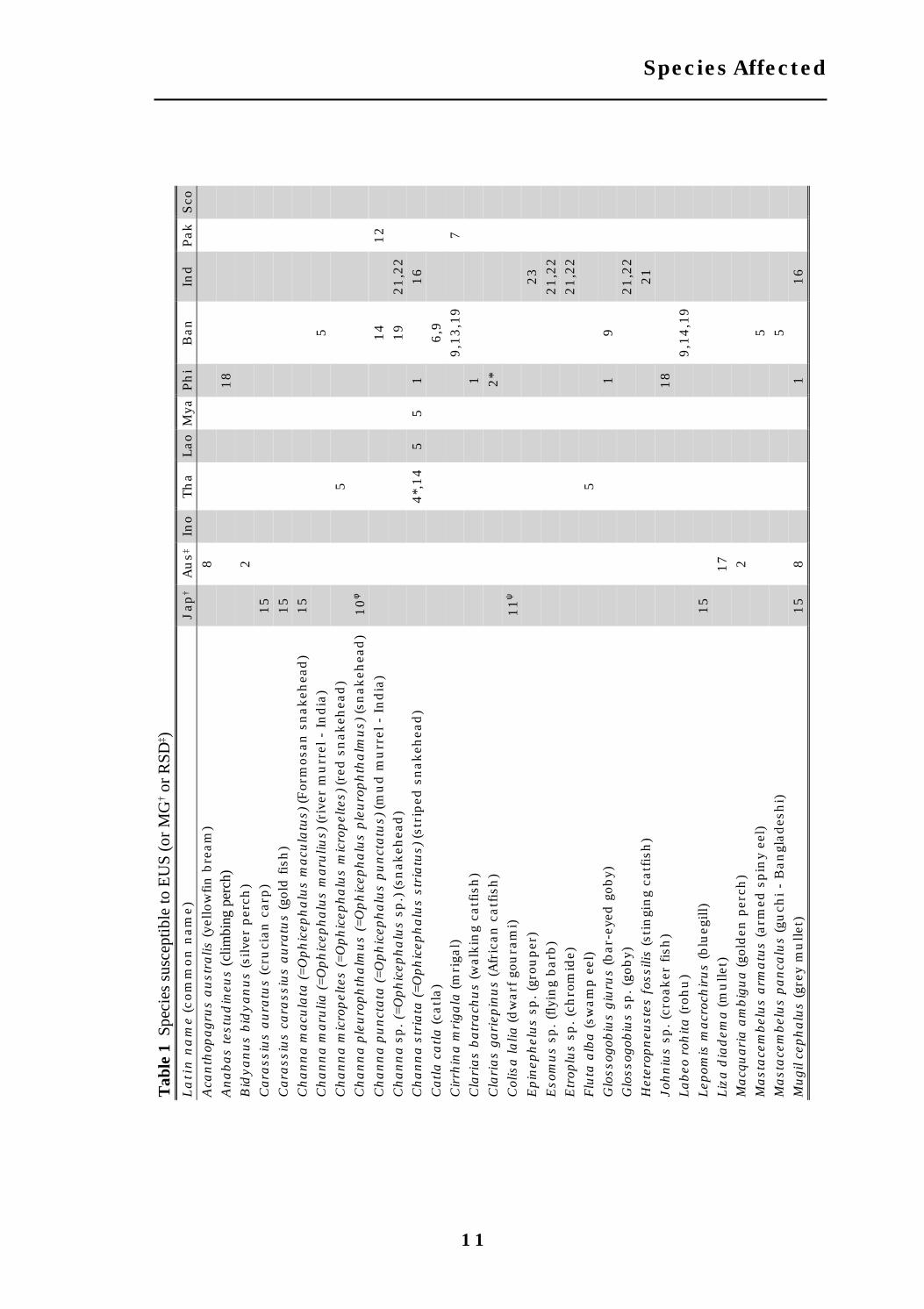
More than 100 fish species have been reported to be affected by EUS (Lilley etal, 1992), but only relatively few reports have been confirmed by demonstratingthe presence of mycotic granulomas in histological section or by isolation ofthe pathogenic Aphanomyces fungus from tissues underlying ulcers. Table 1lists these confirmed cases, including species from MG or RSD outbreaks.
Similarly, some commercially important species are considered to beparticularly resistant to EUS, but few studies have been undertaken toconfirm these observations and investigate the mechanism of resistance.Species reported to be unaffected by EUS outbreaks include Chinese majorcarps, tilapias and milkfish (Chanos chanos). Hatai (1994) experimentallyinjected catfish (Parasilurus asotus), loach (Misgurnus anguillicausatus) andeel (Anguilla japonica) with hyphae of A. invadans and found them to berefractory to infection. Wada et al. (1996) and Shariffpour (1997) experimentallyinjected common carp (Cyprinus carpio) with zoospores of Aphanomyces fromMG and EUS outbreaks respectively, and demonstrated that fungal growthwas suppressed by an intense inflammatory response.
Some authors have commented that the most severely affected species innatural outbreaks are generally bottom dwellers (Llobrera and Gacutan,1987; Chondar and Rao, 1996) or possess air-breathing organs (Roberts et al.,1994b), but examination of Table 1 shows that this is by no means always thecase.
In the case of snakeheads, no particular size group appears to be moresusceptible, with affected fish ranging from 40g to 900g (Cruz-Lacierda andShariff, 1995). However, there is a possibility that size or age may besignificant in other species. For example, Indian major carp, suffer highmortalities as fingerlings (Roberts et al., 1989) but larger fish, althoughappearing ulcerated, are not reported as dying in large numbers (AAHRI,ACIAR, IoA and NACA, 1997).
Some of the EUS-susceptible species listed in Table 1 have a wide geographicaldistribution, beyond the current limits of EUS outbreaks. For example,several snakehead and clariid catfish species occur in Africa and centralAsia. This suggests that there is potential for further spread of the disease tothese areas. However, it should be noted that optimal temperatures forvegetative growth in vitro for A. invadans are in the range 20-30oC (Fraser eta.l, 1992; Lilley and Roberts, 1997) and, probably for this reason, naturaloutbreaks to date have been limited to latitudes between 35oN and 35oS.Experimental injection challenges of native European and American fishspecies have shown that the pathogenic fungus, A. invadans, is capable ofcausing lesions in rainbow trout at 18oC (Thompson et al., in press), but is lessinfective in stickleback (Gasterosteus aculeatus) and roach (Rutilus rutilus) at11-16oC (Khan et al., 1998).
Species affected

10
Epizootic Ulcerative Syndrome (EUS) Technical Handbook
Table 1 Species susceptible to EUS (or MG† or RSD‡) as indicated by thepresence of typical mycotic granulomas in histological section or isolation ofpathogenic Aphanomyces from muscle or internal organs (numbers correspondwith references given below; *denotes artificial challenge)
φφφφφ The two genera Channa and Ophicephalus were united as Channa byMyers and Shapovalov (1931, cited by Clark, 1991)
Ψ Ornamental fish imported from Singapore
COUNTRY KEY: Jap = Japan; Aus = Australia; Ino = Indonesia; Tha = Thailand;Lao = Lao PDR; Mya = Myanmar; Phi = Philippines; Ban = Bangladesh; Ind =India; Pak = Pakistan; Sco = Scotland
REFERENCE KEY:
1 Callinan et al. (1995b)2 Callinan (unpublished)3 Catap (pers. comm.)4 Chinabut et al. (1995)5 Chinabut (unpublished)6 Chowdhury & Chinabut (pers. comm.)7 DFID (1998)8 Fraser et al. (1992)9 Ahmed & Hoque (submitted)10 Hanjavanit et al. (1997)11 Hatai (1994)12 Kanchanakhan (1996a)13 Khan (pers. comm.)14 Lilley and Roberts (1997)15 Miyazaki (1994)16 Mohan and Shankar (1995)17 Pearce (1990)18 Reantaso (1991); S. Chinabut (unpublished)19 Roberts et al. (1989)20 Thompson et al. (in press)21 Vishwanath et al. (1997)22 Vishwanath et al. (1998)23 Viswanath et al. (1997)
11
Species Affected
Tab
le 1
Spe
cies
sus
cept
ible
to E
US
(or
MG
† or
RSD
‡ )L
ati
n n
am
e (c
om
mon
nam
e)
Jap
†A
us‡
Ino
Th
aLao
Mya
Ph
iB
anIn
dPak
Sco
Aca
nth
opagru
s a
ust
ralis
(ye
llow
fin
bre
am
)8
An
ab
as
test
ud
ineu
s (c
limbi
ng p
erch
)18
Bid
ya
nu
s b
idy
an
us
(silve
r per
ch)
2C
ara
ssiu
s a
ura
tus
(cru
cian
car
p)
15
Cara
ssiu
s ca
rass
ius
au
ratu
s (g
old fis
h)
15
Ch
an
na m
acu
lata
(=
Oph
icep
ha
lus
macu
latu
s) (F
orm
osan
snakeh
ead)
15
Ch
an
na m
aru
lia (=
Oph
icep
ha
lus
maru
lius)
(ri
ver
mu
rrel
- I
ndia
)5
Ch
an
na m
icro
pel
tes
(=O
ph
icep
ha
lus
mic
rop
elte
s) (re
d s
nak
ehea
d)
5C
ha
nn
a p
leu
roph
tha
lmu
s (=
Oph
icep
ha
lus
ple
uro
ph
tha
lmu
s) (sn
akeh
ead)
10
φ
Ch
an
na p
un
cta
ta (=
Oph
icep
ha
lus
pu
nct
atu
s) (m
ud m
urr
el -
India
)14
12
Ch
an
na s
p.
(=O
ph
icep
ha
lus
sp.)
(sn
akeh
ead)
19
21,2
2C
ha
nn
a s
tria
ta (=O
ph
icep
ha
lus
stri
atu
s) (st
riped
snakeh
ead)
4*,
14
55
116
Ca
tla
ca
tla
(ca
tla)
6,9
Cir
rhin
a m
riga
la (m
riga
l)9,1
3,1
97
Cla
ria
s b
atr
ach
us
(wal
kin
g ca
tfis
h)
1C
lari
as
ga
riep
inu
s (A
fric
an
catf
ish
)2*
Col
isa
la
lia (dw
arf
gou
ram
i)11
ψ
Ep
inep
hel
us
sp. (g
rou
per
)23
Eso
mu
s sp
. (fly
ing
barb
)21,2
2E
trop
lus
sp. (c
hro
mid
e)21,2
2F
luta
alb
a (sw
am
p e
el)
5G
loss
ogob
ius
giu
rus
(bar
-eye
d g
oby)
19
Glo
ssog
obiu
s sp
. (g
oby)
21,2
2H
eter
opn
eust
es f
ossi
lis (st
ingi
ng
catf
ish
)21
Joh
niu
s sp
. (c
roak
er fis
h)
18
Lab
eo r
ohita
(ro
hu
)9,1
4,1
9Lep
omis
macr
och
iru
s (b
lueg
ill)
15
Liz
a d
iad
ema
(mu
llet)
17
Ma
cqu
ari
a a
mb
igu
a (go
lden
per
ch)
2M
ast
ace
mbel
us
arm
atu
s (a
rmed
spin
y ee
l)5
Ma
sta
cem
bel
us
pa
nca
lus
(gu
chi -
Ban
glades
hi)
5M
ugil
cep
ha
lus
(gre
y m
ullet
)15
81
16

12
Epizootic Ulcerative Syndrome (EUS) Technical Handbook
Tab
le 1
(C
ont’
d) S
peci
es s
usce
ptib
le to
EU
S (o
r M
G† o
r R
SD‡ )
La
tin
na
me
(com
mon
nam
e)
Jap
†A
us‡
Ino
Th
aLao
Mya
Ph
iB
an
Ind
Pak
Sco
Mu
gil
sp. (m
ullet
)21,2
2M
yst
us
sp. (c
atf
ish
)22
Not
opte
rus
not
opte
rus
(gre
y fe
ath
erback
)5
On
corh
yn
chu
s m
ykis
s (r
ain
bow
tro
ut)
- m
arg
inally
susc
epti
ble
11*
20*
Osp
hro
nem
us
gor
am
y (pla
raet
- T
hai)
5O
xyel
eotr
is m
arm
ora
tus
(san
d g
oby)
14
Oxy
eleo
tris
sp (gu
dge
on)
Pla
tyce
ph
alu
s fu
scu
s ( du
sky
flath
ead)
2Pla
tyce
ph
alu
s sp
. (fla
thea
d)
22
Ple
coglo
ssu
s a
ltiv
elis
(ayu
)11,1
5Pse
ttod
es s
p. (s
pin
y tu
rbot
)18
Pu
ntiu
s gon
ion
otu
s (s
ilve
r barb
)5
13
Pu
ntiu
s so
ph
ore
(pu
nti
- B
an
glad
esh
i)9
5Pu
ntiu
s sp
(pu
nti
us)
19
16,2
2R
hod
eus
ocel
latu
s (t
air
iku
-bara
tan
ago
- J
apan
ese)
11*
Roh
tee
sp (ket
i -
Ban
glades
hi)
5S
card
iniu
s er
yth
roph
tha
lmu
s (r
udd) -
marg
inally
susc
epti
ble
11*
Sca
toph
agu
s arg
us
(spot
ted s
cat)
18
Sca
toph
agu
s sp
. (s
cat)
21,2
2S
illago
cilia
ta (sa
nd w
hit
ing)
8,3
*S
illago
sp. (s
illa
go)
21,2
2Ter
apon
sp. (t
her
apon
)21,2
2Tri
chog
ast
er p
ecto
ralis
(sn
akes
kin
gou
ram
i)5
Tri
chog
ast
er t
rich
opte
rus
(3-s
pot
gou
ram
i)10
ψ3*
14
Tri
den
tiger
obsc
uru
s ob
scu
rus
(Jap
an
ese
trid
ent
gob
y)15
Upen
eus
ben
sai (
goatf
ish
)18
Vala
mu
gil
sp. (m
ullet
)21,2
2W
allago
att
u (w
allago
)22
Xen
ento
don
can
cila
(ro
un
d-t
ailed
garf
ish
)14

13
The most severe impact of EUS has probably been on small-scale, mixed-species fisheries and aquaculture activities in rice-fields and rural waterways.It is estimated that 250 million families in the Asian-Pacific region dependon rice as a main crop and much of the incidental fish harvests from thesepaddies are an important part of the families’ diet (Macintosh, 1986). It shouldbe noted that the chief months for harvesting rice paddy fish are fromSeptember to February, the period when most ulcerative disease episodesoccur. In these circumstances, any figure on the financial cost of EUS mayunderestimate the full impact of the disease to these communities.
Estimates of the economic value of fish losses to commercial fish traders aregiven in Table 2. These figures do not, however, take into account indirectsocio-economic costs due to market rejection of harvested ulcerated fish, orin some cases, even unaffected fish. In the 1980s, in some communities, awidespread, but unfounded, fear of disease transmission to consumers led toa drastic decrease in market demand for all food fish. Confidence in freshwaterfish farming, particularly among potential investors and financial agencies,was badly affected.
In the Philippines, the average daily income of fishers (approximately US$4)declined to US$1.50 during disease outbreaks in Laguna de Bay due to therejection of affected fish (ADB/NACA, 1991). Bangladesh suffered severelosses from EUS in 1988 and 1989, and extensive local media coverage aboutthe disease fuelled the public’s fear of health risks from fish consumption,resulting in initial price reductions of up to 75% and high losses to fishtraders. Nepal has no marine fish resources and therefore relies heavily onEUS-susceptible species. It was reported that 15-20% of total fish productionwas lost in Nepal during initial EUS outbreaks (ADB/NACA, 1991). Theoccurrence of EUS in cultured major carp fingerlings gave rise to fears of apotentially crippling effect on the expansion of carp culture in the subcontinentregion. Bhaumik et al. (1991) reported that 73% of the culture ponds in WestBengal were affected at that time, and most of these were reported to have lostbetween 30-40% of their stock. In their report giving details of losses to inlandfishworkers in Kerala, the ICSF (1992) quote the official figure of Rs 20million, but commented that newspapers reported losses up to ten times thisfigure.
The EUS pandemic has demonstrated to national authorities the ability offish disease to cause major financial losses, and as a result, one positiveimpact of EUS has been the increased funding allocated to fish diseaseresearch and diagnostic facilities in Asia by governments and internationalorganisations.
Socio-economics

14
Epizootic Ulcerative Syndrome (EUS) Technical Handbook
EC
ON
OM
IC L
OS
SC
OU
NT
RY
YE
AR
LO
CA
L C
UR
RE
NC
YU
S$
(appro
x.)
RE
FE
RE
NC
E
East
ern
Au
stra
lia
an
nu
ally
Au
st $
1 m
illion
700,0
00
Callin
an e
t a
l. (1996)
Indon
esia
1980-8
3
1984-8
7
- -
119,0
00
116,0
00
AD
B/N
AC
A (1991)
AD
B/N
AC
A (1991)
Th
ailan
d1982-8
3
1983-9
3
B 2
00 m
illion
-
5.5
million
100 m
illion
Ton
guth
ai (1
985)
Ch
inab
ut
(1994)
Ban
glad
esh
1988
1989
Tk 1
18 m
illion
Tk 8
8.2
million
2.8
million
2 m
illion
Baru
a (1990)
Baru
a (1990)
Sri
Lan
ka
1988-8
9
upto
1993
Rs
1 m
illion
Rs
20-4
0 m
illion
20,0
00
4-8
00,0
00
AD
B/N
AC
A (1991)
Bala
suri
ya (1994)
East
ern
Nep
al
1989-9
0R
s 30 m
illion
550,0
00
AD
B/N
AC
A (1991)
India
- B
ihar
- O
riss
a
- K
erala
1990
1989-9
1
1991-9
2
Rs
4.8
million
Rs
3 m
illion
Rs
20 m
illion
150,0
00
95,0
00
625,0
00
Das
(1994)
Das
(1994)
Das
(1994)
Pak
ista
n -
Pu
nja
b1996
-300,0
00
AA
HR
I,
AC
IAR
, Io
A
an
dN
AC
A, 1997)
Tab
le 2
E
stim
ate
d e
con
omic
los
ses
from
fis
h m
orta
liti
es d
ue
to E
US

15
Prior to the initial EUS outbreaks, most countries in the region had notexperienced a fish disease epizootic on such a large scale and, not surprisingly,there has been a great deal of local apprehension as to the consequences ofconsuming diseased fish or using affected waters for domestic or agriculturalpurposes. The concurrent deaths of ducks, cattle and other animals wereattributed to the occurrence of EUS. There is however, no scientific evidencethat the disease itself causes any human or animal illness. Rahman et al.(1988) were unable to induce any disease symptoms in ducks fed EUS-infected fish or even injected with Aeromonas hydrophila cultures. Thereforeit is important to take public educational measures and allay the naturalfears of farmers, fishers and consumers about any wider effects of EUS.However, it must be stressed that good hygiene practices should be adheredto. In particular, dead fish should not be collected for sale or consumption, notbecause of ulcerative disease as such, but because bacteria or toxins presentin decomposing, EUS-affected fish may cause human illness.
The uncontrolled use of chemotherapeutants to treat EUS or other diseasesin intensive culture systems is also a matter of public health concern.Chloramphenicol for instance, is used in treating typhoid in humans andthere is a risk that the build up of bacterial resistance in treated fish(Poonsuk et al., 1983) may be transferred to humans. Of greater concern tofarmers is the possibility of severe allergic reactions affecting farm workersin contact with the drug. There is also the danger that consumers may beexposed to drug residues in marketed fish that had been hurriedly harvestedbefore the recommended withdrawal period had been completed. Althoughthese are issues that affect aquaculture in general, the occurrence of EUShas underlined the need to develop appropriate guidelines and legislation toprotect farmers and consumers against the indiscriminate use ofchemotherapeutants.
Public health

16
Epizootic Ulcerative Syndrome (EUS) Technical Handbook

17
Diseased fish, particularly those with cutaneous ulcers, are vulnerable toinfection by opportunistic pathogens and, in long standing cases, it is oftendifficult to identify the cause of the initial lesion. Given the wide geographicalarea, and the diverse range of habitats in which EUS-affected fish occur, aparticularly diverse mix of microbiological agents have been recovered fromaffected fish. Some of these agents may significantly contribute to a diseasecomplex in a particular outbreak, but it is important to distinguish them fromthe factor (or factors) essential in all EUS outbreaks. A description of fungi,viruses, bacteria and parasites found associated with EUS lesions is givenhere, along with comment on their importance in EUS outbreaks.
Fungi
Recent work has confirmed that a single species of Aphanomyces “fungus”1 isa necessary cause2 of EUS, i.e. it occurs in all outbreaks, and in someoutbreaks (e.g. in Australian estuaries), may be the only biological factorrequired for the disease to occur.
The pathogenic Aphanomyces fungus
Fungi have been known to be involved in the aetiology of EUS in SoutheastAsia since the initial outbreaks in Thailand. Limsuwan and Chinabut (1983)described a “severe chronic granulomatous mycosis” in histological sectionsof affected fish. However, the dominance of saprophytic fungal contaminantson the surface of EUS lesions led to the identification of Achlya and Saprolegniaspp. from affected fish (Pichyangkura and Bodhalamik, 1983; Limsuwan andChinabut, 1983). These were soon recognised as secondary agents (Tonguthai,1985), but it was also assumed that this may be the case for all mycoticinvolvement in EUS.
As described in the History section, before the first appearance of EUS inSoutheast Asia, the pathogenic Aphanomyces piscicida had been isolated fromMG-affected fish in Japan (Hatai et al., 1977), but MG had not yet beenrecognised as synonymous with EUS. An Aphanomyces fungus wassubsequently obtained from RSD outbreaks in Australia in 1989 (Fraser et al.,1992) and, independently, from EUS outbreaks in Thailand in 1991-1992
Aetiology
1 The genus Aphanomyces is contained within the family Saprolegniaceae and theclass Oomycetes, and it should be noted here that the Oomycetes are no longerregarded as true fungi, but rather fungal-like protists. They are now often classedalongside diatoms, brown algae and xanthophytes within the phylum Heterokontaas part of the third botanical kingdom, the Chromista. They are sometimes calledpseudofungi, either as a general term or a formal taxon (Cavalier-Smith, 1987). Theyare, however, still commonly referred to as fungi and this term will be used for thepurpose of this review.2For a definition of "necessary cause" see Epidemiology section.

18
Epizootic Ulcerative Syndrome (EUS) Technical Handbook
(Roberts et al., 1993). These isolates were shown to be capable of reproducingtypical EUS lesions when injected below the dermis of susceptible fish. All ofthese pathogenic MG, RSD and EUS isolates were shown to be slow-growingand thermo-labile in culture. Similar isolates have also been obtained fromthe Philippines, Indonesia, Bangladesh (Lilley and Roberts, 1997) and India(I. Karunasagar, pers. comm.). Recently, pathogenic Aphanomyces culturesfrom most of these countries have been compared directly, and shown bymeans of protein banding profiles (Callinan et al., 1995b; Lilley et al., 1997b),growth characteristics (Lilley and Roberts, 1997) and chemical susceptibility(Lilley and Inglis, 1997) to be all the same species. Genetic fingerprintingtechniques have also been used to show that the various isolates weregenetically all very similar (Lilley et al., 1997a). This is proof that the isolatesare not long-term residents in each locality, as would be expected ofopportunistic fungi. Instead, they are part of one fungal strain that hascolonised much of Asia and Australia in a matter of decades, and resulted inthe spread of EUS.
The pathogenic Aphanomyces has been named variously as Aphanomycespiscicida (Hatai, 1980), Aphanomyces invaderis (Willoughby et al., 1995) andERA (EUS-related Aphanomyces sp.: Lumanlan-Mayo et al., 1997). As isolatesin each case have been shown to be conspecific, however, one species nameis required to describe all these isolates. As A. invadans is the only valid taxonname according to the International Code of Botanical Nomenclature (ICBN),this is the name that will be adopted here.
A. invadans is known to grow fastest in culture at temperatures between 26-30oC (Hatai and Egusa, 1978; Fraser et al., 1992; Lilley and Roberts, 1997), andhas been shown to grow in snakehead muscle tissue between 19-31oC(Chinabut et al., 1995). However, further investigation has revealed thatsnakeheads are able to recover from A. invadans infection at highertemperatures (26, 30oC), but are unable to prevent fungal invasion andeventually succumb to the disease at lower temperatures (19oC) (Chinabut etal., 1995). The humoral and cellular immune response of fish are known tobe supressed at low temperatures (Avtalion et al., 1980; Bly and Clem, 1991),which may explain why mortalities from EUS occur when water temperaturesare low. Naturally and artificially infected snakeheads have been shown toproduce an antibody response against A. invadans (Thompson et al., 1997), andthe cellular macrophage response is also considered to be important inenabling fish to resist infection (Wada et al., 1996).
A summary of the various published descriptions of the characteristics of A.invadans from EUS, MG and RSD outbreaks is given in Table 3. Techniquesfor isolating A. invadans from fish and water, and identifying candidatecultures to the genus Aphanomyces are given in the Annex. As with othersaprolegniacean fungi, A. invadans, is aseptate and produces two zoosporeforms, the secondary form being free-swimming and laterally biflagellate. Nosexual reproductive structures have been observed in any of the isolates fromEUS, MG or RSD outbreaks. The lack of sexual structures is considered to bea particularly common phenomenon among the more pathogenic membersof the Saprolegniaceae (Alderman and Polglase, 1988).

19
Aetiology
Figure 2 SporulatingAphanomyces invadansculture showing clusters ofencysted primary zoosporesfollowing discharge fromlateral evacuation tubes (fromRoberts et al. 1993)

20
Epizootic Ulcerative Syndrome (EUS) Technical Handbook
Ch
ara
cte
rist
icD
esc
ripti
on
Hyp
hal dia
met
erV
ari
able
. W
ider
in
fis
h t
issu
e (1
2-3
0 µ
m) th
an
in
art
ific
ial cu
ltu
re (5-2
0 µ
m o
n G
PY
aga
r). In
cu
ltu
re, h
yph
ae h
ave
rou
nded
tip
s and b
ran
ch a
lmos
t at
righ
t an
gles
to
the
main
axi
s.R
adia
l gr
owth
Gro
ws
at
tem
per
atu
res
bet
wee
n 5
-36
o C, an
d s
alin
ity
bel
ow 1
0ppt
NaC
l.N
o gr
owth
on
cor
nm
eal aga
r, m
alt
ext
ract
aga
r or
Sabou
rau
d d
extr
ose
aga
r.O
n G
PY
aga
r (m
m p
er 2
4h
):
0.8
at
10
o C1.9
at
14
o C2.8
at
18
o C3.9
at
22
o C4.6
at
26
o C4.6
at
30
o C3.4
at
34
o Cn
o gr
owth
at
37
oC
Oog
onia
Not
obse
rved
Zoo
spor
an
gia
Equ
al dia
met
er t
o m
ycel
ium
(abou
t 10 µ
m)
Zoo
spor
an
gial ty
pe
Ter
min
al or
in
terc
alary
. C
omple
x sp
oran
gia h
ave
4 late
ral ev
acu
ati
on t
ubes
(630-9
30
µm lon
g), 3 t
ubes
(430-5
40 µ
m lon
g) o
r 2 o
r 1 t
ube
(330-4
70 µ
m lon
g)Z
oosp
oran
gial re
new
al
Sym
pod
ial bra
nch
ing
bel
ow e
mpty
spor
an
giu
m1
o z
oosp
ore
Sin
gle
row
con
nec
ted b
y th
in s
tran
d o
f cy
topla
sm1
o z
oosp
ore
cyst
clu
ster
sA
chly
oid. U
sually
30-5
0 1
o zo
ospor
e cy
sts
1o z
oosp
ore
cyst
Usu
ally
6-1
0 µ
m d
iam
eter
2o z
oosp
ore
Mot
ile,
su
bsp
her
ical
, bifla
gellate
abou
t 6 µ
m in
dia
met
erR
elea
sed w
ith
in 1
2 h
ours
of sp
oran
gial dev
elop
men
t at
22
o C.
No
spor
ula
tion
abov
e 2ppt
NaC
l2
o zo
ospor
e cy
stA
bou
t 6.5
µm
in
dia
met
er, so
met
imes
“gi
an
t cy
sts”
pro
du
ced u
p t
o 27 µ
m in
dia
met
erD
emon
stra
tes
lim
ited
pol
ypla
net
ism
(re
pea
ted z
oosp
ore
emer
gen
ce, en
cyst
men
t an
dre
-em
erge
nce
) in
th
e pre
sen
ce o
f n
utr
ien
t m
edia
Tab
le 3
Ch
ara
cter
isti
cs o
f A
ph
an
omyce
s in
vad
an
s in
cu
ltu
re
21
Aetiology
Involvement of other saprophytic fungi
Lilley and Roberts (1997) ruled out the possibility that multiple opportunisticfungal species are responsible for the mycotic granulomas typical of EUS, byshowing that a number of saprophytic Saprolegnia, Achlya and Aphanomycesspp. from EUS-affected areas were incapable of sustained growth in snakeheads,even when injected directly into muscle tissue. Nonetheless, saprophyticSaprolegnia, Achlya and Aphanomyces spp. are commonly observed on thesurface of EUS lesions (Pichyangkura and Tangtrongpiros, 1985; Willoughbyand Lilley, 1992; Qureshi et al., 1995), and may contribute to the disease asopportunistic wound parasites.
Reports of saprophytic Aphanomyces spp. acting as wound parasites on fish arenot uncommon (Shanor and Saslow, 1944; Hoshina et al., 1960; Srivastava,1979; Ogbonna and Alabi, 1991; Khulbe et al., 1995). Aphanomyces spp. havealso been reported from freshwater dolphins (Fowles, 1976) and soft shellturtles (Valairatana and Willoughby, 1994), but these isolates can all be easilydistinguished from A. invadans in terms of pathogenic and growthcharacteristics, and should not be confused with the EUS pathogen.
Viruses
Prior to recent mycological findings, viruses were considered to be the mostlikely necessary infectious cause of EUS. Several species of viruses havebeen isolated from EUS outbreaks and varying intepretations have beenmade of the pathogenic significance of these isolates. Evidence to datesuggests that at least one of these species may be involved in some EUSoutbreaks, particularly in Thailand, by predisposing fish to infection by A.invadans.
History of isolation of EUS-associated viruses
Following the 1982-3 EUS outbreak in Thailand, virus-like particles weredemonstrated in various tissues of affected fish (Rattanaphani et al., 1983;Wattanavijarn et al., 1983a; b; 1984). These workers subsequently isolatedthe so-called snakehead rhabdovirus (SHRV), which was shown to beserologically distinct from other fish rhabdoviruses (Ahne et al., 1988;Kasornchandra et al., 1992). Between 1985-1989 a major sampling programmeof over 200 fishes in 8 EUS-affected countries was undertaken, and as aresult, 6 rhabdovirus isolates were obtained from Thailand, Myanmar, SriLanka and Australia (Frerichs et al., 1986; 1989a; Roberts et al. 1989; Lilleyand Frerichs, 1994). These isolates, named ulcerative disease rhabdovirus(UDRV), were shown to represent another species that was distinct fromSHRV (Kasornchandra et al., 1992) and other fish-pathogenic rhabdoviruses(Frerichs et al., 1989b). Significantly, during this sampling programme, noviruses were obtained from Bangladesh, Lao PDR, Malaysia, Indonesia or thePhilippines. Later virological surveys of northeast India (Boonyaratpalin,1989a) and Pakistan (AAHRI, ACIAR, IoA and NACA, 1997) also yielded no viralisolates.

22
Epizootic Ulcerative Syndrome (EUS) Technical Handbook
No further isolates of UDRV have been obtained since 1989, but samplingstudies in Thailand have yielded an increasing number of isolates showingmorphological and electrophoretic similarities to SHRV. Two such isolateswere obtained in 1992, nine in 1994, and nine in 1996 (Kanchanakhan,1996b). A further two virus isolates were obtained in 1997, but awaitcharacterisation (Kanchanakhan, unpublished data).
Aside from the rhabdoviruses, several birnaviruses and a single reovirushave also been isolated from ulcerated fish. Among the birnaviruses, sandgoby virus (SGV) from Thailand, and a more recent isolate from Singapore,have both been shown to be distinct from the IPNV reference strains (Hedricket al., 1986; Subramaniam et al., 1993). Two other birnavirus isolates wereconsidered to be more similar to known IPNV strains: these comprisedsnakehead virus (SHV) from Thailand, and another isolate that was furtheridentified as the Sp serotype of IPNV (Saitanu et al., 1986; Wattanavijarn et al.,1988). A reovirus, isolated from a diseased snakehead in 1992 (Frerichs,1995), also appears to be a new, distinct viral strain or species (Riji John,1997).
The heterogeneity of viral isolations and the low recovery rate of viruses ledsome workers to the conclusion that these were adventitious agents whichwould as likely have been isolated from healthy fish (Frerichs, 1995).Kanchanakhan (1996b) has recently revived interest in viruses bydemonstrating that rhabdoviruses can be more readily isolated from fishspecimens collected during the early period of outbreaks in Thailand. Virusescould not be obtained during the middle, late and recovery phases of outbreaks.In artificial challenge studies using a rhabdovirus strain isolated in Thailandin 1994 (T9412), the virus was reisolated from 100% of snakehead fish 3 daysp.i. (post-injection), decreasing to less than 25% of fish 30 days p.i., at 20oC,suggesting that the virus was being partially or entirely eliminated by thehost defence system (Kanchanakhan, 1996b). Successful virus isolation alsorequires that only freshly killed fish are sampled, and that tissue extracts areprepared immediately thereafter. The advised procedure for virus isolation isgiven in Annex 6.
Pathogenicity of EUS-associated viruses
Pathogenicity trials with most EUS-associated viruses have usuallydemostrated little more than scale damage or occasional development ofminor skin lesions. Frerichs et al. (1993) were unable to show any consistentlesion in snakeheads immersed or injected i.p. (intra peritoneally) with anisolate of UDRV. Of the birnaviruses, only SHV has been tested in challengestudies. Saitanu et al. (1986) reported that i.p. injections of SHV resulted inscale damage in 80% of small snakeheads, but not at all in larger fish. RijiJohn (1997) demonstrated that the reovirus was not pathogenic to juvenilesnakeheads in injection challenges.
More recent work by Kanchanakhan (1996b) showed that rhabdovirus strainT9412 can result in substantial lesions in striped snakeheads, particularlyusing challenges by i.m. (intra muscular) injection. The virulence of T9412was shown to be dependent on temperature, fish species and fish age. All

23
Aetiology
snakehead fry died when challenged at 20oC, but no mortality was recordedat 29oC, or in other species of fish (including EUS-susceptible fish) at eithertemperature.
If viruses have a role in the pathogenicity of EUS, their most likely effect isto cause skin lesions sufficient to allow entry of the fungus, A. invadans.Kanchanakhan (1996b) subjected snakehead juveniles to i.m. injections ofT9412 rhabdovirus or L15 medium, followed by bath challenges with A.invadans spores at 20oC. EUS was induced in 100% of fish given rhabdovirusinfections and only 35% in fish given control L15 injections. This providessome evidence that T9412 may help to predispose fish to infection by A.invadans, but co-immersion challenges with the virus as well as the fungusare required to demonstrate that this can occur under more natural conditions.
Parasites
Several metazoan (Dactylogyrus sp., Gyrodactylus sp.) and protozoan (Chilodonellasp., Trichodina sp., Costia sp., Henneguya sp., Ichthyophthirius sp.) parasiteswere identified from 273 EUS infected fish during the 1982-3 epizootic inThailand (Reungprach et al., 1983) Several fish examined before the secondoutbreak, and thought to be at an early stage of the disease, showed tiny redspots on the skin. Examination revealed a large number of Epistylis sp.protozoans (Tonguthai, 1985).
In Australia, Callinan and Keep (1989) and Pearce (1990) found protozoan andmetazoan parasites present on some affected fish, but concluded that noparasite species was intimately associated with lesions and there was noevidence to suggest that parasites initiate ulcers. In their survey of affectedcountries in southeast Asia, Roberts et al. (1986) found that diseased fishcarried no more than the expected parasite load for wild rice paddy or riverinefish.
It therefore appears unlikely that any parasite acts either as a pathogen ora vector for a pathogen of EUS. However, parasites may at times induce stressin fish and predispose them to infection. For example, Subasinghe (1993)demonstrated a clear association between parasite burden of Trichodina sp.on gills and susceptibility of striped snakeheads to EUS infection. It is alsopossible that external parasites may, in some circumstances, induce mildskin lesions which would allow propagules of the fungal pathogen, Aphanomycesinvadans, to attach and infect the fish host.
Bacteria
Available evidence suggests that bacteria may be important, but not essential,at two distinct stages in the pathogenesis of EUS.
1. Current evidence indicates Aphanomyces invadans must attach to thedermis before it can invade underlying tissues. Cutaneous bacterialinfections (e.g. Flexibacter) may predispose fish to EUS by inducing skinlesions which provide an entry for the fungus (Figure 3).

24
Epizootic Ulcerative Syndrome (EUS) Technical Handbook
It is possible that cutaneous bacterial infections may damage areas ofepidermis and expose dermis, thereby allowing A. invadans to attachand invade underlying tissues. However, to date there are no reportsconfirming bacterial involvement in such a process, suggesting this isnot a common means of EUS lesion induction. Although some workershave suggested that bacteria such as Vibrio anguillarum (Rodgers andBurke, 1981) or nocardioform bacteria (Chakrabarty and Dastidar,1991) are necessary causes of EUS, several studies (Callinan and Keep,1989; Boonyaratpalin, 1989b; Pearce, 1990) have failed to consistentlyassociate any bacterial species with all, or even a large proportion of,ulcers on affected fish, suggesting bacteria are not necessary causes.This suggestion is supported by the observation that bacteria are onlyrarely visible in histological sections of EUS ulcers.
2. There is strong evidence that many EUS-affected fish die as a result ofsepticaemias caused by opportunist bacterial pathogens. It is likelythat these bacteria first colonise the surface of established ulcers andthen invade the bloodstream to induce lethal septicaemia (Figure 3).
Aeromonas spp., notably A. hydrophila (Llobrera and Gacutan, 1987; Paland Pradhan, 1990), can often be isolated from ulcers or internal organsof EUS-affected fish. Some of these A. hydrophila strains have beencharacterised as virulent (Torres et al., 1990; Suthi, 1991; Karunasagaret al., 1995) or cytotoxic (Yadav et al., 1992).

25
Environmental Factors
Current findings indicate that normal skin defences must be compromisedin some way before Aphanomyces invadans can attach to the skin and invadeunderlying tissues. Given that EUS outbreaks are usually seasonally recurrent,it is likely that a number of biotic and/or abiotic factors, influenced byseasonal changes, play a role in lesion induction and/or in the availability ofinfective forms of the fungus.
Several studies have examined possible associations between EUS outbreaksand changes in seasonal factors and water quality variables.
Temperature
Both low and high temperatures appear to influence outbreak occurrence andit is likely that these influences at least partially explain the seasonallyrecurrent pattern of EUS outbreaks.
Low temperatures appear to influence the severity of EUS lesions, and hencethe severity of an outbreak, by impairing the ability of individual fish tocontain and inactivate the invasive fungus. Chinabut et al. (1995) injectedstriped snakehead with A. invadans zoospores and showed that theinflammatory response was less pronounced, fungal invasion was moreextensive, and mortality rates were higher, in fish kept at 19oC comparedwith fish kept at 26oC and 31oC.
Field studies also suggest that low temperatures are an important determinantfor some, but not all, EUS outbreaks. Rodgers and Burke (1981) associatedmaximum EUS prevalence in estuarine fish populations with seasonalaggregations of fish stressed by low or rapidly changing water temperaturesand rapid or prolonged depressions of salinity. Some EUS outbreaks infreshwater fish in Asia have occurred during periods of declining and/orunstable temperatures. During 1988 and 1989, outbreaks at sites inBangladesh, China, India and Lao PDR occurred during months in which themean daily temperature was below the annual mean daily temperature(Phillips and Keddie, 1990). However, outbreaks in the Philippines andThailand have also been recorded in warmer months (Phillips and Keddie,1990) suggesting there is no consistent relationship between EUS outbreaksand low temperatures. Diurnal temperature fluctuations of 10oC were recordedduring outbreaks in both Bangladesh and the Philippines (Phillips andKeddie, 1990).
Studies in the Philippines (Lumanlan-Mayo et al., 1997) suggested thatoutbreaks in rice-fish plots will not occur when maximum diurnal watertemperatures remain at >30oC. It is likely that the causative fungus issubstantially inactive at these temperatures. A. invadans hyphae grow onlypoorly at temperatures above 31oC and do not grow at 37oC (Hatai and Egusa,

26
Epizootic Ulcerative Syndrome (EUS) Technical Handbook
1978; Fraser et al., 1992; Roberts et al., 1993). Zoospores are more sensitivethan hyphae to temperature effects and zoospore production is inhibited at35oC (Campbell, unpublished).
Rainfall and related water quality variables
EUS outbreaks in estuarine fish in Australia follow major rainfall events inthe lower catchment (Virgona, 1992; Callinan et al., 1995). It is likely thatthese events influence EUS occurrence in at least 3 ways.
1. The influx of fresh water into the estuary reduces salinity at outbreaksites to < 2 ppt (Rodgers and Burke, 1981; Costa and Wijeyaratne, 1989;Virgona, 1992), thereby allowing A.invadans to sporulate (Fraser et al.,1992).
2. Acidified runoff water from acid sulfate soil areas in the coastalfloodplain flows into the estuary (Sammut et al., 1996). Fish sublethallyexposed to this water develop areas of epidermal necrosis. A. invadanszoospores attach to, and invade, dermis exposed when this necroticepidermis sloughs, thereby initiating EUS lesions (Callinan, 1997).
3. Organic matter, carried into the estuary with runoff water from thecoastal floodplain, is broken down by microbial agents in the daysfollowing the major rainfall event, thereby reducing dissolved oxygenconcentrations to <1 ppm for several days (Callinan, 1997). Fishsublethally exposed to this water may develop areas of epidermalnecrosis (Plumb et al., 1976) and underlying dermis may be colonised asabove by A. invadans propagules.
Detailed environmental monitoring programs have linked EUS outbreaks infreshwater fish in Asia with rainfall events and associated low and/ordecreasing water temperatures, alkalinity, hardness and chlorideconcentrations (Phillips and Keddie, 1990; Bondad-Reantaso et al., 1992;Catap unpublished). However, in a study of EUS outbreaks in 4 ponds inIndonesia (Bastiawan unpublished), there was no consistent relationshipbetween outbreak occurrence and rainfall, water temperature, hardness,alkalinity or any other measured water quality variable. Similarly, in a studyof EUS in the Philippines, Palisoc and Aralar (1995) found that while outbreaksin Laguna Lake were associated with temperature, depth, Secchi disctransparency, alkalinity and chloride, outbreaks in Lake Naujan wereassociated with temperature only.
Flooding
Floods are thought to spread infection by aiding the spread of infected fish andthe causal fungus. It is suggested that floods in Bangladesh in 1988 resultedin the rapid spread of EUS in that country.

27
Environmental Factors
Site characteristics
Source of infection
An EUS outbreak can occur only when susceptible fish, infective forms of thefungus and suitable environmental conditions are present at the site. Ahmedand Rab (1995) associated EUS outbreaks in Bangladesh with farming ofsusceptible fish species in ponds which had previously been derelict, or pondswhich had been treated with piscicides to remove predators and otherundesirable fish prior to stocking. Their findings indicate that the fungusmust have survived in these ponds, either within surviving infected fish orin the environment, possibly as an encysted spore. Outbreaks in silver perchBidyanus bidyanus in freshwater ponds in Australia are always associatedwith the presence of wild EUS-susceptible fish in the ponds or in the ponds’water supply (Callinan and Rowland, unpublished). These wild fish are a likelysource of fungal propagules.
Soil or sediment characteristics
As noted above, EUS outbreaks in estuarine fish are often associated withrecent acidified runoff from acid sulfate soil areas. It is also possible that soiland/or sediment characteristics influence outbreak occurrence in freshwaterponds, although no definite associations have yet been identified. Macintoshand Phillips (1986) found that sediments at many outbreak sites were slightlyacidic and had low calcium content. They suggested that such soils wouldaccount for the poorly buffered acidic water and high levels of aluminium andiron in water samples from such sites. Ahmed and Rab (1995) noted anassociation between EUS outbreaks and ponds having reddish sandy soils,and suggested the associated relatively high turbidities in these ponds mayhave been stressful to fish.
Conclusion
Taken together, the available evidence suggests that a diverse group of bioticand abiotic agents, including viruses, bacteria, cutaneous ectoparasites, lowpH and low dissolved oxygen concentrations, may initiate skin lesions infreshwater and estuarine fish and that these non-specific lesions aresubsequently colonised by A. invadans. It is therefore unlikely that anyspecific environmental determinant is always associated with EUS outbreaksin freshwater or estuarine fish. It is more likely that environmentaldeterminants will vary from outbreak to outbreak, depending on the agentinitiating the non-specific skin lesions, the aquatic environment at the siteand the fish populations at risk. Further studies are needed to identify theserelationships in more detail.

28
Epizootic Ulcerative Syndrome (EUS) Technical Handbook

29
Correct diagnosis of EUS is important to avoid confusions with other ulcerativeconditions. Positive diagnosis of EUS is made by demonstrating the presenceof mycotic granulomas in histological section and isolating Aphanomycesinvadans from internal tissues. Techniques for the isolation andcharacterisation of A. invadans are described in the Annex. The followinggives some information on clinical signs and histopathology of EUS. Table 4summarises these general findings. A glossary of technical terms is given atthe end of this handbook.
Clinical signs
Studies on the pathology of EUS in Asia have tended to focus on the stripedsnakehead (Channa striata) as this is the species most commonly and mostseverely affected. However, significant differences with other species havebeen noted. In general, lesions on EUS-affected fish can be separated into 3groups, on the basis of gross appearance (Viswanath et al., 1997).
Clinical signs in the early stage of the disease are similar. Appetite isreduced or absent and fish become lethargic, either floating just beneath thesurface or swimming with the head out of the water.
Gross pathology
Pinhead-sized, red spots develop on the body surface, head and fins, caudalpeduncle, dorsum or operculum with no noticeable haemorrhages or ulcers.In the early stages these may simply be areas of acute dermatitis formingrosacea.
The intermediate stage lesions are represented by small (2-4 cm) dermalulcers, with associated loss of scales, haemorrhage and oedema. Roberts etal. (1989) noted that in Puntius spp., gouramies and other midwater fish,ulcers are particularly dark and usually circular; often only one largesuperficial lesion occurs on the flank or dorsum. Most species, other thansnakeheads and mullet, will die at this stage.
The advanced stage lesions appear on other parts of the fishes body andexpand into large necrotic open ulcers; resulting eventually in death. Someaffected species, e.g. striped snakeheads, can survive with much moresevere, chronic lesions that may have completely destroyed the caudalpeduncle or eroded deep into the cranium or abdominal cavity sometimesexposing the swim bladder. Head tissue erosion is a particularly commonfeature of diseased striped snakeheads and specimens have been found withexposed optic nerves or loosened articular bones such as maxillae andmandibles.
Diagnosis

30
Epizootic Ulcerative Syndrome (EUS) Technical Handbook
EUS-affected fish in India
(from Viswanath et al., 1997)
EUS of mullet (Mugil cephalus) inAustralia
(from Callinan et al ., 1989)
Erythematous dermatitis: Yellow skinwith irregular reddening. Scalesfractured. Usually <1 cm in diameter.Epidermis present at margins andirregularly over lesion. Epidermishyperplastic, oedematous and infiltratedby mononuclear cells. Stratumspongiosum mild to severe congestion,oedema and mononuclear cellinfiltration. Other tissues normal. Nohyphae or granulomas.
Type I. Early lesions. Pinhead sized redspots on the body surface. No noticeablehaemorrhage or ulceration. Skin aroundthe spots is normal with nodiscolouration. Sections show focalinflammatory changes. There are severalnodular structures in the epidermis,sometimes associated with fungalhyphae. Dermis and skeletal muscle arenormal, without evidence of fungalinvasion.
Intermediate-type dermatitis:Approximately 1 cm in diameter.Epidermis absent over lesion, thoughsometimes evidence of regeneration,scales usually retained. Mild tomoderate chronic active dermatitis withsome fungal hyphae and granulomaswithin skeletal muscle. Often musclenecrosis.
Type II. Moderately advanced lesions.Approximately 2-4 cm in diameter,raised, circular, discoloured areas on thebody surface. These areas are soft withrelatively intact skin and scales. Insections, mycotic granulomas seen inepidermis, dermis and skeletalmusculature, associated with numerous,non-septate fungal hyphae. Significantnecrotising dermatitis and myositis dueto fungal invasion. In most of theselesions, scales and epidermis notcompletely lost.
Necrotising dermatitis: yellowish-greyto red, ovoid domed areas (1-4 cmdiameter). Epidermis and scales usuallyabsent, dermis swollen and macerated.Few hyphae trailed from lesion (notcotton-wool-like). Moderate to severe,locally extensive, necrotising,granulomatous dermatitis. Largenumber of sparsely-branching, aseptatehyphae (12-18 µm in diameter) usuallywithin granulomas extending to skeletalmuscle. Severe floccular degeneration ofmuscle.
Type III. Advanced lesions. Circular oroval, open dermal ulcers, extending intothe skeletal musculature. Characterisedby large haemorrhagic and necrotic openulcers on the body surface, devoid ofepidermis and scales, with loss ofdermis at the site of the ulcer. In mostcases the underlying musculature isexposed and largely replaced by fungalgranulomas and host inflammatorytissue. Considerable myofibrillarnecrosis. Fungal hyphae extend in alldirections from the focus of the dermalulcer. Necrotic muscle fibres and fungalhyphae often found within granulomas.
Dermal ulcer: About 1-4 cm indiameter. Margins sharply defined.Skeletal muscle exposed up to 1 cmbelow surface. In some cases bone orviscera exposed. Moderate to severediffuse granulomatous myositis.Hyphae, within granulomas, rarelypenetrated internal organs. Some dermalulcers showed evidence of healing.
Table 4 Progressive diagnostic symptoms of EUS

31
Diagnosis
Diseased striped snakeheads, with moderately advanced lesions, placed inimproved water quality conditions often recover. Similarly, lesions on estuarinefish such as mullet appear to heal quickly when fish move into brackish ormarine environments. Healing ulcers are characterised by a conspicuousdark colour caused by increased numbers of melanophores.
Histopathology
A general description of the typical histopathological developments that occurin EUS-diseased fish is given here with reference to some observations inparticular species.
The early skin lesions of some samples have been observed and found to beprincipally areas of epithelial necrosis with surrounding oedema,haemorrhaging of the underlying dermis and some inflammatory cellinfiltration. It has not been possible to confirm fungal involvement in most ofthese early samples but a few have harboured a small number of hyphae. Thepresence of fungal hyphae was demonstrated in the epidermis of some earlystages of infected fish from India (Viswanath et al., 1997). Similarly Robertset al. (1989) were able to study early lesions during an EUS outbreak in acaptive population of Indian major carp. They observed an acute necrotisingmyopathy, more severe than is usually seen in wild fishes, spread over a widearea below the active skin lesion. The epidermis at the margins of the ulceritself was degenerate and thickened, and contained only a very small numberof fungal hyphae enclosed within an epithelioid capsule. The blood vessels ofthe dermis were very hyperaemic and some had a collar of lymphoid ormyeloid cells which might be associated with virus infection although noviral inclusion bodies were detected.
Subsequent pathological developments in all infected fish species involvesignificant degenerative changes in skin and muscle tissue with minimaldisruption of internal organs.
In advanced lesions there is massive necrotising granulomatous mycosis ofthe underlying muscle fibres, involving the distinctive branching aseptate,invasive fungal mycelium. Large numbers of bacteria may be present on thesurface of some advanced lesions. With advancing age of the lesion, fungalcells become progressively enveloped by thick sheaths of host epithelioidcells, and some areas may show evidence of myophagia and healing. In someadvanced lesions, fungal hyphae can be seen invading the abdominal viscera,which would almost certainly be the ultimate cause of death. A large numberof mycotic granulomas have been demonstrated in the kidney, liver anddigestive tract of several fishes including spiny eels, Cirrhinus mrigal, Colisalalia, Channa sp., Puntius sp., Esomus sp., Mugil sp., Valamugil sp., Therapon sp.,Glossogobius sp. and Sillago sp. (Chinabut, 1990; Ahmed and Hoque, 1998;Wada et al., 1994; Viswanath et al., 1998). Wada et al. (1994) also found mycoticgranumlomas in the abdominal adipose tissue, pancreas, gonad, spleen,central nervous system and heart of dwarf gourami; and Vishwanath et al.(1998) further demonstrated fungus penetrating the oesophagus and spinalcord of mullet and intermuscular bones of Puntius.

32
Epizootic Ulcerative Syndrome (EUS) Technical Handbook
The internal organs of diseased fish, other than those invaded by fungalhyphae show only mild histopathological changes which, Roberts et al. (1986)pointed out, may sometimes be the result of background pathology. Palisoc(1990) observed minimal tissue disruption in the kidney of striped snakeheadsin terms of an increased number of melanomacrophage centres, haemosiderinpigments and few mitotic figures. Spleen sections showed a marked increaseof white pulp production and the heart, liver and gills underwent mildhistopathological changes, but none were observed in the stomach andintestine. Other kidney samples have shown tubular, vacuolar degenerationwith granular occlusion and haematopoetic tissue degeneration or focalproliferation. Chinabut (1990) demonstrated that these features wereconsistently more severe in armed spiny eels. Pancreas samples occasionallyshow acinar necrosis (Callinan et al., 1989) with eosinophil and inflammatorycell infiltration. In the liver, mild focal hepatic cellular degeneration may alsooccur in advance of bacterial/fungal involvement. The only consistenthaematological change in diseased fish is a significantly lower level ofhaemoglobin as a result of extra- and intra-vascular destruction of red bloodcells (Tangtrongpiros et al., 1985).

33
Epidemiology is the study of the distribution and determinants (i.e. causes) ofdisease in populations. Epidemiologists typically take a wide view of causalfactors, defining them as ‘any event, condition or characteristic that plays anessential role in producing an occurrence of disease’. By contrast, manypathologists and microbiologists may consider, for example, a particularinfectious agent to be the cause of a disease, and may relegate all othercontributions to “contributing” or “predisposing” factors.
For most diseases, including EUS, there is strong evidence that outbreaksoccur only when a number of causal factors combine. Many of the causalfactors that have been identified or suggested, on the basis of reasonableevidence, for EUS may be represented in a causal web (Figure 3). Note thatthere are several levels within the web and that a number of factors may actat the same level (but not necessarily at the same time or intensity). Note alsothat, for EUS to occur, combinations of causal factors must ultimately lead toexposure of dermis, attachment to it by A. invadans, and subsequent invasionby the fungus of dermis and muscle. The resulting mycotic granulomatousdermatitis and myositis are, by definition, EUS.
The multifactorial nature of EUS causation can also be represented using theconcepts of necessary cause, component cause and sufficient cause. Eachcombination of various causal factors (‘component causes’) which togethercause a disease is known collectively as a ‘sufficient cause’ for that disease(Figure 4). It is important to recognise that, under different circumstances,different combinations of ‘component causes’ may constitute sufficientcause for a disease. Moreover, all sufficient causes for a particular diseasehave in common at least one component cause, known as a ‘necessarycause’. This necessary cause must always be present for that disease tooccur.
Epidemiology
Figure 4. Schematic representation of the sufficient cause of a multifactorialdisease. Note that factor A is the only component cause common to allsufficient causes, and is therefore the only necessary cause.
A
E D
C
B
A
J I
F
C
A
H G
F
B
Sufficient cause 1 Sufficient cause 2 Sufficient cause 3

34
Epizootic Ulcerative Syndrome (EUS) Technical Handbook
For EUS, recent studies have suggested there are a number of sufficientcauses, each made up of its component causes (Figure 5). Note that each ofthese sufficient causes includes, amongst its component causes, the onlycurrently recognised necessary cause, A. invadans propagules.
Figure 5. Sufficient causes for EUS established experimentally.
A. invadans
Susceptible fish species
eg. M. cephalus
Dermis exposed
by mechanicalabrasion
A. invadans
Susceptible fish species(C. striata )
i.m.injection ofrhabdovirus
A. invadans
Susceptible fish species
(S. ciliata)
Sublethalexposure toASS runoff
water
Sufficient cause 1(aquarium trials)
Sufficient cause 2(aquarium trials)
Sufficient cause 3(aquarium trials)
T9412

35
Epidemiology
EU
S C
ausa
l Web
AC
ID
exc
ess
SU
LF
AT
ER
AIN
FA
LL
org
anic
SO
IL m
atte
r
b
acte
ria
ec
to-
VIR
US
( Vib
rio
spp
., A
. in
vada
ns A
. in
vada
ns
ency
sted
LO
W p
Hlo
w D
Op
aras
ites
(T94
12)
Aer
omo
nas
spp
.)sp
ora
ng
ia o
n s
po
ran
gia
on
A. i
nva
dans
infe
cted
fis
hn
on
-fis
h s
ub
stra
te
sp
ore
s
E
PID
ER
MA
L N
EC
RO
SIS
A. I
NV
AD
AN
SA
ND
EX
PO
SU
RE
OF
DE
RM
ISP
RO
PA
GU
LE
S
AT
TA
CH
ME
NT
TO
DE
RM
IS
BY
A. I
NV
AD
AN
S
FIS
H S
PE
CIE
S
fish
ag
eIN
VA
SIO
N O
F D
ER
MIS
AN
Dim
mu
ne
stat
us
MU
SC
LE
BY
A. I
NV
AD
AN
S
WA
TE
R T
EM
PE
RA
TU
RE
EU
S
SE
VE
RE
TIS
SU
Ese
con
dar
y b
acte
rial
seve
re o
smo
tic
LE
SIO
N D
ES
TR
UC
TIO
N
sep
tica
emia
im
bal
ance
RE
PA
IR
D
EA
TH
d
eath
d
eath
R
EC
OV
ER
Y
PR
OV
EN
PA
TH
WA
Y (
AL
L U
PP
ER
CA
SE
)h
ypo
thet
ical
lin
k
Fig
ure
3

36
Epizootic Ulcerative Syndrome (EUS) Technical Handbook

37
Now that research on EUS has conclusively identified some causal factors,rationally based control measures can be developed and implemented.
Prevention
Given that EUS occurs, and is often most damaging, in wild fish stocks, it canbe very difficult to control outbreaks within a local area. Therefore, whereEUS is not endemic, the most effective means of control would be to preventthe disease entering the country, or zone within the country (e.g. an EUS-freeisland), in the first place. Known EUS-susceptible fish are common in Africaand central Asia, and potentially susceptible fish occur in most othercountries that are presently EUS-free. Quarantine and health certificationguidelines for the movement of live fish between countries or regions arecurrently being proposed (Humphrey et al. 1997) and these may prove to be aneffective means of preventing the spread of EUS to new areas. Publication ofstandard diagnostic techniques for important fish diseases such as EUS, andthe development of pathogen-host databases will further assistimplementation of these guidelines.
For areas where EUS is presently considered endemic, prevention programsshould include the following activities :
· Eradication
· Exclusion
· Management
· Surveillance
· Treatment
Eradication
The aim here is to eradicate A. invadans from an already infected site (e.g.farm or pond). Although little is known about how the fungus survivesbetween outbreaks, available evidence suggests that infection may persist,usually at low prevalence, in susceptible fish populations remaining at thesite between crops. It is also possible, but less likely, that the fungus maysurvive in the aquatic environment either as encysted spores or on non-fishsubstrate.
Accordingly, measures to eradicate the fungus should include :
· Removal of all fish (particularly all susceptible species) fromponds, reservoirs and water supply channels prior to re-stocking;
· Drying out and liming of ponds;
· Disinfection of contaminated equipment.
Control of EUS

38
Epizootic Ulcerative Syndrome (EUS) Technical Handbook
Exclusion
Once the fungus has been eradicated from a site, it is important to preventre-introduction. It is likely that infection is spread by affected or carrier fish,as well as by contaminated water or equipment. Accordingly, the followingmeasures should be considered, taking account of local conditions and likelyEUS prevalence in wild stocks.
· Seed stock, broodstock etc, should be obtained, if possible, fromEUS-free locations and prophylactically treated for external fungalinfection (e.g. with a 1%NaCl bath treatment) prior to introductionto the site.
· All wild fish must be rigorously excluded from farms in endemicareas.
· If possible, water should be obtained only from an EUS-freesource, e.g. from a well or bore. If water from a potentiallycontaminated site must be used, it should be passed through finescreens at the supply inlet (to minimise risk of entry of wild fish)and stored in a fish-free reservoir for at least 10 days (Mathewsand Reynolds, 1990, showed that A. astaci spores remained viablefor 6-9 days at 10oC and a similar length of time has beenestablished for A. invadans (Campbell, unpublished)). If the risk ofintroduction of infected wild fish or of A. invadans propagules isconsidered high (e.g. there is an EUS outbreak in progress in wildfish), serious consideration should be given to treating this storedwater with piscicides and a disinfectant prior to use in ponds.
· Equipment which may have been used at infected sites must bedisinfected, using standard hypochlorite or iodophor treatments.
Management
Epidemiological evidence suggests EUS outbreaks in farmed fish are moresevere when stocking densities are high. During high risk periods, e.g. whenEUS prevalence is high in adjacent wild fish populations, stocking densitiesshould be kept as low as possible and farmed populations subjected to minimalstress. In particular, fish should be monitored (see below) to ensure thatbacterial and parasitic skin pathogens do not cause problems during highrisk periods, as such agents are likely to provide opportunities for the fungusto establish infections. Similarly, the pond environment should be monitoredto ensure that abiotic factors which may induce skin damage, e.g. lowdissolved oxygen concentrations, are kept within acceptable limits.
A simple and effective form of prevention, which may be acceptable in someendemic areas, is to farm species which are resistant to EUS. For example,EUS has never been reported in tilapia or milkfish, and very rarely in Chineseor European carps.

39
Control of EUS
Surveillance
It is important that the general health, as well as the EUS status, ofsusceptible fish populations is monitored regularly during the growoutperiod. As suggested above, if a significant proportion of fish with skin damageis detected during periods when A. invadans is likely to be available, thecause(s) must be identified and appropriate action taken. Although samplingmethods for fish in ponds have not been accurately defined, it is suggestedthat a representative sample of fish from each population at risk should beexamined at least weekly. The best means to achieve this will vary from farmto farm and depend on the species being grown. In some cases, it may besufficient to closely observe the fish during feeding, bearing in mind that adiseased component of the population may not feed. In general, potentialstress, particularly the risk of causing skin damage, to the fish arising fromcollecting a representative sample must be balanced against the need toobserve them.
Treatment
A small number of studies has identified potentially useful treatments forpreventing transmission of EUS in populations of farmed fish.
· Aquarium trials have shown that the following chemicals preventinduction of EUS lesions in abraded African catfish (Clariasgariepinus) fingerlings exposed to A. invadans propagules : 25 mg/L formalin, 5 ppt sodium chloride, 5 ppm Coptrol (a chelated coppercompound) and 0.1 mg/L malachite green (Callinan, unpublished).
· Pond trials have shown that 5 ppm Coptrol prevented induction ofEUS lesions in abraded African catfish fingerlings exposed to A.invadans propagules. Malachite green (0.1 mg/L) was only partlyeffective in preventing induction, while formalin (25 mg/L) wasineffective (Callinan, unpublished).
· Pond studies in Bangladesh (Ahmed and Rab, 1995) suggestedthat addition of agricultural lime to ponds during the cultureperiod decreased the severity of EUS outbreaks. However, insubsequent pond trials in the Philippines, addition of lime at 2kg/100 m2 during the culture period failed to prevent EUS lesioninduction in abraded African catfish (Callinan, unpublished).
· In vitro trials have suggested that malachite green, hydrogenperoxide and Proxitane 0510 (containing 5% peracetic acid inhydrogen peroxide) may have useful fungicidal activity against A.invadans (Lilley and Inglis, 1997).
Although no published accounts of effective curative treatments for establishedEUS lesions on farmed fish are available, Indian workers claim that aproprietary mixture, “CIFAX”, is curative.

40
Epizootic Ulcerative Syndrome (EUS) Technical Handbook
FISH STOCK
1. Use disease-free stock2. Treat
EQUIPMENT FISH TANKS
SteriliseSterilise
POND / HATCHERY
BIRDS & MANAGEMENT POINTS: Disinfect with FISHMAMMALS Exclude Pond preparation lime or bleaching PONDS
Maintain water quality powderGood nutrition and optimalfeeding rateLow stocking densityMonitor stock & environment
Exclude Use tube-wellwater or treat
WILD FISH WATERSUPPLY
Disinfect hands & Disinfect hands &feet / boots feet / boots
FARM VISITORS FARM WORKERS
Figure 6 - Diagram summarising methods of preventing entry of infectiousagents to the farm environment

1
Epizootic Ulcerative Syndrome (EUS) Technical Handbook
Moderate dermatitis in snakehead followingintra-muscular injection of rhabdovirusisolate T9412. This virus is considered aprobable component cause of EUS.
Aphanomyces invadans hyphaedemonstrating ability to grow invasivelythrough snakehead muscle. (Grocott's stain).
Typical EUS-like dermal ulceration insnakehead following intra-muscular injectionof rhabdovirus T9412 followed by bathchallenge with Aphanomyces invadanszoospores.
Lesions caused by Lernaea infestation, whichmay predispose fish to EUS.
Cutaneous Flexibacter bacterial infectionwhich may predispose fish to EUS.
Biological agents associated with EUS infection
Squash preparation of fungus showingtypical Aphanomyces characteristics.

2
Epizootic Ulcerative Syndrome (EUS) Technical Handbook
Giant gouramy showing ulcers caused bydisease unrelated to EUS.
EUS-affected snakehead, swimming with it'shead out of the water.
EUS snakehead fish kill in Suphanburi,Thailand.
EUS-affected rohu showing moderatelyadvanced lesions.
EUS snakehead with typical dermalulceration.
EUS snakehead with severe erosion of headtissues.
Clinical signs and gross pathology
––

3
Epizootic Ulcerative Syndrome (EUS) Technical Handbook
Early infection demonstrating fungal hyphaewithin small areas of necrosis.
Mild sarcolysis in a moderate EUS lesion.
Fungal hyphae in the kidney of EUS infectedfish.
Unrelated granulomas (left and right sides)and mycotic granulomas (centre) in the kidneyof EUS infected fish.
Severe mycotic granulomas in a muscle lesionfrom an advanced case of EUS.
Fungal hyphae (stained black) within mycoticgranulomas in a muscle lesion from anadvanced case of EUS. (Grocott's stain).
Histopathology
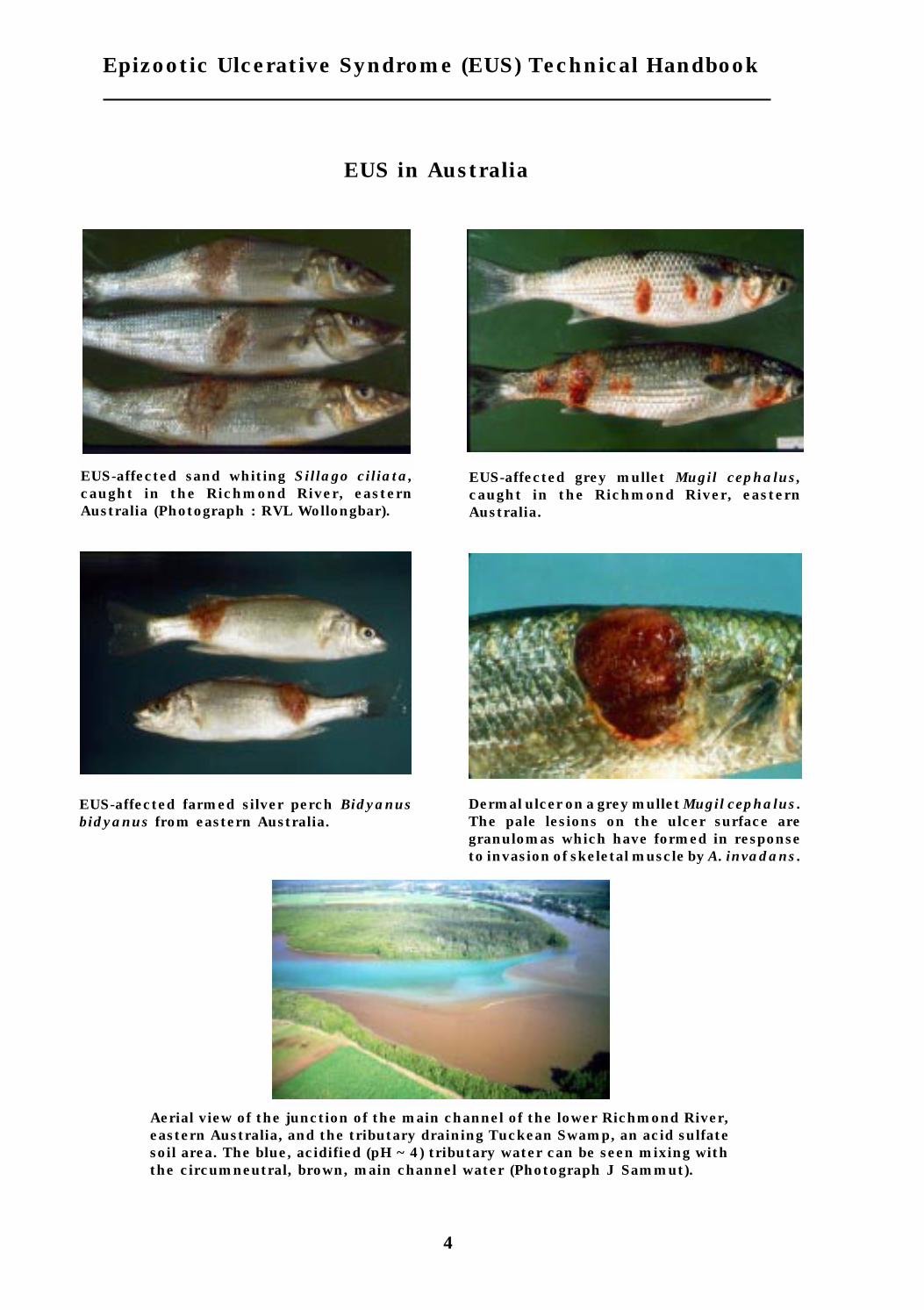
4
Epizootic Ulcerative Syndrome (EUS) Technical Handbook
EUS-affected sand whiting Sillago ciliata,caught in the Richmond River, easternAustralia (Photograph : RVL Wollongbar).
EUS-affected grey mullet Mugil cephalus,caught in the Richmond River, easternAustralia.
Dermal ulcer on a grey mullet Mugil cephalus.The pale lesions on the ulcer surface aregranulomas which have formed in responseto invasion of skeletal muscle by A. invadans.
EUS-affected farmed silver perch Bidyanusbidyanus from eastern Australia.
Aerial view of the junction of the main channel of the lower Richmond River,eastern Australia, and the tributary draining Tuckean Swamp, an acid sulfatesoil area. The blue, acidified (pH ~ 4) tributary water can be seen mixing withthe circumneutral, brown, main channel water (Photograph J Sammut).
EUS in Australia

41
Pale, raised lesions which have not yet completely ulcerated are mostsuitable for fungal isolation attempts. Yellow to red focal skin lesions orhealing ulcers are unsuitable. Fish should be killed by decapitation andpinned, with the lesion uppermost, to a dissecting board. The scales aroundthe periphery of the lesion should be removed and underlying skin searedwith a red-hot spatula so as to sterilise the surface. If possible, the fish andboard should then be removed to a laminar flow cabinet containing filtered airfree of fungal elements. Using a sterile scalpel blade and sterile, fine pointed,rat tooth forceps, cut through stratum compactum underlying the seared areaand, by then cutting horizontally and reflecting superficial tissues, exposeunderlying muscle. Ensure the instruments do not contact the contaminatedexternal surface and otherwise contaminate the underlying muscle. Usingaseptic technique, carefully excise up to 4 pieces of affected muscle,approximately 2 mm3 and place them on a Petri dish containing the isolationmedium.
Where a suitable lesion is found on the tail of a small fish (<20 cm), cut thefish in two using a sterile scalpel, by slicing a cross-section through the fishat the edge of the lesion. Flame the scalpel until red-hot and use this tosterilise the exposed surface of the muscle. Use a small-bladed sterile scalpelto cut out a circular block of muscle (2-4 mm3) from beneath the lesion. Usesterile, fine pointed forceps to remove the block and place it on the isolationmedium. In this way, it should be easy to prevent the instruments contactingthe contaminated external surface of the fish.
Two different isolation media have been used successfully to obtain A.invadans cultures. The use of Czapek Dox agar with penicillin G (100 units/ml) and oxolinic acid (100 µg/ml) was reported by Fraser et al. (1992), and anadapted version of Willoughby and Roberts (1994) GP-PenOx broth is detailedbelow. Inoculated media are incubated at approximately 25°C and examinedunder a microscope (preferably an inverted microscope) within 12 hours.Emerging hyphal tips may be repeatedly transferred to fresh plates of GP-PenStrep agar until cultures are free of bacterial contamination. They maythen be subcultured on GP agar at intervals of no greater than 5 days.
The fungus is subcultured by aseptically cutting a block of agar, 3-4 mm indiameter, from the periphery of a colony and placing this upside-down onto aPetri dish of fresh agar. Agar dishes should be inoculated within 24 hours ofpreparation and the surface should not be dried before use.
Basic GP (glucose-peptone) broth3 g/l glucose1 g/l peptone0.128 g/l MgSO4.7H2O0.014 g/l KH2PO4
Annex 1 - Isolation of Aphanomycesinvadans from EUS-affected fish

42
Epizootic Ulcerative Syndrome (EUS) Technical Handbook
0.029 g/l CaCl2.2H2O2.4 mg/l FeCl3.6H2O1.8 mg/l MnCl2.4H2O3.9 mg/l CuSO4.5H2O0.4 mg/l ZnSO4.7H2OAutoclave at 121°C for 15 minutes
GP-PenOx brothprepare GP broth as above, autoclave, cool to 50oC and add:100 units/ml penicillin-K10 µg/ml oxolinic acid
GP agaras GP broth with:12 g/l technical agar
GP-PenStrep agarprepare GP agar and after autoclaving and cooling to 50oC add:100 units/ml penicillin-K10 µg/ml streptomycin sulphate

43
The method involves collection of a 1 litre pooled sample (10 x 100 ml aliquots)from the pond and performance of a plate count on a 200 ml sample. Gelatinand selected metal ions are added to the subsample as nutrient supplementsand antibiotics are added to reduce growth of contaminating bacteria andzygomycete fungi.
The method is a modification of aquatic fungal propagule count methodsdescribed by Anon (1992), Willoughby et al. (1984)and Celio and Padgett (1989).These previously described methods proved unsatisfactory for Aphanomycesinvadans as they failed to support growth of isolates while allowing abundantgrowth of other fungi and bacteria. The modifications are based on theobservations that A. invadans is one of a limited number of aquatic fungi thatcan utilise complex protein sources and that a large proportion of coloniesattach to plastic surfaces. During the counting procedure, most of the non-A.invadans colonies fail to attach and are removed by washing. Bacterialcontamination is minimised by the low concentrations of complex protein,absence of glucose and addition of antibiotics.
After incubation of plate count cultures, colonies consistent with A. invadansare identified on the basis of attachment to the plastic surface, and hyphaland sporangial morphology. Representative A. invadans-like colonies aresubcultured for further characterisation
The technique has been trialled successfully in several artificially infectedpond waters, but to date it has been possible to trial the method only onceduring a natural EUS outbreak. Counts of A. invadans propagules in pondwater during that outbreak have ranged from 10 - 30 per litre.
Reagents
GelatinPimaricin (2.5%)Sodium benzyl penicillinOxolinic acidPotassium dihydrogen orthophosphate (KH2PO4)Calcium chloride (CaCl2.2H2O)Magnesium sulphate (MgSO4.7H2O)Zinc sulphate (ZnSO4.7H2O)Ferric chloride (FeCl3.6H2O)Distilled water
The chemicals used should be reagent (analytical) grade. To avoidcontaminating the stock pimaricin suspension, aseptically dispense a 1 mlaliquot for day to day use. All antibiotics to be held at 4°C.
Annex 2 - Count method for Aphanomycesinvadans propagules in pond water

44
Epizootic Ulcerative Syndrome (EUS) Technical Handbook
Glassware
All glassware used throughout this procedure must be scrupulously clean.Minimum requirement is as follows:
Soak in laboratory detergentRinse 3 times in tap waterRinse 5 times in distilled waterMark 1 litre glass bottles at 200 ml capacity.
Sample collection bottle
The sample collection bottle used is a modified 100 ml glass Schott bottle. Theinlet (a small glass filter funnel) is covered with nylon mesh of 200 mmaperture size. The collection bottle is secured to a 2 m long stick so that whenthe stick touches the bottom sediment the inlet will be about 5 cm above thesediment. The stick is used to push the bottle to the bottom as rapidly aspossible. Plastic bottles and tubing are not suitable.
Pipettes
By preference, all pipetting should be done with air displacement pipetteswith disposable tips. Two micropipettesare used, one capable of 40 - 200 mland the other 200 - 1000 ml. Where this is not possible glass capillary plungertype pipettes (Socorex) should suffice if capillaries are discarded after use. Ifglass pipettes have to be used they must be carefully washed. Where smallvolumes are required e.g. pimaricin, it may be necessary to prepareintermediate dilutions.
Filtered pond water (FPW)
Collect 3 litres or more of pond water from one of the untreated ponds to betested. For each pond pooled water sample approximately 250 ml of FPW willbe required. Filter through No 1 filter paper and autoclave at 121°C for 15minutes. Store at room temperature. Before use, this water sample should betested using the FPW positive control system described below to ensure that100% of A. invadans colonies sporulate satisfactorily.
Stock metal ion solutions
Use sterile glassware to make up solutions. Keep the solutions in separatesterile disposable plastic containers at 4°C.

45
Annex 2 - Count method for Aphanomyces invadans
Dissolve the following amounts carefully in specified quantity of steriledistilled water before dispensing into storage container.
Distilled H2OKH2PO4 0.267 g 20 mlCaCl2.2H2O 0.588 g 20 mlMgSO4.7H2O 2.533 g 20 mlZnSO4.7H2O 0.110 g 250 mlFeCl3.6H2O 0.600 g 250 ml
Gelatin nutrient solution
The following formula is sufficient for 5 pond water pooled samples. Adjust byproportion as necessary.
Gelatin
Gelatin 0.4 g 100 ml FPW
Prepare fresh gelatin solution immediately before use. Use a clean sterilebottle and measuring cylinder to make up the solution. Weigh gelatin andtransfer to sterile bottle. Add approximately 35 ml of the FPW and bring tosimmering point (a microwave oven is convenient). Dissolve gelatin byvigorously shaking. When gelatin is dissolved add remaining FPW and cool toroom temperature in the freezer.
Metal ions
Add 1.20 ml of each ion solution (K, Mg, Ca, Zn, Fe) to the gelatinsolution.
Antibiotics
Penicillin 0.080 gOxolinic acid 0.080 gPimaricin 120 µl
Immediately before use add antibiotics as above to cooled gelatin solution(100 ml). Shake the bottle vigorously to dissolve as much of the antibiotics aspossible. (The pimaricin and oxolinic acid will remain in suspension and thenutrient medium must therefore be shaken before each aliquot is removed).
Collecting a pooled water sample
Using the collection bottle collect ten aliquots of approximately 100 ml fromrandom sites in the pond. Each aliquot should be collected as close to thebottom as possible without disturbing the sediment. The 1 litre bottle for thepooled sample must be glass and both it and the collection bottle should bethoroughly rinsed before use in the water to be tested. The pooled samplesshould be held at room temperature and processed as soon as possible,certainly within 1 hour of collection. The delay between collection andprocessing should be as constant as possible for all ponds.
46
Epizootic Ulcerative Syndrome (EUS) Technical Handbook
Processing the pooled sample
This procedure should be carried out close to an incubator maintained at27°C. Mix the pooled sample by gentle inversion and rolling and immediatelydispense 200 ml of pooled sample into a sterile 1 litre bottle marked at 200 ml.Shake the gelatin nutrient solution and add 18 ml of solution to thesubsample (use a sterile 25 or 50 ml measuring cylinder). Mix the subsampleand nutrients gently by rolling and inversion, and immediately dispense theentire sample into 7 disposable plastic Petri dishes, mixing as above betweendishes. By eye keep the volume in each dish as equal as possible. Transferplates to the 27°C incubator as soon as they have been dispensed. Incubatefor 48 hours in the dark. After 24 hours gently turn the plates through 360°to detach larger colonies that may be growing, otherwise do not disturb them.
Positive controls
These provide comparative material to aid recognition of A. invadans coloniesin the test sample and ensure that reagents and glassware are satisfactory.The results of the pond water control may allow a correction factor to becalculated at the end of the experiment. Each test day set up a pond waterpositive control and an FPW positive control.
Approximately 14 hours before use, wash 3 x 4-day-old GPY broth mats of avigorous A. invadans culture in 5 changes of FPW. Try to keep the delaybetween washing and use as constant as possible. Immediately before use,remove the mats with a sterile wire. Dispense 9.9 ml of FPW into a sterileglass bottle and add 100 µl of gently mixed spore suspension. Mix gently andtake a 1 ml sample for a Sedgewick-Rafter count. Count motile and totalspores in 30 x 1 µl squares. There should be approximately 100 spores per 100µl and more than 50% of these should be motile.
To one 1 litre bottle add 200 ml of FPW and to another add 200 ml of the samplepond water from the same pond from which the FPW was obtained. To eachcontrol add 150 µl of the 1:100 spore suspension and proceed with addition ofgelatin nutrient solution as above.
If 1:100 spore preparation contains less than 50, or more than 150 spores per0.1 ml, adjust the volume added so that about 150 motile spores will be addedto each 1 litre bottle.
Repeat the count on the 1:100 spore suspension immediately after adding thespores to the control samples. Record the number of spores (the mean of the2 counts) added to each 1 litre bottle and the time elapsed since the mats werefirst washed.
Counting and Identification
Examine the positive control plates first. Gently discard the fluid and anyfloating fungal colonies in the plates and fill the dishes with sterile distilledwater. Allow the dishes to stand for 5 minutes before discarding all but about5 ml of wash fluid.

47
Annex 2 - Count method for Aphanomyces invadans
With a felt tip pen, close to but not obscuring the colony, mark on the plasticdish colonies resembling A. invadans. The colonies will be loosely adherentto the bottom and will measure 3-5 mm in diameter.
Examine the marked colonies with the 10x and 20x objectives of an invertedmicroscope. Mark with a circle, colonies that have the characteristic rightangle branching, 10 µm hyphal diameter and the rounded tip. Hyphaldiameter can be estimated with a calibrated eyepiece graticule.
At this stage hyphal tips from representative colonies in the test samples canbe removed with a sharp scalpel and inoculated deep into agar plates (CzapekDox with 100 units/ml penicillin and 100 mg/ml oxolinic acid, or GP-PenStrep: see Annex 1). Examine the plates daily with a stereo microscope forat least 5 days, and subculture fungal tips as soon as possible. Recovered fungican be identified by sporulation features, hyphal diameter, growth rate at22oC and failure to grow at 37oC (see section on Fungal Aetiology). Count onlythose colonies typical of A. invadans.
Spores per litre = A. invadans colony count (total for all 7 plates) x 5
If less than 100% of colonies sporulate satisfactorily in the FPW positivecontrol then it may be necessary to repeat the counts after adjusting ionicstrength of FPW by diluting up to 1 part FPW with 2 parts distilled water.Determine the optimum dilution by titration in the FPW positive controlsystem.
Recording
Record the colony count for each sample and for the FPW and pond waterpositive control. For the latter subtract any A. invadans that were detected inthe test sample.
Record the number of colonies subcultured, and for each subculture recordthe results of sporulation, hyphal diameter and 22oC and 37°C growth tests.Maintain axenic cultures of representative probable A. invadans colonies forpossible pathogenicity studies.

48
Epizootic Ulcerative Syndrome (EUS) Technical Handbook

49
A. invadans cultures can be maintained in flasks of 200 ml GP broth (seeAnnex 1) at 10oC for only 6 weeks before subculturing is required. This is dueto the rapid staling of the growth medium (Willoughby and Chinabut, 1996).The advantage of this technique, however, is that any bacterial contaminationcan be easily recognised as clouding of the medium.
Cultures can be maintained for longer periods on agar slopes in universaltubes, with sterile light paraffin oil covering the entire slope, as described bySmith and Onions (1994). Particular care should be taken to avoidcontamination, as bacterial growth is not readily apparent in these cultures.GPY agar can be used for this procedure, but A. invadans have been sustainedfor longer periods (over 6 months at 20oC) using a buffered medium developedfor A. astaci (PG-1).
GPY (glucose-peptone-yeast) agar
As GP agar (Annex 1) with:0.5 g/l yeast extract
PG-1 (peptone-glucose-1) agar
3 g/l glucose6 g/l peptone0.17 g/l MgCl2.6H2O0.15 g/l CaCl2.2H2O0.37 g/l KCl0.02 g/l FeCl3.6H2O0.044 g/l Na2 EDTA12 g/l technical agar
Buffer with 13 mM sodium phosphate. Adjust pH to 6.3. Autoclave the glucoseand sodium phosphate buffer separately from the other ingredients.
sodium phosphate buffer
make up stocks of:
31.2 g/l solution A - NaH2PO4.2H2O - store at 4oC71.7 g/l solution B - Na2HPO4.12H2O - store at room temperature
407.5 ml solution A, 92.5 ml solution B and 500 ml distilled water arecombined to make 1000 ml phosphate buffer (100 mM). 130 ml of this bufferis used in 1000 ml PG-1.
Annex 3 - Maintenance of Aphanomycesinvadans cultures

50
Epizootic Ulcerative Syndrome (EUS) Technical Handbook

51
The induction of asexual reproductive structures is necessary in order toidentify fungal cultures as members of the genus Aphanomyces. To inducesporulation, place an agar plug (3-4 mm in diameter) of actively growingmycelium in a Petri dish containing GPY broth and incubate for 4 days atapproximately 20oC. Wash the nutrient agar out of the resulting mat bysequential transfer through 5 Petri dishes containing autoclaved pond water(APW), and leave overnight at 20oC in APW. After about 12 hours, the formationof achlyoid clusters of primary cysts and the release of motile secondaryzoospores should be apparent under the microscope. Features that distinguishsporulating cultures of Aphanomyces from Saprolegnia and Achlya are given inAnnex 5.
GPY broth
as GP broth (Annex 1) with:0.5 g/l yeast extract
APW (autoclaved pond water)
Sample pond/lake water known to support fungal growth, and with pH 6-7.Filter through Whatman 541 filter paper. Combine one part pond water withtwo parts distilled water and autoclave.
Annex 4 - Inducing sporulation inAphanomyces invadans cultures

52
Epizootic Ulcerative Syndrome (EUS) Technical Handbook

53
The Saprolegniaceae are aseptate, eucarpic fungi that typically demonstratetwo zoospore forms. The secondary zoospores are characteristically reniformand laterally biflagellated. The two flagellae differ in type (heterokont), withone anteriorly-directed tinsel-type flagellum and one posteriorly-directedwhiplash-type flagellum.
Saprolegniacean genera are distinguished primarily by asexual characters,particularly zoosporangial shape, method of zoospore release and method ofzoosporangial renewal. The production of asexual characters can be inducedas described in Annex 4. The variation in these characters between the threemain saprolegniacean fungi associated with fish disease (Aphanomyces,Achlya and Saprolegnia) are illustrated in Figure 7. Identification of thesefungi to the species level usually depends on the production of sexualstructures, but these are commonly absent from fish-parasitic species, andunknown from A. invadans.
The zoosporangia of Aphanomyces spp. are typically no wider than the hyphae.A single row of primary zoospores are formed within a zoosporangium andreleased from an apical tip, or from lateral evacuation tubes, at which timethey immediately encyst and form achlyoid clusters. The primary zoospore istherefore not fully released from the sporangium. The main free-swimmingstage of Aphanomyces spp. is the secondary zoospore which is discharged fromthe encysted primary zoospores. The secondary zoospore remains motile fora period depending on environmental conditions and location of a host orsubstratum. Typically the zoospore encysts and germinates to produce newhyphae, although further tertiary generations of zoospores may be releasedfrom cysts (polyplanetism). Specific identification of the EUS pathogen, A.invadans, is discussed in the Fungal Aetiology section.
Achlya spp. zoosporangia are usually formed from terminal hyphal swellingswhich differentiate into the primary zoospores. These encyst, as withAphanomyces, in an achlyoid manner, but only at the apical tip of thezoosporangium. Zoosporangial renewal is typically sympodial, branching fromthe hypha below the basal septum delimiting the spent zoosporangium.
The zoosporangia of Saprolegnia spp. are, as with Achlya spp., short terminalhyphal swellings. However in saprolegnians, the primary zoospore is fullyreleased from the zoosporangium and remains motile for a short period beforeencysting and releasing secondary zoospores. Polyplanetism can be particularlypronounced among fish-parasitic Saprolegnia spp. Zoosporangial renewal istypically by internal proliferation, i.e. the secondary zoosporangium developswithin the previously emptied primary zoosporangium.
Annex 5 - Identification of saprolegniaceanfungal cultures

54
Epizootic Ulcerative Syndrome (EUS) Technical Handbook
Figure 7 - Zoosporangia formation and dehiscence in Aphanomyces, Achlyaand Saprolegnia (reproduced with kind permission of Dr L.G. Willoughby)

55
The isolation of viruses from EUS-diseased specimens can be very difficult,even with access to specialised virological facilities. Two factors have beenidentified as critical for successful isolation. Firstly, the diseased fish needto be collected during the early period of an outbreak (i.e. within 1-2 weeks ofthe disease being noticed); and secondly, the specimens need to be alive justbefore collection of the tissue samples.
Tissue sampling and handling
Diseased fish with early skin lesions should be collected. Fish are sacrificedand wiped clean with tissue paper. Approximately 1g is taken of each tissuesampled. For muscle samples, tissue debris and surface fungus on theulcerated lesions are removed using a clean razor blade. Pieces of muscletissue are taken from beneath the lesions. For internal organ samples, theabdomen is carefully opened using clean scissors, and small pieces of tissuefrom kidney, spleen, intestine and pancreas are taken and pooled. If the fishare very small, the entire viscera can be taken. Tissue samples from up to5 fish can be pooled and processed as 1 tissue extract. Tissue samples can bestored up to 48 h in HBSS (Hank’s balanced salt solution) supplemented with2% FCS (foetal calf serum), 500 IU/ml penicillin, 500 mg/ml streptomycinand 10 mg/ml amphotericin B (fungizone) at 4°C.
Samples are then homogenised using a sterile, pre-cooled pestle and mortaruntil a smooth paste is obtained. Sterile fine sand is added to facilitatehomogenisation. Samples are diluted 1:10 by the addition of 9 ml HBSScontaining 2% serum. After mixing well, the samples are transferred tosterile centrifuge tubes and spun at 1000 x g at 4°C for 15 min to separate celldebris, sand and possibly contaminating micro-organisms from the fluidextract. A further 1:5 dilution is carried out by filling 5 ml sterile disposablesyringes with 4 ml HBSS (with 2% serum) and then drawing up 1 mlsupernatant. These 1:50 final dilutions are mixed well and then filter-sterilised through 0.45 mm disposable filter units. The filtrates or tissueextracts are kept in 5 ml sterile bottles at 4°C which are ready to be inoculateddirectly onto fish cell lines or, if necessary, transported to the fish virologylaboratory.
Virus isolation
Simultaneous cell culture and sample inoculation should be carried outusing BF-2 and/or SSN-1 cell lines. Tests are conducted in 24-well plates.Each plate is first seeded with a single cell suspension of the indicator cellline in L-15 medium containing 2% serum and 1x antibiotics (100 IU/mlpenicillin and 100 mg/ml streptomycin). Each well receives 1.3-1.4 ml of cellsuspension. Cell density should be sufficient to produce a 80-90% confluent
Annex 6 - Isolation of viruses

56
Epizootic Ulcerative Syndrome (EUS) Technical Handbook
monolayer 1 day after seeding. Tissue extracts (1:50 dilution) are immediatelyinoculated into 2 replicate wells. The inoculum volume is 200 µl/well. Anequal number of inoculated wells as negative control well should be allocatedfor each plate. Cells are incubated at 23-25°C and observed daily for CPE(cytopathic effect) for at least 14 days. The first blind passage of culture fluidsis performed between day 7-10 by transferring 200 µl of supernatant from eachwell to fresh culture wells and observing the plates for a further 14 days. Asecond and third blind passage should also be carried out.
Samples showing CPE in which the cell monolayer changes (e.g. disintegrates,sloughs off the surface of the tissue culture wells, or results in cell lysis)should be passaged to provide larger quantities of the suspected virus. Twohundred microlitres of supernatant from a single well exhibiting CPE isinoculated into 25 cm2 flasks containing a 80-90% confluent cell monolayer.The suspected virus is allowed to adsorb for 1 h. The cells are washed oncewith 5 ml PBS (phosphate buffered saline), then 7 ml of maintenance medium(L-15 with 2% serum) is added. Flasks are incubated at 23-25°C together withun-inoculated control flasks for comparison. When the cells show completeCPE, they are spun at 1000 x g at 4°C for 15 min. The supernatant is collectedand divided into 1 ml aliquots. Some tubes are kept at 4°C for furthercharacterisationwithin 6 months and others stored at -20°C or -70°C for longterm storage.

57
An outbreak can be defined as a short term epidemic or a series of diseaseevents clustered in space and time. Such disease events are usually newcases of a disease occurring at a higher frequency than is normally expected.Throughout this section, the terms outbreak and epidemic are used more orless interchangeably.
An outbreak investigation is a systematic procedure to help identify causesand sources of epidemics, with a view to controlling the existing epidemic andpreventing future ones. Usually, the primary objective of an epidemic ordisease outbreak investigation is to identify ways of preventing furthertransmission of the disease-causing agent. The epidemiological approach tooutbreak investigations is based on the premise that cases of a disease arenot distributed randomly, but occur in patterns within the at-risk population.It is the role of the epidemiologist to record and analyse these patterns to helpmeet the primary objective.
Little is known of the means whereby EUS spreads within and betweenregions, although movements of subclinically affected fish are probablyimportant in transmitting A. invadans infection. Until effective control andprevention measures are implemented, it is likely that the disease willcontinue to spread and that outbreaks will continue to recur in endemicareas. Comprehensive investigations of initial outbreaks in previously EUS-free areas as well as of recurrent outbreaks in previously endemic areas areurgently needed and will contribute important information on causal factorsfor EUS.
Investigation procedure
The procedure for an outbreak investigation follows 9 basic steps. Not all thesteps are necessarily included in every investigation, nor do they alwaysfollow the same sequence. In practice, several steps will be undertakensimultaneously.
The 9 basic steps are :
1. Establish a diagnosis.
The initial provisional diagnosis in an EUS outbreak is usually based onspecies of fish affected, clinical signs, gross pathology and, perhaps,seasonality. Whenever possible, laboratory tests should be undertaken toverify the provisional diagnosis. Since some laboratory procedures (e.g.histopathology, fungal isolation) may take weeks, the implementation ofcontrol measures is often based on the provisional diagnosis.
Annex 7 - Investigation of EUS outbreaks

58
Epizootic Ulcerative Syndrome (EUS) Technical Handbook
2. Define a “case”.
Depending on the type of investigation, an EUS case might be an individualaffected fish or an aggregation of individuals such as the population in anaffected pond. A useful case definition at the individual animal level might be‘a fish with necrotising granulomatous dermatitis and myositis associatedwith highly invasive non-septate fungal hyphae’.
3. Confirm that an outbreak is actually occurring.
This step may seem unnecessary but in many instances it is required,particularly in areas where EUS is already endemic. The disease may beexpected to occur at low prevalence at certain times, but even a moderateprevalence increase, especially if ulceration is severe and/or toxigenic A.hydrophila is present, will lead to substantial production losses if not recognisedearly. Moreover, dermal ulceration caused by other agents is common in fishpopulations and is often macroscopically very difficult to distinguish fromEUS. Laboratory confirmation of a diagnosis of EUS will usually be necessary.
4. Characterise the outbreak in terms of time, affected/unaffected fish, and place.
From an epidemiologic viewpoint, it is important to characterise the outbreakin terms of the above 3 variables. This characterisation must be done in sucha way that hypotheses can be developed regarding the source, mode oftransmission and duration of the outbreak. The information is organised inan attempt to find answers to the following kinds of questions:
Time
What is the exact period of the outbreak?
Given the diagnosis, what is the probable period of exposure?
Is the outbreak most likely common source, propagated or both?
Fish
Are there any characteristics about fish for which specific attack rates vary?
Which groups have the highest and which have the lowest attack rates?
Place
What are the significant features of the geographical distribution of cases?
What are the relevant attack rates?
Time
There are 3 basic time spans used to describe disease temporal patterns: theepidemic period, which is of variable length depending on the particularepidemic; a 12 month period to describe seasonal patterns; and an indefinitelylong period of years to identify long-term trends. A knowledge of seasonal

59
Investigationof EUS outbreaks
patterns and long-term trends is important when deciding whether or not anepidemic exists in the present period and in predicting future epidemics.
The temporal pattern of an outbreak is described in terms of its epidemiccurve. The epidemic curve is a graph showing the onset of cases of thedisease either as a bar graph or frequency polygon. The first case identifiedfor a particular outbreak is referred to as the ‘index’ case. For infectiousdiseases such as EUS, information about the index case can be valuable inidentifying the source of the outbreak.
In general, an epidemic curve has 4 and sometimes 5 segments:
i. the endemic level
ii. an ascending branch
iii. a peak or plateau
iv. a descending branch
v. a secondary peak
The slope of the ascending branch can indicate the type of exposure (propagatingor common source) or the mode of transmission and incubation period. Iftransmission is rapid and the incubation period is short, then the ascendingbranch will be steeper than if transmission is slow or if the incubation periodis long.
The length of the plateau and slope of the descending branch are related tothe availability of susceptible animals which in turn is dependant on manyfactors such as stocking densities, the changing importance of differentmechanisms of transmission and the proportion of resistant or immune fishin the population at risk.
The interval of time chosen for graphing the cases is important to thesubsequent interpretation of the epidemic curve. The time interval should beselected on the basis of the incubation or latency period and the period overwhich the cases are distributed. A common error in this regard is theselection of a time interval which is too long, which may obscure subtledifferences in temporal patterns. A general rule is to make the intervalbetween one eighth and one quarter of the incubation period. Accordingly, forEUS, which has an incubation period of approximately 10 days, the incidence/prevalence in the population should be measured every 2 days.
Outbreaks are often referred to as being either ‘common source’ (casesresulting from exposure to a common source, as in intoxications) or ‘propagatedsource’ (animal-to-animal transmission as in most infectious diseases). Insome EUS outbreaks, it is conceivable that both types of sources could beinvolved, the initial cases resulting from exposure to a common source (suchas contaminated water or equipment) and secondary cases resulting fromfish-to-fish spread.

60
Epizootic Ulcerative Syndrome (EUS) Technical Handbook
The duration of an outbreak is influenced by:
· the number of susceptible animals exposed to a source of infectionwhich become infected;
· the period of time over which susceptible animals are exposed tothe infection source;
· the minimum and maximum incubation periods of the disease.
Fish
Although the word ‘fish’ is used here, the terms ‘cases’ and ‘non-cases’ shouldbe used to embrace the wider definitions where ‘cases’ might be ponds, farms,etc. For simplicity, the discussion will be restricted to individual fish only.
Species, age, sex and geographical origin are often associated with varyingrisk of disease. However, it should be kept in mind that fish-level patterns canbe closely linked to temporal and spatial patterns of disease.
To describe patterns of disease by fish categories, it is first necessary tooutline what measures of disease frequency are used in outbreakinvestigations. The basic measure of disease frequency in outbreaks is the‘attack rate’ (AR). An attack rate is a special form of an incidence rate wherethe period of observation is relatively short. An attack rate is the number ofcases of the disease divided by the number of animals at risk at the beginningof the outbreak. Where different risk factors for the disease are to beevaluated, attack rates specific for the particular factor must be calculated.For example, suppose there were deaths due to suspected EUS in a pond andit appeared that small fish were at greater risk of having EUS than larger fish.We might make the following calculations:
Number with EUSFor small fish, AR1 = —————————-
Total small fish
Number with EUSFor large fish, AR2 = —————————-
Total large fish
Using some hypothetical numbers, say 300 out of 1000 small fish, and 100 outof 1000 large fish, in the pond had EUS during the outbreak. The attack rateshere are 30% and 10% respectively, suggesting that small fish were 3 timesmore likely to develop EUS than large fish. Such a finding could lend supportto a hypothesis, for example, that nutritionally stressed fish are moresusceptible to EUS.
Formal measures to compare attack rates among groups of fish with differentcharacteristics are described in the next section.
With EUS, of course, the total numbers of fish in different putative riskcategories would have to be estimated through representative sampling.Another problem could be the estimation of the numbers of mortalities andwhen they died. When investigating an outbreak of EUS, where it is postulated

61
Investigationof EUS outbreaks
that some epidermal insult (such as rhabdovirus infection) precedes infectionwith A. invadans, examination of samples of fish at different times during theoutbreak should attempt to characterise the different types of lesions presentand the rhabdovirus-infection status of individual fish. Thus, the ‘spectrum’of disease is important here.
Place
Describing the outbreak in terms of place may lead to identification of thecause. For farmed fish, this may involve looking at the pattern in differentponds. It is often useful to consider time and place together. This can be doneby drawing a plan of the ponds and recording the dates when cases occurred.Such a diagram may also give a lead to whether the outbreak is a commonsource or propagating. For larger scale epidemics, spot maps are useful.
5. Analysing the data.
Factor-specific attack rates for such factors as species, age, sex, pond,management system, etc are calculated and arranged in an ‘attack ratetable’. A theoretical example is shown below for EUS where size indicatingnutritional stress is suspected:
In the above table, attack rates are expressed as percentages. The second lastcolumn is the difference in attack rates (sometimes called the ‘AttributableRisk’) and the last column is the ‘Relative Risk’ which is the ratio of the attackrates.
The higher the attack rate difference and the relative risk, the moreimportant the specific factor is in increasing the risk of disease. The analysisbecomes more complicated when trying to sort out the interactions andconfounding among factors. Stratified and multivariate analyses are used toinvestigate these phenomena but such methods are beyond the scope of thishandbook.
It should also be noted in the above hypothetical outbreak example that smallfish were 3 times more likely to have EUS than medium sized fish and 6 timesmore likely than large fish. Also, medium sized fish were at twice the risk oflarger fish. This dose-response phenomenon when relating size to attackrate would lend support to a hypothesis that nutritional stress as manifestedin size is a ‘component cause’ of EUS.
With factor Without factor
EUS Total AR EUS Total AR AR Diff RR
Small 30 100 30% 35 500 7% 23% 4.3
Medium 20 200 10% 45 400 11% -1% 1
Large 15 300 5% 50 300 17% -12% 0.3

62
Epizootic Ulcerative Syndrome (EUS) Technical Handbook
6. Working hypotheses
Based on the analysis of time, place and fish data, working hypotheses aredeveloped for further investigation. These may concern one or more of thefollowing:
· whether the outbreak is common source or propagating;
· if a common source, whether it is point or multiple exposure;
· the mode of transmission - contact, vehicle or vector.
Any hypothesis should be compatible with all the facts.
Corrective action can be taken based on the more realistic hypotheses.
7. Intensive follow-up
This includes clinical, pathological, and microbiological examinations,together with examinations of water quality data and recent meteorologicaldata. Epidemiologic follow-up will include detailed analyses of these data aswell as the search for additional cases on other premises. Flow charts ofmanagement and movements of fish, water and equipment, for example, maybe required as part of this process.
Transmission trials may be necessary where additional infectious agents,such as bacteria or ectoparasites, are suspected as component causes of theoutbreak.
8. Control and prevention
Hopefully, the investigation will contribute to the termination of the outbreakand information gained will ensure that the risk of similar occurrences isreduced. Strategies to stop the epidemic must be put in place as soon aspossible and will often be undertaken in the absence of conclusive findings.
Much further work is required before effective methods for treating EUSoutbreaks in ponds can be developed. Detailed investigation of a number ofoutbreaks could provide valuable information about possibly important‘component’ causes.
9. Reporting
For small outbreaks, this may take the form of a brief discussion with thefarm manager outlining the important features and actions required toprevent further occurrences. However, it is wise to always produce some formof written report so that a permanent record of events exists for future use.For large outbreaks, findings should be published in scientific literature.
For substantial investigations the report should contain the following sections:background, methods, results, hypotheses, financial impact (whereappropriate), recommendations and appendices containing laboratory reportsetc.

63
Annex 8 - EUS Sampling Datasheets
To make information available for the regionwide collaborative EUSprogramme, please complete as fully as possible and send with formalin-fixedfish samples (see Annex 9) to: The Aquatic Animal Health Research Institute,Kasetsart University, Jatujak, Bangkok 10900, Thailand. (*Sections markedwith an asterix are optional, depending on resources available).
EUS SAMPLING DATASHEET 1 SITE NAME:
Date:Collected by:
Current EUS outbreakIs the present outbreak: (a) restricted to sampling site?
(b) occurring throughout the local area?(c) a national problem?
Date present outbreak started:Estimated number/weight of fish lost from present outbreak:Value of losses from present outbreak:Conditions 3-12 days before outbreak (e.g. temperature, rainfall):
*Fish market price:Species Price/kg of unaffected fish Price/kg of affected fish
Site descriptionCountry:Province:District:Town:Village:
Type of water body at site:
Farm Lake ReservoirCanal River RicefieldSwamp Other:
Describe site:
Previous history of EUS at the site:

64
Epizootic Ulcerative Syndrome (EUS) Technical Handbook
EUS SAMPLING DATASHEET 2 SITE NAME:
Sampling pointHow and where on the site were fish sampled:
Was selection of sampling point random? If not give reason:
Size of pond:Depth of pond:Fish species:Stocking rate:Are there wild fish in the pond?Were fish introductions made just prior to outbreak?What is the source of the water to this pond?Describe attempted treatments/control strategies:
Perceived importance of the problem to local farmers/fishermen:
*Water quality data
TimeTurbidity
TemperaturepHAlkalinityHardness
ConductivityNH4
*#Fish population dataSpecies No. of fish No. infected % infected Severity of infection
#when estimating EUS prevalence in the population, a random sample of at least 100 fish should be taken and examined forlesions

65
EUS Sampling Datasheets
EU
S S
AM
PL
ING
DA
TA
SH
EE
T 3
SIT
E N
AM
E:
Fis
h s
amp
ling
dat
a
*Pho
toC
ode
Spe
cies
Leng
thD
escr
iptio
n of
clin
ical
sig
nsIn
dica
te w
hich
tiss
ue s
ampl
ed*M
icro
scop
ic e
xam
inat
ion
No
(cm
)H
isto
logy
*Myc
olog
y*B
acte
riolo
gy*F
ungu
s*S
kin
para
site
s

66
Epizootic Ulcerative Syndrome (EUS) Technical Handbook

67
1. Complete EUS sampling datasheet (Annex 8) for each site, recording fulldetails for each fish sampled.
2. Sample only live specimens of diseased fish. If fish with clinical signsof EUS are readily apparent, several samples of each species should becollected, preferably at different stages of infection.
3. Dissect large fish and take samples of skin/muscle (<1cm3), spleen,kidney and liver. The muscle section should include the lesion and thesurrounding tissue. Small fish (<3cm in length) can be slit along theabdomen and preserved whole.
3. Fix the tissues immediately in cold 10% formalin. The amount offormalin in the jar should be 15-20 times the volume of the tissue to befixed.
4. Gently agitate the fixative 2-3 times over the first hour after adding thetissue.
5. The selected site should be sampled repeatedly over the outbreakperiod and specimens sent to a centralised diagnostic facility. If anappropriate facility is not available in-country, or to make theinformation available for the regionwide collaborative EUS programme,send to The Aquatic Animal Health Research Institute, KasetsartUniversity, Jatujak, Bangkok 10900, Thailand, along with a copy of thesampling datasheet.
Annex 9 - Procedure for samplingfish for histological examination

68
Epizootic Ulcerative Syndrome (EUS) Technical Handbook

69
AAHRI - Aquatic Animal Health Research Institute, ThailandACIAR - Australian Centre for International Agriculture
ResearchAPW - autoclaved pond waterBF-2 - bluegill fry cell lineCDA - Czapek Dox agarCPE - cytopathic effectDFID - Department for International Development, United
Kingdom Government, formally ODAEUS - epizootic ulcerative syndromeFAO - Food and Agriculture Organisation of the United NationsFCS - foetal calf serumFME - fish meat-extract (Hatai et al., 1977)GP - glucose-peptone mediumGP-PenOx - glucose-peptone-penicillin-oxolinic acid mediumGP-PenStrep - glucose-peptone-penicillin-streptomycin mediumGPY - glucose-peptone-yeast mediumGY - glucose yeast medium (Dykstra et al., 1986)HBSS - Hank’s balanced salt solutionICBN - International Code of Botanical NomenclatureICSF - International Collective in Support of Fishworkersi.m. - intra-muscular injectionIoA - Institute of Aquaculture, Stirling University, Scotlandi.p. - intraperitoneal injectionIPNV - infectious pancreatic necrosis virusMG - mycotic granulomatosisNACA - Network of Aquaculture Centres in Asia and the Pacific,
BangkokNSW - New South Wales, AustraliaODA - Overseas Development Administration of the United
Kingdom, present name DFIDPG-1 - peptone-glucose-1 mediump.i. - post-injectionPNG - Papua New GuineaRSD - redspot diseaseSEAADCP - South East Asia Aquatic Disease Control ProjectSGV - sand goby virusSHRV - snakehead fish rhabdovirusSHV - snakehead fish virusSSN-1 - striped snakehead cell lineUDRV - ulcerative disease rhabdovirusUM - ulcerative mycosis (menhaden disease)
Abbreviations

70
Epizootic Ulcerative Syndrome (EUS) Technical Handbook

71
achlyoid: referring to type of zoospore discharge fromzoosporangium: primary zoospores encyst asthey emerge from the exit pore, forming a loose,spherical cluster. Characteristic of Oomycetegenera Aphanomyces and Achlya.
acinar necrosis: irreversible degeneration of acinar tissue(exocrine pancreatic cells)
aetiology: the science of the cause or origin of disease
alkalinity: a measure of anions (e.g. HCO3-, CO3
=, OH-) in asolution (an alkaline solution would give a pHreaction above 7)
axenic: cultures of microorganisms which are notcontaminated by, or are completely free of, thepresence of other organisms.
birnavirus: a group of non-enveloped isometric viruses(including IPNV) with a genome of doublestranded RNA in two segments
chlamydospore/gemma: asexual spherical structure of fungi originatingby differentiation of a hyphal segment(s) usedprimarily for perennation, not dissemination
complex zoosporangium: zoosporangium with more than one evacuationtubes from which zoospores are released
cytopathic effect (CPE): cell degeneration caused by viral growth, thepathological changes in cell culture are oftenvirus-specific and can form the basis of avirological diagnosis
dermatitis: inflamation of the skin
ectoparasiticide: a substance capable of destroying externalparasites
enterotoxigenic: producing or containing a toxin specific for thecells of the intestinal mucosa
eosinophil: a leucocyte with a bilobed nucleus and coarsegranular cytoplasm that stains readily withacidic dyes such as eosin
epidemiological: relating to the occurrence, transmission andcontrol of epidemic diseases
epithelioid: like epithelium
Glossary

72
Epizootic Ulcerative Syndrome (EUS) Technical Handbook
epizootic: the sudden outbreak and rapid spread of adisease affecting a large number of animalsover a wide area (“epidemic” is now used insteadof epizootic)
erythematous: relating to, or causing, erythema: a disease ofthe skin, in which a diffused inflammationforms rose-coloured patches of variable size
eucarpic: referring to fungi that develop reproductivestructures on limited portions of the thallus,such that the residual nucleate protoplasmremains capable of further mitotic growth andregeneration
exophthalmia: abnormal protrusion of the eyeball
focal proliferation: cell division and growth limited to a specific partof the tissue
granular occlusion: blockage or obstruction of a duct or blood vesselby grain-like particles
granulomatous: composed of a tumour-like mass or nodule ofgranulation tissue due to a chronicinflammatory response
haemoglobin: respiratory pigment in red blood cells, aconjugated protein that can combine reversiblywith oxygen
haemorrhage: escape of blood from the vascular system
haemosiderin: an insoluble form of storage iron, visiblemicroscopically
hardness: a measure of calcium, magnesium and othermetals in freshwater expressed as mg/litre ofcalcium carbonate
heterokont: Cell with flagellae (or other motile organelles)unequal in length or unlike in movement
hyperaemic: a superabundance or congestion of blood, due toincreased flow of blood to an area, or due toobstruction in the return of blood from an area
hyperplastic: pertaining to hyperplasia: an abnormal increasein the number of cells
intercalary zoosporangia: zoosporangia forming in the middle of a hyphalsegment
lateral evacuation tubes: zoosporangial tube from which zoospores arereleased

73
Glossary
lymphoid: of or resembling lymph or lymphatic tissue
melanomacrophagecentre (MMC): a discrete group of large phagocytic cells
within haemopoetic and other soft tissues ofteleost fish; yellow-brown or black in colourdepending on species, age and health of thefish
mitotic figures: cells seen in histological preparations to beundergoing mitosis, sometimes an indicationof neoplastic changes or cell regeneration as aresult of a previous toxic insult.
monogenean: an ectoparasitic flatworm of the class Trematodawith a direct lifecycle and a single host
mycosis: disease resulting from infection with a fungus
myeloid: having the appearance of myelocytes (aprecursor cell of blood granulocytes)
myofibrillar: pertaining to myofibrils: long cylindricalorganelle of striated muscle, composed of regulararrays of thick and thin filaments andconstituting the contractile apparatus
myopathy: disease of muscle tissue
myophagia: atrophy or wasting away of muscular tissue
myositis: inflammation of the muscles
myxosporidian: a spore-producing, parasitic protozoan
necrosis: the sum of the morphological changes indicativeof cell death and caused by the progressivedegradative action of enzymes; it may affect asingle cell, a group of cells or part of a structureor organ
occlusion: closure or blockage of an orifice or tube
oedema: accumulation of body fluids in the tissues,generally causing swelling of a part of the body
oomycete: class of fungal-like protists, typically giving riseto biflagellate, heterokont zoospores. Hyphaeare aseptate and the cell wall is believed inmost species to lack chitin and contain cellulose
polyplanetism: phenomenon in successive generations ofsecondary zoospores are formed by repeatedcycles of encystment and excystment

74
Epizootic Ulcerative Syndrome (EUS) Technical Handbook
proliferative
zoosporangial renewal: the development of a secondary zoosporangiumwithin a previously emptied primary one. Thesupporting hyphae may also grow through theprimary sporangium before forming thesecondary sporangium. Common in theoomycete genus Saprolegnia
prophylactic: disease-preventing
reniform: kidney-shaped
rhabdovirus: a group of bullet-shaped viruses 130-380nmlong and 60-95nm wide with a genome of singlestranded RNA
rosacea: a disease of the skin characterised by redcolouration and caused by chronic dilation ofcapillaries
septicaemia: systemic disease associated with the presenceand persistence of pathogenic microorganismsor their toxins in the blood
simple zoosporangium: zoosporangium with a single evacuation porefrom which zoospores are released
sporulation: sporogenesis. Formation of spores that involvesdivision of a large cell into many small spores
sympodial zoosporangial
renewal: lateral branching of a fungal hypha below thebasal septum of a delimited zoosporangium, sothat the lateral branch then becomes the primaryaxis. Common in the oomycete genus Achlya
syndrome: a group of symptoms or signs, which, whenconsidered together, characterise a disease
telangiectasis: abnormal dilation of capillaries in the secondarylamellae of gills
terminal zoosporangium: zoosporangium forming at the end tip of a hyphalsegment
therapeutic: relating to the treatment of disease
ulcer: an interuption of continuity of an epithelialsurface, with an inflammed base
zoosporangium(plural: zoosporangia): fungal asexual reproductive structure in which
zoospores are produced, and from which theyare released

75
AAHRI, ACIAR, IoA and NACA (1997) Epizootic ulcerative syndrome (EUS) offishes in Pakistan. A report of the findings of an ACIAR/ DFID-fundedmission to Pakistan. 9-19 March 1997.
ADB/NACA (1991) Fish Health Management in Asia-Pacific. Report on aRgional Study and Workshop on Fish Disease and Fish HealthManagement. ADB Agriculture Department Report Series No. 1.Network of Aquaculture Centres in Asia-Pacific, Bangkok.
Ahmed, M. and Rab, M.A. (1995) Factors affecting outbreaks of epizooticulcerative syndrome in farmed and wild fish in Bangladesh. Journalof Fish Diseases 18, 263-271.
Ahmed, G.U. and Hoque, A. (submitted) Mycotic involvement in epizooticulcerative syndrome of freshwater fishes of Bangladesh: ahistopathological study. In: Proceedings of the Fifth Asian FisheriesForum, Chiang Mai, Thailand, 11-14 November 1998. Asian FisheriesSociety, Manila.
Ahne, W., Jørgensen, P.E.V., Olesen, N.J. and Wattanavijarn, W (1988)Serological examination of a rhabdovirus isolated from snakeheadfish (Ophicephalus striatus) in Thailand with ulcerative syndrome.Journal of Applied Ichthyology 4, 194-196.
Alderman, D.J. and Polglase, J.L. (1988) Pathogens, parasites and commensals.In: Holdich, D.M. and Lowery, R.S. (Eds) Freshwater Crayfish: Biology,Management and Exploitation. Pp. 167-212.
Anon. (1992) Notes on mycological procedure. Workshop on MycologicalAspects of Fish and Shellfish Disease, Bangkok, Thailand, January1992. Aquatic Animal Health Research Institute.
Avtalion, R.R., Wichkovsky, A. and Katz, D. (1980) Regulatory effect of tempratureon specific supression and enhancement of the humoral response infish. In: Manning, M.J. (Ed) Phylogeny of Immunological Memory. Pp.113-121. Elsevier, Amsterdam.
Balasuriya, L.K.S.W. (1994) epizootic ulcerative syndrome in fish in SriLanka, country status report. In: Roberts, R.J., Campbell, B. andMacRae, I.H. (Eds) Proceedings of the ODA Regional Seminar onEpizootic Ulcerative Syndrome, 25-27 January 1994. Pp. 39-47. AquaticAnimal Health Research Institute, Bangkok.
Barua, G. (1990) Bangladesh report. In: Phillips, M.J. and Keddie, H.G. (Eds)Regional Research Programme on Relationships Between EpizooticUlcerative Syndrome in Fish and the Environment. A Report on theSecond Technical Workshop, 13-26 August 1990. Network ofAquaculture Centres in Asia-Pacific, Bangkok.
Barua, G. (1994) The status of epizootic ulcerative syndrome of fish ofBangladesh. In: Roberts, R.J., Campbell, B. and MacRae, I.H. (Eds),Proceedings of the ODA Regional Seminar on Epizootic UlcerativeSyndrome, 25-27 January 1994. Pp.13-20. Aquatic Animal HealthResearch Institute, Bangkok.
References

76
Epizootic Ulcerative Syndrome (EUS) Technical Handbook
Bhaumik, U., Pandit, P.K. and Chatterjee, J.G. (1991) Impact of epizooticulcerative syndrome on the fish yield, consumption and trade in WestBengal. Journal of the Inland Fisheries Society of India 23(1), 45-51.
Bly, J. E. and Clem, L. W. (1991) Temperature-mediated processes in teleostimmunity: In vitro immunosuppression induced by in vivo lowtemperature in channel catfish. Veterinary Immunology andImmunopathology 28(3-4), 365-377.
Bondad-Reantaso, M.G., Lumanlan, S.C., Natividad, J.M. and Phillips, M.J.(1992) Environmental monitoring of the epizootic ulcerative syndrome(EUS) in fish from Munoz, Nueva Ecija in the Philippines. In: Shariff,M., Subasinghe, R.P. and Arthur, J.R. (Eds) Diseases in AsianAquaculture 1. Pp. 475-490. Fish Health Section, Asian FisheriesSociety, Manila.
Bondad-Reantaso, M.G., Paclibare, J.O., Lumanlan-Mayo, S.C. and Catap, E.S.(1994) EUS outbreak in the Philippines: a country report. In: Roberts,R.J., Campbell, B. and MacRae, I.H. (Eds), Proceedings of the ODARegional Seminar on Epizootic Ulcerative Syndrome, 25-27 January1994. Pp. 61-67. Aquatic Animal Health Research Institute, Bangkok.
Boonyaratpalin, S. (1989a) A report on epizootic ulcerative sysndrome of fishin India. Paper code NACA/WP/89/80, Network of Aquaculture Centresin Asia, Bangkok.
Boonyaratpalin, S. (1989b) Bacterial pathogens involved in the epizooticulcerative of fish in southeast Asia. Journal of Aquatic Animal Health1, 272-276.
Burkholder, J.M., Noga, E.J., Hobbs, C.H. and Glasgow Jr., H.B. (1992) Newphantom dinoflagellate is the caustive agent of major estuarine fishkills. Nature 358, 407-410
Callinan, R.B. (1994a) A comparative review of Aphanomyces species associatedwith epizootic ulcerative syndrome, red spot disease and mycoticgranulomatosis. In: Roberts, R.J., Campbell, B. and MacRae, I.H. (Eds)Proceedings of the ODA Regional Seminar on Epizootic UlcerativeSyndrome, AAHRI, Bangkok, 25-27 January 1994. Pp. 248-252. AquaticAnimal Health Research Institute, Bangkok.
Callinan, R.B. (1994b) Red spot disease - EUS in Australia. In: Roberts, R.J.,Campbell, B. and MacRae, I.H. (Eds) Proceedings of the ODA RegionalSeminar on Epizootic Ulcerative Syndrome, 25-27 January 1994. Pp.82-88. Aquatic Animal Health Research Institute, Bangkok.
Callinan, R.B. (1997) Pathogenesis of red spot disease (epizootic ulcerativesyndrome) in estuarine fish in eastern Australia and the Philippines.Ph.D. Thesis, University of Queensland. 232 pp.
Callinan, R.B. and Keep, J.A. (1989) Bacteriology and parasitology of red spotdisease in sea mullet, Mugil cephalus L., from eastern Australia.Journal of Fish Diseases 12, 349-356.
Callinan, R.B., Fraser, G.C. and Virgona, J.L. (1989) Pathology of red spotdisease in sea mullet, Mugil cephalus L., from eastern Australia.Journal of Fish Diseases 12, 467-479.

77
References
Callinan, R.B., Paclibare, J.O., Bondad-Reantaso, M.G., Chin, J.C. andGogolewski, R.P. (1995a) Aphanomyces species associated with epizooticulcerative syndrome (EUS) in the Philippines and red spot disease(RSD) in Australia: preliminary comparative studies. Diseases ofAquatic Organisms 21, 233-238.
Callinan, R.B., Paclibare, J.O., Reantaso, M.B., Lumanlan-Mayo, S.C., Fraser,G.C. and Sammut, J. (1995b) In: Shariff, M., Arthur, J.R. andSubasinghe, R.P. (Eds) EUS outbreaks in estuarine fish in Australiaand the Philippines: associations with acid sulphate soils, rainfall andAphanomyces. Diseases in Asian Aquaculture II. Pp. 291-298. FishHealth Section, Asian Fisheries Society, Manila.
Callinan, R.B., Sammut, J. and Fraser G.C. (1996) Epizootic ulcerativesyndrome (red spot disease) in estuarine fish - confirmation thatexposure to acid sulfate soil runoff and an invasive aquatic fungus,Aphanomyces sp., are causative factors. Proceedings of the SecondNational Conference on Acid Sulfate Soils. Roberts J Smith andAssociates and ASSMAC, Australia.
Cavalier-Smith, T. (1987) The origin of Fungi and pseudofungi. In: Rayner,A.D.M., Brasier, C.M. and Moore, D. (Eds), Evolutionary Biology of theFungi. Pp. 339-353. Cambridge University Press.
Celio, D.A. and Padgett, D.E. (1989) An improved method of quantifying watermould spores in natural water columns. Mycologia 81(3), 459-460.
Chakrabarty, A.N. and Dastidar, S.G. (1991) Repeated isolation ofchemoautotrophic nocardioform bacteria from fish epizootic ulcerativesyndrome. Indian Journal of Experimental Biology 29, 623-627.
Chinabut, S. (1990) The histopathology of EUS in Asia. In: Phillips, M.J. andKeddie, H.G. (Eds) Regional Research Programme on RelationshipsBetween Epizootic Ulcerative Syndrome in Fish and the Environment.A Report on the Second Technical Workshop, 13-26 August 1990. P. 75Network of Aquaculture Centres in Asia-Pacific, Bangkok.
Chinabut, S. (1994) EUS in Thailand. In: Roberts, R.J., Campbell, B. andMacrae, I.H. (Eds), Proceedings of the ODA Regional Seminar onEpizootic Ulcerative Syndrome, 25-27 January 1994. Pp. 58-60 AquaticAnimal Health Research Institute, Bangkok, Thailand.
Chinabut, S., Roberts, R.J., Willoughby, L.G. and Pearson, M.D. (1995)Histopathology of snakehead, Channa striatus (Bloch), experimentallyinfected with the specific Aphanomyces fungus associated with epizooticulcerative syndrome (EUS) at different temperatures. Journal of FishDiseases 18, 41-47.
Chondar, S.L. and Rao, P.S. (1996) Epizootic ulcerative syndrome disease tofish and its control: a review. World Aquaculture 1996, Queen SirikitNational Convention Centre, Bangkok, 29 Jan - 2 Feb 1996. Book ofAbstracts. P. 77. World Aquaculture Society.
Chulalongkorn University. (1983) The Symposium on Fresh Water FishesEpidemic: 1982-1983, Chulalongkorn University, Bangkok, 23-24June 1983.

78
Epizootic Ulcerative Syndrome (EUS) Technical Handbook
Chulalongkorn University. (1985) Proceedings of the Technical Conferenceon Living Aquatic Resources, Chulalongkorn University, Bangkok, 7-8 March 1985.
Chulalongkorn University. (1987) Proceedings of the Second Seminar onLiving Aquatic Resources, Chulalongkorn University, Bangkok, 17-18 December 1987.
Clark, S. (1991) Snakeheads. The species and their distribution. Aquaristand Pondkeeper 56(8), 17-22.
Coates, D., Nunn, M.J., and Uwate, K.R. (1989) Epizootic Ulcerative Diseaseof freshwater fish in Papua New Guinea. Science in New Guinea 15,1-11.
Costa, H.H. and Wijeyaratne, M.J.S. (1989) Epidemiology of epizootic ulcerativesyndrome occurring for the first time among fish in Sri Lanka.Journal of Applied Ichthyology 1, 48-52.
Cruz-Lacierda, E.R. and Shariff, M. (1995) Experimental transmission ofepizootic ulcerative syndrome (EUS) in snakehead (Ophicephalusstriatus). In: Shariff, M., Arthur, J.R. and Subasinghe, R.P. (Eds)Diseases in Asian Aquaculture II. Pp. 327-336. Fish Health Section,Asian Fisheries Society, Manila.
Das, M.K. (1994) Outbreak of the fish disease, epizootic ulcerative syndromein India - an overview. In: Roberts, R.J., Campbell, B. and Macrae, I.H.(Eds), Proceedings of the ODA Regional Seminar on Epizootic UlcerativeSyndrome, 25-27 January 1994. Pp. 21-38 Aquatic Animal HealthResearch Institute, Bangkok, Thailand.
Das, M.K. and Das, R.K. (1993) A review of the fish disease epizootic ulcerativesyndrome in India. Environment and Ecology 11(1), 134-145.
DFID (1998) A report of the second mission to investigate epizootic ulcerativesyndrome (EUS) in Pakistan. 19-30 April 1998.
Dykstra, M.J., Noga, E.J., Levine, J.F., Moye, D.W. and Hawkins, J.H. (1986)Characterization of the Aphanomyces species involved with ulcerativemycosis (UM) in menhaden. Mycologia 78(4), 664-672
Egusa, S. and Masuda, N. (1971) A new fungal disease of Plecoglossus altivelis.Fish Pathology 6, 41-46.
FAO. (1986) Report of the expert consultation on ulcerative fish diseases inthe Asia-Pacific region. (TCP/RAS/4508). Bangkok. 5-9 August 1986.FAO, Regional Office for Asia and the Pacific, Bangkok.
Fowles, B. (1976) Factors affecting growth and reproduction in selectedspecies of Aphanomyces. Mycologia 68, 1221-1232.
Fraser, G.C., Callinan, R.B. and Calder, L.M. (1992) Aphanomyces speciesassociated with red spot disease: an ulcerative disease of estuarinefish from eastern Australia. Journal of Fish Diseases 15, 173-181.
Frerichs, G.N. (1995) Viruses associated with the epizootic ulcerative syndrome(EUS) of fish in south-east Asia. Veterinary Research 26, 449-454.
Frerichs, G.N., Hill, B.J. and Way, K. (1989a) Ulcerative disease rhabdovirus:cell-line susceptibility and serological comparison with other fishrhabdoviruses. Journal of Fish Diseases 12, 51-56.

79
References
Frerichs, G.N., Millar, S.D. and Alexander, M. (1989b) Rhabdovirus infectionof ulcerated fish in South-East Asia. In: Ahne, W. and Kurstak, E. (Eds)Viruses of Lower Vertebrates. Pp. 396-410. Springer-Verlag, Berlin.
Frerichs, G.N., Millar, S.D. and Chinabut, C. (1993) Clinical response ofsnakeheads (Ophicephalus striatus) to experimental infection withsnakehead fish rhabdovirus and snakehead cell line retrovirus.Aquaculture. 116, 297-301.
Frerichs, G.N., Millar, S.D. and Roberts, R.J. (1986) Ulcerative rhabdovirus infish in South-East Asia. Nature 322, 216.
Guizhen, J. (1990) Peoples Republic of China report. In: Phillips, M. J. andKeddie, H. G. (Eds) Regional Research Programme on RelationshipsBetween Epizootic Ulcerative Syndrome in Fish and the Environment.A Report on the Second Technical Workshop, 13-26 August 1990.Network of Aquaculture Centres in Asia-Pacific, Bangkok.
Haines, A.K. (1983) Fish fauna and ecology. In: Petr, T. (Ed), The Purari tropicalenvironment of high rainfall river basin Pp. 367-384. Dr W. Junk.
Hanjavanit, C., Suda, H. and Hatai, K. (1997) Mycotic granulomatosis found intwo species of ornamental fishes imported from Singapore.Mycoscience 38(4), 433-436.
Hatai, K. (1980) Studies on pathogenic agents of saprolegniasis in fresh waterfishes. Special Report of Nagasaki Prefectural Institute of FisheriesNo.8, Matsugae-cho, Nagasaki, Japan. (In Japanese).
Hatai, K. (1994) Mycotic granulomatosis in ayu (Plecoglossus altivelis) due toAphanomyces piscicida. In: Roberts, R.J., Campbell, B. and MacRae,I.H. (Eds), Proceedings of the ODA Regional Seminar on EpizooticUlcerative Syndrome, 25-27 January 1994. Aquatic Animal HealthResearch Institute, Bangkok.
Hatai, K. and Egusa, S. (1978) Studies on the pathogenic fungus of mycoticgranulomatosis - II. Some of the note on the MG-fungus. Fish Pathology13(2), 85-89. (In Japanese, English abstract)
Hatai, K., Egusa, S., Takahashi, S. and Ooe, K. (1977) Study on the pathogenicfungus of mycotic granulomatosis - I. Isolation and pathogenicity ofthe fungus from cultured ayu infected with the disease. Fish Pathology11(2), 129-133.
Hatai, K., Nakamura, K., Rha, S.A., Yuasa, K. and Wada, S. (1994) Aphanomycesinfection in dwarf gourami (Colisa lalia). Fish Pathology 29(2), 95-99.
Hedrick, R.P., Eaton, W.D., Fryer, J.L., Groberg, W.G. Jr. and Boonyaratpalin,S. (1986) Characteristics of a birnavirus isolated from cultured sandgoby, Oxyeleotris marmoratus. Diseases of Aquatic Organisms 1, 219-225.
Hoshina, T., Sano, T. and Sunayama, M. (1960) Studies on the saprolegniasisof eel. J. Tokyo Univ. Fish. 47, 59-79.
Humphrey, J., Arthur, J.R., Subasinghe, R.P. and Phillips, M.J. (1997) AquaticAnimal Quarantine and Health Certification in Asia. Proceedings ofthe Regional Workshop on Health and Quarantine Guidelines for theResponsible Movement (Introduction and Transfer) of AquaticOrganisms. Bangkok. 28 January 1996. (FAO Fisheries TechnicalPaper No. 373). FAO, Rome.

80
Epizootic Ulcerative Syndrome (EUS) Technical Handbook
ICSF (1992) Enigma of EUS. Consultation on Epizootic Ulcerative SyndromeVis-A-Vis the Environment and the People. 25-26 May 1992.Trivandrum, Kerala. International Collective in Support of Fishworkers,Madras, India. 40pp.
Jhingran, A.G. and Das, M.K. (1990) Epizootic ulcerative syndrome in fishes.Bulletin of the Central Inland Capture Fisheries Research Institute(No.65). CIFRI, Barrackpore, India.
Jothy, A.A. (1981) Preliminary report on the outbreak of wabak kudisonfreshwater fish in paddy growing areas in Peninsular Malaysia.Report to the Ministry of Agriculture, Malaysia. December 1981. 17pp.
Kanchanakhan, S. (1996a) Epizootic ulcerative syndrome (EUS): a new lookat the old story. The AAHRI Newsletter 5(1), 2-3.
Kanchanakhan, S. (1996b) Field and laboratory studies on rhabdovirusesassociated with epizootic ulcerative syndrome (EUS) of fishes. Ph.D.Thesis, University of Stirling, Scotland. 278 pp.
Kasornchandra, J., Engelking, H.M., Lannan, C.N., Rohovec, J.S. and Fryer,J.L. (1992) Characterization of the rhabdovirus from snakehead fishOphicephalus striatus. Diseases of Aquatic Organisms 13, 89-94.
Karunasagar, I., Sugumar, G., and Karunasagar, I. (1995) Virulence charactersof Aeromonas spp isolated from EUS-affected fish. In: Shariff, M.,Arthur, J.R. and Subasinghe, R.P. (Eds), Diseases in Asian AquacultureII Pp. 307-314. Fish Health Section, Asian Fisheries Society, Manila.
Khan, M. H., Marshall, L., Thompson, K. D., Campbell, R. E., and Lilley, J. H.(1998) Susceptibility of five fish species (Nile tilapia, rosy barb,rainbow trout, stickleback and roach) to intramuscular injection withthe Oomycete fish pathogen, Aphanomyces invadans. Bulletin of theEuropean Association of Fish Pathologists 18(6), 192-197.
Khulbe, R.D., Joshi, C. and Bisht, G.S. (1995) Fungal diseases of fish in NanakSagar, Nainai Tal, India. Mycopathologia 130, 71-74.
Kumar, D., Dey, R.K. and Sinha, A. (1991) Outbreak of epizootic ulcerativesyndrome of fish in India. In: Sinha, V.R.P. and Srivastava, H.C. (Eds),Aquaculture Productivity Pp. 345-356. Oxford and IBH PublishingCompany, New Delhi.
Levine, J.F., Hawkins, J.H., Dykstra, M.J., Noga, E.J., Moye, D.W. and Cone,R.S. (1990a) Species distribution of ulcerative lesions on finfish in theTar-Pamlico River Estuary, North Carolina. Diseases of AquaticOrganisms 8(1), 1-5.
Levine, J.F., Hawkins, J.H., Dykstra, M.J., Noga, E.J., Moye, D.W. and Cone,R.S. (1990b) Epidemiology of ulcerative mycosis in Atlantic menhadenin the Tar-Pamlico River Estuary, North Carolina. Journal of AquaticAnimal Health 2(3), 162-171.
Lian, C.X. (1990) Peoples Republic of China report. In: Phillips, M.J. andKeddie, H.G. (Eds) Regional Research Programme on RelationshipsBetween Epizootic Ulcerative Syndrome in Fish and the Environment.A Report on the Second Technical Workshop, 13-26 August 1990.Network of Aquaculture Centres in Asia-Pacific, Bangkok.

81
References
Lilley, J.H. and Frerichs, G.N. (1994) Comparison of rhabdoviruses associatedwith epizootic ulcerative syndrome (EUS) with respect to their structuralproteins, cytopathology and serology. Journal of Fish Diseases 17, 513-522.
Lilley, J.H. and Inglis, V. (1997) Comprative effects of various antibiotics,fungicides and disinfectants on Aphanomyces invaderis and othersaprolegniceous fungi. Aquaculture Research 28, 461-469.
Lilley, J.H. and Roberts, R.J. (1997) Pathogenicity and culture studiescomparing the Aphanomyces involved in epizootic ulcerative syndrome(EUS) with other similar fungi. Journal of Fish Diseases 20, 135-144.
Lilley, J.H., Hart, D., Richards, R.H., Roberts, R.J., Cerenius L. and Söderhäll,K. (1997a) Pan-Asian spread of single fungal clone results in largescale fish-kills. Veterinary Record 140, 653-654.
Lilley, J.H., Thompson, K.D. and Adams, A. (1997b) Characterization ofAphanomyces invadans by electrophoretic and Western blot analysis.Diseases of Aquatic Organisms 30, 187-197.
Lilley, J.H., Phillips, M.J. and Tonguthai, K. (1992) A review of epizooticulcerative syndrome (EUS) in Asia. Aquatic Animal Health ResearchInstitute and Network of Aquaculture Centres in Asia-Pacific, Bangkok.
Limsuwan, C., and Chinabut, S. (1983) Histological changes of some freshwaterfishes during 1982-83 disease outbreak. In: Proceedings of theSymposium on Freshwater Fish Epidemic 1982-3, Bangkok, 23-24June 1983. P. 255 Chulalongkorn University, Bangkok.
Llobrera, A. and Gacutan, R.Q. (1987) Aeromonas hydrophila associated withulcerative disease epizootic in Laguna de Bay, Philippines. Aquaculture67, 273-278.
Lumanlan-Mayo, S.C., Callinan, R.B., Paclibare, J.O., Catap, E.S. and Fraser,G.C. (1997) Epizootic ulcerative syndrome (EUS) in rice-fish culturesystems: an overview of field experiments 1993-1995. In: Flegel, T.W.and MacRae, I.H. (Eds), Diseases in Asian Aquaculture III. Pp. 129-138. Fish Health Section, Asian Fisheries Society, Manila.
Macintosh, D.J. (1986) Environmental background to the ulcerative diseaseconditionin South-east Asia. Pp. 61-113. In: Roberts, R.J., Macintosh,D.J., Tonguthai, K., Boonyaratpalin, S., Tayaputch, N., Phillips, M.J.and Millar, S.D. (Eds), Field and Laboratory Investigations into UlcerativeFish Diseases in the Asia-Pacific Region. Technical Report of FAOProject TCP/RAS/4508. Bangkok.
Matthews, M. and Reynolds, J.D. (1990) Laboratory investigations of thepathogenicity of Aphanomyces astaci for Irish freshwater crayfish.Hydrobiologia 203, 121-126.
McGarey, D.J., Beatty, T.K., Alberts, V.A., Te Strake, D. and Lim, D.V. (1990)Investigations of potential microbial pathogens associated withulcerative disease syndrome (UDS) of Florida fish. Pathology in MarineScience.
McKenzie, R.A. and Hall, W.T.K. (1976) Dermal ulceration of mullet (Mugilcephalus). Australian Veterinary Journal 52, 230-231

82
Epizootic Ulcerative Syndrome (EUS) Technical Handbook
Miyazaki, T. (1994) Comparison among mycotic granulomatosis,saprolegniasis and anaaki-byo in fishes: a Japanese experience. In:Roberts, R.J., Campbell, B. and MacRae, I.H. (Eds) Proceedings of theODA Regional Seminar on Epizootic Ulcerative Syndrome, AquaticAnimal Health Research Institute, Bangkok, 25-27 January 1994. Pp.253-270. AAHRI, Bangkok.
Miyazaki, T. and Egusa, S. (1972) Studies on mycotic granulomatosis in freshwater fishes - I. Mycotic granulomatosis in goldfish. Fish Pathology 7,15-25 (In Japanese).
Miyazaki, T. and Egusa, S. (1973) Studies on mycotic granulomatosis in freshwater fishes - II. Mycotic granulomatosis in ayu. Fish Pathology 7,125-133. (In Japanese).
Miyazaki, T. and Egusa, S. (1973) Studies on mycotic granulomatosis in freshwater fishes - III. Mycotic granulomatosis in blue-gill. Fish Pathology8, 41-43 (In Japanese).
Miyazaki, T. and Egusa, S. (1973) Studies on mycotic granulomatosis in freshwater fishes - IV. Mycotic granulomatosis in wild fishes. Fish Pathology8, 44-47. (In Japanese).
Mohan, C.V. and Shankar, K.M. (1994) Epidemiological analysis of epizooticulcerative syndrome of fresh and brackishwater fishes of Karnataka,India. Current Science 66(9), 656-658
Mohan, C.V. and Shankar, K.M. (1995) Role of fungus in epizootic ulcerativesyndrome of fresh- and brackishwater fishes of India: a histopathologicalassessment. In: Shariff, M., Arthur, J.R. and Subasinghe, R.P. (Eds).Diseases in Asian Aquaculture II. Pp. 299-305. Fish Health Section,Asian Fisheries Society, Manila.
Munday, B.L. (1985) Ulcer disease in cod (Pseudophycis bachus) from theTamar River. Tasmanian Fisheries Research 27, 15-18.
Myers, G.S. and Shapovalov. (1931) On the identity of Ophicephalus andChanna, two genera of labyrinth fishes. Peking Natural History Bulletin6(2), 33-37.
National Workshop on Ulcerative Disease Syndrome in Fish. (1990)Proceedings of the National Workshop on Ulcerative Disease Syndromein Fish, Calcutta, India, 6-7 March 1990.
Noga, E.J. (1993) Fungal diseases of marine and estuarine fishes. Pathobiologyof Marine and Estuarine Organisms Pp. 85-110. CRC Press, Inc.
Noga, E.J. (1994) Epidemic ulcerative diseases recently affecting estuarinefishes of the western Atlantic ocean. In: Roberts, R.J., Campbell, B.and MacRae, I.H. (Eds) Proceedings of the ODA Regional Seminar onEpizootic Ulcerative Syndrome, 25-27 January 1994. Pp. 89-100.Aquatic Animal Health Research Institute, Bangkok.
Noga, E.J. and Dykstra, M.J. (1986) Oomycete fungi associated with ulcerativemycosis in menhaden, Brevoortia tyrannus (Latrobe). Journal of FishDiseases 9, 47-53.
Noga, E.J., Khoo, L., Stevens, J.B., Fan, Z. and Burkholder, J.M. (1996) Noveltoxic dinoflagellate causes epidemic disease in estuarine fish. MarinePollution Bulletin, 32(2), 219-224.

83
References
Noga, E.J., Wright, J.F., Levine, J.F., Dykstra, M.J. and Hawkins, J.H. (1991)Dermatological diseases affecting fishes of the Tar-Pamlico Estuary,North Carolina. Diseases of Aquatic Organisms 10(2), 87-92.
Ogbonna, C.I.C. and Alabi, R.O. (1991) Studies on species of fungi associatedwith mycotic infections of fish in a Nigerian freshwater fish pond.Hydrobiologia 220, 131-135.
Paclibare, J.O., Catap, E.S. and Callinan, R.B. (1994) Fungal isolation fromEUS-affected fish in the Philippines. In: Roberts, R.J., Campbell, B.and MacRae, I.H. (Eds) Proceedings of the ODA Regional Seminar onEpizootic Ulcerative Syndrome, Aquatic Animal Health ResearchInstitute, Bangkok, 25-27 January 1994. Pp. 238-243. AAHRI, Bangkok.
Pal, J. and Pradhan, K. (1990) Bacterial involvement in ulcerative conditionof air-breathing fish from India. Journal of Fish Biology 36, 833-839.
Palisoc, F.P. (1990) Histopathology of the epizootic ulcerative syndrome (EUS)positive snakehead, Ophicephalus striatus, from Laguna Lake,Philippines. P. 22. Abstracts from the Symposium on Diseases inAsian Aquaculture. 26-29 November 1990. Bali, Indonesia. FishHealth Section, Asian Fisheries Society, Manila.
Palisoc, F. and Aralar, E. (1995) Relationships between the environment inLaguna Lake and Lake Naujan and the epizootic ulcerative syndrome(EUS) in fish. Abstracts from the Fourth Asian Fisheries Forum, 16-20 October 1995. P. 36. Asian Fisheries Society, Manila and ChinaSociety Fisheries, Beijing.
Pearce, M. (1990) Epizootic ulcerative syndrome technical report December1987 - September 1989. Northern Territory Department of PrimaryIndustry and Fisheries. Fisheries Report No.22. Northern Territory,Australia. 82pp.
Phillips, M.J. (1989) A report on the NACA workshop on the regional researchprogramme on ulcerative syndrome in fish and the environment. 20-24 March 1989. Network of Aquaculture Centres in Asia-Pacific,Bangkok.
Phillips, M.J. and Keddie, H.G. (1990) Regional Research Programme onRelationships Between Epizootic Ulcerative Syndrome in Fish and theEnvironment. A Report on the Second Technical Workshop, 13-26August 1990. 133pp. Network of Aquaculture Centres in Asia-Pacific,Bangkok.
Pichyangkura, S. and Tangtrongpiros, J. (1985) The relationship betweenmicroscopic exam of Achlya sp. infection and characteristic of lesionsOphicephalus striatus. Pp. 19-23. Proceedings of the Living AquaticResources. 7-8 March 1985. Chulalongkorn University, Bangkok. (InThai, English abstract).
Pichyangkura, S. and Bodhalamik, V. (1983) The study of Achlya sp of fishdisease in Ophicephalus striatus. The Symposium on Fresh WaterFishes Epidemic: 1982-1983. 23-24 June 1983. Pp. 197-205.Chulalongkorn University, Bangkok. (In Thai, English abstract).
Plumb, J.A., Grizzle, J.M. and Defigueriedo, J. (1976) Necrosis and bacterialinfection in channel catfish (Ictaluris punctatus) following hypoxia.Journal of Wildlife Diseases, 12, 247-253.

84
Epizootic Ulcerative Syndrome (EUS) Technical Handbook
Poonsuk, K., Saitanu, K., Navephap, O. and Wongsawang, S. (1983)Antimicrobial susceptibility testing of Aeromonas hydrophila: strainisolated from fresh water fish infection. Pp. 436-438. The Symposiumon Fresh Water Fishes Epidemic: 1982-1983. 23-24 June 1983.Chulalongkorn University, Bangkok. (In Thai, English abstract).
Qureshi, T.A., Chouhan, R., Prasad, Y. and Mastan, S.A. (1995) Mycologicalstudies on EUS affected catfish, Mystus cavasius. P.38. Abstracts of theFourth Asian Fisheries Forum. 16-20 October 1995. Asian FisheriesSociety and China Society Fisheries. Beijing.
Raman, R.P. (1992) EUS strikes in the brackishwaters of Chilka Lagoon inIndia. Fish Health Section Newsletter 3(2), 3-4. Asian FisheriesSociety, Manila.
Rahman, T., Choudhury, B. and Barman, N. (1988) Role of UDS affected fishin the health of ducks in Assam: an experimental study. In: Proceedingsof the Symposium on Recent Outbreak of Fish Diseases in NorthEastern India. 30 December 1988. P. 10 Guwahati, Assam, India.
Rattanaphani, R., Wattanavijarn, W., Tesprateep, T., Wattanodorn, S.,Sukolopong, V., Vetchagarun, S. and Eongpakornkeaw, A. (1983)Preliminary report on fish virus in snakehead fish. Thai Journal ofVeterinary Medicine 13, 44-50.
Reantaso, M.B. (1991) EUS in brackishwaters of the Philippines. Fish HealthSection Newsletter 2(1), 8-9. Asian Fisheries Society, Manila.
Reungprach, H., Boonyaratpalin, S., Supamatya, K., Kesornchandra, J.,Polsheivin, W. and Sadvakdee, J. (1983) Special Report of the FishDisease Outbreak Committee in Thailand, Ministry of Agricultureand Cooperatives, Thailand. 64 pp. (In Thai)
Riji John, K. (1997) Characterisation of reovirus-like agents associated withsnakehead fish and cell culture. Ph.D. Thesis, University of Stirling,Scotland.
Roberts, R.J., Campbell, B., and MacRae, I.H. (1994a) Proceedings of theRegional Seminar on Epizootic Ulcerative Syndrome. 25-27 January1994. The Aquatic Animal Health Research Institute, Bangkok.
Roberts, R.J., Frerichs, G.N., Tonguthai, K. and Chinabut, S. (1994b) EpizooticUlcerative Syndrome of Farmed and Wild Fishes. In: Muir, J.F. andRoberts, R.J. (Eds) Pp. 207-239. Chapter 4. Recent Advances inAquaculture V. Blackwell Science.
Roberts, R.J., Macintosh, D.J., Tonguthai, K., Boonyaratpalin, S., Tayaputch,N., Phillips, M.J. and Millar, S.D. (1986) Field and laboratoryinvestigations into ulcerative fish diseases in the Asia-Pacific region.Technical Report of FAO Project TCP/RAS/4508. Bangkok. 214pp.
Roberts, R.J., Willoughby, L.G. and Chinabut, S. (1993) Mycotic aspects ofepizootic ulcerative syndrome (EUS) of Asian fishes. Journal of FishDiseases 16, 169-183.
Roberts, R.J., Wootten, R., MacRae, I., Millar, S. and Struthers, W. (1989)Ulcerative disease survey, Bangladesh. Final Report to the Governmentof Bangladesh and the Overseas Development Administration. Instituteof Aquaculture, Stirling.

85
References
Rodgers, L.J. and Burke, J.B. (1981) Seasonal variation in the prevalence ofred spotdisease in estuarine fish with particular reference to the seamullet, Mugil cephalus L. Journal of Fish Diseases 4, 297-307.
Rukyani, A. (1994) Status of epizootic ulcerative disease in Indonesia. In:Roberts, R.J., Campbell, B. and MacRae, I.H. (Eds) Proceedings of theODA Regional Seminar on Epizootic Ulcerative Syndrome, 25-27January 1994. Aquatic Animal Health Research Institute, Bangkok.
Saitanu, K., Wongsawang, S., Sunyascotcharee, B. and Sahaphong, S. (1986)Snakehead fish virus isolation and pathogenicity studies. In: Maclean,J.L., Dizon, L.B. and Hosillos, L.V. (Eds.) The First Asian FisheriesForum Pp. 327-330. Asian Fisheries Society, Manila.
Sammut, J., White, I. and Melville, M.D. (1996) Acidification of an estuarinetributary in eastern Australia due to drainage of acid sulfate soils.Marine and Freshwater Research, 47(5), 669-684.
Sanjeevaghosh, D. (1991) EUS ravages Kerala inland fisheries. FishingChimes 11(9), 47-49.
Shanor, L. and Saslow, H.B. (1944) Aphanomyces as a fish parasite. Mycologia36, 413-415.
Shariff, M. and Law, A.T. (1980) An incidence of fish mortality in Bekok River,Johore, Malaysia. In: Proceedings of the International Symposium onConservation Input from Life Sciences. 27-30 October 1980. Pp. 153-162. Universiti Kebangsaan, Bangi, Selangor, Malaysia.
Shariff, M. and Saidin, T.H. (1994) Status of epizootic ulcerative syndrome inMalaysia since 1986. In: Roberts, R.J., Campbell, B. and MacRae, I.H.(Eds) Proceedings of the ODA Regional Seminar on Epizootic UlcerativeSyndrome, 25-27 January 1994. P.48. Aquatic Animal Health ResearchInstitute, Bangkok.
Sharifpour, I. (1997) Histology of the inflammatory response of the carp(Cyprinus carpio L.) to various stimuli. Ph.D. Thesis, University ofStirling, Scotland.
Shrestha, G.B. (1994) Status of epizootic ulcerative syndrome (EUS) and itseffects on aquaculture in Nepal. In: Roberts, R.J., Campbell, B. andMacRae, I.H. (Eds) Proceedings of the ODA Regional Seminar onEpizootic Ulcerative Syndrome, 25-27 January 1994. Pp.49-57. AquaticAnimal Health Research Institute, Bangkok.
Smith, D. and Onions, A.H.S. (1994) The preservation and maintenance ofliving fungi. International Mycological Institute, Technical HandbooksNo.2. 122pp.
Soe, U.M. (1990) Myanmar report. In: Phillips, M. J. and Keddie, H. G., (Eds)Regional Research Programme on Relationships Between EpizooticUlcerative Syndrome in Fish and the Environment. A Report on theSecond Technical Workshop, 13-26 August 1990. Pp. 35-38 Network ofAquaculture Centres in Asia-Pacific, Bangkok.
Srivastava, R.C. (1979) Aphanomycosis - a new threat to fish population.Mykosen 22(1), 25-29.

86
Epizootic Ulcerative Syndrome (EUS) Technical Handbook
Subasinghe, R.P. (1993) Effects of controlled infections of Trichodina sp ontransmission of epizootic ulcerative syndrome (EUS) to naivesnakehead, Ophicephalus striatus Bloch. Journal of Fish Diseases 16,161-164.
Subramaniam, S., Chew-lim, M., Chong, S.Y., Howe, J., Ngoh, G.H. and Chan,Y.C. (1993) Molecular and electron microscopic studies of infectiouspancreatic necrosis virus from snakehead. In: IXth InternationalCongress of Virology. Glasgow, UK (Abstract), P. 368.
Suthi, G. (1991) Pathogenicity of motile aeromonads for Puntius schwanfeldiand Oreochromis niloticus with particular reference to the ulcerativedisease syndrome (EUS). M.Sc. Thesis, University of Stirling, Scotland.71pp.
Tangtrongpiros, J., Charoennetisart, P., Wongsatayanont, B., Tavatsin, A.and Chaisiri, N. (1985) Haematological change in snakehead fishduring 1984 outbreak. Proceedings of the Technical Conference onLiving Aquatic Resources. 7-8 March 1985. Chulalongkorn University,Bangkok. (In Thai, English abstract).
Thompson, K.D., Lilley, J.H., Chinabut, S. and Adams, A. (1997) The antibodyresponse of snakehead, Channa striata Bloch, to Aphanomyces invaderis.Fish & Shellfish Immunology 7(5), 349-353.
Thompson, K.D., Lilley, J.H., Chen, S.-C., Adams, A. and Richards, R.H. (inpress) The immune response of rainbow trout (Oncorhynchus mykiss)against Aphanomyces invadans. Fish & Shellfish Immunology.
Tonguthai, K. (1985) A preliminary account of ulcerative fish diseases in theIndo-Pacific region (a comprehensive study based on Thai experiences).National Inland Fisheries Institute, Bangkok. 39pp.
Torres, J.L., Shariff, M. and Law, A.T. (1990) Identification and virulencescreening of Aeromonas spp isolated from healthy and epizooticulcerative syndrome (EUS)-infected fish. In: Hirano, R. and Hanyu, I.(Eds) Proceedings of the Second Asian Fisheries Forum, Tokyo, Japan,17-22 April 1989. Pp. 663-667. Asian Fisheries Society, Manila.
Ulcerative Fish Disease Committee. (1983) Practical Report of the UlcerativeFish Disease Committee 1982-1983, Bangkok.
Valairatana, W. and Willoughby, L.G. (1994) The aquatic fungi Aphanomycesand Pythium, as wound pathogens on a soft shell turtle (Trionyxcartilogineus). The AAHRI Newsletter 3(1), 2.
Virgona, J.L. (1992) Environmental factors influencing the prevalence of acutaneous ulcerative disease (red spot) in the sea mullet Mugilcephalus L., in the Clarence River, New South Wales, Austalia.Journal of Fish Diseases 15, 363-378
Vishwanath, T.S., Mohan, C.V. and Shankar, K.M. (1997) Mycoticgranulomatosis and seasonality are the consistent features of epizooticulcerative syndrome of fresh and brackishwater fishes of Karnataka,India. Asian Fishery Science 10, 155-160.
Vishwanath, T.S., Mohan, C.V. and Shankar, K.M. (1998) Epizootic ulcerativesyndrome (EUS), associated with a fungal pathogen, in Indian fishes:histopathology - “a cause for invasiveness”. Aquaculture 165, 1-9.

87
References
Viswanath, T.S., Mohan, C.V. and Shankar, K.M. (1997) Clinical andhistopathological characterization of different types of lesionsassociated with epizootic ulcerative syndrome (EUS). Journal ofAquaculture in the Tropics 12(1), 35-42.
Wada, S., Rha, S.-A., Kondoh, T., Suda, H., Hatai, K. and Ishii, H. (1996)Histopathological comparison between ayu and carp artificially infectedwith Aphanomyces piscicida. Fish Pathology 31(2), 71-80.
Wada, S., Yuasa, K., Rha, S., Nakamura, K. and Hatai, K. (1994) Histopathologyof Aphanomyces infection in dwarf gourami (Colisa lalia). Fish Pathology29(4), 229-237.
Wattanavijarn, W., Ousavaplangchai, L., Rattanaphani, R., Sukolapong, V.,Tesaprateep, T., Eongpakornkeaw, A., Tangtrongpiros, J.,Vetchangarun, S. and Thirapatsakun, T. (1983a) Virus-like particlesin the skeletal muscle, capillaries and spleen of sick snakehead fish(Ophicephalus striatus) during a disease epidemic. Thai Journal ofVeterinary Medicine 13(2), 122-130.
Wattanavijarn, W., Tesaprateep, T., Wattanodorn, S., Sukolapong, V.,Vetchangarun, S. and Eongpakornkeaw, A. (1983b) Virus-like particlesin the liver snakehead fish. Thai Journal of Veterinary Medicine13(1), 51-57.
Wattanavijarn, W., Tangtrongpiros, J., Rattanaphani, R., Sukolapong, V.,Eongpakornkeaw, A. and Vetchangarun, S. (1984) Examination of sickcatfish by scanning and transmission electron microscopy. ThaiJournal of Veterinary Medicine 14(1), 31-38.
Wattanavijarn, W., Torchy, C., Tangtrongpiros, J. and de Kinkelin, P. (1988)Isolation of a birnavirus belonging to Sp serotype, from southeast Asiafishes. Bulletin of the European Association of Fish Pathologists 8(5),106-108.
Widagdo, D. (1990) Indonesia report. In: Phillips, M.J. and Keddie, H.G. (Eds)Regional Research Programme on Relationships Between EpizooticUlcerative Syndrome in Fish and the Environment. A Report on theSecond Technical Workshop, 13-26 August 1990. Network ofAquaculture Centres in Asia-Pacific, Bangkok.
Willoughby, L.G. (1995) Aphanomyces invaderis, the fungal pathogen of EUS. C/N ratios and morphogenesis. The AAHRI Newsletter 4(1), 1-2.
Willoughby, L.G. and Chinabut, S. (1996) Self-staling in Aphanomyces invaderis,the fungal pathogen of freshwater, tropical fish affected by epizooticulcerative syndrome (EUS). The AAHRI Newsletter 5(2), 2-3.
Willoughby, L.G. and Lilley, J. (1992) The ecology of aquatic fungi in Thailand,and the fish disease relationship. The AAHRI Newsletter 1(1), 5-6.
Willoughby, L.G. and Roberts, R.J. (1994) Improved methodology for isolationof the Aphanomyces fungal pathogen of epizootic ulcerative syndrome(EUS) in Asian fish. Journal of Fish Diseases 17, 541-543.
Willoughby, L.G., Pickering, A.D. and Johnson, H.G. (1984) Polycell-gel assayof water for spores of Saprolegniaceae (fungi), especially those of theSaprolegnia pathogen of fish. Hydrobiologia 114, 237-248.

88
Epizootic Ulcerative Syndrome (EUS) Technical Handbook
Willoughby, L.G., Roberts, R.J. and Chinabut, S. (1995) Aphanomyces invaderissp. nov., the fungal pathogen of freshwater tropical fishes affected byepizootic ulcerative syndrome (EUS). Journal of Fish Diseases 18,273-275.
Wilson, K.D. and Lo, K.S. (1992) Fish Disease in Hong Kong and the potentialrole of the veterinarian. 23rd Annual Conference of the InternationalAssociation for Aquatic Animal Medicine (IAAAM), Hong Kong, 18-22May 1992.
Xuan, T.T. (1990) Vietnam report. In: Phillips, M.J. and Keddie, H.G. (Eds)Regional Research Programme on Relationships Between EpizooticUlcerative Syndrome in Fish and the Environment. A Report on theSecond Technical Workshop, 13-26 August 1990. Network ofAquaculture Centres in Asia-Pacific, Bangkok.
Yadav, M., Indira, G. and Ansary, A. (1992) Cytotoxin elaboration by Aeromonashydrophila isolated from fish with epizootic ulcerative syndrome.Journal of Fish Diseases 15(2), 183-189.
Zoological Society of Assam. (1988) Proceedings of the Symposium on RecentOutbreak of Fish Diseases in North Eastern India. 30 December 1988.Organised by Zoological Society of Assam, Guwahati, Assam, India.23pp.