Embed Size (px)
Citation preview

CELLS AND TISSUES
Carry out all chemical activities needed to sustain life
Cells are the building blocks of all living things
Tissues are groups of cells that are similar in structure and function

ANATOMY OF THE CELL
Cells are not all the same All cells share general structures Cells are organized into three main
regions Nucleus Cytoplasm Plasma membrane
Figure 3.1a

THE NUCLEUS
Control center of the cell Contains
genetic material (DNA)
Three regions Nuclear
membrane Nucleolus Chromatin
Figure 3.1b
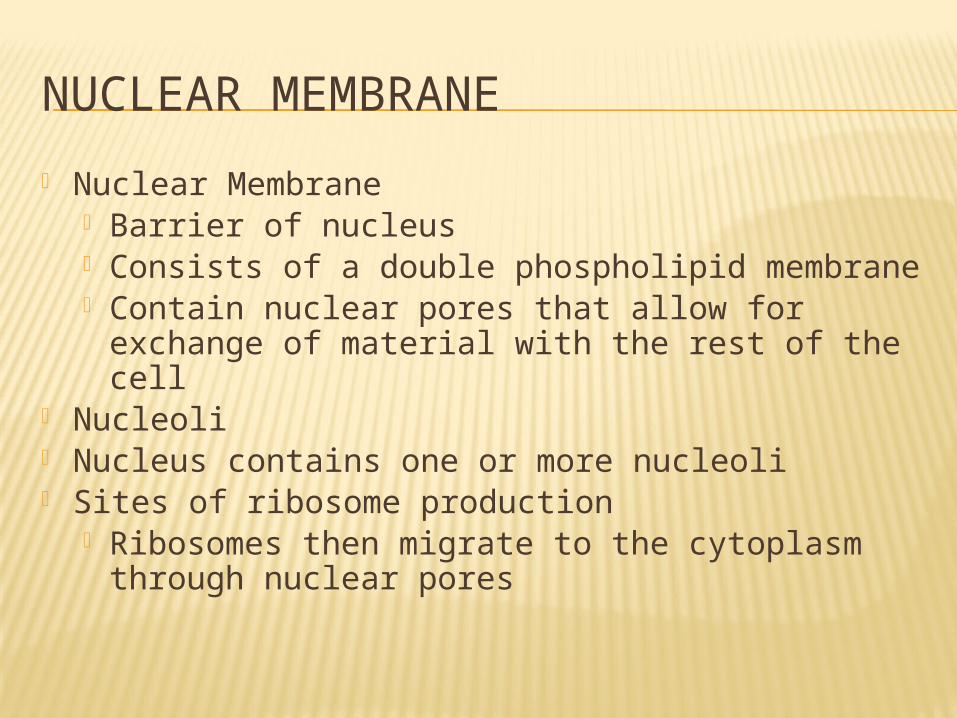
NUCLEAR MEMBRANE
Nuclear Membrane Barrier of nucleus Consists of a double phospholipid membrane Contain nuclear pores that allow for
exchange of material with the rest of the cell Nucleoli Nucleus contains one or more nucleoli Sites of ribosome production
Ribosomes then migrate to the cytoplasm through nuclear pores

CHROMATIN
Composed of DNA and protein Scattered throughout the nucleus Chromatin condenses to form
chromosomes when the cell divides
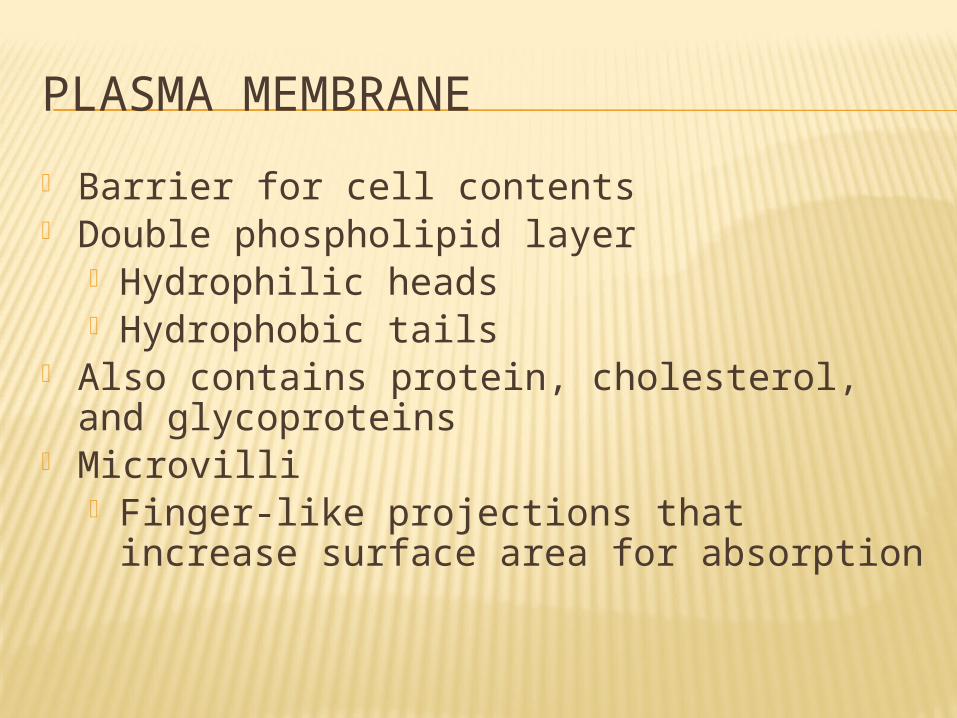
PLASMA MEMBRANE
Barrier for cell contents Double phospholipid layer
Hydrophilic heads Hydrophobic tails
Also contains protein, cholesterol, and glycoproteins
Microvilli Finger-like projections that increase surface
area for absorption

PLASMA MEMBRANE
Figure 3.2
Extracellular fluid(watery environment)
Sugargroup
Polar heads ofphospholipidmolecules
Bimolecularlipid layer containingproteins
Nonpolar tailsof phospholipidmolecules
Glycoprotein
Proteins Filaments ofcytoskeleton Cytoplasm
(watery environment)
Channel
Cholesterol
Glycolipid

Plasmamembranes ofadjacent cells
Desmosome(anchoring junction)
Tight(impermeable) junction
Microvilli
Gap(communicating) junction
Extracellularspace betweencells
Underlyingbasementmembrane
Connexon
Figure 3.3

CYTOPLASM Material outside the nucleus and inside
the plasma membrane Cytosol
Fluid that suspends other elements Organelles
Metabolic machinery of the cell “Little organs”
Inclusions Non-functioning units (stored nutrients, cell
products, etc.)
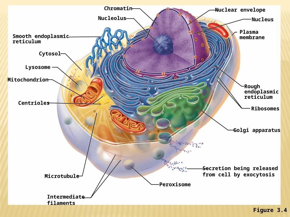
Ribosomes
Golgi apparatus
Secretion being releasedfrom cell by exocytosisMicrotubule
Centrioles
Mitochondrion
Lysosome
Cytosol
Smooth endoplasmicreticulum
Chromatin
Nucleolus
Nuclear envelope
Nucleus
Plasmamembrane
Roughendoplasmicreticulum
Intermediatefilaments
Peroxisome
Figure 3.4

CYTOPLASMIC ORGANELLES
Ribosomes Made of protein and RNA Sites of protein synthesis Found at two locations
Free in the cytoplasm Attached to rough endoplasmic reticulum

CYTOPLASMIC ORGANELLES Endoplasmic reticulum (ER)
Fluid-filled tubules for carrying substances Two types of ER
Rough Endoplasmic Reticulum Studded with ribosomes Site where building materials of cellular
membrane are formed Smooth Endoplasmic Reticulum
Functions in cholesterol synthesis and breakdown, fat metabolism, and detoxification of drugs

CYTOPLASMIC ORGANELLES
Golgi apparatus Modifies and packages proteins Produces different types of packages
Secretory vesicles Cell membrane components Lysosomes

Figure 3.20
Protein-containing vesicles pinch off rough ERand migrate to fuse with membranes ofGolgi apparatus.
Proteins aremodified withinthe Golgi compartments.
Proteins arethen packagedwithin differentvesicle types, depending ontheir ultimatedestination.
Plasmamem-brane
Secretion byexocytosis
Vesicle becomeslysosome
Golgiapparatus
Rough ER ERmembrane
Phagosome
Proteins incisterna
Pathway B:Vesicle membraneto be incorporatedinto plasmamembranePathway A:
Vesicle contentsdestined for exocytosis Extracellular fluid
Secretoryvesicle
Pathway C:Lysosome containing acid hydrolaseenzymes
1
3
2

CYTOPLASMIC ORGANELLES
Lysosomes Contain enzymes that digest nonusable
materials within the cell Packaged by Golgi apparatus
Peroxisomes Membranous sacs of oxidase enzymes
Detoxify harmful substances Break down free radicals
(highly reactive chemicals) Replicate by pinching in half

CYTOPLASMIC ORGANELLES
Mitochondria “Powerhouses” of the cell Change shape continuously Carry out reactions where oxygen is used
to break down food Provides ATP for cellular energy

CYTOPLASMIC ORGANELLES
Cytoskeleton Network of protein structures that extend
throughout the cytoplasm Provides the cell with an internal
framework
Figure 3.7a

Figure 3.7a-c
(a) Microfilaments (b) Intermediate filaments (c) Microtubules
Actin subunit
7 nm 10 nm
Fibrous subunitsTubulin subunits
25 nm
Microfilaments form the bluenetwork surrounding the pinknucleus.
Intermediate filaments formthe purple batlike network.
Microtubules appear as goldnetworks surrounding thecells’ pink nuclei.

CYTOPLASMIC ORGANELLES
Centrioles Rod-shaped bodies Direct formation of mitotic spindle during
cell division

CELLULAR PROJECTIONS
Not found in all cells Used for movement
Cilia moves materials across the cell surface
Flagellum propels the cell

CELL DIVERSITY
Figure 3.8a–b
CELL DIVERSITY
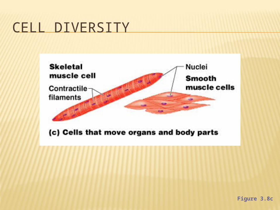
Figure 3.8c

CELL DIVERSITY
Figure 3.8d–e

CELL DIVERSITY
Figure 3.8f–g


![A Molecular Biology: Open Access - Open Access Journals€¦ · the molecular basis of carcinogenesis [1]. Cancer cells, unlike normal cells, sustain proliferative signaling to proliferate](https://img.pdfslide.us/doc/110x75/5fc153b832b30222ce574aa6/a-molecular-biology-open-access-open-access-journals-the-molecular-basis-of-carcinogenesis.jpg)