Embed Size (px)
Citation preview

THE FUTURE OF AUTOMATED MOBILE EYE DIAGNOSIS
Cassie A. Ludwig, BS1, Mia X. Shan, BS, BAH1, Nam Phuong H. Nguyen1, Daniel Y. Choi, MD1,
Victoria Ku, BS1, Carson K. Lam, MD1
1Byers Eye Institute, Stanford University School of Medicine 2405 Watson Drive, Palo Alto, CA, USA 94305
Corresponding Author: [email protected]
The current model of ophthalmic care requires the ophthalmologist’s involvement in data collection,diagnosis, treatment planning, and treatment execution. We hypothesize that ophthalmic datacollection and diagnosis will be automated through mobile devices while the education, treatmentplanning, and fine dexterity tasks will continue to be performed at clinic visits and in the operatingroom by humans. Comprehensive automated mobile eye diagnosis includes the following steps:mobile diagnostic tests, image collection, image recognition and interpretation, integrativediagnostics, and user-friendly, mobile platforms. Completely automated mobile eye diagnosis willrequire improvements in each of these components, particularly image recognition and interpreta-tion and integrative diagnostics. Once polished and integrated into greater medical practice,automated mobile eye diagnosis has the potential to increase access to ophthalmic care with reducedcosts, increased efficiency, and increased accuracy of diagnosis.
Journal MTM 5:2:44�50, 2016 doi:10.7309/jmtm.5.2.7 www.journalmtm.com
Introduction
Current Model of Eye Care Data Collection
In a typical eye care clinic visit, a combination of an
ophthalmic technician, nurse, optometrist, orthop-
tist, and/or ophthalmologist takes an ocular history
then measures the patient’s visual acuity, intraocu-
lar pressure, pupil shape and diameter, presence of
relative afferent pupillary defect, and the range of
extraocular movements in each eye. Tools used
include a slit lamp to view the external eye, anterior,
and posterior chamber, a gonioscope to measure
the iridocorneal angle, and a direct and/or indirect
ophthalmoscope to view the fundus. If necessary,
additional testing includes marginal reflex distance
and levator function measurement, color vision test-
ing, and optical coherence tomography, among others.
Once a diagnosis is made, a physician helps the
patient decide between different treatment options,
and provides education, medication, and/or a pro-
cedure as treatment.
Future Model of Eye Care Data Collection
With the dramatic rise in demand for ophthalmic
care relative to the small increase in ophthalmolo-
gists, how can ophthalmologists continue to increase
access to care? We hypothesize that ophthalmic
diagnosis will be automated through mobile devices,
with the help of support staff for education, treat-
ment planning, and fine dexterity tasks. Transition-
ing to mobile as opposed to current fixed forms of
diagnosis will increase accessibility to ophthalmic
services, particularly in low-resource settings. There
are 2.6 billion smartphone subscriptions today and
81% of health care professionals owned a smart-
phone in 2010*making the smartphone a highly
accessible platform for automated diagnosis through-
out the world.1,2 The result of the delegation of
diagnosis to mobile devices may allow for faster,
remote, low-cost diagnosis and disease monitoring.
Automated mobile eye diagnosis will require the
integration of data captured from mobile software
and hardware technology to create evidence-based
PERSPECTIVE PIECE
#JOURNAL OF MOBILE TECHNOLOGY IN MEDICINE VOL. 5 | ISSUE 2 | JULY 2016 44
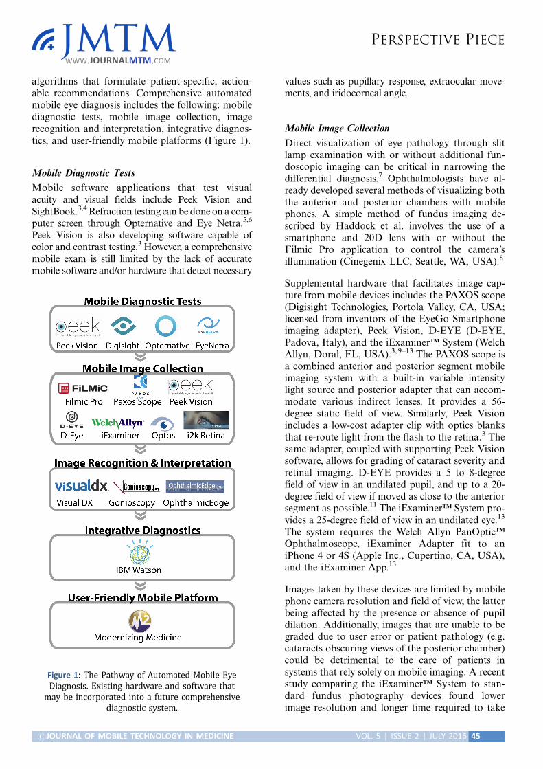
algorithms that formulate patient-specific, action-able recommendations. Comprehensive automatedmobile eye diagnosis includes the following: mobilediagnostic tests, mobile image collection, imagerecognition and interpretation, integrative diagnos-tics, and user-friendly mobile platforms (Figure 1).
Mobile Diagnostic Tests
Mobile software applications that test visualacuity and visual fields include Peek Vision andSightBook.3,4 Refraction testing can be done on a com-puter screen through Opternative and Eye Netra.5,6
Peek Vision is also developing software capable ofcolor and contrast testing.3 However, a comprehensivemobile exam is still limited by the lack of accuratemobile software and/or hardware that detect necessary
values such as pupillary response, extraocular move-ments, and iridocorneal angle.
Mobile Image Collection
Direct visualization of eye pathology through slitlamp examination with or without additional fun-doscopic imaging can be critical in narrowing thedifferential diagnosis.7 Ophthalmologists have al-ready developed several methods of visualizing boththe anterior and posterior chambers with mobilephones. A simple method of fundus imaging de-scribed by Haddock et al. involves the use of asmartphone and 20D lens with or without theFilmic Pro application to control the camera’sillumination (Cinegenix LLC, Seattle, WA, USA).8
Supplemental hardware that facilitates image cap-ture from mobile devices includes the PAXOS scope(Digisight Technologies, Portola Valley, CA, USA;licensed from inventors of the EyeGo Smartphoneimaging adapter), Peek Vision, D-EYE (D-EYE,Padova, Italy), and the iExaminerTM System (WelchAllyn, Doral, FL, USA).3, 9�13 The PAXOS scope isa combined anterior and posterior segment mobileimaging system with a built-in variable intensitylight source and posterior adapter that can accom-modate various indirect lenses. It provides a 56-degree static field of view. Similarly, Peek Visionincludes a low-cost adapter clip with optics blanksthat re-route light from the flash to the retina.3 Thesame adapter, coupled with supporting Peek Visionsoftware, allows for grading of cataract severity andretinal imaging. D-EYE provides a 5 to 8-degreefield of view in an undilated pupil, and up to a 20-degree field of view if moved as close to the anteriorsegment as possible.11 The iExaminerTM System pro-vides a 25-degree field of view in an undilated eye.13
The system requires the Welch Allyn PanOpticTM
Ophthalmoscope, iExaminer Adapter fit to aniPhone 4 or 4S (Apple Inc., Cupertino, CA, USA),and the iExaminer App.13
Images taken by these devices are limited by mobilephone camera resolution and field of view, the latterbeing affected by the presence or absence of pupildilation. Additionally, images that are unable to begraded due to user error or patient pathology (e.g.cataracts obscuring views of the posterior chamber)could be detrimental to the care of patients insystems that rely solely on mobile imaging. A recentstudy comparing the iExaminerTM System to stan-dard fundus photography devices found lowerimage resolution and longer time required to take
Figure 1: The Pathway of Automated Mobile Eye
Diagnosis. Existing hardware and software that
may be incorporated into a future comprehensive
diagnostic system.
PERSPECTIVE PIECE
#JOURNAL OF MOBILE TECHNOLOGY IN MEDICINE VOL. 5 | ISSUE 2 | JULY 2016 45

images using the smartphone setup.14 However, astudy comparing smartphone ophthalmoscopy withthe D-EYE device to dilated retinal slit-lamp exami-nation found exact agreement between the twomethods in 204 of 240 eyes on grade of diabeticretinopathy.15 Notably the latter study was perfor-med with an iPhone 5 (8-megapixel iSight camera)and the former with an iPhone 4 (5-megapixel stillcamera). Enhanced mobile imaging hardware willcontinue to improve the resolution,16 and wider-field imaging can be provided by pupil dilation(requiring a technician or nurse), laser scan imaging(Optos PLC, Marlborough, MA) or by integrationwith software that patches retina images together intoa mosaic, such as i2k Retina software (DualAlignLLC, Clifton Park, New York, USA).17 User errorcan be overcome with increasing experience withmobile imaging. However, patient pathology thatobscures views of the posterior chamber willultimately limit mobile diagnosis.
Furthermore, current mobile imaging does not re-place the scleral depressed indirect ophthalmos-copic exam that allows stereoscopic views of theindented retina anteriorly beyond the peripheralretina to the ora serrata and pars plana. This capa-bility would be needed for evaluation of flashesor floaters*a common presentation in which thediagnosis of retinal tear, hole, or detachment mustbe ruled out over multiple visits.
Another limitation in mobile image capture is thelack of a slit-lamp device for assessing individualcorneal layers and the anterior chamber, limitingthe precision of diagnosis of corneal pathology andof cell and flare diagnostic of uveitis. Automatedmobile eye diagnosis will require hardware orsoftware that allows for large-field image captureof the fundus and visualization of the corneal layersand the anterior chamber.
Image Recognition and Interpretation
Following image capture, automated mobile diag-nosis requires an interpretation system that detectsmultiple features of the anterior and posteriorsegments. The ideal system would recognize atypicalcolor, contrast, shape, and size of all visible compo-nents of the eye. Anteriorly, it would be able to dis-tinguish the lids from the lashes, sclera, conjunctiva,limbus, iris, cornea, pupil, and lens, and posteriorly,it should be able to distinguish between the opticdisc, macula, vascular arcades, and peripheral retina.After recognizing the component parts of the eye,
so called ‘‘segmentation’’, the system should then beable to label the type of abnormality and its locationwithin the anatomy of the eye. Lastly, for a systemto expand healthcare delivery and access, it wouldneed to provide some level of instruction. On thesimplest level, this could involve determining if apatient should be referred to an ophthalmologist or
screened again at a later date. As we will discuss,significant progress has already been made inaccomplishing this.
One method of expanding access to specialty care isto build tools that enable primary care providers tomake decisions that typically require training in amedical specialty. One strategy is creating systemsthat eliminate prior knowledge as a prerequisite fordiagnosis. For example, the identification of a skinlesion usually requires the clinician to have studied thepresentation, shape, color, texture and location ofvarious skin lesions as well as have a sense of diseasevariation and overlap. VisualDx is a subscription-
based website that walks users through step-by-stepvisual diagnosis of dermatologic conditions, includ-ing some overlap with ophthalmologic diagnoses.Three other similar sites include gonioscopy.org,oculonco.com, and ophthalmicedge.org.24�26
Peek Vision’s software automates one componentof image recognition, optic cup:disc ratio calcula-tion, important for diagnosis of glaucomatous opticneuropathy.20 Additionally, a team has automatedthe quantification of the number, morphology, andreflective properties of drusen based on spectraldomain-optical coherence tomography.21
An alternative solution is crowdsourcing. In twostudies, researchers demonstrated the utility ofcrowdsourcing of untrained people looking at retinaimages in automated diagnosis.28�30 However, vari-ation in human interpretation, the number and
experience of reviewers prevent its application.
Several research groups are developing algorithmsfor automated diagnosis. The most developedapplication of these algorithms is in the area of
diabetic retinopathy screening in which many pro-posed algorithms reach sensitivity and specificitypercentiles in the 90s.23 One of the most publishedalgorithms is the Iowa Detection System, that hasas its input a retina color photograph, and as itsoutput, a number between 0 and 1. The closer theoutput is to 1, the more likely the patient has a stageof diabetic retinopathy that should be referred toan ophthalmologist or that the photograph is of
PERSPECTIVE PIECE
#JOURNAL OF MOBILE TECHNOLOGY IN MEDICINE VOL. 5 | ISSUE 2 | JULY 2016 46

insufficient quality to determine stage of diabetic
retinopathy.22 It database includes numerous retinal
images of racially diverse individuals taken using
different camera types, and the Iowa Detection
System been found to perform comparably to retina
specialists.16 The best algorithm, however, remains a
debatable issue as the algorithms are tested against
human interpreters and with limited datasets where
the gold standard is determined by another human
interpreter or consensus of interpreters.23 Develop-
ment of an algorithm capable of classifying disease
outside of one spectrum of disease or of identifying
the clinical significance of retinal findings remain
an area of active research. We are living amidst a
turning point in computing that has already revolu-
tionized the field of computer vision and is set to
change medical imaging.
Neural networks improve upon the algorithms for
automated image recognition and interpretation.
Neural networks are biologically inspired algo-
rithms that learn to approximate a function (Figure 2).
Their inputs can be composed of text, numbers or
images. They contain interconnected layers of func-
tions called ‘‘neurons’’ that receive inputs from
neurons in the layer adjacent to themselves and
pass their outputs to the next layer of neurons. The
ultimate function that the neural network approx-
imates is encoded in the strength of connection
between neurons. These connections are fine tuned
by training the neural network with correct inputs
and outputs. For example, a retinal photograph
with the correct diagnosis as the output. Neural
networks have been in development since the 1940s,
and many research groups have already applied these
algorithms to interpretation of ophthalmic images.18,19
However recent advances in the availability of high
performance computing hardware has made feasible
the construction and training of neural networks
that contain several layers, various schemes of inter-
connectedness, and several neurons in each layer.
Due to the repeated stacking of many layers of
neurons, this form of computation has been termed
‘‘deep learning’’ and lies at the core of artificial
intelligence software such as Google self driving
cars (Google Inc., Mountain View, CA, USA), voice
recognition in phones, and Facebook facial recog-
nition (Facebook Inc., Palo Alto, CA, USA).
Most algorithms proposed thus far for image
diagnosis, even those that use neural networks, are
composed of specific, coded features used in combi-
nation for image recognition. Ophthalmic diagnosis,
however, is a complex task that takes into account
the location of lesions, macroscopic and microscop-
ic structural changes, and textures difficult to
describe even by ophthalmologists. Deep learning,
although flawed by drawbacks such as overfitting
and need for large training sets, makes few prior
assumptions about the features needed to recognize
Figure 2: General Architecture of Neural Network. General architecture of neural network with arbitrary number of
neurons in each layer, number of layers, different schemes of interconnection between neurons in one layer and neurons in
adjacent layer. It is fully connected, meaning that every neuron in a layer is connected to every neuron in its adjacent layer.
Inputs are values such as pixel values from an image, and layers can be a one-dimensional row of neurons such as in the
example or a three-dimensional volume such as in convolutional neural networks.
PERSPECTIVE PIECE
#JOURNAL OF MOBILE TECHNOLOGY IN MEDICINE VOL. 5 | ISSUE 2 | JULY 2016 47

images. Deep learning learns features from the datawith which it is trained. In the next five years, therewill be a wave of literature describing the complextasks these algorithms can perform in automatedophthalmic diagnosis.30,31
Integrative Diagnostics
Ultimately, all data points gathered need to beintegrated into one mobile system for diagnosis,such as with artificial intelligence software. IBMWatson, made famous by its Jeopardy! win, is anexample of software that can integrate evidence-based medical knowledge accurately and consistentlyfor automated diagnosis.32 IBM Watson’s servers canprocess 500 gigabytes of information per second*the equivalent of 1 million books.32 IBM Watson’sconversion of this information into evidence-basedalgorithms for diagnosis would provide the most up-to-date diagnostic programming possible.
User-Friendly Mobile Platform
To deliver the final information to the patient and/or physician, a user-friendly interface between themobile phone and user will be necessary. An ex-ample of such an interface is Modernizing Medi-cine’s electronic medical assistant (EMA) iPadapplication for ophthalmology, which integratespublished healthcare information and providesphysicians with treatment options and outcomemeasures.33,34 While a physician-mobile deviceinterface may be useful in guiding treatment plans,a patient-directed interface may ultimately provideactionable steps for the patient to take before everseeing a physician for treatment.
DiscussionThe future of automated mobile eye diagnosis liesin improvements in each of the above components,particularly image recognition and interpretationand integrative diagnostics. With automated mobileeye diagnosis, patients will have faster access toinformation about their conditions to guide theirnext steps. Automated diagnosis will be a reliable,cost-effective, and accessible tool for individualsacross demographics to gather ophthalmologicinformation necessary to understand and managetheir conditions.
Benefits of Automated Eye Diagnosis
Given the increased availability of wireless networksand advancements in technology, automated eyediagnosis will prove to be more cost-effective,
accessible, and reliable than specialist diagnoses.35,36
Automated diagnosis will have the highest yieldin low-resource settings where both physical andeconomic barriers limit access to specialist diagno-sis. In a 2007 study, researchers in Scotland foundthat automated grading of images for diabeticretinopathy reduced both the workload and associ-ated costs of care.37 Automated grading for a cohortof 160,000 patients lowered the total cost of grading by47% as compared to manual grading, a saving of US$0.25 per patient. Similar studies on automateddiabetic retinopathy and cataracts services in Canada,US, and rural South Africa have also shown that earlyscreening programs save programs between $1,206 and$3,900 per sight per year in diagnosis, treatment, andreferrals.38�41 With this decrease in cost of automatedeye diagnosis, the service can more easily spread tosmaller clinics in rural areas for early detection andtriage of eye pathology.
Additionally, unlike humans, computers can rapidlyincorporate new scientific information into theiralgorithms. The processing power of IBM Watson,for example, is inevitably impossible for any humanspecialist.32 Furthermore, humans maintain variousbiases during diagnosis, such as anchoring bias(relying too heavily on the first piece of informationgiven) and framing bias (being prejudiced based onthe way a statement is phrased).36 Automated eyediagnostic programs can continuously add newinformation to their database and algorithms whileavoiding biases to make decisions.
Drawbacks to Automated Eye Diagnosis
Potential drawbacks to automated eye diagnosisinclude initial costs as well as concerns about itsreliability and sensitivity. The design phase involvesinitial investment in hardware for image capture,programming software, and program developers.Implementation requires distribution of hardwareand software to the appropriate users. Maintenanceentails ongoing costs for the programmers and per-sonnel running the service. However, as mentionedpreviously with automated diabetic retinopathyand cataracts diagnosis, the savings can eventuallytrump the initial costs as screening services expandto more patients.37�41
The accuracy of every step of automated eye di-agnosis is critical to its success. In the Scotlanddiabetic retinopathy study, automated grading of14,406 images from 6,722 patients missed three morecases of referable disease compared with manualgrading.37 However, these were non-sight-threatening
PERSPECTIVE PIECE
#JOURNAL OF MOBILE TECHNOLOGY IN MEDICINE VOL. 5 | ISSUE 2 | JULY 2016 48

maculopathy instead of referable or proliferativeretinopathy. Additionally, in 2010, researchers foundthat the algorithm for automated detection of diabeticretinopathy lagged only slightly behind the sensitivityand specificity of retinal specialists.37 As algorithmsimprove and are conducted on larger datasets, webelieve that automation will outperform experts insensitivity and specificity.
Lastly, the transmission of patient information overmobile devices necessitates strict and establishedprotocols in patient consent and personal healthinformation security. Software developers mustalways consider the security of patient information.
Next Steps
The first step in shifting the roles of ophthalmolo-gists away from data collection will be trainingancillary providers to collect the data necessary fordiagnosis. These providers can then submit a digitalrepresentation of the information to software thatgives instructions on treatment options and furthertesting, shifting the focus of ophthalmologiststowards education, treatment planning, and treat-ment implementation.
ConclusionWe believe that automated mobile eye diagnosisusing evidence-based algorithms will increase patientsafety, improve access to ophthalmic services, andfacilitate timely referrals. However, completely auto-mated eye diagnosis will require improvements inimage capture and recognition as well as automatedintegrative diagnostics. Once polished and integratedinto greater medical practice, automated eye diagno-sis has the potential to become a powerful tool toincrease access to ophthalmic services worldwide.
AcknowledgementsThe project described herein was conducted withsupport for Cassie A. Ludwig from the TL1 compo-nent of the Stanford Clinical and TranslationalScience Award to Spectrum (NIH TL1 TR 001084).
References1. Ericsson. Ericsson Mobility Report 2015; http://www.
ericsson.com/res/docs/2015/mobility-report/ericsson-
mobility-report-nov-2015.pdf. (accessed 7 Dec 2015).
2. Asurvey of mobile phone usage by health professionals
in the UK. 2010; http://www.d4.org.uk/research/
survey-mobile-phone-use-health-professionals-UK.pdf. (accessed 7 Dec 2015).
3. Peek Vision. 2015; http://www.peekvision.org/.
(accessed 20 Mar 2015).
4. DigiSight. 2014; https://www.digisight.net/digisight/
index.php. (accessed 20 Mar 2015).
5. Opternative. 2015; https://www.opternative.com/.
(accessed 20 Mar 2015).
6. EyeNetra. 2013; http://eyenetra.com/aboutus-company.
html. (accessed 20 Mar 2015).
7. Image Analysis and Modeling in Ophthalmology.
USA: CRC Press; 2014.
8. Haddock LJ, Kim DY, Mukai S. Ssimple inexpensive
technique for high-quality smartphone fundus photo-
graphy in human and animal eyes. 2013;2013:518479.
9. Myung A, Jais A, He L, Blumenkranz MS, Chang
RT. 3D Printed Smartphone Indirect Lens Adapter
for Rapid, High Quality Retinal Imaging. Journal of
Mobile Technology in Medicine 2014;3(1):9�15.
10. Myung A, Jais A, He L, Chang RT. Simple, Low-
Cost Smartphone Adapter for Rapid, High Quality
Ocular Anterior Segment Imaging: A Photo Diary.
Journal of Mobile Technology in Medicine 2014;3(1):
2�8.
11. D-EYE. 2015; http://www.d-eyecare.com/. (accessed
7 Dec 2015).
12. Paxos Scope. 2014; https://www.digisight.net/digi-
sight/paxos-scope.php. (accessed 7 Dec 2015).
13. WelchAllyn. iEXAMINER. 2015; http://www.wel-
challyn.com/en/microsites/iexaminer.html. (accessed
7 Dec 2015).
14. Darma S, Zantvoord F, Verbraak FD. The quality
and usability of smartphone and hand-held fundus
photography, compared to standard fundus photo-
graphy. Acta ophthalmologica 2015;93(4):310�1.
15. Russo A, Morescalchi F, Costagliola C, Delcassi L,
Semeraro F. Comparison of smartphone ophthalmo-
scopy with slit-lamp biomicroscopy for grading dia-
betic retinopathy. American journal of ophthalmology
2015;159(2):360�364.e361.
16. Abramoff MD, Niemeijer M, Russell SR. Auto-
mated detection of diabetic retinopathy: barriers to
translation into clinical practice. Expert review of
medical devices 2010;7(2):287�96.
17. Maamari RN, Keenan JD, Fletcher DA, Margolis
TP. A mobile phone-based retinal camera for por-
table wide field imaging. The British journal of
ophthalmology 2014;98(4):438�41.
PERSPECTIVE PIECE
#JOURNAL OF MOBILE TECHNOLOGY IN MEDICINE VOL. 5 | ISSUE 2 | JULY 2016 49

18. Gardner GG, Keating D, Williamson TH, Elliott AT.
Automatic detection of diabetic retinopathy using
an artificial neural network: a screening tool. The
British journal of ophthalmology 1996;80(11):940�4.
19. Yun WL, Acharya UR, Venkatesh YV, et al. Identi-
fication of different stages of diabetic retinopathy
using retinal optical images. Information Sciences
2008;178(1):106�21.
20. Giardini ME, Livingstone IA, Jordan S, et al. A
smartphone based ophthalmoscope. Conference pro-
ceedings: . . . Annual International Conference of the
IEEE Engineering in Medicine and Biology Society.
IEEE Engineering in Medicine and Biology Society.
Annual Conference 2014;2014:2177�80.
21. de Sisternes L, Simon N, Tibshirani R, Leng T,
Rubin DL. Quantitative SD-OCT imaging biomar-
kers as indicators of age-related macular degenera-
tion progression. Investigative ophthalmology & visual
science 2014;55(11):7093�103.
22. Hansen MB, Abramoff MD, Folk JC, et al. Results
of Automated Retinal Image Analysis for Detection
of Diabetic Retinopathy from the Nakuru Study,
Kenya. PloS one 2015;10(10).
23. Mookiah MR, Acharya UR, Chua CK, et al.
Computer-aided diagnosis of diabetic retinopathy:
A review. Computers in biology and medicine
2013;43(12):2136�55.
24. VisualDx. 2015; http://www.visualdx.com/. (accessed
20 Mar 2015).
25. WLM A. Gonioscopy.org. 2015; http://gonioscopy.
org/. (accessed 20 Mar 20).
26. B D. Oculonco. 2015; http://www.oculonco.com/.
(accessed 20 Mar 2015).
27. OphthalmicEdge. 2014; ophthalmicedge.org
(accessed 20 Mar 2015).
28. Mitry D, Peto T, Hayat S, et al. Crowdsourcing as a
novel technique for retinal fundus photography
classification: analysis of images in the EPIC Norfolk
cohort on behalf of the UK Biobank Eye and Vision
Consortium. PloS one 2013;8(8):71154.
29. Brady CJ, Villanti AC, Pearson JL, et al. Rapid
grading of fundus photographs for diabetic retino-
pathy using crowdsourcing. Journal of medical Inter-
net research 2014;16(10):e233.
30. Lim G, Lee ML, Hsu W, et al. Transformed
Representations for Convolutional Neural Networks
in Diabetic Retinopathy Screening. InWorkshops
at the Twenty-Eighth AAAI Conference on Artificial
Intelligence 2014 Jun 18.
31. Krizhevsky A, Sutskever I, Hinton G. Imagenet
classification with deep convolutional neural net-
works. Advances in Neural Information Processing
Systems 2012;25:1106�14.
32. IBM Watson. 2015; http://www.ibm.com/smarterpla-
net/us/en/ibmwatson/. (accessed 20 Mar 2015).
33. Dattoli K CJ. IBM Reveals New Companies Develop-
ing Watson-Powered Apps: Healthcare, Retail and
Financial Services Firms To Debut Mobile Applications
Later This Year. 2014. http://www-03.ibm.com/press/
us/en/pressrelease/43936.wss. (accessed 20 Mar 2015).
34. Modernizing Medicine. 2014; https://www.modmed.
com/. (accessed 20 Mar 2015).
35. Hansmann U ML, Nicklous MS, Stober Th. Perva-
sive Computing: The Mobile World. 2 ed: Springer-
Verlag Berlin Heidelberg; 2003.
36. Dilsizian SE, Siegel EL. Artificial intelligence in
medicine and cardiac imaging: harnessing big dataand advanced computing to provide personalized
medical diagnosis and treatment. Current cardiology
reports 2014;16(1):441.
37. Scotland GS, McNamee P, Philip S, et al. Cost-
effectiveness of implementing automated grading
within the national screening programme for diabetic
retinopathy in Scotland. The British journal of
ophthalmology 2007;91(11):1518�23.
38. Khan T, Bertram MY, Jina R, et al. Preventing
diabetes blindness: cost effectiveness of a screening
programme using digital non-mydriatic fundus
photography for diabetic retinopathy in a primary
health care setting in South Africa. Diabetes research
and clinical practice. 2013;101(2):170�6.
39. Benbassat J, Polak BC. Reliability of screeningmethods for diabetic retinopathy. Diabetic medicine:
a journal of the British Diabetic Association 2009;
26(8):783�90.
40. Whited JD, Datta SK, Aiello LM, et al. A modeled
economic analysis of a digital tele-ophthalmology
system as used by three federal health care agencies
for detecting proliferative diabetic retinopathy. Tele-
medicine journal and e-health: the official journal of the
American Telemedicine Association 2005;11(6):641�51.
41. Li Z, Wu C, Olayiwola JN, Hilaire DS, Huang JJ.
Telemedicine-based digital retinal imaging vs stan-
dard ophthalmologic evaluation for the assessment
of diabetic retinopathy. Connecticut medicine
2012;76(2):85�90.
PERSPECTIVE PIECE
#JOURNAL OF MOBILE TECHNOLOGY IN MEDICINE VOL. 5 | ISSUE 2 | JULY 2016 50