Embed Size (px)
Citation preview
© 2013 Pearson Education, Inc.
Force of Muscle Contraction
• Force of contraction depends on number of cross bridges attached, which is affected by
• Number of muscle fibers stimulated (recruitment)• Relative size of fibers—hypertrophy of cells
increases strength• Frequency of stimulation• Degree of muscle stretch
© 2013 Pearson Education, Inc.
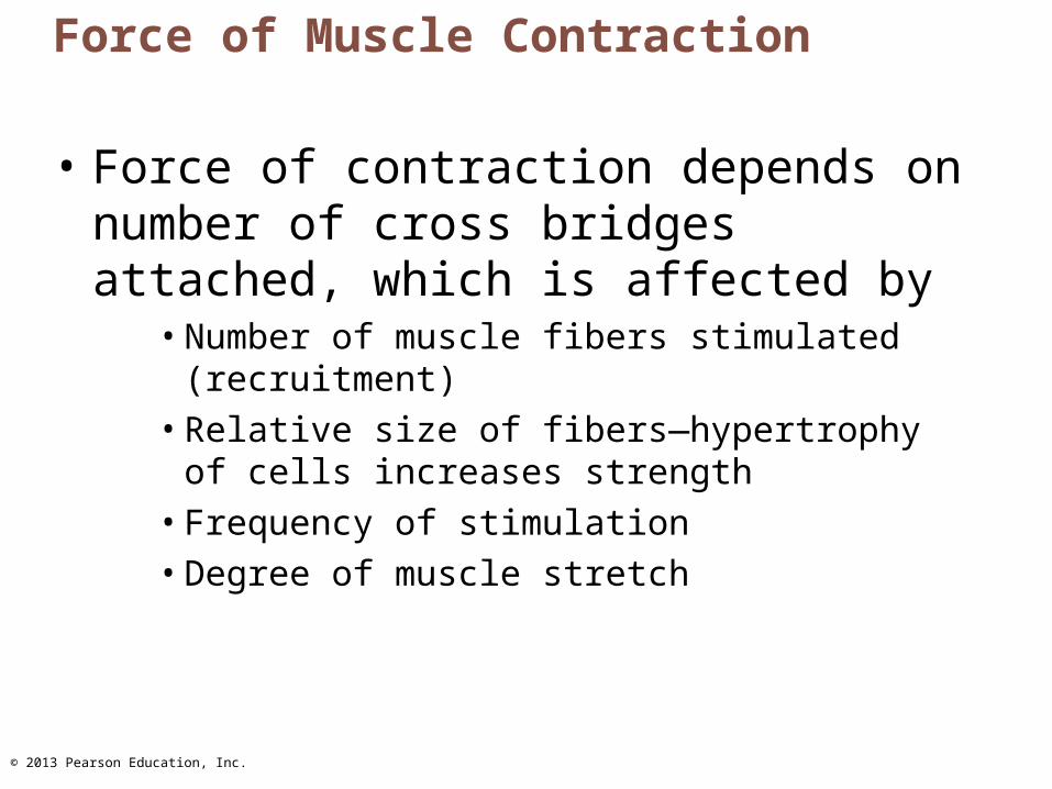
Figure 9.21 Factors that increase the force of skeletal muscle contraction.
Largenumber of
musclefibers
recruited
Largemusclefibers
Highfrequency ofstimulation
(wavesummation
and tetanus)
Muscle andsarcomere
stretched toslightly over 100%of resting length
Contractile force (more cross bridges attached)
© 2013 Pearson Education, Inc.
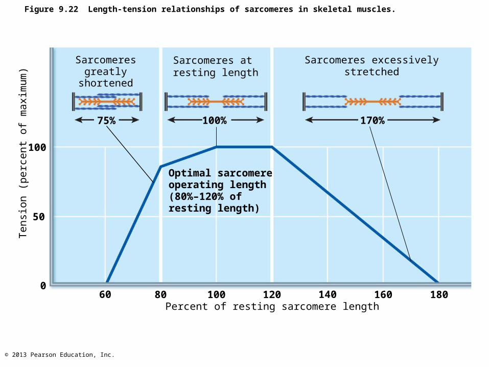
Figure 9.22 Length-tension relationships of sarcomeres in skeletal muscles.
Sarcomeresgreatly
shortened
Sarcomeres atresting length
Sarcomeres excessivelystretched
Optimal sarcomereoperating length(80%–120% ofresting length)
Tensi
on (
perc
ent
of
maxim
um
)
100
50
060 80 100 120 140 160 180
Percent of resting sarcomere length
75% 100% 170%
© 2013 Pearson Education, Inc.
Velocity and Duration of Contraction
• Influenced by:– Muscle fiber type– Load– Recruitment
© 2013 Pearson Education, Inc.
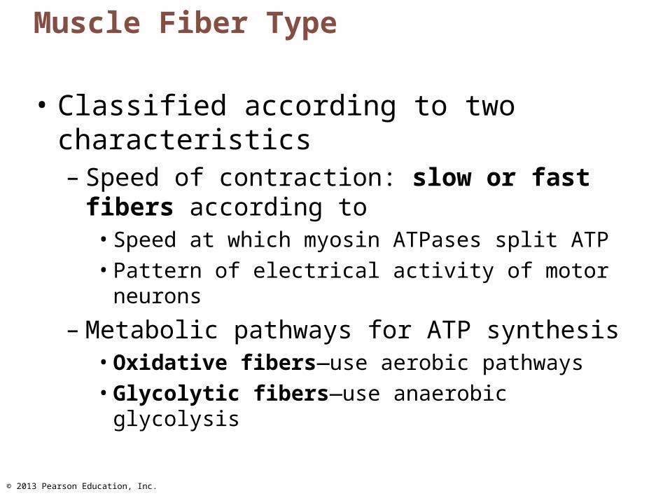
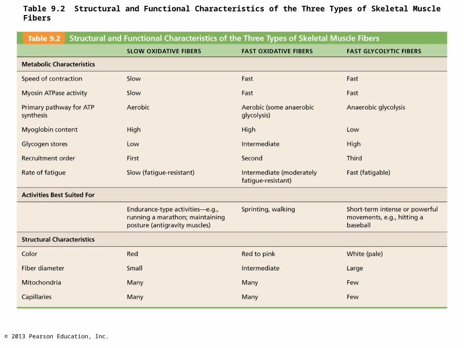
Muscle Fiber Type
• Classified according to two characteristics– Speed of contraction: slow or fast fibers
according to• Speed at which myosin ATPases split ATP• Pattern of electrical activity of motor neurons
– Metabolic pathways for ATP synthesis• Oxidative fibers—use aerobic pathways• Glycolytic fibers—use anaerobic glycolysis
© 2013 Pearson Education, Inc.
Muscle Fiber Type
• Three types – Slow oxidative fibers; Fast oxidative
fibers; Fast glycolytic fibers
• Most muscles contain mixture of fiber types range of contractile speed, fatigue resistance– All fibers in one motor unit same type– Genetics dictate individual's percentage of
each
© 2013 Pearson Education, Inc.
Table 9.2 Structural and Functional Characteristics of the Three Types of Skeletal Muscle Fibers
© 2013 Pearson Education, Inc.
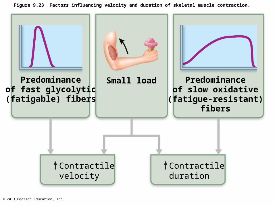
Figure 9.23 Factors influencing velocity and duration of skeletal muscle contraction.
Predominanceof fast glycolytic(fatigable) fibers
Contractilevelocity
Small load Predominanceof slow oxidative(fatigue-resistant)
fibers
Contractileduration
© 2013 Pearson Education, Inc.
Influence of Load
• Muscles contract fastest when no load added
load latent period, slower contraction, and duration of contraction
© 2013 Pearson Education, Inc.
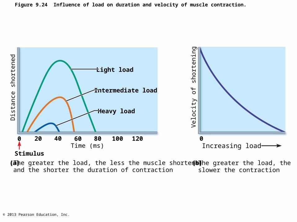
Figure 9.24 Influence of load on duration and velocity of muscle contraction.
Light load
Intermediate load
Heavy load
Dis
tance
short
ened
0 20 40 60 80 100 120
StimulusTime (ms)
The greater the load, the less the muscle shortensand the shorter the duration of contraction
The greater the load, the slower the contraction
Increasing load0
Velo
city
of
short
enin
g
© 2013 Pearson Education, Inc.
Influence of Recruitment
• Recruitment faster contraction and duration of contraction
© 2013 Pearson Education, Inc.
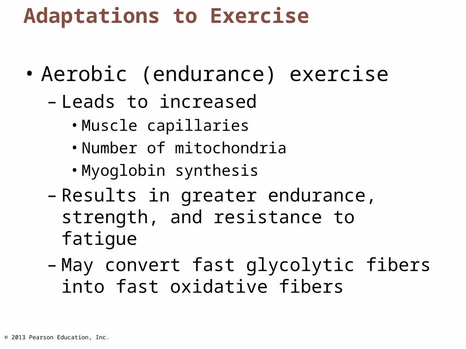
Adaptations to Exercise
• Aerobic (endurance) exercise– Leads to increased
• Muscle capillaries• Number of mitochondria• Myoglobin synthesis
– Results in greater endurance, strength, and resistance to fatigue
– May convert fast glycolytic fibers into fast oxidative fibers
© 2013 Pearson Education, Inc.
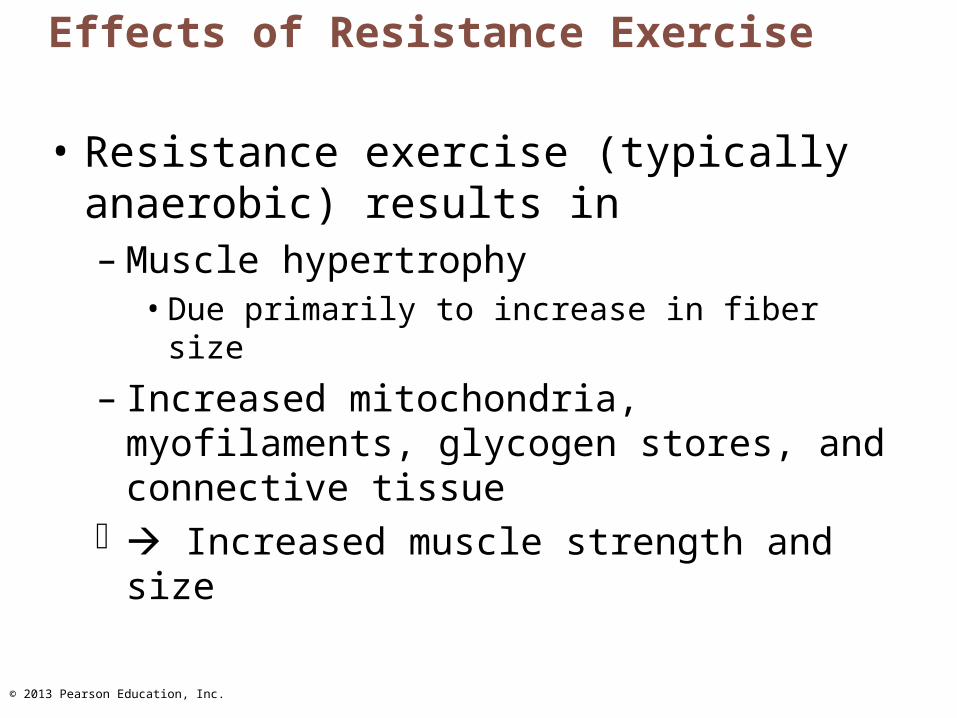
Effects of Resistance Exercise
• Resistance exercise (typically anaerobic) results in– Muscle hypertrophy
• Due primarily to increase in fiber size
– Increased mitochondria, myofilaments, glycogen stores, and connective tissue
Increased muscle strength and size
© 2013 Pearson Education, Inc.
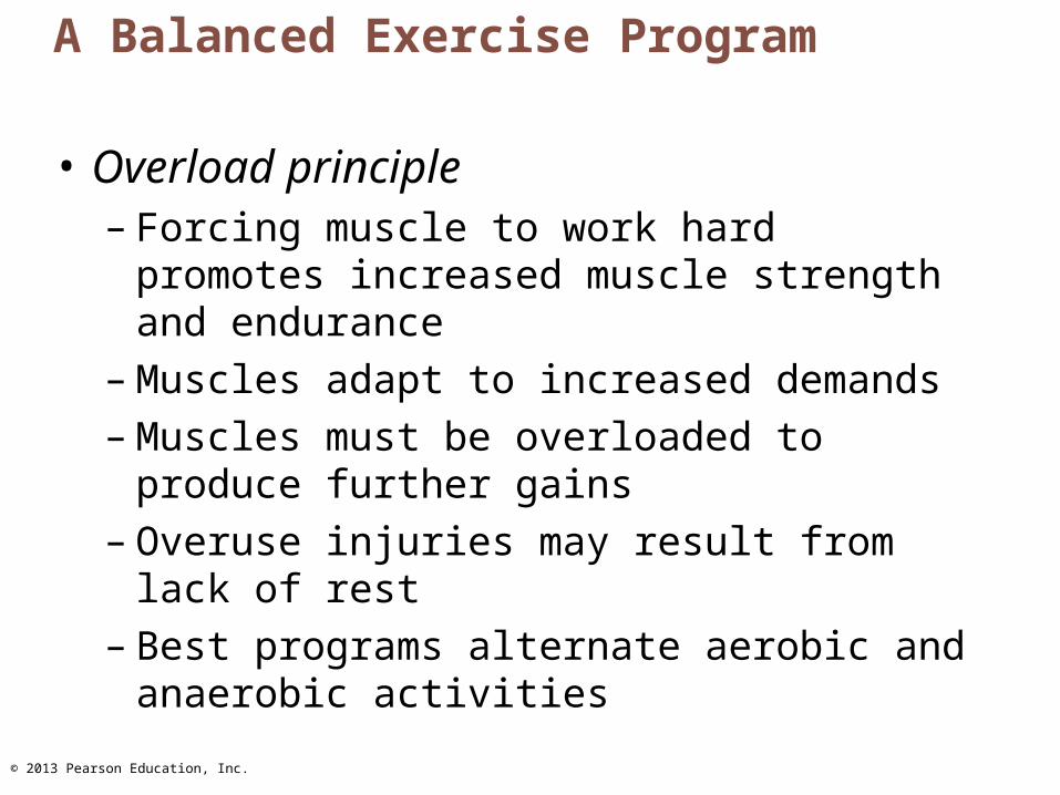
A Balanced Exercise Program
• Overload principle– Forcing muscle to work hard promotes
increased muscle strength and endurance– Muscles adapt to increased demands– Muscles must be overloaded to produce
further gains– Overuse injuries may result from lack of rest– Best programs alternate aerobic and
anaerobic activities
© 2013 Pearson Education, Inc.
Homeostatic Imbalance
• Disuse atrophy– Result of immobilization– Muscle strength declines 5% per day
• Without neural stimulation muscles atrophy to ¼ initial size– Fibrous connective tissue replaces lost
muscle tissue rehabilitation impossible
© 2013 Pearson Education, Inc.
Smooth Muscle
• Found in walls of most hollow organs(except heart)
• Usually in two layers (longitudinal and circular)
© 2013 Pearson Education, Inc.
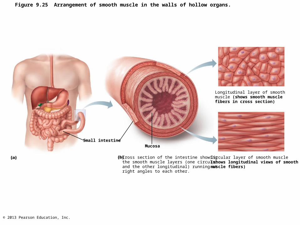
Figure 9.25 Arrangement of smooth muscle in the walls of hollow organs.
Small intestineMucosa
Cross section of the intestine showing the smooth muscle layers (one circular and the other longitudinal) running at right angles to each other.
Circular layer of smooth muscle(shows longitudinal views of smoothmuscle fibers)
Longitudinal layer of smooth muscle (shows smooth musclefibers in cross section)
© 2013 Pearson Education, Inc.
Microscopic Structure
• Spindle-shaped fibers - thin and short compared with skeletal muscle fibers; only one nucleus; no striations
• Lacks connective tissue sheaths; endomysium only
• SR - less developed than in skeletal muscle • Pouchlike infoldings (caveolae) of sarcolemma
sequester Ca2+ - most calcium influx from outside cell; rapid
• No sarcomeres, myofibrils, or T tubules
© 2013 Pearson Education, Inc.
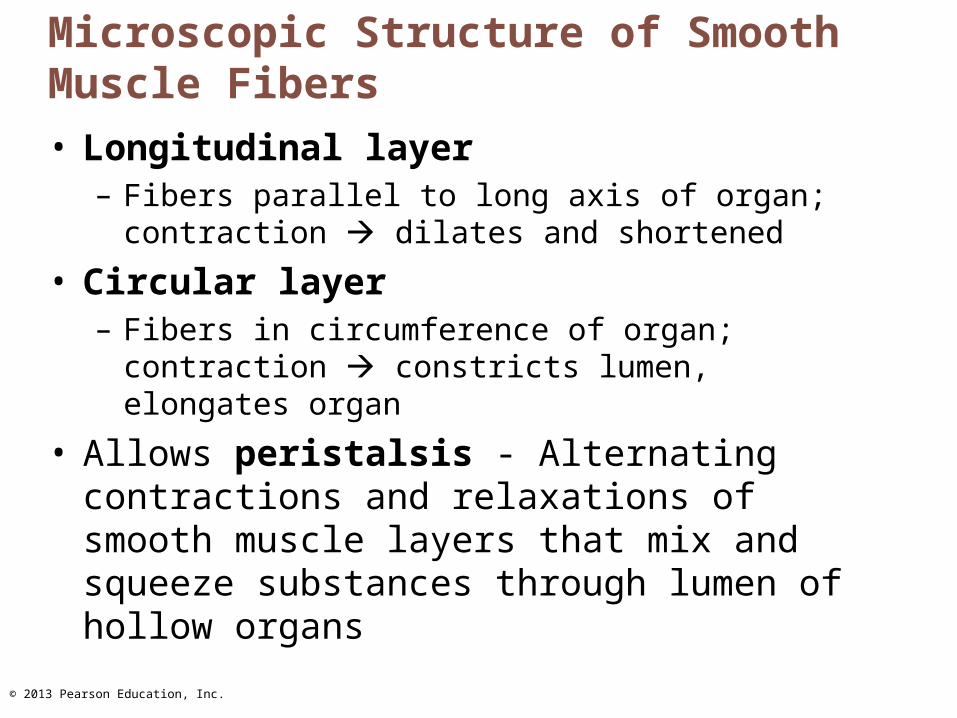
Microscopic Structure of Smooth Muscle Fibers
• Longitudinal layer– Fibers parallel to long axis of organ; contraction
dilates and shortened
• Circular layer– Fibers in circumference of organ; contraction
constricts lumen, elongates organ
• Allows peristalsis - Alternating contractions and relaxations of smooth muscle layers that mix and squeeze substances through lumen of hollow organs
© 2013 Pearson Education, Inc.
Innervation of Smooth Muscle
• No NMJ as in skeletal muscle
• Autonomic nerve fibers innervate smooth muscle at diffuse junctions
• Varicosities (bulbous swellings) of nerve fibers store and release neurotransmitters into diffuse junctions
© 2013 Pearson Education, Inc.
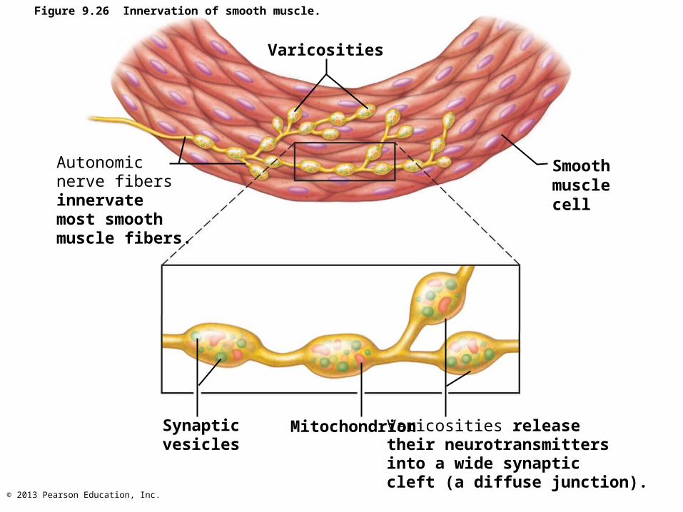
Figure 9.26 Innervation of smooth muscle.
Varicosities
Autonomicnerve fibersinnervatemost smoothmuscle fibers.
Smoothmusclecell
Synapticvesicles
Mitochondrion Varicosities releasetheir neurotransmittersinto a wide synaptic cleft (a diffuse junction).
© 2013 Pearson Education, Inc.
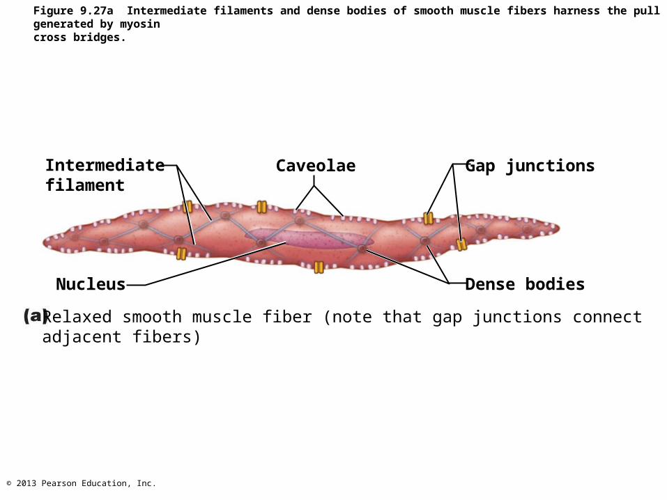
Myofilaments in Smooth Muscle
• Myofilaments are spirally arranged, causing smooth muscle to contract in corkscrew manner
• Dense bodies– Proteins that anchor noncontractile
intermediate filaments to sarcolemma at regular intervals
– Correspond to Z discs of skeletal muscle
© 2013 Pearson Education, Inc.
Intermediatefilament
Caveolae Gap junctions
Nucleus Dense bodies
Relaxed smooth muscle fiber (note that gap junctions connect adjacent fibers)
Figure 9.27a Intermediate filaments and dense bodies of smooth muscle fibers harness the pull generated by myosincross bridges.
© 2013 Pearson Education, Inc.
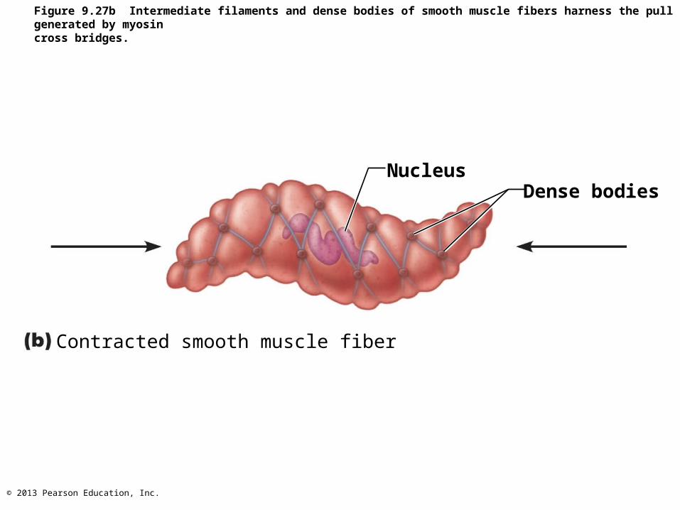
NucleusDense bodies
Contracted smooth muscle fiber
Figure 9.27b Intermediate filaments and dense bodies of smooth muscle fibers harness the pull generated by myosincross bridges.
© 2013 Pearson Education, Inc.
Contraction of Smooth Muscle
• Slow, synchronized contractions
• Cells electrically coupled by gap junctions– Action potentials transmitted from fiber to fiber
• Some cells self-excitatory (depolarize without external stimuli); act as pacemakers for sheets of muscle – Rate and intensity of contraction may be
modified by neural and chemical stimuli
© 2013 Pearson Education, Inc.
Contraction of Smooth Muscle
• Actin and myosin interact by sliding filament mechanism
• Final trigger is intracellular Ca2+
– Ca2+ is obtained from the SR and extracellular space
• ATP energizes sliding process
© 2013 Pearson Education, Inc.
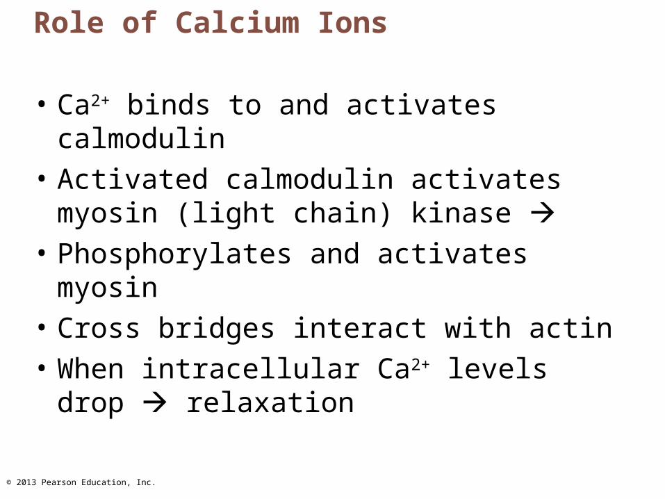
Role of Calcium Ions
• Ca2+ binds to and activates calmodulin
• Activated calmodulin activates myosin (light chain) kinase
• Phosphorylates and activates myosin
• Cross bridges interact with actin
• When intracellular Ca2+ levels drop relaxation
© 2013 Pearson Education, Inc.
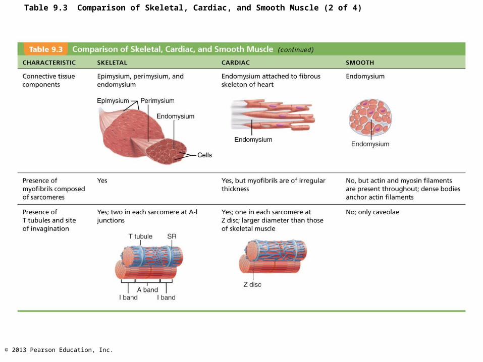
Table 9.3 Comparison of Skeletal, Cardiac, and Smooth Muscle (2 of 4)
© 2013 Pearson Education, Inc.
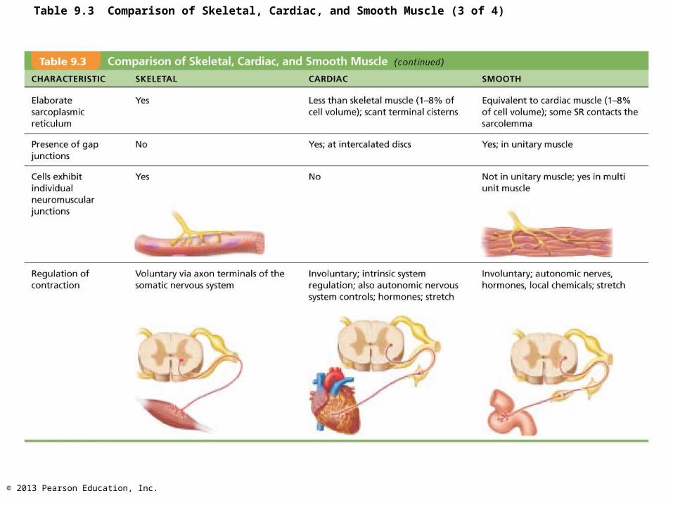
Table 9.3 Comparison of Skeletal, Cardiac, and Smooth Muscle (3 of 4)
© 2013 Pearson Education, Inc.
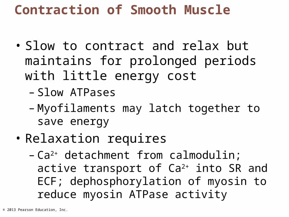
Contraction of Smooth Muscle
• Slow to contract and relax but maintains for prolonged periods with little energy cost– Slow ATPases– Myofilaments may latch together to save
energy
• Relaxation requires– Ca2+ detachment from calmodulin; active
transport of Ca2+ into SR and ECF; dephosphorylation of myosin to reduce myosin ATPase activity
© 2013 Pearson Education, Inc.
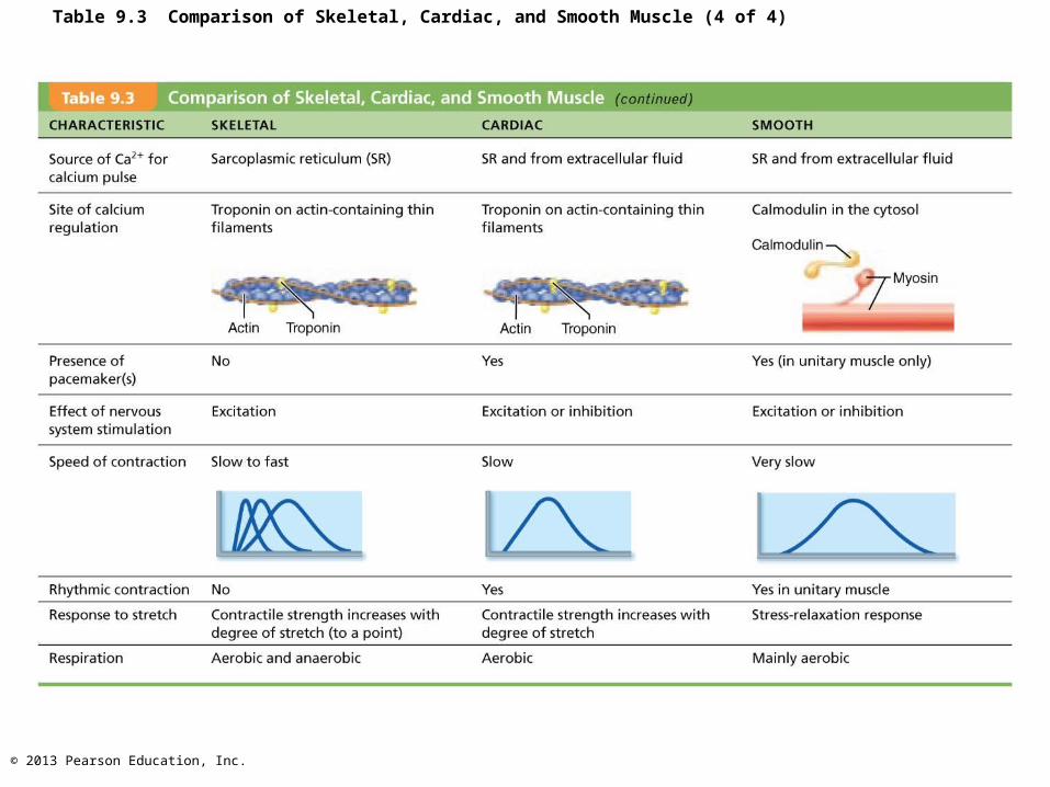
Table 9.3 Comparison of Skeletal, Cardiac, and Smooth Muscle (4 of 4)
© 2013 Pearson Education, Inc.
Developmental Aspects
• ~ All muscle tissue develops from myoblasts• Cardiac and skeletal muscle become amitotic,
but can lengthen and thicken in growing child• Myoblast-like skeletal muscle satellite cells have
limited regenerative ability• Cardiomyocytes can divide at modest rate, but
injured heart muscle mostly replaced by connective tissue
• Smooth muscle regenerates throughout life
© 2013 Pearson Education, Inc.
Developmental Aspects
• Muscular development reflects neuromuscular coordination
• Development occurs head to toe, and proximal to distal
• Peak natural neural control occurs by midadolescence
• Athletics and training can improve neuromuscular control
© 2013 Pearson Education, Inc.
Developmental Aspects
• Female skeletal muscle makes up 36% of body mass
• Male skeletal muscle makes up 42% of body mass, primarily due to testosterone
• Body strength per unit muscle mass same in both sexes
© 2013 Pearson Education, Inc.
Developmental Aspects
• With age, connective tissue increases and muscle fibers decrease
• By age 30, loss of muscle mass (sarcopenia) begins
• Regular exercise reverses sarcopenia
• Atherosclerosis may block distal arteries, leading to intermittent claudication and severe pain in leg muscles
© 2013 Pearson Education, Inc.
Muscular Dystrophy
• Group of inherited muscle-destroying diseases; generally appear in childhood
• Muscles enlarge due to fat and connective tissue deposits
• Muscle fibers atrophy and degenerate
© 2013 Pearson Education, Inc.
Muscular Dystrophy
– No cure– Prednisone improves muscle strength and
function– Myoblast transfer therapy disappointing– Coaxing dystrophic muscles to produce more
utrophin (protein similar to dystrophin) successful in mice
– Viral gene therapy and infusion of stem cells with correct dystrophin genes show promise


![This monthchild bearing. Although venous disease is rarely a life- or ... gravitational hydrostatic force, and hydrodynamic force due to muscular contraction [8]. Venous unction is](https://img.pdfslide.us/doc/110x75/609cc863c542dd02e2241620/this-month-child-bearing-although-venous-disease-is-rarely-a-life-or-gravitational.jpg)