Embed Size (px)
Citation preview
© 2011, 2007, 2003 Pearson Education, Inc.All Rights Reserved.
Chapter 3Anatomy and Physiology Related to Speech, Hearing, and Language

© 2011, 2007, 2003 Pearson Education, Inc.All Rights Reserved.
ANATOMY AND PHYSIOLOGY
The Physiological Subsystems Supporting Speech
The Speech Production Process The Nervous System Motor Speech Control
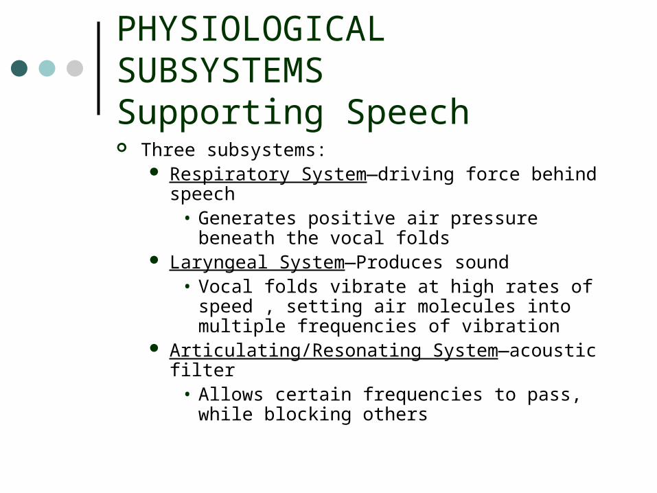
PHYSIOLOGICAL SUBSYSTEMS Supporting Speech Three subsystems:
Respiratory System—driving force behind speech• Generates positive air pressure beneath the vocal
folds Laryngeal System—Produces sound
• Vocal folds vibrate at high rates of speed , setting air molecules into multiple frequencies of vibration
Articulating/Resonating System—acoustic filter• Allows certain frequencies to pass, while blocking
others

© 2011, 2007, 2003 Pearson Education, Inc.All Rights Reserved.
THE RESPIRATORY SYSTEM Primary biological function
Supply oxygen to blood and remove excess carbon dioxide Physiology of quiet breathing
Rate and depth of breaths determined by body’s oxygen needs• Inhalation and exhalation durations are equal
Muscles of inhalation expand thorax Passive recoil forces control exhalation
• Air pressure in lungs must equal atmospheric pressure• ½ liter of air exchanged during tidal breathing
Generating source for speech production Structures
Pulmonary apparatus• Lungs, trachea (2 bronchi), pulmonary airways
Chest wall• Rib cage wall, abdominal wall, abdominal content, diaphragm

THE RESPIRATORY SYSTEM - Structures
Pulmonary Apparatus Lungs: Pair of air-filled elastic sacs that are cone-shaped,
porous, and spongy. Trachea: A cartilaginous and membranous tube that runs down
the neck into the torso. Split into two bronchi. Pulmonary airways: Result of continuous divisions of the
bronchi, resulting in an intricate network in the lungs.
Chest Wall Rib cage wall: Framework of bone and cartilage that surrounds
the lungs. Abdominal wall: The framework for the lower half of the torso. Abdominal wall covered by two broad sheets of connective tissue,
the abdominal aponeurosis and lumbodorsal fascia. Abdominal content: Stomach and intestines Diaphragm

© 2011, 2007, 2003 Pearson Education, Inc.All Rights Reserved.
THE RESPIRATORY SYSTEM

© 2011, 2007, 2003 Pearson Education, Inc.All Rights Reserved.
THE RESPIRATORY SYSTEM Muscles of the Respiratory System
Inspiratory muscles are generally above the diaphragm Expiratory muscles are generally below the diaphragm
Inspiratory Muscles Diaphragm
• Main muscle of inspiration• Separates the thorax from the abdomen• Contracts during inspiration, pulling down and forward, increasing lung
volume Numerous thoracic and neck muscles contribute to inspiration (Figure 3.5).
• Examples: External intercostals, pectoralis major, pectoralis minor, serratus anterior, levatores costarum, sternocleidomastoid.
Muscles of Expiration Internal intercostals
• Help control the descent of the rib cage during expiration for speech. Muscles of the abdomen (external oblique, internal oblique, transverse
abdominis, rectus abdominis)• Contraction pulls lower ribs and sternum downward, forces abdominal wall
inward.
© 2011, 2007, 2003 Pearson Education, Inc.All Rights Reserved.
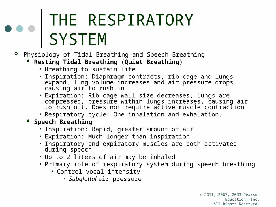
THE RESPIRATORY SYSTEM
Physiology of Tidal Breathing and Speech Breathing Resting Tidal Breathing (Quiet Breathing)
• Breathing to sustain life• Inspiration: Diaphragm contracts, rib cage and lungs expand, lung
volume increases and air pressure drops, causing air to rush in • Expiration: Rib cage wall size decreases, lungs are compressed,
pressure within lungs increases, causing air to rush out. Does not require active muscle contraction
• Respiratory cycle: One inhalation and exhalation. Speech Breathing
• Inspiration: Rapid, greater amount of air• Expiration: Much longer than inspiration• Inspiratory and expiratory muscles are both activated during speech• Up to 2 liters of air may be inhaled• Primary role of respiratory system during speech breathing
• Control vocal intensity• Subglottal air pressure

© 2011, 2007, 2003 Pearson Education, Inc.All Rights Reserved.
THE RESPIRATORY SYSTEM Lifespan Issues of the Respiratory System
Resting tidal breathing rate decreased from birth to adulthood
• More alveoli Maximum lung capacity reached in early adulthood
• Remains constant until middle age Respiratory function affected by exercise, health, and
smoking

THE RESPIRATORY SYSTEM
What kind of deficits may we have if there is an impaired respiratory system?
How do we treat this?

© 2011, 2007, 2003 Pearson Education, Inc.All Rights Reserved.
THE LARYNGEAL SYSTEM
The Larynx—organ of the laryngeal system An air valve Main sound generator for speech production Composed of
• Cartilages• Muscle • Other tissue
Biological functions Protects against foreign objects entering trachea and
lungs Coughing Closes airway during swallowing Closes airway during physical exertion
• Lifting heavy objects

© 2011, 2007, 2003 Pearson Education, Inc.All Rights Reserved.
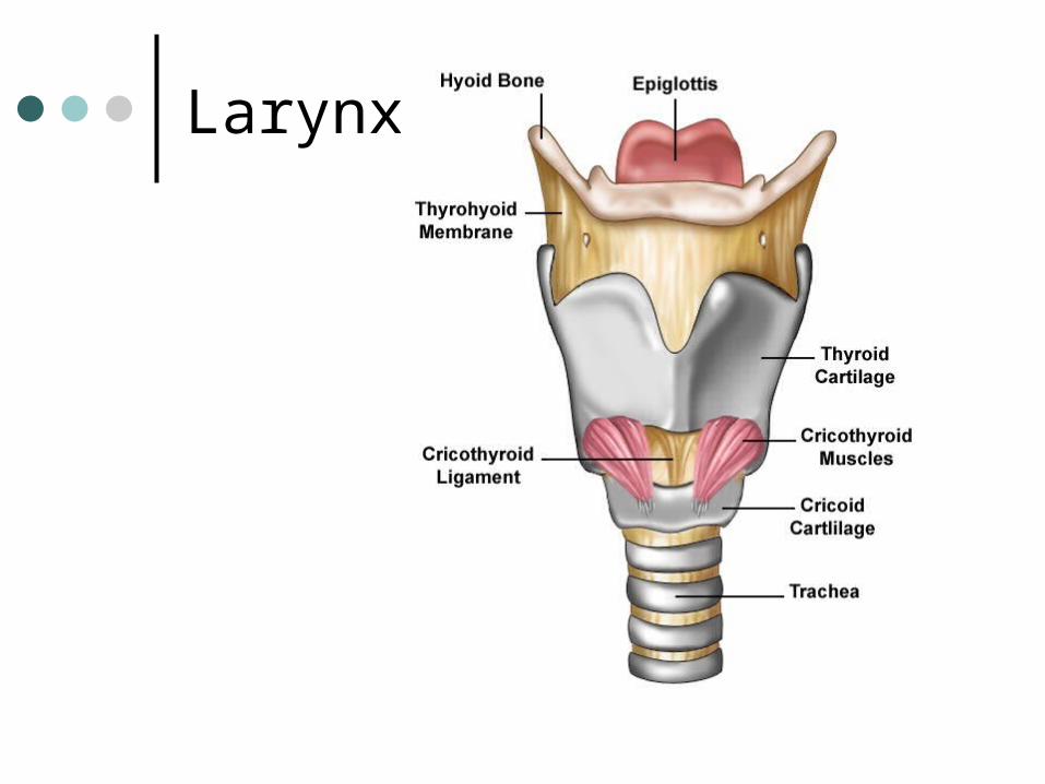
THE LARYNGEAL SYSTEM Structures of the Laryngeal System
Hyoid bone: Horseshoe shaped bone that serves as a main attachment for laryngeal and tongue musculature. Positioned at the top of the larynx and not connected to any other bones.
Cartilages• Thyroid cartilage: Largest laryngeal cartilage. Forms the front and sides
of the laryngeal skeleton. The thyroid prominence is a protrusion just below the thyroid notch. It has two sets of horns, the superior cornua (connect to the hyoid bone), and the inferior cornua (connect to cricoid cartilage).
• Cricoid cartilage: Ring-shaped. Lower aspect of the laryngeal skeleton.• Arytenoid cartilages: Pyramidal-shaped. The base of each has a vocal
process and a muscular process. • Epiglottis: Leaf-shaped cartilage attached to the thyroid cartilage and
hyoid bone.
Muscles of the Larynx• Intrinsic: Critical for phonation and modifying pitch and loudness.
• Posterior cricoarytenoid, lateral cricoarytenoid, interarytenoid, thyroarytenoid, cricothyroid
• Extrinsic: Support and stabilize the larynx.• Sternothyroid, thyrohyoid, inferior constrictor muscles
• Supplementary: Assist in laryngeal elevation (suprahyoid) or depression (infrahyoid).
Larynx

Lateral View

http://greenfield.fortunecity.com/rattler/46/upali4.htm

Vocal Folds
Vocal Folds Front attachment: Midline of the thyroid cartilage Back attachment: Vocal processes of the arytenoid
cartilages via the vocal ligament. Abduct (move apart) during respiration and adduct
(move together) during phonation.
Five layers: • Epithelial tissue, three layers of lamina propria,
and the thyroarytenoid muscle. The body consists of the muscle and deepest layer of lamina propria. The cover is the intermediate and superficial layers of lamina propria and epithelium
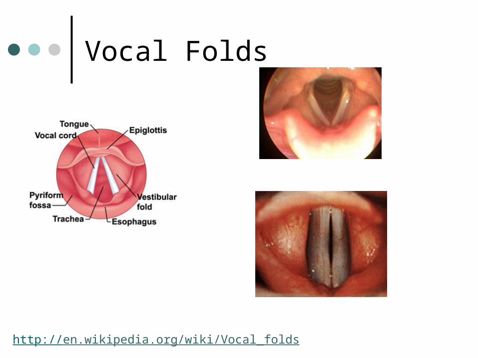
Vocal Folds
http://en.wikipedia.org/wiki/Vocal_folds

© 2011, 2007, 2003 Pearson Education, Inc.All Rights Reserved.
THE LARYNGEAL SYSTEM
Lifespan Issues of the Laryngeal System Newborns
• Larynx small and high in the neck 10-20 years of age
• Larynx reaches final position Laryngeal cartilages become less pliable with age Vocal folds increase in length differentially for males
and females• Become less flexible with age
Laryngeal System
What kind of deficits may exist if there is an impaired laryngeal system?
How do we treat this?

© 2011, 2007, 2003 Pearson Education, Inc.All Rights Reserved.
ARTICULATORY/RESONATING SYSTEM
Composed of Oral cavity Nasal cavity Pharyngeal cavity (vocal tract)
Vocal tract Acoustic tube that shapes sound energy produced by
respiratory and laryngeal systems into speech sounds

© 2011, 2007, 2003 Pearson Education, Inc.All Rights Reserved.
ARTICULATORY/RESONATING SYSTEM
Structures of the Articulatory/Resonating System Facial skeleton and cranium (22 bones) The mandible articulates with the temporal bone by the temporomandibular joint. Teeth
• Adults have 32, in alveolar processes of the mandible and maxilla (hard palate composed of bone of maxilla)
Tongue• Muscular hydrostat• Five components: Body, root, dorsum, blade, tongue tip• Intrinsic muscles: Superior longitudinal, inferior longitudinal, vertical,
transverse• Extrinsic muscles: Styloglossus, palatoglossus, hyoglossus, genioglossus
Velum• Also called the soft palate• Uvula- termination of the velum• Velopharyngeal closure: Contact of the velum with the lateral and posterior
pharyngeal walls.• Velar elevation is necessary to prevent air escaping through the nose and to
build up air pressure for production of pressure sounds.

© 2011, 2007, 2003 Pearson Education, Inc.All Rights Reserved.
ARTICULATORY/RESONATING SYSTEM


© 2011, 2007, 2003 Pearson Education, Inc.All Rights Reserved.
ARTICULATORY/RESONATING SYSTEM
Lifespan Issues of the Articulatory/Resonating System Bones of the skull reach adult size by 8 years Newborns have 45 separate skull bones that fuse into 22 at
adulthood Lower facial bones reach adult size at 18 years Dentition emerges around 6 months and is complete around 3
years Secondary dentition complete around 18 years Newborn’s tongue occupies most of the oral cavity Tongue reaches adult size around 16 years Length and volume of the oral cavity increases throughout
development• Changes the overall resonant characteristics

ARTICULATORY/RESONATING SYSTEM
What kind of deficits may exist if there is an impaired articulatory/resonating system?
How do we treat this?
© 2011, 2007, 2003 Pearson Education, Inc.All Rights Reserved.
THE SPEECH PRODUCTION PROCESS
Begins with phonation Tracheal/alveolar pressure
Air pressure beneath adducted vocal folds Fundamental frequency: The number of cycles of vocal
fold vibration per second. Movement of the tongue, lips, and larynx change the
shape of the vocal tract and modify sound

SPEECH PRODUCTION PROCESS
Phonation1) vocal folds are adducted2) subglottic air pressure builds3) vocal fold tissue is displaced upward and sideward4) air rushes through the opening, increasing in velocity5) negative air pressure results and pulls vocal folds
together• Natural elasticity of vocal folds helps return to
original position• Contraction of adductor intrinsic laryngeal muscles

VIBRATORY CYCLE

CHANGES TO THE SPEECH MECHANISM Anatomical and physical changes impact
the way speech is produced Tidal breathing rate decreases in the first few
years• Respiratory system’s structures increase in size
and lung capacities increase Position and size of larynx changes
• Changes in vocal folds during puberty Increase in length and volume of oral cavity
impacts the resonance properties of vocal tract as one ages

Nervous System Divisions
Central nervous system (CNS) Brain and spinal cord
Peripheral nervous system (PNS) Nerves that emerge from the brain and the spinal cord to
innervate the rest of the body Innervate: supply of nerves to a particular region or part of
the body Cranial nerves: emerge from brain; 12 pairs Spinal nerves: emerge from spinal cord; 31 pairs Cranial and spinal nerves carry information back and forth
between brain, spine, and rest of body Sensory information carried to the brain via afferent
pathways Motor information carried away from brain via efferent
pathways

Directional and Positional Terms
Proximal: relatively close to a site of reference
Distal: relatively far from a site of reference Anterior: toward the front Posterior: toward the back Superior: toward the top Inferior: toward the bottom External: toward the outside Internal: toward the inside

© 2011, 2007, 2003 Pearson Education, Inc.All Rights Reserved.
THE CENTRAL NERVOUS SYSTEM
CNS: Composed of Brain and Spinal Cord Brain consists of Brainstem, cerebellum, and cerebrum
Brain- chief operator of CNS function Protective Shield- Bone = Skull Cerebrum: Left and right hemispheres
• Motor and sensory functions are contralateral.• Each hemisphere consists of white matter pathways and gray cortical matter.• The cortex has gyri (hills) and sulci/fissures (valleys).• Each hemisphere has four lobes: Frontal, temporal, parietal, and occipital.
Hemispheric Asymmetry• The left hemisphere is dominant for speech and language in 98% of people.• The primary area of asymmetry is in the left temporal lobe.• Left hemisphere language dominance demonstrated as young as age seven.
Subcortical and Lower Brain Structures• Thalamus: Relay stations for incoming and outgoing information. • Basal ganglia: Large subcortical nuclei that regulate motor functioning and
maintain posture and muscle tone. There are direct and indirect pathways.• Brainstem: Composed of the midbrain, pons, and medulla. Important for
regulatory functions, processing information, and contains white matter tracts. • Cerebellum: Left and right hemispheres and vermis. Coordinates fine motor
control, complex motor activities, muscle tone, and participates in motor learning.

CNS continued
Spinal Cord• Protective Shield is the vertebral column• Neuronal cell bodies protected by a myelin
sheath. • Receives sensory info and contains motor
neurons supplying muscles
• Cerebrospinal fluid (CSF): circulates between innermost two layers of meninges; carries chemicals important to metabolic processes and serves as important buffer for any jolts to CNS.
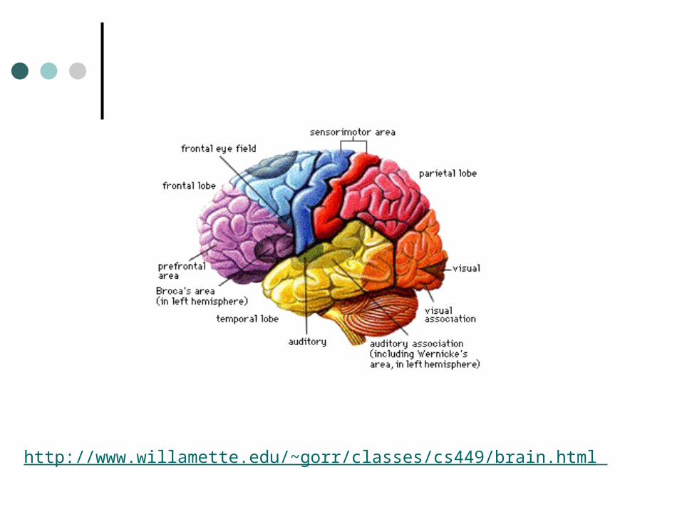
http://www.willamette.edu/~gorr/classes/cs449/brain.html
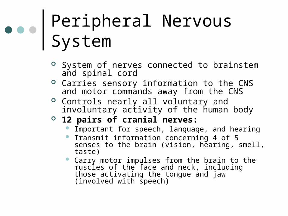
Peripheral Nervous System
System of nerves connected to brainstem and spinal cord
Carries sensory information to the CNS and motor commands away from the CNS
Controls nearly all voluntary and involuntary activity of the human body
12 pairs of cranial nerves: Important for speech, language, and hearing Transmit information concerning 4 of 5 senses to the brain
(vision, hearing, smell, taste) Carry motor impulses from the brain to the muscles of the
face and neck, including those activating the tongue and jaw (involved with speech)
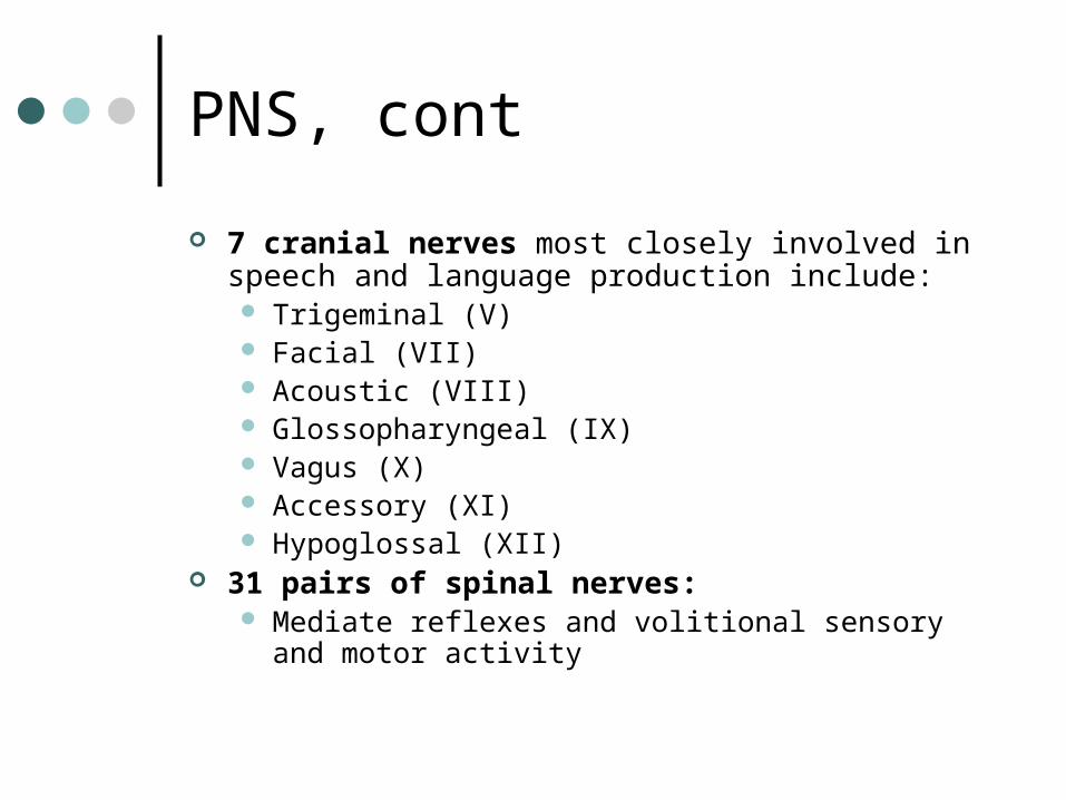
PNS, cont
7 cranial nerves most closely involved in speech and language production include: Trigeminal (V) Facial (VII) Acoustic (VIII) Glossopharyngeal (IX) Vagus (X) Accessory (XI) Hypoglossal (XII)
31 pairs of spinal nerves: Mediate reflexes and volitional sensory and motor
activity

© 2011, 2007, 2003 Pearson Education, Inc.All Rights Reserved.
MOTOR SPEECH CONTROL Motor speech production process
Movement plan/program retrieved from memory Sent to motor control areas Transmitted to muscles and structures of the speech
mechanism Nerve impulses modified throughout the process to
ensure precise, smooth muscle movements Internal and external sensory information allows
monitoring and modification of movements

© 2011, 2007, 2003 Pearson Education, Inc.All Rights Reserved.
ANATOMY AND PHYSIOLOGY OF THE AUDITORY SYSTEM The Outer Ear
Pinna• Enhances sound• Aids localization
External auditory meatus• Elliptical tube lined with skin• Glands that produce cerumen• Resonator

© 2011, 2007, 2003 Pearson Education, Inc.All Rights Reserved.
ANATOMY AND PHYSIOLOGY OF THE AUDITORY SYSTEM
The Middle Ear Tympanic membrane
• Vibrates in response to sound• 3 layers
Middle ear space (tympanic cavity)• Air-filled, lined with mucous membranes, housed in temporal
bone Eustachian tube
• Connects middle ear with nasopharynx Ossicles in ossicular chain
• Malleus, incus, stapes
© 2011, 2007, 2003 Pearson Education, Inc.All Rights Reserved.
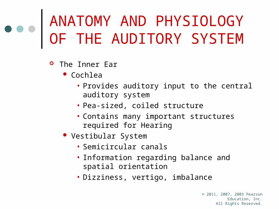
ANATOMY AND PHYSIOLOGY OF THE AUDITORY SYSTEM The Inner Ear
Cochlea• Provides auditory input to the central auditory
system• Pea-sized, coiled structure• Contains many important structures required for
Hearing Vestibular System
• Semicircular canals• Information regarding balance and spatial
orientation• Dizziness, vertigo, imbalance

Anatomy of the Ear