Embed Size (px)
Citation preview
AHA Scientific Statement
1
Background—Acute rheumatic fever remains a serious healthcare concern for the majority of the world’s population despite its decline in incidence in Europe and North America. The goal of this statement was to review the historic Jones criteria used to diagnose acute rheumatic fever in the context of the current epidemiology of the disease and to update those criteria to also take into account recent evidence supporting the use of Doppler echocardiography in the diagnosis of carditis as a major manifestation of acute rheumatic fever.
Methods and Results—To achieve this goal, the American Heart Association’s Council on Cardiovascular Disease in the Young and its Rheumatic Fever, Endocarditis, and Kawasaki Disease Committee organized a writing group to comprehensively review and evaluate the impact of population-specific differences in acute rheumatic fever presentation and changes in presentation that can result from the now worldwide availability of nonsteroidal anti-inflammatory drugs. In addition, a methodological assessment of the numerous published studies that support the use of Doppler echocardiography as a means to diagnose cardiac involvement in acute rheumatic fever, even when overt clinical findings are not apparent, was undertaken to determine the evidence basis for defining subclinical carditis and including it as a major criterion of the Jones criteria. This effort has resulted in the first substantial revision to the Jones criteria by the American Heart Association since 1992 and the first application of the Classification of Recommendations and Levels of Evidence categories developed by the American College of Cardiology/American Heart Association to the Jones criteria.
Conclusions—This revision of the Jones criteria now brings them into closer alignment with other international guidelines for the diagnosis of acute rheumatic fever by defining high-risk populations, recognizing variability in clinical presentation in these high-risk populations, and including Doppler echocardiography as a tool to diagnose cardiac involvement. (Circulation. 2015;131:000-000. DOI: 10.1161/CIR.0000000000000205.)
Key Words: AHA Scientific Statements ◼ acute rheumatic fever ◼ Doppler echocardiography ◼ Jones criteria ◼ rheumatic heart disease ◼ subclinical carditis
© 2015 American Heart Association, Inc.
Circulation is available at http://circ.ahajournals.org DOI: 10.1161/CIR.0000000000000205
The American Heart Association makes every effort to avoid any actual or potential conflicts of interest that may arise as a result of an outside relationship or a personal, professional, or business interest of a member of the writing panel. Specifically, all members of the writing group are required to complete and submit a Disclosure Questionnaire showing all such relationships that might be perceived as real or potential conflicts of interest.
This statement was approved by the American Heart Association Science Advisory and Coordinating Committee on January 28, 2015, and the American Heart Association Executive Committee on March 9, 2015. A copy of the document is available at http://my.americanheart.org/statements by selecting either the “By Topic” link or the “By Publication Date” link. To purchase additional reprints, call 843-216-2533 or e-mail [email protected].
The American Heart Association requests that this document be cited as follows: Gewitz MH, Baltimore RS, Tani LY, Sable CA, Shulman ST, Carapetis J, Remenyi B, Taubert KA, Bolger AF, Beerman L, Mayosi BM, Beaton A, Pandian NG, Kaplan EL; on behalf of the American Heart Association Committee on Rheumatic Fever, Endocarditis, and Kawasaki Disease of the Council on Cardiovascular Disease in the Young. Revision of the Jones criteria for the diagnosis of acute rheumatic fever in the era of Doppler echocardiography: a scientific statement from the American Heart Association. Circulation. 2015;131:•••–•••.
Expert peer review of AHA Scientific Statements is conducted by the AHA Office of Science Operations. For more on AHA statements and guidelines development, visit http://my.americanheart.org/statements and select the “Policies and Development” link.
Permissions: Multiple copies, modification, alteration, enhancement, and/or distribution of this document are not permitted without the express permission of the American Heart Association. Instructions for obtaining permission are located at http://www.heart.org/HEARTORG/General/Copyright-Permission-Guidelines_UCM_300404_Article.jsp. A link to the “Copyright Permissions Request Form” appears on the right side of the page.
Revision of the Jones Criteria for the Diagnosis of Acute Rheumatic Fever in the Era of Doppler Echocardiography
A Scientific Statement From the American Heart Association
Endorsed by the World Heart Federation
Michael H. Gewitz, MD, FAHA, Co-Chair; Robert S. Baltimore, MD, Co-Chair; Lloyd Y. Tani, MD, FAHA; Craig A. Sable, MD, FAHA; Stanford T. Shulman, MD; Jonathan Carapetis, MBBS; Bo Remenyi, MBBS; Kathryn A. Taubert, PhD, FAHA;
Ann F. Bolger, MD, FAHA; Lee Beerman, MD; Bongani M. Mayosi, MBChB; Andrea Beaton, MD; Natesa G. Pandian, MD; Edward L. Kaplan, MD, FAHA; on behalf of the American Heart
Association Committee on Rheumatic Fever, Endocarditis, and Kawasaki Disease of the Council on Cardiovascular Disease in the Young
at FMRP SKANFO INC on May 15, 2015http://circ.ahajournals.org/Downloaded from
2 Circulation May 19, 2015
Although acute rheumatic fever (ARF) has declined in Europe and North America in incidence over the past
4 to 6 decades, the disease remains one of the most impor-tant causes of cardiovascular morbidity and mortality among socially and economically disadvantaged populations all over the world, especially in the developing countries that are home to the majority of the world’s population. Incidence rates in these countries still reach epidemic levels.1 The Jones crite-ria, used for guidance in the diagnosis of ARF since 1944, were last modified by the American Heart Association (AHA) in 1992.2 They were reconfirmed in principle at an AHA-sponsored workshop in 20003 and historically have
represented the clinical standard to establish the diagnosis of ARF. However, in the past few years, developments in several areas have prompted reexamination of the traditional Jones criteria. For example, the limited diagnostic role for echocar-diography in the diagnosis of carditis as expressed in the Jones criteria revision of 19922 is a major area of focus. This posi-tion may no longer be appropriate, because echocardiographic techniques and applications, including quantitative Doppler and color flow mapping, have evolved worldwide during the past 2 decades. Other national and regional guidelines for the diagnosis of ARF have recently included the use of echocar-diography/Doppler methodologies.4,5 Numerous studies from
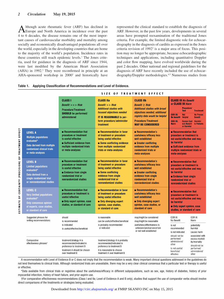
Table 1. Applying Classification of Recommendations and Level of Evidence.
A recommendation with Level of Evidence B or C does not imply that the recommendation is weak. Many important clinical questions addressed in the guidelines do not lend themselves to clinical trials. Although randomized trials are unavailable, there may be a very clear clinical consensus that a particular test or therapy is useful or effective.
*Data available from clinical trials or registries about the usefulness/efficacy in different subpopulations, such as sex, age, history of diabetes, history of prior myocardial infarction, history of heart failure, and prior aspirin use.
†For comparative effectiveness recommendations (Class I and IIa; Level of Evidence A and B only), studies that support the use of comparator verbs should involve direct comparisons of the treatments or strategies being evaluated.
at FMRP SKANFO INC on May 15, 2015http://circ.ahajournals.org/Downloaded from
Gewitz et al Revised Jones Criteria for Acute Rheumatic Fever 3
a broad range of clinical circumstances have suggested that there be more widespread use of echocardiography as a way to diagnose carditis even in the absence of overt clinical findings (“subclinical carditis”).6–30 Furthermore, echocardiography has become a cornerstone in worldwide screening programs to evaluate the prevalence of rheumatic heart disease (RHD).31–35
In addition to consideration of the proper role of echocar-diography in ARF, issues have been raised regarding other clinical areas. For example, whereas in the 1992 version of the Jones criteria,2 monoarticular arthritis was offered for con-sideration when a patient had been treated with nonsteroidal anti-inflammatory drugs before diagnosis, evidence has been published since then that indicates that in selective high-risk populations, monoarticular arthritis may be an indicator of the major manifestation of arthritis.36 Furthermore, previous AHA ARF guidelines did not categorize recommendations using the currently favored Classification of Recommendations and Levels of Evidence categories. The writing group was charged with the task of performing an assessment of the evidence and assigning a Classification of Recommendation according to the American College of Cardiology/AHA classification system.37 The Classification of Recommendations is an estimate of the size of the treatment effect that considers risks versus benefits in addition to evidence and/or agreement that a given treatment or procedure is or is not useful/effective or, alternatively, may cause harm. The Level of Evidence is an estimate of the cer-tainty or precision of the treatment effect. The writing group reviewed and ranked evidence supporting each recommenda-tion, with the weight of evidence ranked as Level of Evidence A, B, or C according to specific definitions that are included in Table 1. For conditions for which inadequate data are available, recommendations are based on expert consensus and clinical experience and are ranked as Level of Evidence C. This system also provides suggested phrases for writing recommendations within each Classification of Recommendations.
Finally, recent perspectives regarding the diagnosis of acute streptococcal pharyngitis itself, as reviewed in the AHA scientific statement of 2009,38 need to be referenced as part of the discus-sion regarding in whom the diagnosis of ARF can be established.
As with past AHA statements concerning the Jones criteria, this revision focuses on the diagnosis of ARF and not on issues concerning the surveillance for and diagnosis of chronic RHD or its consequences.
Epidemiological BackgroundInsight into how to best define the appropriate application of diagnostic criteria for ARF within a given population requires a brief review of the current epidemiology of ARF.
It is well established that during the 20th century, the inci-dence of ARF and the prevalence of RHD declined substan-tially in Europe, North America, and developed nations in other geographic locations.39,40 This decline has been attributed to improved hygiene, improved access to antibiotic drugs and medical care, reduced household crowding, and other social and economic changes.39,41 Changes in the epidemiology of specific group A streptococcal strains that cause infections may also have played a role.42 Although sporadic cases of ARF continue to be seen in affluent nations, the major burden is currently found in low- and middle-income countries and in selected indigenous
populations elsewhere. The pattern of disease in the high-prev-alence regions is often hyperendemic, with cases occurring throughout the year and a virtual absence of outbreaks. This is in contrast to high-income settings, which experience a low back-ground incidence of ARF with periodic outbreaks.28,43
There is also evidence of differences in incidence even in pop-ulations within the same country, which further demonstrates the disproportional disease burden. For example, although the overall mean incidence of ARF in New Zealand rose by 55% over the past 2 decades, the incidence of ARF among the non-Maori/Pacific New Zealand populations declined by 70% over the same period.44 Similar discrepancies in disease burden exist in Australia, where the indigenous population experiences one of the world’s highest reported incidences of ARF at 153 to 380 cases per 100 000 people per year in the 5- to 14-year-old age group,45 whereas in other Australian populations, the incidence approximates European and North American levels.
In summary, the global distribution of ARF/RHD is clearly disproportionate. Certain geographic regions and specific ethnic and socioeconomic groups experience very high rates of ARF incidence, whereas in other regions, the disease has virtually disappeared. This has led to concern regarding the uniform sensitivity of the Jones criteria, even as revised over the years, when applied to geographic areas or to populations within those areas, or elsewhere, where ARF is hyperendemic.
Implications of Epidemiological ConsiderationsBecause the clinical utility of a diagnostic test is determined by a number of factors, including its pretest probability and back-ground disease prevalence, and in view of the heterogeneity in global disease burden noted above, a single set of diagnostic criteria may no longer be sufficient for all population groups and in all geographic regions. To avoid overdiagnosis in low-incidence populations and to avoid underdiagnosis in high-risk populations, variability in applying diagnostic criteria in low-risk compared with high-risk populations is reasonable, as has been promulgated by the Australian rheumatic fever guidelines.4 The epidemiological data appear to indicate the following:
1. It is reasonable to consider individuals to be at low risk for ARF if they come from a setting or popula-tion known to experience low rates of ARF or RHD (Class IIa; Level of Evidence C).
2. It is reasonable that where reliable epidemiologi-cal data are available, low risk should be defined as having an ARF incidence <2 per 100 000 school-aged children (usually 5–14 years old) per year or an all-age prevalence of RHD of ≤1 per 1000 population per year (Class IIa; Level of Evidence C).
3. Children not clearly from a low-risk population are at moderate to high risk depending on their reference population (Class I; Level of Evidence C).
Clinical Manifestations of ARFGenerally, the clinical profile of ARF in low- and middle-income countries closely resembles that of high-income countries.46–48 Universally, the most common major manifestations during the first episode of ARF (the “major criteria” for diagnosis) remain carditis (50%–70%) and arthritis (35%–66%).1,9,28,46–48 These
at FMRP SKANFO INC on May 15, 2015http://circ.ahajournals.org/Downloaded from
4 Circulation May 19, 2015
are followed in frequency by chorea (10%–30%), which has been demonstrated to have a female predominance, and then subcutaneous nodules (0%–10%) and erythema marginatum (<6%), which remain much less common but highly specific manifestations of ARF.9,46–48 Despite this general consistency for each of the classic major manifestations, recent data have suggested the possibility of substantial variability of manifes-tations in specific circumstances and populations.
For example, in very high-risk populations, such as the indigenous Australian population, variability in typical Jones criteria manifestations has been described.9,36,45 As discussed below, these include presentations with aseptic monoarthri-tis, polyarthralgia, and low-grade (as opposed to traditionally considered high-grade) fevers. These variable manifestations were reinforced in the 2012 Australian criteria4 to increase the sensitivity of diagnosis in patients from those specific high-risk populations. To date, however, the applicability of these variable clinical manifestations in low-risk populations has not been tested and is not recommended.
In general, it remains standard practice to maintain continu-ing vigilance in the application of the clinical manifestations for the diagnosis of ARF. Ongoing reassessment of evolv-ing clinical information is important in any specific patient, because there always has been the potential for “diagnosis overlap” in application of the Jones criteria. In addition to the above, much attention has also been focused on the appro-priate role of noninvasive cardiac imaging, namely, echocar-diography combined with Doppler flow assessment, in the diagnosis of carditis in ARF.
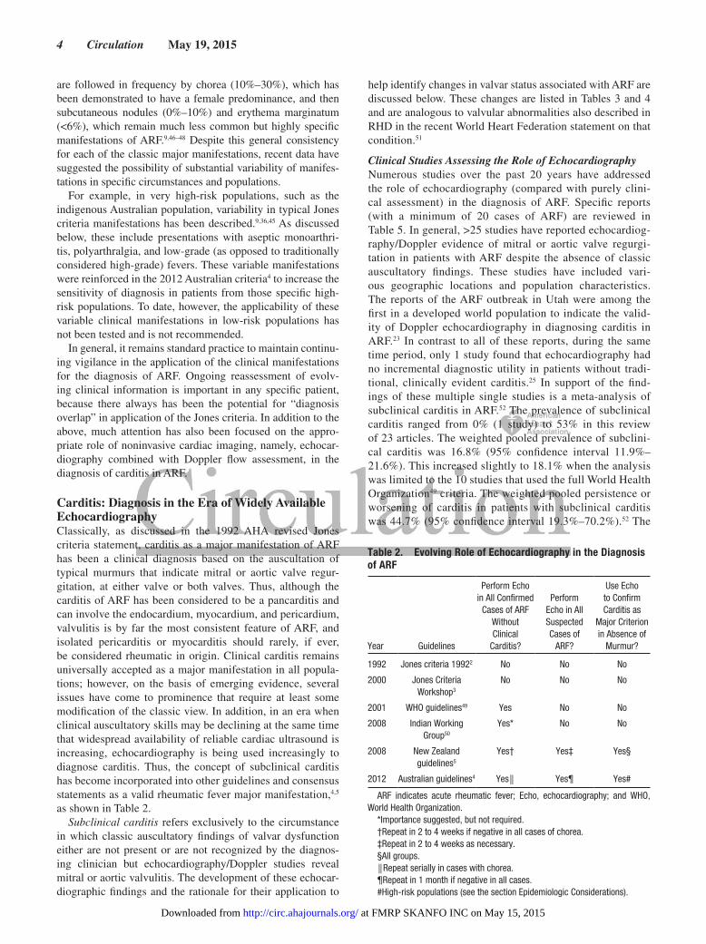
Carditis: Diagnosis in the Era of Widely Available EchocardiographyClassically, as discussed in the 1992 AHA revised Jones criteria statement, carditis as a major manifestation of ARF has been a clinical diagnosis based on the auscultation of typical murmurs that indicate mitral or aortic valve regur-gitation, at either valve or both valves. Thus, although the carditis of ARF has been considered to be a pancarditis and can involve the endocardium, myocardium, and pericardium, valvulitis is by far the most consistent feature of ARF, and isolated pericarditis or myocarditis should rarely, if ever, be considered rheumatic in origin. Clinical carditis remains universally accepted as a major manifestation in all popula-tions; however, on the basis of emerging evidence, several issues have come to prominence that require at least some modification of the classic view. In addition, in an era when clinical auscultatory skills may be declining at the same time that widespread availability of reliable cardiac ultrasound is increasing, echocardiography is being used increasingly to diagnose carditis. Thus, the concept of subclinical carditis has become incorporated into other guidelines and consensus statements as a valid rheumatic fever major manifestation,4,5 as shown in Table 2.
Subclinical carditis refers exclusively to the circumstance in which classic auscultatory findings of valvar dysfunction either are not present or are not recognized by the diagnos-ing clinician but echocardiography/Doppler studies reveal mitral or aortic valvulitis. The development of these echocar-diographic findings and the rationale for their application to
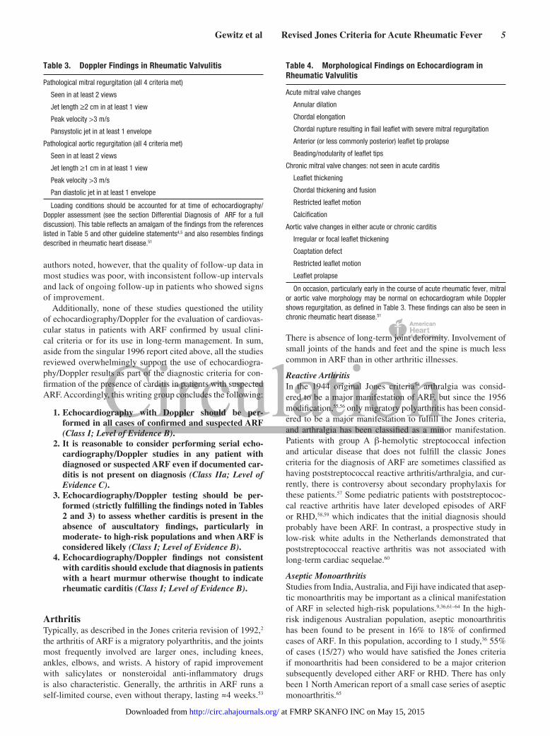
help identify changes in valvar status associated with ARF are discussed below. These changes are listed in Tables 3 and 4 and are analogous to valvular abnormalities also described in RHD in the recent World Heart Federation statement on that condition.51
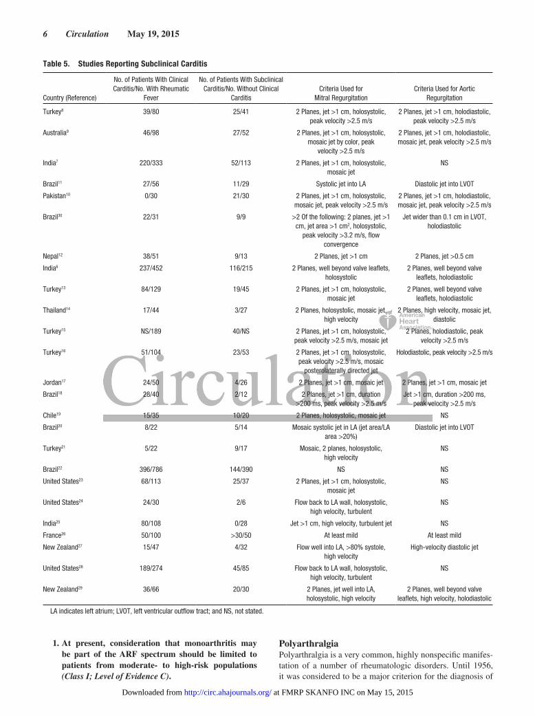
Clinical Studies Assessing the Role of EchocardiographyNumerous studies over the past 20 years have addressed the role of echocardiography (compared with purely clini-cal assessment) in the diagnosis of ARF. Specific reports (with a minimum of 20 cases of ARF) are reviewed in Table 5. In general, >25 studies have reported echocardiog-raphy/Doppler evidence of mitral or aortic valve regurgi-tation in patients with ARF despite the absence of classic auscultatory findings. These studies have included vari-ous geographic locations and population characteristics. The reports of the ARF outbreak in Utah were among the first in a developed world population to indicate the valid-ity of Doppler echocardiography in diagnosing carditis in ARF.23 In contrast to all of these reports, during the same time period, only 1 study found that echocardiography had no incremental diagnostic utility in patients without tradi-tional, clinically evident carditis.25 In support of the find-ings of these multiple single studies is a meta-analysis of subclinical carditis in ARF.52 The prevalence of subclinical carditis ranged from 0% (1 study) to 53% in this review of 23 articles. The weighted pooled prevalence of subclini-cal carditis was 16.8% (95% confidence interval 11.9%–21.6%). This increased slightly to 18.1% when the analysis was limited to the 10 studies that used the full World Health Organization49 criteria. The weighted pooled persistence or worsening of carditis in patients with subclinical carditis was 44.7% (95% confidence interval 19.3%–70.2%).52 The
Table 2. Evolving Role of Echocardiography in the Diagnosis of ARF
Year Guidelines
Perform Echo in All Confirmed
Cases of ARF Without Clinical
Carditis?
Perform Echo in All Suspected Cases of
ARF?
Use Echo to Confirm Carditis as
Major Criterion in Absence of
Murmur?
1992 Jones criteria 19922 No No No
2000 Jones Criteria Workshop3
No No No
2001 WHO guidelines49 Yes No No
2008 Indian Working Group50
Yes* No No
2008 New Zealand guidelines5
Yes† Yes‡ Yes§
2012 Australian guidelines4 Yes║ Yes¶ Yes#
ARF indicates acute rheumatic fever; Echo, echocardiography; and WHO, World Health Organization.
*Importance suggested, but not required.†Repeat in 2 to 4 weeks if negative in all cases of chorea.‡Repeat in 2 to 4 weeks as necessary.§All groups.║Repeat serially in cases with chorea.¶Repeat in 1 month if negative in all cases.#High-risk populations (see the section Epidemiologic Considerations).
at FMRP SKANFO INC on May 15, 2015http://circ.ahajournals.org/Downloaded from
Gewitz et al Revised Jones Criteria for Acute Rheumatic Fever 5
authors noted, however, that the quality of follow-up data in most studies was poor, with inconsistent follow-up intervals and lack of ongoing follow-up in patients who showed signs of improvement.
Additionally, none of these studies questioned the utility of echocardiography/Doppler for the evaluation of cardiovas-cular status in patients with ARF confirmed by usual clini-cal criteria or for its use in long-term management. In sum, aside from the singular 1996 report cited above, all the studies reviewed overwhelmingly support the use of echocardiogra-phy/Doppler results as part of the diagnostic criteria for con-firmation of the presence of carditis in patients with suspected ARF. Accordingly, this writing group concludes the following:
1. Echocardiography with Doppler should be per-formed in all cases of confirmed and suspected ARF (Class I; Level of Evidence B).
2. It is reasonable to consider performing serial echo-cardiography/Doppler studies in any patient with diagnosed or suspected ARF even if documented car-ditis is not present on diagnosis (Class IIa; Level of Evidence C).
3. Echocardiography/Doppler testing should be per-formed (strictly fulfilling the findings noted in Tables 2 and 3) to assess whether carditis is present in the absence of auscultatory findings, particularly in moderate- to high-risk populations and when ARF is considered likely (Class I; Level of Evidence B).
4. Echocardiography/Doppler findings not consistent with carditis should exclude that diagnosis in patients with a heart murmur otherwise thought to indicate rheumatic carditis (Class I; Level of Evidence B).
ArthritisTypically, as described in the Jones criteria revision of 1992,2 the arthritis of ARF is a migratory polyarthritis, and the joints most frequently involved are larger ones, including knees, ankles, elbows, and wrists. A history of rapid improvement with salicylates or nonsteroidal anti-inflammatory drugs is also characteristic. Generally, the arthritis in ARF runs a self-limited course, even without therapy, lasting ≈4 weeks.53
There is absence of long-term joint deformity. Involvement of small joints of the hands and feet and the spine is much less common in ARF than in other arthritic illnesses.
Reactive ArthritisIn the 1944 original Jones criteria54 arthralgia was consid-ered to be a major manifestation of ARF, but since the 1956 modification,55,56 only migratory polyarthritis has been consid-ered to be a major manifestation to fulfill the Jones criteria, and arthralgia has been classified as a minor manifestation. Patients with group A β-hemolytic streptococcal infection and articular disease that does not fulfill the classic Jones criteria for the diagnosis of ARF are sometimes classified as having poststreptococcal reactive arthritis/arthralgia, and cur-rently, there is controversy about secondary prophylaxis for these patients.57 Some pediatric patients with poststreptococ-cal reactive arthritis have later developed episodes of ARF or RHD,58,59 which indicates that the initial diagnosis should probably have been ARF. In contrast, a prospective study in low-risk white adults in the Netherlands demonstrated that poststreptococcal reactive arthritis was not associated with long-term cardiac sequelae.60
Aseptic MonoarthritisStudies from India, Australia, and Fiji have indicated that asep-tic monoarthritis may be important as a clinical manifestation of ARF in selected high-risk populations.9,36,61–64 In the high-risk indigenous Australian population, aseptic monoarthritis has been found to be present in 16% to 18% of confirmed cases of ARF. In this population, according to 1 study,36 55% of cases (15/27) who would have satisfied the Jones criteria if monoarthritis had been considered to be a major criterion subsequently developed either ARF or RHD. There has only been 1 North American report of a small case series of aseptic monoarthritis.65
Table 3. Doppler Findings in Rheumatic Valvulitis
Pathological mitral regurgitation (all 4 criteria met)
Seen in at least 2 views
Jet length ≥2 cm in at least 1 view
Peak velocity >3 m/s
Pansystolic jet in at least 1 envelope
Pathological aortic regurgitation (all 4 criteria met)
Seen in at least 2 views
Jet length ≥1 cm in at least 1 view
Peak velocity >3 m/s
Pan diastolic jet in at least 1 envelope
Loading conditions should be accounted for at time of echocardiography/Doppler assessment (see the section Differential Diagnosis of ARF for a full discussion). This table reflects an amalgam of the findings from the references listed in Table 5 and other guideline statements4,5 and also resembles findings described in rheumatic heart disease.51
Table 4. Morphological Findings on Echocardiogram in Rheumatic Valvulitis
Acute mitral valve changes
Annular dilation
Chordal elongation
Chordal rupture resulting in flail leaflet with severe mitral regurgitation
Anterior (or less commonly posterior) leaflet tip prolapse
Beading/nodularity of leaflet tips
Chronic mitral valve changes: not seen in acute carditis
Leaflet thickening
Chordal thickening and fusion
Restricted leaflet motion
Calcification
Aortic valve changes in either acute or chronic carditis
Irregular or focal leaflet thickening
Coaptation defect
Restricted leaflet motion
Leaflet prolapse
On occasion, particularly early in the course of acute rheumatic fever, mitral or aortic valve morphology may be normal on echocardiogram while Doppler shows regurgitation, as defined in Table 3. These findings can also be seen in chronic rheumatic heart disease.51
at FMRP SKANFO INC on May 15, 2015http://circ.ahajournals.org/Downloaded from
6 Circulation May 19, 2015
1. At present, consideration that monoarthritis may be part of the ARF spectrum should be limited to patients from moderate- to high-risk populations (Class I; Level of Evidence C).
PolyarthralgiaPolyarthralgia is a very common, highly nonspecific manifes-tation of a number of rheumatologic disorders. Until 1956, it was considered to be a major criterion for the diagnosis of
Table 5. Studies Reporting Subclinical Carditis
Country (Reference)
No. of Patients With Clinical Carditis/No. With Rheumatic
Fever
No. of Patients With Subclinical Carditis/No. Without Clinical
CarditisCriteria Used for
Mitral RegurgitationCriteria Used for Aortic
Regurgitation
Turkey8 39/80 25/41 2 Planes, jet >1 cm, holosystolic, peak velocity >2.5 m/s
2 Planes, jet >1 cm, holodiastolic, peak velocity >2.5 m/s
Australia9 46/98 27/52 2 Planes, jet >1 cm, holosystolic, mosaic jet by color, peak
velocity >2.5 m/s
2 Planes, jet >1 cm, holodiastolic, mosaic jet, peak velocity >2.5 m/s
India7 220/333 52/113 2 Planes, jet >1 cm, holosystolic, mosaic jet
NS
Brazil11 27/56 11/29 Systolic jet into LA Diastolic jet into LVOT
Pakistan10 0/30 21/30 2 Planes, jet >1 cm, holosystolic, mosaic jet, peak velocity >2.5 m/s
2 Planes, jet >1 cm, holodiastolic, mosaic jet, peak velocity >2.5 m/s
Brazil30 22/31 9/9 >2 Of the following: 2 planes, jet >1 cm, jet area >1 cm2, holosystolic,
peak velocity >3.2 m/s, flow convergence
Jet wider than 0.1 cm in LVOT, holodiastolic
Nepal12 38/51 9/13 2 Planes, jet >1 cm 2 Planes, jet >0.5 cm
India6 237/452 116/215 2 Planes, well beyond valve leaflets, holosystolic
2 Planes, well beyond valve leaflets, holodiastolic
Turkey13 84/129 19/45 2 Planes, jet >1 cm, holosystolic, mosaic jet
2 Planes, well beyond valve leaflets, holodiastolic
Thailand14 17/44 3/27 2 Planes, holosystolic, mosaic jet, high velocity
2 Planes, high velocity, mosaic jet, diastolic
Turkey15 NS/189 40/NS 2 Planes, jet >1 cm, holosystolic, peak velocity >2.5 m/s, mosaic jet
2 Planes, holodiastolic, peak velocity >2.5 m/s
Turkey16 51/104 23/53 2 Planes, jet >1 cm, holosystolic, peak velocity >2.5 m/s, mosaic
posterolaterally directed jet
Holodiastolic, peak velocity >2.5 m/s
Jordan17 24/50 4/26 2 Planes, jet >1 cm, mosaic jet 2 Planes, jet >1 cm, mosaic jet
Brazil18 28/40 2/12 2 Planes, jet >1 cm, duration >200 ms, peak velocity >2.5 m/s
Jet >1 cm, duration >200 ms, peak velocity >2.5 m/s
Chile19 15/35 10/20 2 Planes, holosystolic, mosaic jet NS
Brazil20 8/22 5/14 Mosaic systolic jet in LA (jet area/LA area >20%)
Diastolic jet into LVOT
Turkey21 5/22 9/17 Mosaic, 2 planes, holosystolic, high velocity
NS
Brazil22 396/786 144/390 NS NS
United States23 68/113 25/37 2 Planes, jet >1 cm, holosystolic, mosaic jet
NS
United States24 24/30 2/6 Flow back to LA wall, holosystolic, high velocity, turbulent
NS
India25 80/108 0/28 Jet >1 cm, high velocity, turbulent jet NS
France26 50/100 >30/50 At least mild At least mild
New Zealand27 15/47 4/32 Flow well into LA, >80% systole, high velocity
High-velocity diastolic jet
United States28 189/274 45/85 Flow back to LA wall, holosystolic, high velocity, turbulent
NS
New Zealand29 36/66 20/30 2 Planes, jet well into LA, holosystolic, high velocity
2 Planes, well beyond valve leaflets, high velocity, holodiastolic
LA indicates left atrium; LVOT, left ventricular outflow tract; and NS, not stated.
at FMRP SKANFO INC on May 15, 2015http://circ.ahajournals.org/Downloaded from
Gewitz et al Revised Jones Criteria for Acute Rheumatic Fever 7
ARF, but as the Jones criteria were modified over the decades to fulfill Dr Jones’ original intention not to overdiagnose ARF, polyarthralgia was reclassified as a minor manifestation. The present writing group has not found compelling evidence to amend this conclusion in low-risk populations.
As noted previously, arthritis caused by ARF is highly responsive to salicylates and nonsteroidal anti-inflammatory agents, which are now readily available worldwide over the counter and therefore have often been used before clinical evaluation. Use of such drugs before diagnosis may mask the development of the classic migratory nature of polyarthritis and underlines the need for a careful history to be taken in all patients with suspected ARF. Additionally, patients suscep-tible to develop ARF are often at elevated risk for other infec-tious and inflammatory diseases that may be associated with arthralgia or arthritis. Therefore, clinicians should be aware of the extensive differential diagnosis for joint problems and should be particularly careful to exclude other causes of arthritis, especially septic arthritis (Table 6).
As noted in other sections of this statement, the positive predictive value of any sign or symptom increases as the inci-dence of disease increases in the population. Thus, children with polyarthralgia are more likely to have ARF if they come from a population with a high incidence of ARF than if they come from a low-incidence population. In the latter case, the writing group affirmed that polyarthralgia is almost always a symptom of an illness other than ARF and favored retaining polyarthralgia as a minor manifestation for low-risk popula-tions, as per the historic Jones criteria.
1. The inclusion of polyarthralgia as a major manifesta-tion is applicable only for moderate- or high-incidence populations and only after careful consideration and exclusion of other causes of arthralgia such as autoim-mune, viral, or reactive arthropathies (Table 6) (Class IIb; Level of Evidence C).
Chorea (Sydenham Chorea)Chorea in ARF is characterized by purposeless, involuntary, nonstereotypical movements of the trunk or extremities.66 It often is associated with muscle weakness and emotional lability. Table 6 reviews the differential diagnosis of cho-rea. In some patients, chorea can be predominantly unilat-eral and may require careful neurological examination to confirm that other neurological disorders are not present. Huntington chorea, systemic lupus erythematous, Wilson disease, and drug reactions are to be excluded, and the movements should be differentiated from tics, athetosis, conversion reaction, and hyperkinesis. Evidence of a recent group A streptococcal infection may be difficult or impos-sible to document because of the long latent period between the inciting streptococcal infection and the onset of chorea. Worsening of choreiform movements in a child with previ-ous low-grade residual chorea may be hard to distinguish from a new attack of chorea.
Skin FindingsErythema marginatum is the unique, evanescent, pink rash seen with pale centers and rounded or serpiginous margins.
The rash usually is present on the trunk and proximal extrem-ities and is not facial. Heat can induce its appearance, and it blanches with pressure. As with other rashes, erythema mar-ginatum may be harder to detect in dark-skinned individuals. Subcutaneous nodules are firm, painless protuberances found on extensor surfaces at specific joints, including the knees, elbows, and wrists, and also are seen in the occiput and along the spinous processes of the thoracic and lumbar vertebrae. They have not been found to have racial or population vari-ability. Nodules are more often observed in patients who also have carditis, and as with erythema marginatum, subcutane-ous nodules almost never occur as the sole major manifesta-tion of ARF.
Other Clinical Features: Minor ManifestationsIn the 196556 revision of the Jones criteria, the authors com-mented that during an episode of ARF, temperature usually exceeds 38°C, and in the 1992 revision,2 that was revised to 39°C. However, in the aforementioned Aboriginal Australian population, a high-risk population, the definition of fever as a temperature >38°C has resulted in improved sensitivity, with 75% of individuals with ARF meeting this criterion com-pared with only 25% when a cutoff value of >39°C was used. A cutoff value of >37.5°C would have allowed the diagnosis of fever in 90% of suspected cases of ARF. This is of poten-tial importance, because 41% of individuals in this particular population who were not diagnosed as having ARF because of the absence of fever when defined as 38°C or 39°C sub-sequently developed ARF or RHD.36 However, in most set-tings, including all low-risk populations, fever associated with ARF usually exceeds 38.5°C orally. As with arthritis, the widespread availability of antipyretic agents requires that a detailed history be taken to put the presentation of fever in the proper context.
Generally, there appear to be no differences in other minor clinical manifestations (raised C-reactive protein, erythrocyte sedimentation rate, prolonged PR interval on ECG, a past his-tory of rheumatic fever or RHD) between that of low- and higher-risk populations and geographies.2,4,45,61 For most popu-lations, an erythrocyte sedimentation rate >60 mm in the first hour and C-reactive protein >3.0 mg/dL are considered typical of ARF.
In ARF, C-reactive protein values should always be higher than the upper limit of normal for any specific laboratory and are commonly >7.0 mg/dL or even higher, depending on the laboratory method used. Some experts, however, consider an erythrocyte sedimentation rate >30 mm/h as consistent with the diagnosis of ARF.4 Normal erythrocyte sedimentation rate and C-reactive protein levels prompt serious reconsid-eration of the diagnosis of ARF, because except for patients with isolated chorea, these values are almost never normal in ARF.
Abdominal pain, rapid sleeping pulse rate, tachycardia out of proportion to fever, malaise, anemia, leukocytosis, epistaxis, and precordial pain also may be noted in patients with ARF. Although these clinical and laboratory features are not diagnostic, they are certainly compatible with the presence of ARF. Because these signs and symptoms fre-quently are noted in many diseases, their usefulness is less
at FMRP SKANFO INC on May 15, 2015http://circ.ahajournals.org/Downloaded from
8 Circulation May 19, 2015
than that of the principal minor manifestations. A family history of rheumatic fever also may heighten the suspicion of this disease.
Evidence of Preceding Streptococcal Infection
Because other illnesses may closely resemble ARF, labo-ratory evidence of antecedent group A streptococcal infec-tion is needed whenever possible, and the diagnosis is in doubt when such evidence is not available. Exceptions to this include chorea, which may be the only manifestation of rheumatic fever at the time of its presentation, and rarely, individuals with chronic, indolent rheumatic carditis with insidious onset and slow progression. This latter problem refers to patients without an identifiable history of ARF who have had subclinical carditis that was not detected previ-ously, and it may be the only manifestation of prior ARF in a patient who presents with cardiovascular sequelae of an ARF attack at a time remote from the initial episode.34 Interpretation of streptococcal serology results can be dif-ficult in populations with endemic skin or upper respiratory group A streptococcal infections. In these settings, a nega-tive streptococcal antibody test helps to exclude a recent infection, but a positive test does not necessarily indicate an infection in the past few months.
Any 1 of the following can serve as evidence of preceding infection, per a recent AHA statement38:
1. Increased or rising anti-streptolysin O titer or other streptococcal antibodies (anti-DNASE B) (Class I; Level of Evidence B).38 A rise in titer is better evidence than a single titer result.
2. A positive throat culture for group A β-hemolytic streptococci (Class I; Level of Evidence B).38
3. A positive rapid group A streptococcal carbohydrate antigen test in a child whose clinical presentation
suggests a high pretest probability of streptococcal pharyngitis (Class I; Level of Evidence B).38
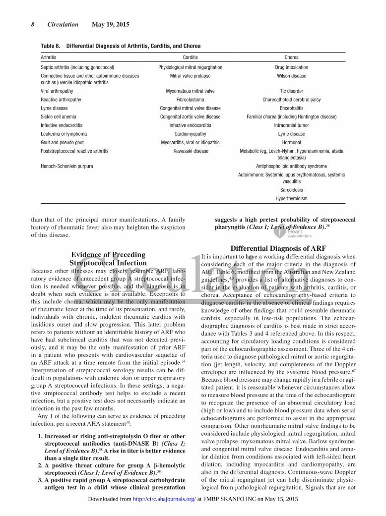
Differential Diagnosis of ARFIt is important to have a working differential diagnosis when considering each of the major criteria in the diagnosis of ARF. Table 6, modified from the Australian and New Zealand guidelines,4,5 provides a list of alternative diagnoses to con-sider in the evaluation of patients with arthritis, carditis, or chorea. Acceptance of echocardiography-based criteria to diagnose carditis in the absence of clinical findings requires knowledge of other findings that could resemble rheumatic carditis, especially in low-risk populations. The echocar-diographic diagnosis of carditis is best made in strict accor-dance with Tables 3 and 4 referenced above. In this respect, accounting for circulatory loading conditions is considered part of the echocardiographic assessment. Three of the 4 cri-teria used to diagnose pathological mitral or aortic regurgita-tion (jet length, velocity, and completeness of the Doppler envelope) are influenced by the systemic blood pressure.67 Because blood pressure may change rapidly in a febrile or agi-tated patient, it is reasonable whenever circumstances allow to measure blood pressure at the time of the echocardiogram to recognize the presence of an abnormal circulatory load (high or low) and to include blood pressure data when serial echocardiograms are performed to assist in the appropriate comparison. Other nonrheumatic mitral valve findings to be considered include physiological mitral regurgitation, mitral valve prolapse, myxomatous mitral valve, Barlow syndrome, and congenital mitral valve disease. Endocarditis and annu-lar dilation from conditions associated with left-sided heart dilation, including myocarditis and cardiomyopathy, are also in the differential diagnosis. Continuous-wave Doppler of the mitral regurgitant jet can help discriminate physio-logical from pathological regurgitation. Signals that are not
Table 6. Differential Diagnosis of Arthritis, Carditis, and Chorea
Arthritis Carditis Chorea
Septic arthritis (including gonococcal) Physiological mitral regurgitation Drug intoxication
Connective tissue and other autoimmune diseases such as juvenile idiopathic arthritis
Mitral valve prolapse Wilson disease
Viral arthropathy Myxomatous mitral valve Tic disorder
Reactive arthropathy Fibroelastoma Choreoathetoid cerebral palsy
Lyme disease Congenital mitral valve disease Encephalitis
Sickle cell anemia Congenital aortic valve disease Familial chorea (including Huntington disease)
Infective endocarditis Infective endocarditis Intracranial tumor
Leukemia or lymphoma Cardiomyopathy Lyme disease
Gout and pseudo gout Myocarditis, viral or idiopathic Hormonal
Poststreptococcal reactive arthritis Kawasaki disease Metabolic (eg, Lesch-Nyhan, hyperalaninemia, ataxia telangiectasia)
Henoch-Schonlein purpura Antiphospholipid antibody syndrome
Autoimmune: Systemic lupus erythematosus, systemic vasculitis
Sarcoidosis
Hyperthyroidism
at FMRP SKANFO INC on May 15, 2015http://circ.ahajournals.org/Downloaded from
Gewitz et al Revised Jones Criteria for Acute Rheumatic Fever 9
holosystolic and peak velocity <3.0 m/s are more likely to be physiological than pathological. The mitral valve prolapse seen in ARF patients differs from the redundant, myxoma-tous mitral valve and prolapse seen with Barlow syndrome.68 In valvulitis from ARF, only the coapting portion of the ante-rior mitral valve leaflet tip prolapses, and there is no billow-ing of the medial portion or body of the leaflet. This leaflet tip prolapse results in abnormal leaflet coaptation, a regurgi-tant orifice, and a jet of mitral regurgitation that is typically directed posterolaterally.
Isolated congenital mitral valve abnormalities are relatively uncommon but are in the differential diagnosis of newly iden-tified mitral regurgitation. These include cleft mitral valve, double-orifice mitral valve, parachute mitral valve variants, and fibroelastomas. Congenital aortic valve anomalies should be in the differential diagnosis of newly identified aortic regurgitation; however, isolated aortic regurgitation is rarely the sole valvular finding in rheumatic carditis. Congenital diagnoses to consider include bicuspid aortic valve, spontane-ously closed ventricular septal defect with aortic valve pro-lapse, subaortic membrane, and syndromic-related aortic root dilation. Infective endocarditis can be mistaken for rheumatic carditis if there is no obvious vegetation and valve damage has already occurred.
Rheumatic Fever RecurrencesAs stated in the 1992 guidelines,2 patients who have a his-tory of ARF or RHD are at high risk for “recurrent” attacks if reinfected with group A streptococci. Such an attack is con-sidered a new episode of ARF, but one in which the complete set of Jones criteria, even as revised, may not be completely fulfilled.
1. With a reliable past history of ARF or established RHD, and in the face of documented group A streptococcal infection, 2 major or 1 major and 2 minor or 3 minor manifestations may be sufficient for a presumptive diagnosis (Class IIb; Level of Evidence C).
2. When minor manifestations alone are present, the exclusion of other more likely causes of the clinical pre-sentation is recommended before a diagnosis of an ARF recurrence is made (Class I; Level of Evidence C).
“Possible” Rheumatic FeverIn some circumstances, a given clinical presentation may not fulfill these updated Jones criteria, but the clinician may still have good reason to suspect that ARF is the diagno-sis. This may occur in high-incidence settings where, for
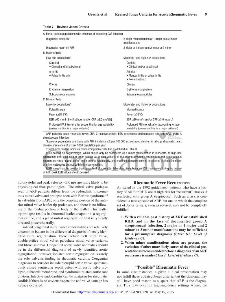
Table 7. Revised Jones Criteria
A. For all patient populations with evidence of preceding GAS infection
Diagnosis: initial ARF 2 Major manifestations or 1 major plus 2 minor manifestations
Diagnosis: recurrent ARF 2 Major or 1 major and 2 minor or 3 minor
B. Major criteria
Low-risk populations* Moderate- and high-risk populations
Carditis† • Clinical and/or subclinical
Carditis • Clinical and/or subclinical
Arthritis • Polyarthritis only
Arthritis • Monoarthritis or polyarthritis • Polyarthralgia‡
Chorea Chorea
Erythema marginatum Erythema marginatum
Subcutaneous nodules Subcutaneous nodules
C. Minor criteria
Low-risk populations* Moderate- and high-risk populations
Polyarthralgia Monoarthralgia
Fever (≥38.5°C) Fever (≥38°C)
ESR ≥60 mm in the first hour and/or CRP ≥3.0 mg/dL§ ESR ≥30 mm/h and/or CRP ≥3.0 mg/dL§
Prolonged PR interval, after accounting for age variability (unless carditis is a major criterion)
Prolonged PR interval, after accounting for age variability (unless carditis is a major criterion)
ARF indicates acute rheumatic fever; CRP, C-reactive protein; ESR, erythrocyte sedimentation rate; and GAS, group A streptococcal infection.
*Low-risk populations are those with ARF incidence ≤2 per 100 000 school-aged children or all-age rheumatic heart disease prevalence of ≤1 per 1000 population per year.
†Subclinical carditis indicates echocardiographic valvulitis as defined in Table 3.‡See section on polyarthralgia, which should only be considered as a major manifestation in moderate- to high-risk
populations after exclusion of other causes. As in past versions of the criteria, erythema marginatum and subcutaneous nodules are rarely “stand-alone” major criteria. Additionally, joint manifestations can only be considered in either the major or minor categories but not both in the same patient.
§CRP value must be greater than upper limit of normal for laboratory. Also, because ESR may evolve during the course of ARF, peak ESR values should be used.
at FMRP SKANFO INC on May 15, 2015http://circ.ahajournals.org/Downloaded from
10 Circulation May 19, 2015
example, laboratory tests for acute phase reactants or for confirmation of recent streptococcal infection are not avail-able, documentation of clinical features is not clear, or the history is not considered to be reliable. In such situations, clinicians should use their discretion and clinical acumen to make the diagnosis that they consider most likely and man-age the patient accordingly.
1. Where there is genuine uncertainty, it is reason-able to consider offering 12 months of secondary prophylaxis followed by reevaluation to include a careful history and physical examination in addi-tion to a repeat echocardiogram (Class IIa; Level of Evidence C).
2. In a patient with recurrent symptoms (particularly involving the joints) who has been adherent to pro-phylaxis recommendations but lacks serological evi-dence of group A streptococcal infection and lacks echocardiographic evidence of valvulitis, it is reason-able to conclude that the recurrent symptoms are not likely related to ARF, and discontinuation of antibi-otic prophylaxis may be appropriate (Class IIa; Level of Evidence C).
Impact of Modifications of Jones Criteria in High-Risk Populations
A retrospective study in North Queensland, Australia, investigated the impact of the addition of subclinical car-ditis, monoarthritis, and low-grade fever (>37.5°C) to the 1992 revised Jones criteria.36 Of the 98 cases with a clinical
diagnosis of ARF, only 71.4% met the revised Jones criteria. Modification of the criteria, as discussed above, increased the proportion of the cases that satisfied diagnostic criteria to 91.8%. Of the 28 people who did not meet the traditional Jones criteria, 12 (42%) developed evidence of chronic RHD. This study, if confirmed, may suggest that the addi-tion of monoarthritis and subclinical carditis as major mani-festations and low-grade fever as a minor manifestation to the Jones criteria could increase sensitivity when applied specifically to high-risk populations. Additionally, study of the impact of the application of the New Zealand guidelines resulted in a 16% increase in the diagnosis of ARF com-pared with the 1992 revision of the Jones criteria.29 There are no additional data that corroborate these results in popu-lations with a lower incidence of ARF.
In summary, in the context of the previous discussion, revision of the Jones criteria to meet current technologi-cal advances and clinical needs is warranted. Thus, strict application of echocardiography/Doppler findings (Tables 3 and 4) may be used to fulfill the major criterion of carditis, even in the absence of classic auscultatory findings, provid-ing that ambient loading conditions are taken into consid-eration. In addition, monoarthritis or polyarthralgia could be accepted as fulfilling the major criterion of arthritis, but only in moderate- to high-risk populations. For low-risk populations, monoarthritis is not included, and polyarthral-gia remains a minor criterion. Similarly, the requirement for the presence of fever can be fulfilled with oral, tympanic, or rectal temperature documented at 38°C in moderate- to high-risk populations, but only at ≥38.5°C in others. The
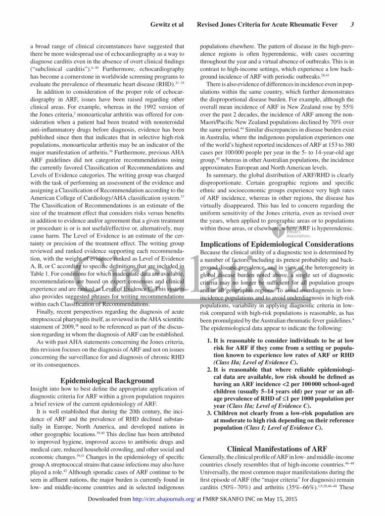
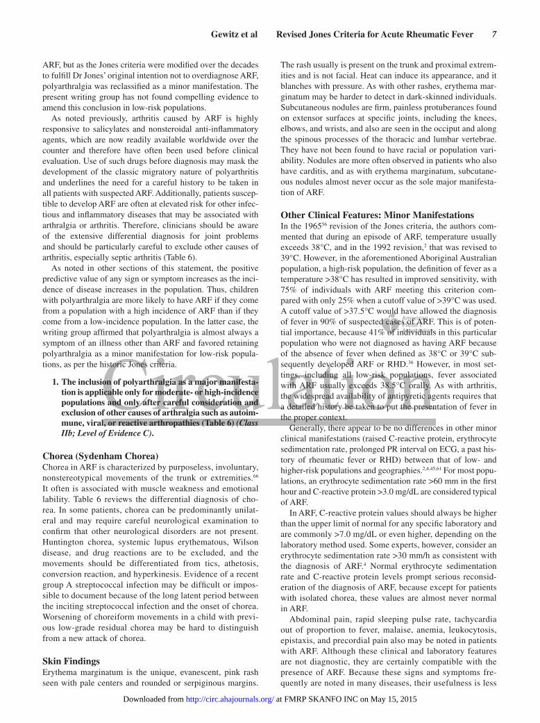
A B
C D
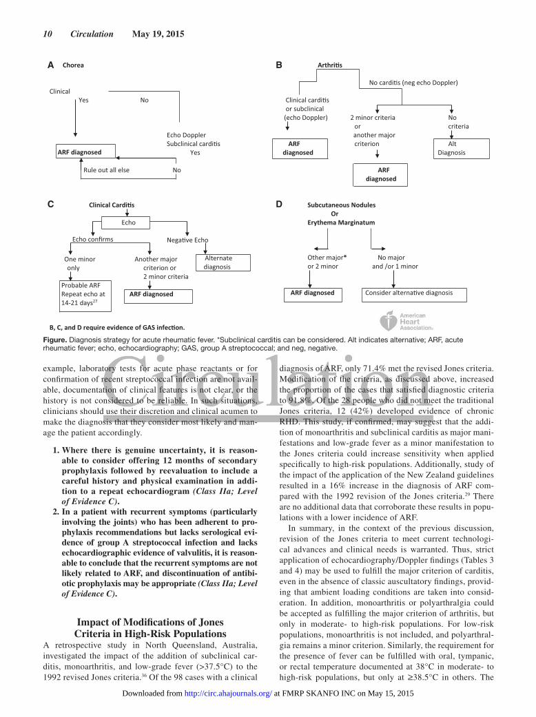
Figure. Diagnosis strategy for acute rheumatic fever. *Subclinical carditis can be considered. Alt indicates alternative; ARF, acute rheumatic fever; echo, echocardiography; GAS, group A streptococcal; and neg, negative.
at FMRP SKANFO INC on May 15, 2015http://circ.ahajournals.org/Downloaded from
Gewitz et al Revised Jones Criteria for Acute Rheumatic Fever 11
writing group confirms the appropriateness of retaining the time-honored approach initially advocated by Dr Jones that favors low sensitivity and high specificity in assessing the criteria for the diagnosis of ARF in low-risk populations. Table 7 and the Figure summarize diagnostic strategies using these revised criteria.
Future ConsiderationsIn addition to the broad epidemiological issues and the widespread careful application of echocardiography that have led to the suggested revisions in the Jones criteria described in this statement, recent findings suggesting
genetic susceptibility factors in ARF69–71 may one day point to a totally new set of diagnostic tools. Future revisions should continue to honor Dr Jones’ initial goal, particu-larly in low-risk populations, to avoid overdiagnosis and its consequences.54
AcknowledgmentsThe authors acknowledge the support and critical input of committee members not on the writing group (Larry Baddour, MD; Jane Burns, MD; Marianne Jackson, MD; Mathew Levison, MD; Peter Lockhart, DDS; Brian McCrindle, MD; Patrick T. O’Gara, MD; and Walter Wilson, MD) and the assistance of Patty Libby in preparation of this document.
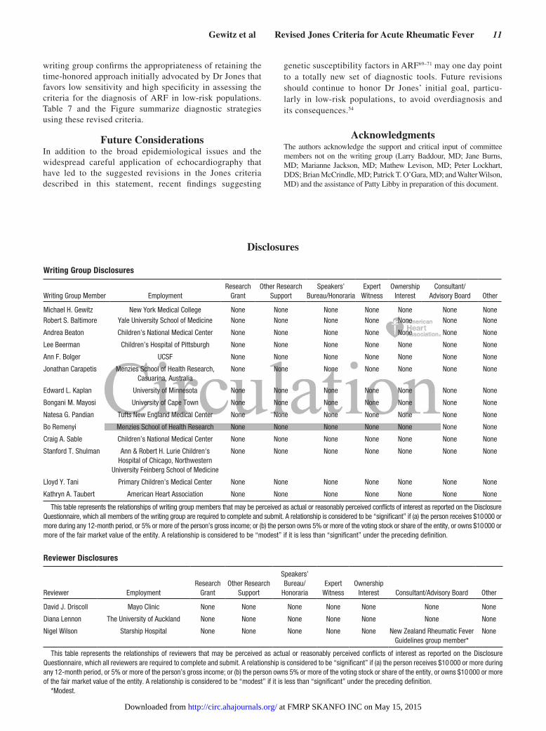
Writing Group Disclosures
Writing Group Member EmploymentResearch
GrantOther Research
SupportSpeakers’
Bureau/HonorariaExpert
WitnessOwnership
InterestConsultant/
Advisory Board Other
Michael H. Gewitz New York Medical College None None None None None None NoneRobert S. Baltimore Yale University School of Medicine None None None None None None None
Andrea Beaton Children’s National Medical Center None None None None None None None
Lee Beerman Children’s Hospital of Pittsburgh None None None None None None None
Ann F. Bolger UCSF None None None None None None None
Jonathan Carapetis Menzies School of Health Research, Casuarina, Australia
None None None None None None None
Edward L. Kaplan University of Minnesota None None None None None None None
Bongani M. Mayosi University of Cape Town None None None None None None None
Natesa G. Pandian Tufts New England Medical Center None None None None None None None
Bo Remenyi Menzies School of Health Research None None None None None None None
Craig A. Sable Children’s National Medical Center None None None None None None None
Stanford T. Shulman Ann & Robert H. Lurie Children's Hospital of Chicago, Northwestern
University Feinberg School of Medicine
None None None None None None None
Lloyd Y. Tani Primary Children’s Medical Center None None None None None None None
Kathryn A. Taubert American Heart Association None None None None None None None
This table represents the relationships of writing group members that may be perceived as actual or reasonably perceived conflicts of interest as reported on the Disclosure Questionnaire, which all members of the writing group are required to complete and submit. A relationship is considered to be “significant” if (a) the person receives $10 000 or more during any 12-month period, or 5% or more of the person’s gross income; or (b) the person owns 5% or more of the voting stock or share of the entity, or owns $10 000 or more of the fair market value of the entity. A relationship is considered to be “modest” if it is less than “significant” under the preceding definition.
Reviewer Disclosures
Reviewer EmploymentResearch
GrantOther Research
Support
Speakers’ Bureau/
HonorariaExpert
WitnessOwnership
Interest Consultant/Advisory Board Other
David J. Driscoll Mayo Clinic None None None None None None None
Diana Lennon The University of Auckland None None None None None None None
Nigel Wilson Starship Hospital None None None None None New Zealand Rheumatic Fever Guidelines group member*
None
This table represents the relationships of reviewers that may be perceived as actual or reasonably perceived conflicts of interest as reported on the Disclosure Questionnaire, which all reviewers are required to complete and submit. A relationship is considered to be “significant” if (a) the person receives $10 000 or more during any 12-month period, or 5% or more of the person’s gross income; or (b) the person owns 5% or more of the voting stock or share of the entity, or owns $10 000 or more of the fair market value of the entity. A relationship is considered to be “modest” if it is less than “significant” under the preceding definition.
*Modest.
Disclosures
at FMRP SKANFO INC on May 15, 2015http://circ.ahajournals.org/Downloaded from
12 Circulation May 19, 2015
References 1. Seckeler MD, Hoke TR. The worldwide epidemiology of acute rheumatic
fever and rheumatic heart disease. Clin Epidemiol. 2011;3:67–84. doi: 10.2147/CLEP.S12977.
2. Dajani AS, Ayoub E, Bierman FZ, Bisno AL, Denny FW, Durack DT, Ferrieri P, Freed M, Gerber M, Kaplan EL, Karchmer AW, Markowitz M, Rahimtoola SH, Shulman ST, Stollerman G, Takahashi M, Taranta A, Taubert KA, Wilson W, Durack; Special Writing Group of the Committee on Rheumatic Fever, Endocarditis, and Kawasaki Disease of the Council on Cardiovascular Disease in the Young of the American Heart Association. Guidelines for the diagnosis of rheumatic fever: Jones crite-ria, 1992 update [published correction appears in JAMA. 1993;269:476]. JAMA. 1992;268:2069–2073.
3. Ferrieri P; for the Jones Criteria Working Group. Proceedings of the Jones criteria workshop. Circulation. 2002;106:2521–2523.
4. RHDAustralia (ARF/RHD Writing Group), National Heart Foundation of Australia, Cardiac Society of Australia and New Zealand. The Australian Guideline for Prevention, Diagnosis, and Management of Acute Rheumatic Fever and Rheumatic Heart Disease (2nd ed). Casuarina, Australia: RHDAustralia; 2012.
5. Atatoa-Carr P, Lennon D, Wilson N; New Zealand Rheumatic Fever Guidelines Writing Group. Rheumatic fever diagnosis, manage-ment, and secondary prevention: a New Zealand guideline. N Z Med J. 2008;121:59–69.
6. Vijayalakshmi IB, Mithravinda J, Deva AN. The role of echocardiogra-phy in diagnosing carditis in the setting of acute rheumatic fever. Cardiol Young. 2005;15:583–588. doi: 10.1017/S1047951105001745.
7. Vijayalakshmi IB, Vishnuprabhu RO, Chitra N, Rajasri R, Anuradha TV. The efficacy of echocardiographic criterions for the diagnosis of carditis in acute rheumatic fever. Cardiol Young. 2008;18:586–592. doi: 10.1017/S1047951108003107.
8. Ozdemir O, Işık S, Abacı A, Hızlı S, Akelma AZ, Kışlal FM, Celik A, Razi CH, Koçak M. Silent enemy in acute rheumatic fever: subclinical carditis [in Turkish]. Turk Kardiyol Dern Ars. 2011;39:41–46.
9. Cann MP, Sive AA, Norton RE, McBride WJ, Ketheesan N. Clinical presentation of rheumatic fever in an endemic area. Arch Dis Child. 2010;95:455–457. doi: 10.1136/adc.2008.157107.
10. Beg A, Sadiq M. Subclinical valvulitis in children with acute rheu-matic fever. Pediatr Cardiol. 2008;29:619–623. doi: 10.1007/s00246- 007-9173-0.
11. Caldas AM, Terreri MT, Moises VA, Silva CM, Len CA, Carvalho AC, Hilário MO. What is the true frequency of carditis in acute rheumatic fever? A prospective clinical and Doppler blind study of 56 children with up to 60 months of follow-up evaluation. Pediatr Cardiol. 2008;29:1048–1053. doi: 10.1007/s00246-008-9242-z.
12. Rayamajhi A, Sharma D, Shakya U. Clinical, laboratory and echocar-diographic profile of acute rheumatic fever in Nepali children. Ann Trop Paediatr. 2007;27:169–177. doi: 10.1179/146532807X220271.
13. Ozer S, Hallioğlu O, Ozkutlu S, Celiker A, Alehan D, Karagöz T. Childhood acute rheumatic fever in Ankara, Turkey. Turk J Pediatr. 2005;47:120–124.
14. Panamonta M, Chaikitpinyo A, Kaplan EL, Pantongwiriyakul A, Tassniyom S, Sutra S. The relationship of carditis to the initial attack of Sydenham’s chorea. Int J Cardiol. 2004;94:241–248. doi: 10.1016/j.ijcard.2003.04.020.
15. Ozkutlu S, Hallioglu O, Ayabakan C. Evaluation of subclinical valvar dis-ease in patients with rheumatic fever. Cardiol Young. 2003;13:495–499.
16. Karaaslan S, Demirören S, Oran B, Baysal T, Başpinar O, Uçar C. Criteria for judging the improvement in subclinical rheumatic valvitis. Cardiol Young. 2003;13:500–505.
17. Khriesat I, Najada A, Al-Hakim F, Abu-Haweleh A. Acute rheumatic fever in Jordanian children. East Mediterr Health J. 2003;9:981–987.
18. Lanna CC, Tonelli E, Barros MV, Goulart EM, Mota CC. Subclinical rheu-matic valvitis: a long-term follow-up. Cardiol Young. 2003;13:431–438.
19. Figueroa FE, Fernández MS, Valdés P, Wilson C, Lanas F, Carrión F, Berríos X, Valdés F. Prospective comparison of clinical and echocardio-graphic diagnosis of rheumatic carditis: long term follow up of patients with subclinical disease. Heart. 2001;85:407–410.
20. Hilário MO, Andrade JL, Gasparian AB, Carvalho AC, Andrade CT, Len CA. The value of echocardiography in the diagnosis and followup of rheu-matic carditis in children and adolescents: a 2 year prospective study. J Rheumatol. 2000;27:1082–1086.
21. Elevli M, Celebi A, Tombul T, Gökalp AS. Cardiac involvement in Sydenham’s chorea: clinical and Doppler echocardiographic findings. Acta Paediatr. 1999;88:1074–1077.
22. da Silva CH; Pediatric Committee, Sao Paulo Pediatric Rheumatology Society. Rheumatic fever: a multicenter study in the state of Sao Paulo. Rev Hosp Clin Fac Med Sao Paolo. 1999;54:85–90.
23. Minich LL, Tani LY, Pagotto LT, Shaddy RE, Veasy LG. Doppler echo-cardiography distinguishes between physiologic and pathologic “silent” mitral regurgitation in patients with rheumatic fever. Clin Cardiol. 1997;20:924–926.
24. Hoffman TM, Rhodes LA, Pyles LA, Balian AA, Neal WA, Einzig S. Childhood acute rheumatic fever: a comparison of recent resurgence areas to cases in West Virginia. W V Med J. 1997;93:260–263.
25. Vasan RS, Shrivastava S, Vijayakumar M, Narang R, Lister BC, Narula J. Echocardiographic evaluation of patients with acute rheumatic fever and rheumatic carditis. Circulation. 1996;94:73–82.
26. Maheu B, Costes P, Lionet P, Kamblock J, Papouin G, Mansourati J, Genet L, Blanc JJ. Contribution of Doppler echocardiography to the diagnosis of the first attack of acute rheumatic fever [in French]. Arch Mal Coeur Vaiss. 1995;88:1833–1839.
27. Abernethy M, Bass N, Sharpe N, Grant C, Neutze J, Clarkson P, Greaves S, Lennon D, Snow S, Whalley G. Doppler echocardiography and the early diagnosis of carditis in acute rheumatic fever. Aust N Z J Med. 1994;24:530–535.
28. Veasy LG, Tani LY, Hill HR. Persistence of acute rheumatic fever in the intermountain area of the United States. J Pediatr. 1994;124:9–16.
29. Wilson NJ, Voss L, Morreau J, Stewart JM, Lennon D. New Zealand guidelines for the diagnosis of acute rheumatic fever: small increase in the incidence of definite cases compared to the America Heart Association Jones criteria. N Z Med J. 2013;126:50–59.
30. Caldas AM, Terreri MT, Moises VA, Silva CM, Carvalho AC, Hilário MO. The case for utilizing more strict quantitative Doppler echocardiographic criterions for diagnosis of subclinical rheumatic carditis. Cardiol Young. 2007;17:42–47. doi: 10.1017/S1047951106001296.
31. Marijon E, Ou P, Celermajer DS, Ferreira B, Mocumbi AO, Jani D, Paquet C, Jacob S, Sidi D, Jouven X. Prevalence of rheumatic heart disease detected by echocardiographic screening. N Engl J Med. 2007;357:470–476. doi: 10.1056/NEJMoa065085.
32. Saxena A, Ramakrishnan S, Roy A, Seth S, Krishnan A, Misra P, Kalaivani M, Bhargava B, Flather MD, Poole-Wilson PP. Prevalence and out-come of subclinical rheumatic heart disease in India: the RHEUMATIC (Rheumatic Heart Echo Utilisation and Monitoring Actuarial Trends in Indian Children) study. Heart. 2011;97:2018–2022. doi: 10.1136/heartjnl-2011-300792.
33. Paar JA, Berrios NM, Rose JD, Cáceres M, Peña R, Pérez W, Chen-Mok M, Jolles E, Dale JB. Prevalence of rheumatic heart disease in children and young adults in Nicaragua. Am J Cardiol. 2010;105:1809–1814. doi: 10.1016/j.amjcard.2010.01.364.
34. Beaton A, Okello E, Lwabi P, Mondo C, McCarter R, Sable C. Echocardiography screening for rheumatic heart disease in Ugandan schoolchildren. Circulation. 2012;125:3127–3132. doi: 10.1161/CIRCULATIONAHA.112.092312.
35. Webb RH, Wilson NJ, Lennon DR, Wilson EM, Nicholson RW, Gentles TL, O’Donnell CP, Stirling JW, Zeng I, Trenholme AA. Optimising echo-cardiographic screening for rheumatic heart disease in New Zealand: not all valve disease is rheumatic. Cardiol Young. 2011;21:436–443.
36. Carapetis JR, Currie BJ. Rheumatic fever in a high incidence population: the importance of monoarthritis and low grade fever. Arch Dis Child. 2001;85:223–227.
37. Gibbons RJ, Smith S, Antman E. American College of Cardiology/American Heart Association clinical practice guidelines: part 1: where do they come from? Circulation 2003;107:2979–2986. doi: 10.1161/01.CIR.0000063682.20730.A5.
38. Gerber MA, Baltimore RS, Eaton CB, Gewitz M, Rowley AH, Shulman ST, Taubert KA. Prevention of rheumatic fever and diagnosis and treat-ment of acute streptococcal pharyngitis: a scientific statement from the American Heart Association Rheumatic Fever, Endocarditis, and Kawasaki Disease Committee of the Council on Cardiovascular Disease in the Young, the Interdisciplinary Council on Functional Genomics and Translational Biology, and the Interdisciplinary Council on Quality of Care and Outcomes Research. Circulation. 2009;119:1541–1551. doi: 10.1161/CIRCULATIONAHA.109.191959.
39. Gordis L. The virtual disappearance of rheumatic fever in the United States: lessons in the rise and fall of disease: T. Duckett Jones Memorial Lecture. Circulation. 1985;72:1155–1162.
40. Levinson SS, Bearfield JL, Ausbrook DK, Muriel H, Shireman L, Pacelli C, Stanton H, Masterson J. The Chicago rheumatic fever program: a 20 plus year history. J Chronic Dis. 1982;35:199–206.
at FMRP SKANFO INC on May 15, 2015http://circ.ahajournals.org/Downloaded from
Gewitz et al Revised Jones Criteria for Acute Rheumatic Fever 13
41. Markowitz M. The decline of rheumatic fever: role of medical intervention: Lewis W. Wannamaker Memorial Lecture. J Pediatr. 1985;106:545–550.
42. Shulman ST, Stollerman G, Beall B, Dale JB, Tanz RR. Temporal changes in streptococcal M protein types and the near-disappearance of acute rheu-matic fever in the United States. Clin Infect Dis. 2006;42:441–447. doi: 10.1086/499812.
43. Pastore S, De Cunto A, Benettoni A, Berton E, Taddio A, Lepore L. The resurgence of rheumatic fever in a developed country area: the role of echocardiography. Rheumatology (Oxford). 2011;50:396–400. doi: 10.1093/rheumatology/keq290.
44. Milne RJ, Lennon DR, Stewart JM, Vander Hoorn S, Scuffham PA. Incidence of acute rheumatic fever in New Zealand children and youth. J Paediatr Child Health. 2012;48:685–691. doi: 10.1111/j.1440-1754.2012.02447.x.
45. Parnaby MG, Carapetis JR. Rheumatic fever in indigenous Australian children. J Paediatr Child Health. 2010;46:527–533. doi: 10.1111/j.1440-1754.2010.01841.x.
46. Jamal M, Abbas KA. Clinical profile of acute rheumatic fever in children. J Trop Pediatr. 1989;35:10–13.
47. Vinker S, Zohar E, Hoffman R, Elhayany A. Incidence and clinical mani-festations of rheumatic fever: a 6 year community-based survey. Isr Med Assoc J. 2010;12:78–81.
48. Grassi A, Fesslovà V, Carnelli V, Boati E, Dell’era L, Salice P, Bardare M, Corona F. Clinical characteristics and cardiac outcome of acute rheumatic fever in Italy in the last 15 years. Clin Exp Rheumatol. 2009;27:366–372.
49. World Health Organization. Rheumatic Fever and Rheumatic Heart Disease: Report of a WHO Expert Consultation, Geneva, 29 October–1 November 2001. Geneva, Switzerland: World Health Organization; 2001. WHO Technical Report Series 923. http://www.who.int/cardiovascular_diseases/resources/en/cvd_trs923.pdf. Accessed May 18, 2011.
50. Working Group on Pediatric Acute Rheumatic Fever and Cardiology Chapter of Indian Academy of Pediatrics. Consensus guidelines on pedi-atric acute rheumatic fever and rheumatic heart disease. Indian Pediatr. 2008;45:565–573.
51. Reményi B, Wilson N, Steer A, Ferreira B, Kado J, Kumar K, Lawrenson J, Maguire G, Marijon E, Mirabel M, Mocumbi AO, Mota C, Paar J, Saxena A, Scheel J, Stirling J, Viali S, Balekundri VI, Wheaton G, Zühlke L, Carapetis J. World Heart Federation criteria for echocardiographic diagnosis of rheumatic heart disease: an evidence-based guideline. Nat Rev Cardiol. 2012;9:297–309. doi: 10.1038/nrcardio.2012.7.
52. Tubridy-Clark M, Carapetis JR. Subclinical carditis in rheumatic fever: a systematic review. Int J Cardiol. 2007;119:54–58. doi: 10.1016/j.ijcard.2006.07.046.
53. Jaggi P. Rheumatic fever and post group-A streptococcal arthritis. Pediatr Infect Dis J. 2011;30:424–425.
54. Jones TD. Diagnosis of rheumatic fever. JAMA. 1944;126:481–484. doi: 10.1001/jama.1944.02850430015005.
55. Jones criteria (modified) for guidance in the diagnosis of rheumatic fever: report of the Committee on Standards and Criteria for Programs of Care. Circulation. 1956;13:617–620.
56. Committee Report. Jones criteria (revised) for guidance in the diagnosis of rheumatic fever. Circulation 1965;32:664–668.
57. De Cunto CL, Giannini EH, Fink CW, Brewer EJ, Person DA. Prognosis of children with poststreptococcal reactive arthritis. Pediatr Infect Dis J. 1988;7:683–686.
58. Merino Muñoz R, Viota Losada F, Sancho Madrid B, Castro Gussoni C, García-Consuegra Molina J. Rheumatic fever and post-strep-tococcal arthritis: clinical review [in Spanish]. An Esp Pediatr. 1991;35:239–242.
59. Koçak G, Imamoğlu A, Tutar HE, Atalay S, Türkay S. Poststreptococcal reactive arthritis: clinical course and outcome in 15 patients. Turk J Pediatr. 2000;42:101–104.
60. van Bemmel JM, Delgado V, Holman ER, Allaart CF, Huizinga TW, Bax JJ, van der Helm-van Mil AH. No increased risk of valvular heart disease in adult poststreptococcal reactive arthritis. Arthritis Rheum. 2009;60:987–993. doi: 10.1002/art.24401.
61. Tani LY. Rheumatic fever and rheumatic heart disease. In: Allen HD, Driscoll MD, Shaddy RE, Feltes TF, eds. Moss and Adams’ Heart Disease in Infants, Children, and Adolescents. 8th ed. Philadelphia, PA: Lippincott Williams & Wilkins; 2013:1303–1330.
62. Parks T, Kado J, Colquhoun S, Carapetis J, Steer A. Underdiagnosis of acute rheumatic fever in primary care settings in a developing country [published correction appears in Trop Med Int Health. 2010;15:384]. Trop Med Int Health. 2009;14:1407–1413. doi: 10.1111/j.1365-3156. 2009.02385.x.
63. Noonan S, Zurynski YA, Currie BJ, McDonald M, Wheaton G, Nissen M, Curtis N, Isaacs D, Richmond P, Ramsay JM, Elliott EJ, Carapetis JR. A national prospective surveillance study of acute rheumatic fever in Australian children. Pediatr Infect Dis J. 2013;32:e26–e32. doi: 10.1097/INF.0b013e31826faeb3.
64. Sanyal SK, Thapar MK, Ahmed SH, Hooja V, Tewari P. The initial attack of acute rheumatic fever during childhood in North India: a prospective study of the clinical profile. Circulation. 1974;49:7–12.
65. Harlan GA, Tani LY, Byington CL. Rheumatic fever presenting as mono-articular arthritis. Pediatr Infect Dis J. 2006;25:743–746. doi: 10.1097/01.inf.0000227726.44519.00.
66. Markowitz M, Kuttner AG. Rheumatic Fever. Philadelphia, PA: WB Saunders; 1972.
67. Stout KK, Verrier ED. Acute valvular regurgitation. Circulation. 2009;119:3232–3241. doi: 10.1161/CIRCULATIONAHA.108.782292.
68. Anyanwu AC, Adams DH. Etiologic classification of degenerative mitral valve disease: Barlow’s syndrome and fibroelastic deficiency. Semin Thorac Cardiovasc Surg. 2007;19:90–96.
69. Guilherme L, Köhler KF, Postol E, Kalil J. Genes, autoimmunity and pathogenesis of rheumatic heart disease. Ann Pediatr Cardiol. 2011;4:13–21. doi: 10.4103/0974-2069.79617.
70. Bryant PA, Robins-Browne R, Carapetis JR, Curtis N. Some of the peo-ple, some of the time: susceptibility to acute rheumatic fever. Circulation. 2009;119:742–753. doi: 10.1161/CIRCULATIONAHA.108.792135.
71. Engel ME, Stander R, Vogel J, Adeyemo AA, Mayosi BM. Genetic sus-ceptibility to acute rheumatic fever: a systematic review and meta-anal-ysis of twin studies. PLoS One. 2011;6:e25326. doi: 10.1371/journal.pone.0025326.
at FMRP SKANFO INC on May 15, 2015http://circ.ahajournals.org/Downloaded from
M. Mayosi, Andrea Beaton, Natesa G. Pandian and Edward L. KaplanJonathan Carapetis, Bo Remenyi, Kathryn A. Taubert, Ann F. Bolger, Lee Beerman, Bongani Michael H. Gewitz, Robert S. Baltimore, Lloyd Y. Tani, Craig A. Sable, Stanford T. Shulman,
Doppler Echocardiography: A Scientific Statement From the American Heart AssociationRevision of the Jones Criteria for the Diagnosis of Acute Rheumatic Fever in the Era of
Print ISSN: 0009-7322. Online ISSN: 1524-4539 Copyright © 2015 American Heart Association, Inc. All rights reserved.
is published by the American Heart Association, 7272 Greenville Avenue, Dallas, TX 75231Circulation published online April 23, 2015;Circulation.
http://circ.ahajournals.org/content/early/2015/04/23/CIR.0000000000000205World Wide Web at:
The online version of this article, along with updated information and services, is located on the
http://circ.ahajournals.org//subscriptions/
is online at: Circulation Information about subscribing to Subscriptions:
http://www.lww.com/reprints Information about reprints can be found online at: Reprints:
document. Permissions and Rights Question and Answer this process is available in the
click Request Permissions in the middle column of the Web page under Services. Further information aboutOffice. Once the online version of the published article for which permission is being requested is located,
can be obtained via RightsLink, a service of the Copyright Clearance Center, not the EditorialCirculationin Requests for permissions to reproduce figures, tables, or portions of articles originally publishedPermissions:
at FMRP SKANFO INC on May 15, 2015http://circ.ahajournals.org/Downloaded from