Embed Size (px)
DESCRIPTION
Greg Crowther's talk at the Seattle Parasitology Conference (May, 2010).
Citation preview
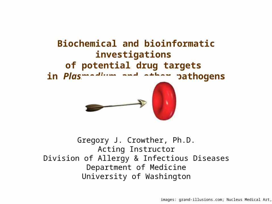
Biochemical and bioinformatic investigations of potential drug targets
in Plasmodium and other pathogens
Gregory J. Crowther, Ph.D.Acting Instructor
Division of Allergy & Infectious DiseasesDepartment of MedicineUniversity of Washington
images: grand-illusions.com; Nucleus Medical Art, Inc.

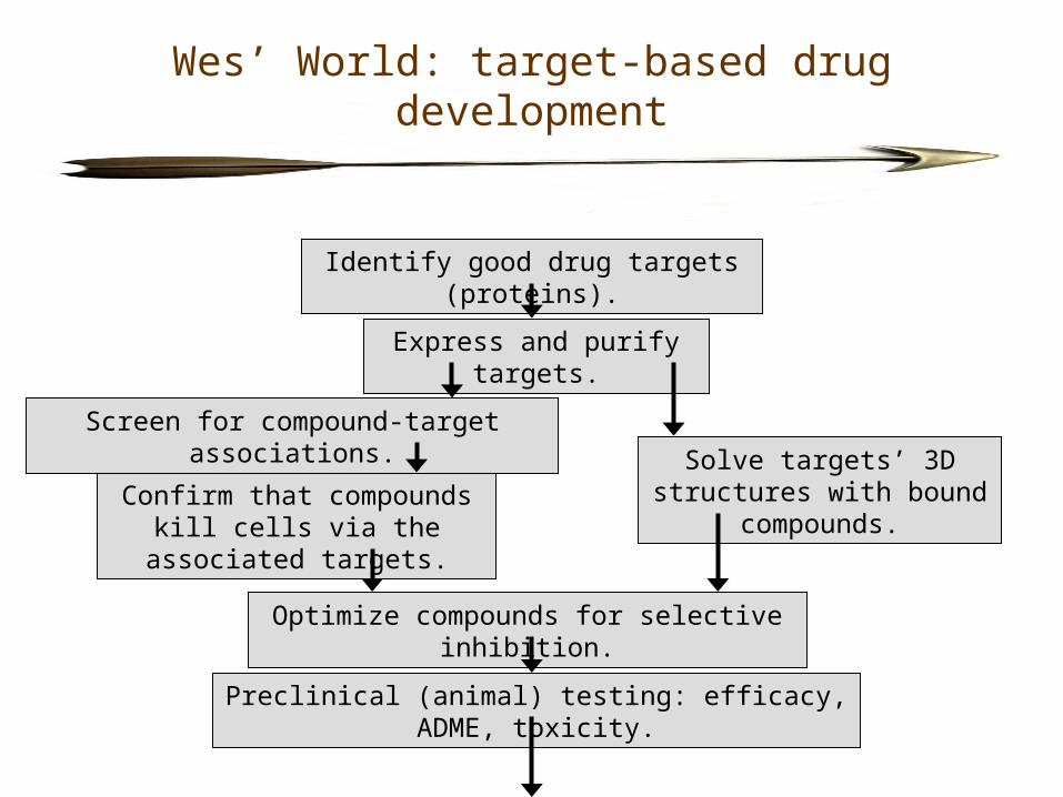
Wes’ World: target-based drug development
• Distinct from “cell-based” (or “phenotypic”) drug development, which proceeds without knowledge of compounds’ specific subcellular targets.
• Targeting a specific protein (often with a known 3D structure) should enable rational, rapid development of selective inhibitors.
• Sequencing/analysis of genomes should bring many new targets into play.
• Few examples so far of drugs created via target-based approaches:- Relenza (zanamivir) – for influenza virus- difluoromethylornithine – for T. brucei- protease inhibitors – for HIV
Nelfinavir in the active site of HIV-1 protease. Image from C.M. Henry, C&EN 79: 69, 2001.
Wes’ World: target-based drug development
Identify good drug targets (proteins).
Express and purify targets.
Solve targets’ 3D structures with bound compounds.Confirm that compounds kill
cells via the associated targets.
Preclinical (animal) testing: efficacy, ADME, toxicity.
Screen for compound-target associations.
Optimize compounds for selective inhibition.
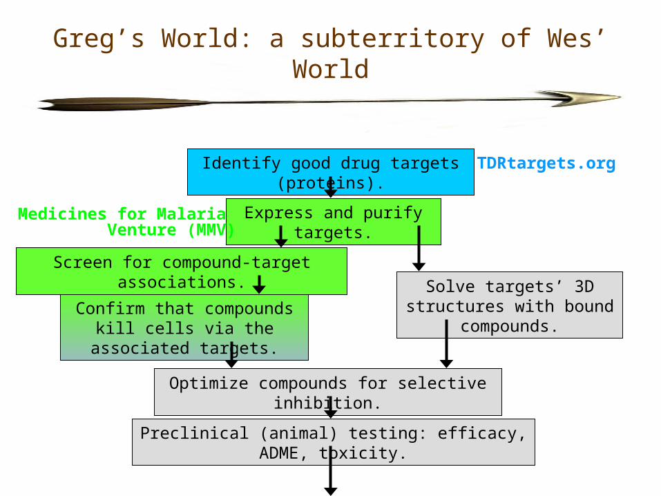
Greg’s World: a subterritory of Wes’ World
Identify good drug targets (proteins).
Express and purify targets.
Solve targets’ 3D structures with bound compounds.Confirm that compounds kill
cells via the associated targets.
Preclinical (animal) testing: efficacy, ADME, toxicity.
Screen for compound-target associations.
Optimize compounds for selective inhibition.
TDRtargets.org
Medicines for Malaria Venture (MMV)
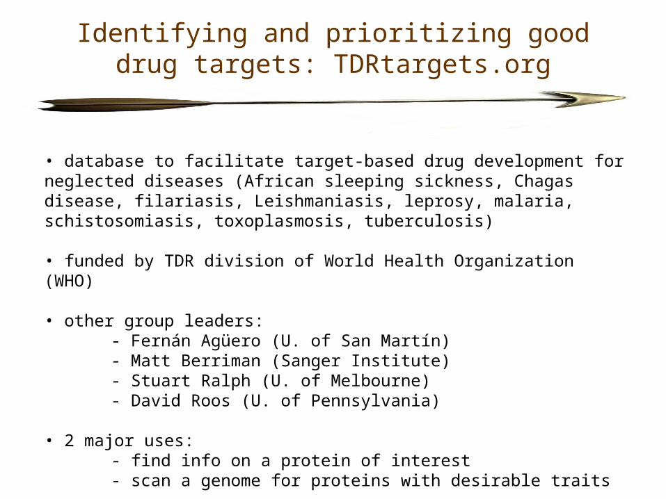
Identifying and prioritizing good drug targets: TDRtargets.org
• database to facilitate target-based drug development for neglected diseases (African sleeping sickness, Chagas disease, filariasis, Leishmaniasis, leprosy, malaria, schistosomiasis, toxoplasmosis, tuberculosis)
• funded by TDR division of World Health Organization (WHO)
• other group leaders:- Fernán Agüero (U. of San Martín)- Matt Berriman (Sanger Institute)- Stuart Ralph (U. of Melbourne)- David Roos (U. of Pennsylvania)
• 2 major uses:- find info on a protein of interest- scan a genome for proteins with desirable traits
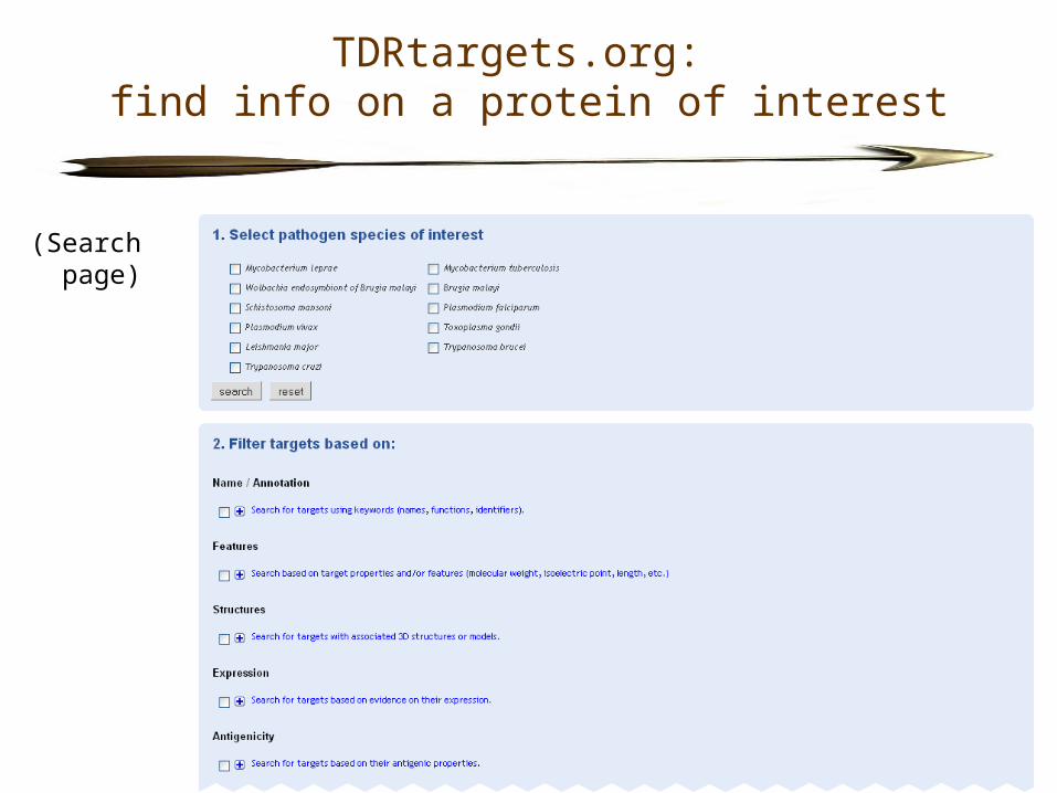
TDRtargets.org: find info on a protein of interest
(Search page)

TDRtargets.org: find info on a protein of interest
(Example of a gene page)
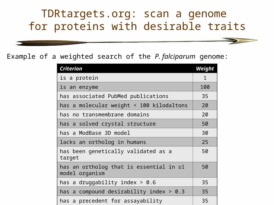
TDRtargets.org: scan a genome for proteins with desirable traits
Example of a weighted search of the P. falciparum genome:
Criterion Weight
is a protein 1
is an enzyme 100
has associated PubMed publications 35
has a molecular weight < 100 kilodaltons 20
has no transmembrane domains 20
has a solved crystal structure 50
has a ModBase 3D model 30
lacks an ortholog in humans 25
has been genetically validated as a target 50
has an ortholog that is essential in ≥1 model organism 50
has a druggability index > 0.6 35
has a compound desirability index > 0.3 35
has a precedent for assayability 35
is present in both P. falciparum and P. vivax 25
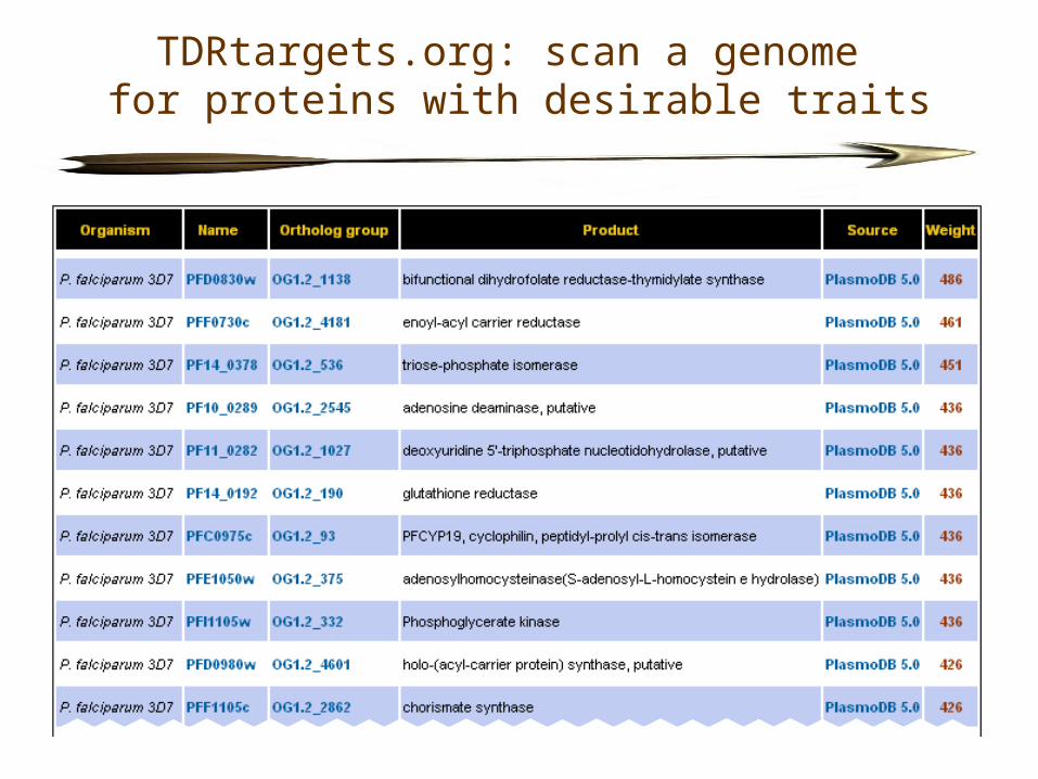
TDRtargets.org: scan a genome for proteins with desirable traits
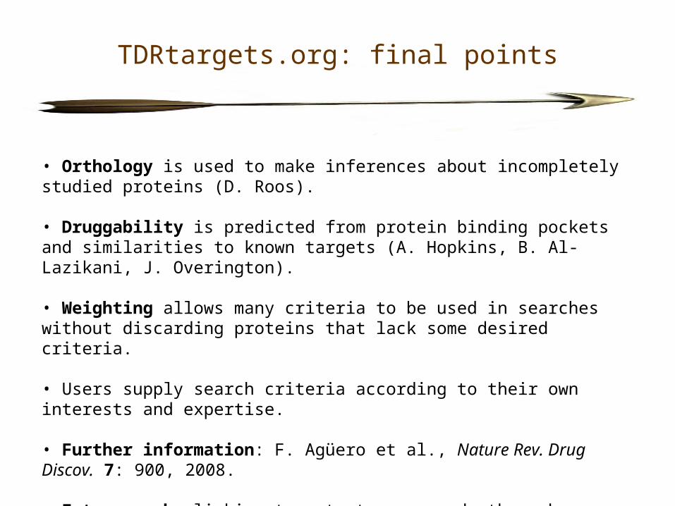
TDRtargets.org: final points
• Orthology is used to make inferences about incompletely studied proteins (D. Roos).
• Druggability is predicted from protein binding pockets and similarities to known targets (A. Hopkins, B. Al-Lazikani, J. Overington).
• Weighting allows many criteria to be used in searches without discarding proteins that lack some desired criteria.
• Users supply search criteria according to their own interests and expertise.
• Further information: F. Agüero et al., Nature Rev. Drug Discov. 7: 900, 2008.
• Future work: linking targets to compounds through informatics.
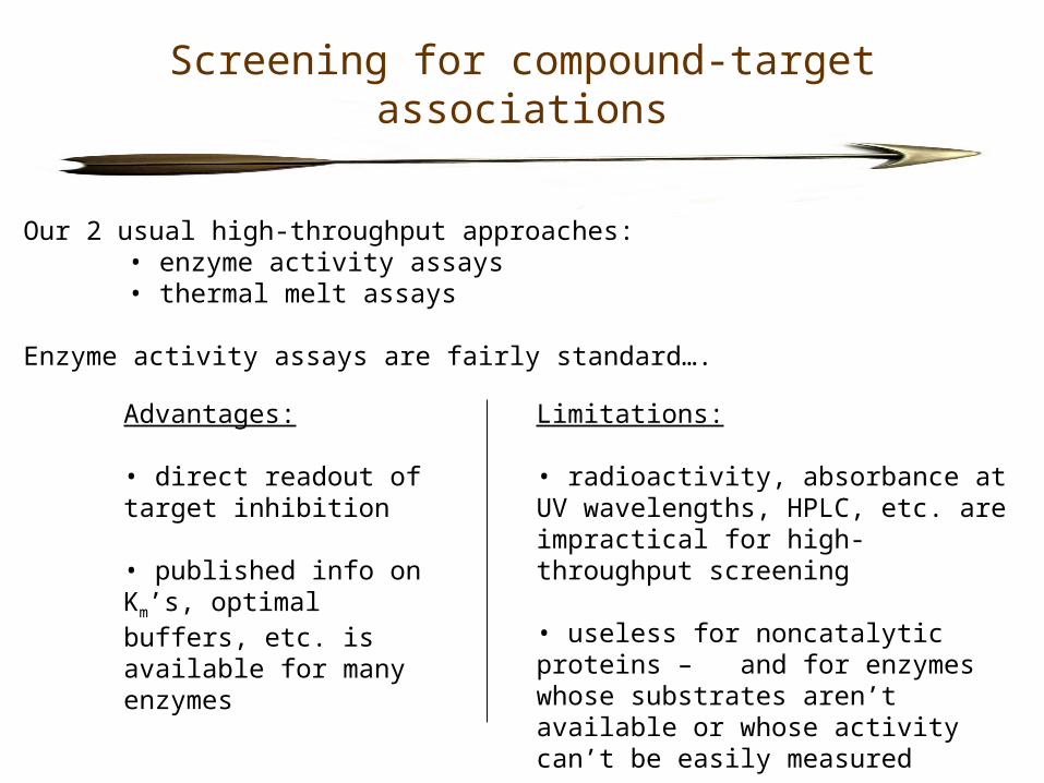
Screening for compound-target associations
Our 2 usual high-throughput approaches: • enzyme activity assays• thermal melt assays
Enzyme activity assays are fairly standard….
Advantages:
• direct readout of target inhibition
• published info on Km’s, optimal buffers, etc. is available for many enzymes
Limitations:
• radioactivity, absorbance at UV wavelengths, HPLC, etc. are impractical for high-throughput screening
• useless for noncatalytic proteins – and for enzymes whose substrates aren’t available or whose activity can’t be easily measured
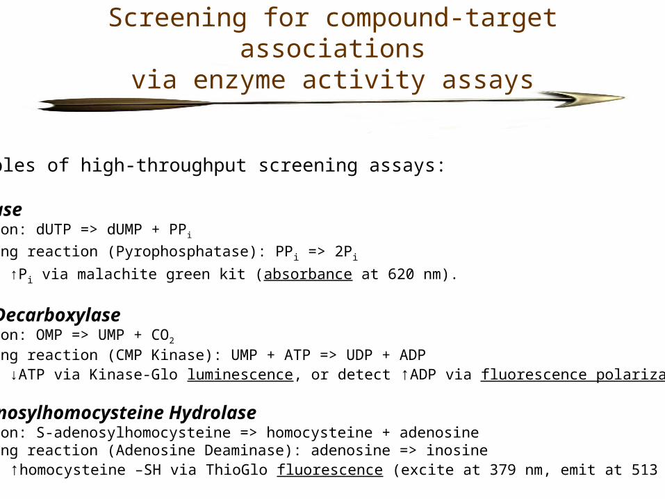
Screening for compound-target associationsvia enzyme activity assays
Examples of high-throughput screening assays:
dUTPaseReaction: dUTP => dUMP + PPi
Coupling reaction (Pyrophosphatase): PPi => 2Pi
Detect ↑Pi via malachite green kit (absorbance at 620 nm).
OMP DecarboxylaseReaction: OMP => UMP + CO2
Coupling reaction (CMP Kinase): UMP + ATP => UDP + ADPDetect ↓ATP via Kinase-Glo luminescence, or detect ↑ADP via fluorescence polarization.
S-Adenosylhomocysteine HydrolaseReaction: S-adenosylhomocysteine => homocysteine + adenosineCoupling reaction (Adenosine Deaminase): adenosine => inosineDetect ↑homocysteine –SH via ThioGlo fluorescence (excite at 379 nm, emit at 513 nm).
Screening for compound-target associations via enzyme activity assays
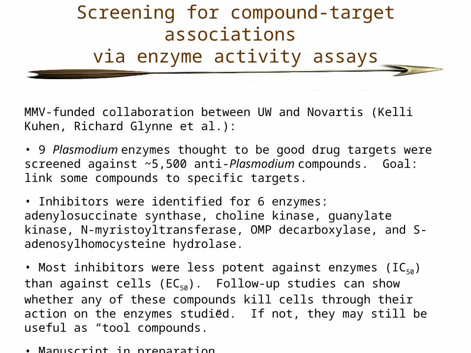
MMV-funded collaboration between UW and Novartis (Kelli Kuhen, Richard Glynne et al.):
• 9 Plasmodium enzymes thought to be good drug targets were screened against ~5,500 anti-Plasmodium compounds. Goal: link some compounds to specific targets.
• Inhibitors were identified for 6 enzymes: adenylosuccinate synthase, choline kinase, guanylate kinase, N-myristoyltransferase, OMP decarboxylase, and S-adenosylhomocysteine hydrolase.
• Most inhibitors were less potent against enzymes (IC50) than against cells (EC50). Follow-up studies can show whether any of these compounds kill cells through their action on the enzymes studied. If not, they may still be useful as “tool compounds.”
• Manuscript in preparation….
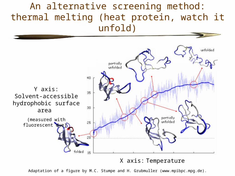
An alternative screening method:thermal melting (heat protein, watch it unfold)
X axis: Temperature
Y axis:Solvent-accessible
hydrophobic surface area
(measured with fluorescent dye)
Adaptation of a figure by M.C. Stumpe and H. Grubmuller (www.mpibpc.mpg.de).
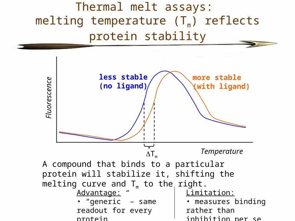
Thermal melt assays: melting temperature (Tm) reflects protein stability
Flu
ores
cenc
e
Temperature
less stable(no ligand)
more stable(with ligand)
Tm
A compound that binds to a particular protein will stabilize it, shifting the melting curve and Tm to the right.
Advantage:• “generic” – same readout for every protein
Limitation:• measures binding rather than inhibition per se
Thermal melt assays:application to screens of pathogen proteins
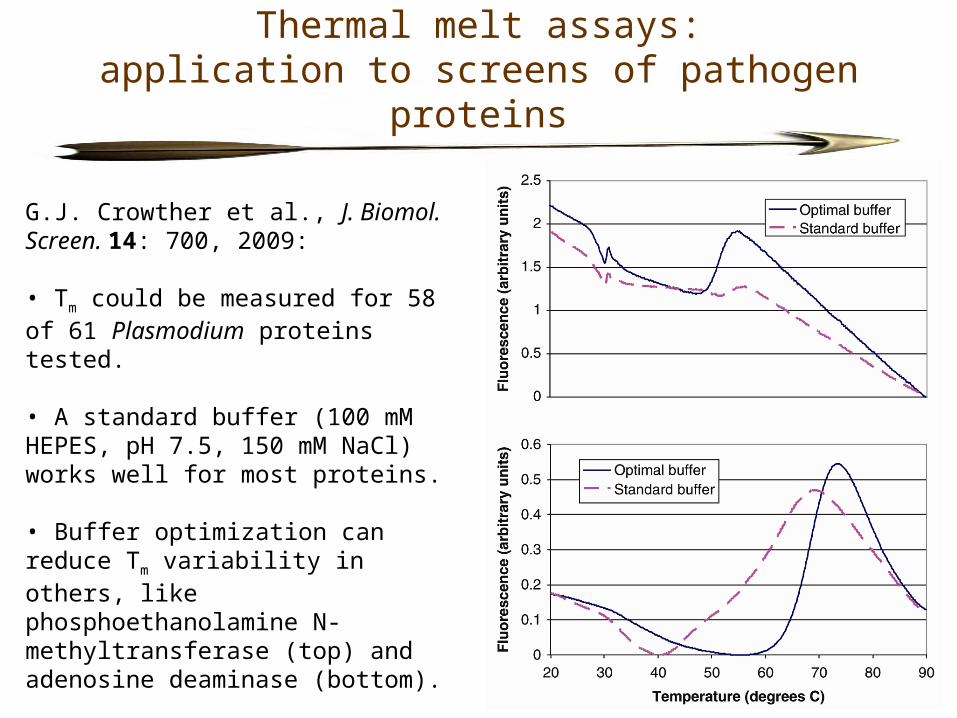
G.J. Crowther et al., J. Biomol. Screen. 14: 700, 2009:
• Tm could be measured for 58 of 61 Plasmodium proteins tested.
• A standard buffer (100 mM HEPES, pH 7.5, 150 mM NaCl) works well for most proteins.
• Buffer optimization can reduce Tm variability in others, like phosphoethanolamine N-methyltransferase (top) and adenosine deaminase (bottom).
Thermal melt assays:application to screens of pathogen proteins
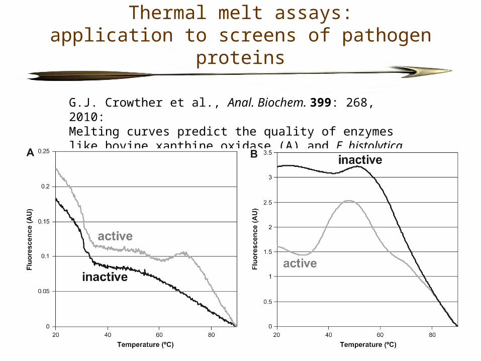
G.J. Crowther et al., Anal. Biochem. 399: 268, 2010:Melting curves predict the quality of enzymes like bovine xanthine oxidase (A) and E. histolytica cysteine protease 1 (B).
Thermal melt assays:application to screens of pathogen proteins
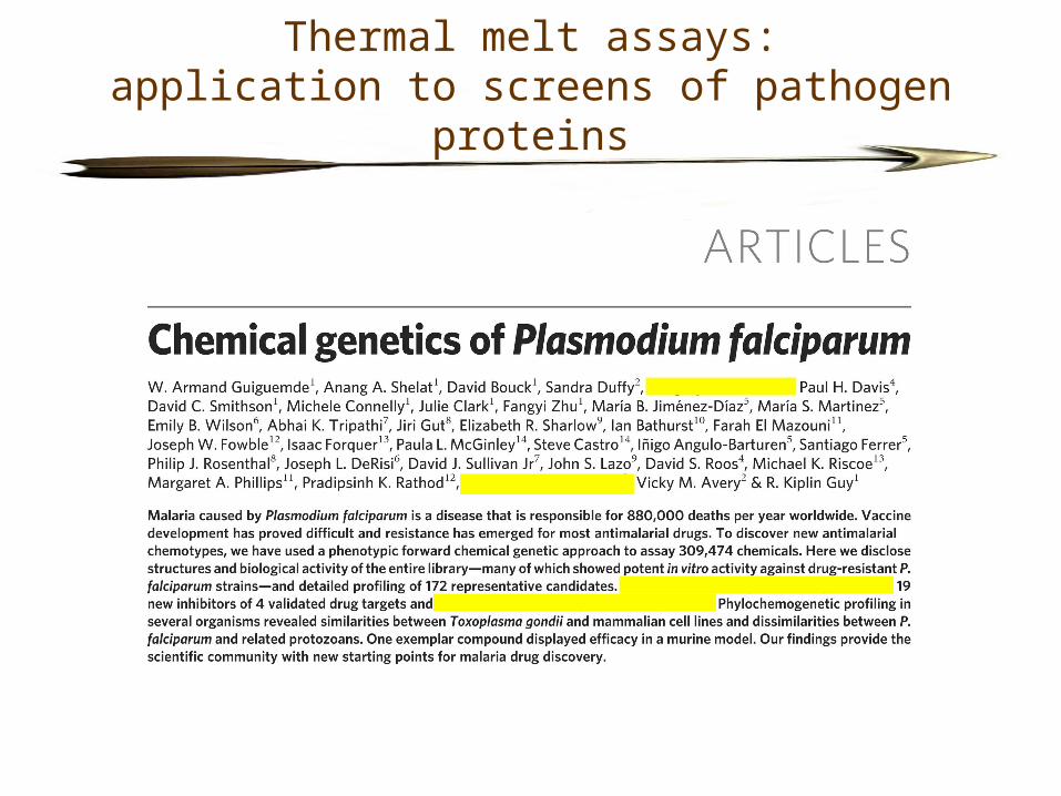
Collaborative project led by Kip Guy at St. Jude (Memphis):
• A screen of ~300,000 compounds revealed 172 new potent inhibitors of blood-stage P. falciparum (EC50 < 2 µM).
• UW contribution: these 172 compounds were screened against 61 Plasmodium proteins via thermal melt assays.
• Ligands (possible inhibitors) were identified for 7 proteins: 6-phosphogluconolactonase, 6-pyruvoyltetrahydropterin synthase, choline kinase, D-ribulose-5-phosphate 3-epimerase, glycogen synthase kinase, and thioredoxin.
• Paper will be published in Nature on May 20….
Thermal melt assays:application to screens of pathogen proteins
Conclusion
• Target-based drug discovery has not yet led to many new drugs for neglected diseases.
• Nevertheless there are reasons for optimism.
- New genomic/bioinformatic data (e.g., via TDRtargets.org): ○ more possible protein targets○ better prioritization of targets
- New screening methods (e.g., thermal melt assays): ○ more “screenable” proteins
- New 3D protein structures (e.g., via MSGPP and SSGCID): ○ more structure-based drug design
- New private-sector involvement (e.g., Novartis): ○ better compound libraries○ more screening horsepower○ more piggy-backing opportunities
Thank you . . . “Parasites! Excellent!”
. . . to my assay team:- Diana Chung- Panqing He- Kuzma Kovzun- Phil Rodenbough- Andrew Thomas
. . . to other inhabitants of Wes’ World:- Lynn Barrett (lab manager)- Fred Buckner’s lab- Mike Gelb (Dept. of Chemistry)- Wim Hol and MSGPP- Alberto Napuli’s expression/purification group
. . . to collaborating groups around the world:- Kip Guy (St. Jude)- Ray Hui (SGC, U. of Toronto)- Kelli Kuhen and Richard Glynne (Novartis)- TDR network (Fernán Agüero, Matt Berriman, Stuart Ralph, David Roos)- Roger Weigand and Dyann Wirth (Broad Institute)
. . . to funding agencies:- Medicines for Malaria Venture (MMV)- WHO/TDR

The End
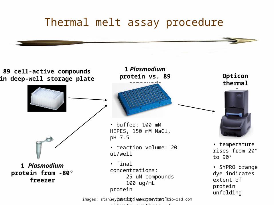
Thermal melt assay procedure
89 cell-active compoundsin deep-well storage plate
1 Plasmodium protein vs. 89 compounds Opticon
thermal cycler
• temperature rises from 20° to 90°
• SYPRO orange dye indicates extent of protein unfolding
• buffer: 100 mM HEPES, 150 mM NaCl, pH 7.5
• reaction volume: 20 uL/well
• final concentrations: 25 uM compounds 100 ug/mL protein
• positive control: citrate synthase +/- oxaloacetate
images: stanleylab.org; eenzyme.com; bio-rad.com
1 Plasmodium protein from -80° freezer
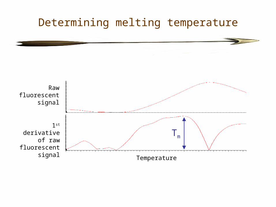
Determining melting temperature
Temperature
Raw fluorescent
signal
1st derivative of raw fluorescent
signalTm
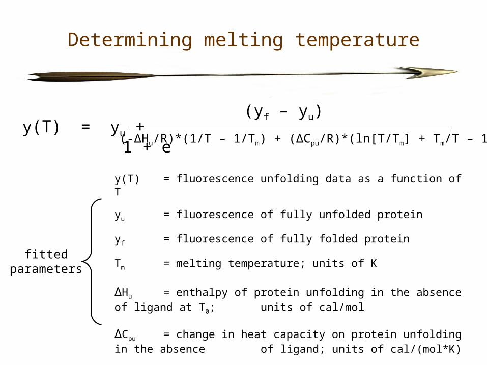
Determining melting temperature
y(T) = yu +1 + e
(-ΔHu/R)*(1/T – 1/Tm) + (ΔCpu/R)*(ln[T/Tm] + Tm/T – 1)
y(T) = fluorescence unfolding data as a function of T
yu = fluorescence of fully unfolded protein
yf = fluorescence of fully folded protein
Tm = melting temperature; units of K ΔHu = enthalpy of protein unfolding in the absence of ligand at T0;
units of cal/mol
ΔCpu = change in heat capacity on protein unfolding in the absence of ligand; units of cal/(mol*K)
R = gas constant; units of cal/(mol*K)
(yf – yu)
fittedparameters
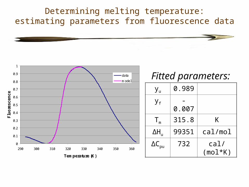
Determining melting temperature:estimating parameters from fluorescence data
0
0.1
0.2
0.3
0.4
0.5
0.6
0.7
0.8
0.9
1
290 300 310 320 330 340 350 360
Temperature (K)
Flu
ore
sc
en
ce
data
model
yu 0.989
yf -0.007
Tm 315.8 K
ΔHu 99351 cal/mol
ΔCpu 732 cal/(mol*K)
Fitted parameters:
Determining melting temperature
310
315
320
325
330
335
340
345
350
310 315 320 325 330 335 340 345 350
Tm estimated by parameter fitting (K)
Tm
es
tim
ate
d b
y 1
st
de
riv
ati
ve
(K
)
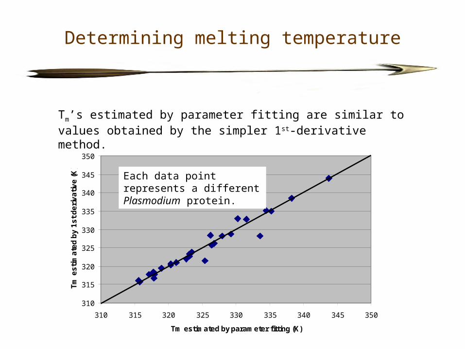
Each data point represents a different Plasmodium protein.
Tm’s estimated by parameter fitting are similar to values obtained by the simpler 1st-derivative method.
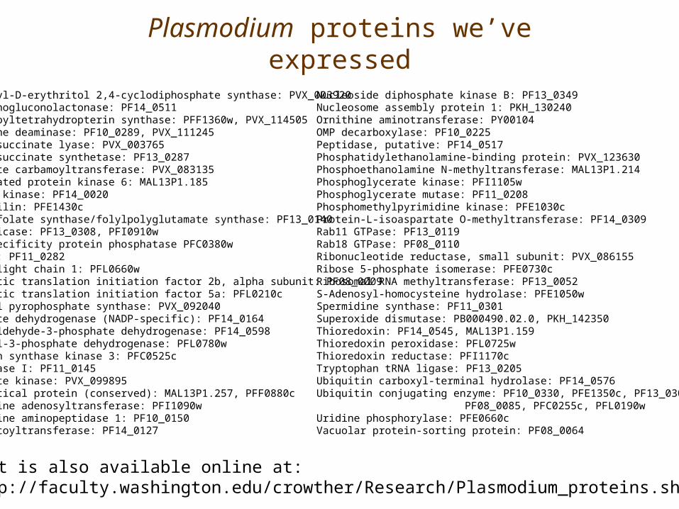
Plasmodium proteins we’ve expressed
List is also available online at:http://faculty.washington.edu/crowther/Research/Plasmodium_proteins.shtml
2C-methyl-D-erythritol 2,4-cyclodiphosphate synthase: PVX_003920 6-phosphogluconolactonase: PF14_05116-pyruvoyltetrahydropterin synthase: PFF1360w, PVX_114505 Adenosine deaminase: PF10_0289, PVX_111245Adenylosuccinate lyase: PVX_003765Adenylosuccinate synthetase: PF13_0287 Aspartate carbamoyltransferase: PVX_083135 CDK-related protein kinase 6: MAL13P1.185 Choline kinase: PF14_0020Cyclophilin: PFE1430c Dihydrofolate synthase/folylpolyglutamate synthase: PF13_0140 DNA helicase: PF13_0308, PFI0910w Dual-specificity protein phosphatase PFC0380w dUTPase: PF11_0282 Dynein light chain 1: PFL0660w Eukaryotic translation initiation factor 2b, alpha subunit: PF08_0009 Eukaryotic translation initiation factor 5a: PFL0210c Farnesyl pyrophosphate synthase: PVX_092040 Glutamate dehydrogenase (NADP-specific): PF14_0164 Glyceraldehyde-3-phosphate dehydrogenase: PF14_0598 Glycerol-3-phosphate dehydrogenase: PFL0780w Glycogen synthase kinase 3: PFC0525c Glyoxalase I: PF11_0145 Guanylate kinase: PVX_099895 Hypothetical protein (conserved): MAL13P1.257, PFF0880c Methionine adenosyltransferase: PFI1090w Methionine aminopeptidase 1: PF10_0150N-myristoyltransferase: PF14_0127
Nucleoside diphosphate kinase B: PF13_0349 Nucleosome assembly protein 1: PKH_130240 Ornithine aminotransferase: PY00104 OMP decarboxylase: PF10_0225 Peptidase, putative: PF14_0517 Phosphatidylethanolamine-binding protein: PVX_123630 Phosphoethanolamine N-methyltransferase: MAL13P1.214 Phosphoglycerate kinase: PFI1105w Phosphoglycerate mutase: PF11_0208 Phosphomethylpyrimidine kinase: PFE1030c Protein-L-isoaspartate O-methyltransferase: PF14_0309 Rab11 GTPase: PF13_0119 Rab18 GTPase: PF08_0110 Ribonucleotide reductase, small subunit: PVX_086155 Ribose 5-phosphate isomerase: PFE0730c Ribosomal RNA methyltransferase: PF13_0052 S-Adenosyl-homocysteine hydrolase: PFE1050w Spermidine synthase: PF11_0301 Superoxide dismutase: PB000490.02.0, PKH_142350 Thioredoxin: PF14_0545, MAL13P1.159 Thioredoxin peroxidase: PFL0725w Thioredoxin reductase: PFI1170c Tryptophan tRNA ligase: PF13_0205 Ubiquitin carboxyl-terminal hydrolase: PF14_0576 Ubiquitin conjugating enzyme: PF10_0330, PFE1350c, PF13_0301,
PF08_0085, PFC0255c, PFL0190w Uridine phosphorylase: PFE0660c Vacuolar protein-sorting protein: PF08_0064

![Life Sciences...76 3 Contribution of Natural Products to Drug Discovery in Tropical Diseases mosquito [2]. Plasmodium falciparum, Plasmodium vivax, Plasmodium ovale, Plasmodium malariae,andPlasmodium](https://img.pdfslide.us/doc/110x75/6049cbda4f3447749747f712/life-sciences-76-3-contribution-of-natural-products-to-drug-discovery-in-tropical.jpg)