Embed Size (px)
Citation preview
CELL CELL STRUCTURE STRUCTURE
AND FUNCTIONAND FUNCTION
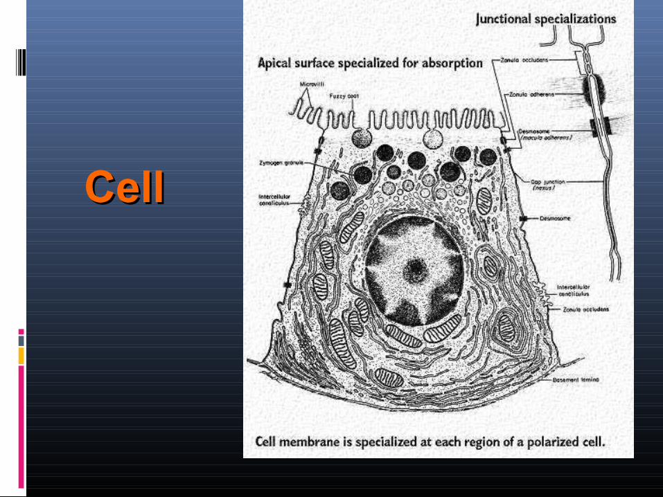
CellCell
PLASMA MEMBRANEPLASMA MEMBRANE
PLASMA MEMBRANEPLASMA MEMBRANE
It is necessary that for the proper functioning the cells and cell components should be separated from the outside environment.
PLASMA MEMBRANEPLASMA MEMBRANE
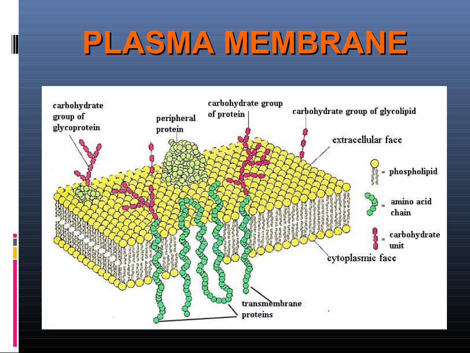
A lipid/protein/carbohydrate complex.
Provides a barrier to the cell
Forms a boundary between the cell and its environment
Contains transport and signaling systems
Membranes bound organelles
Segregation of biochemical activities within the cell
PLASMA MEMBRANEPLASMA MEMBRANE
The lipids have
hydrophilic polar heads
pointing out and the
hydrophobic portion
forming the core.
PLASMA MEMBRANEPLASMA MEMBRANE
Lipid bilayers are fluid.Individual phospholipids diffuse rapidly throughout the two dimensional surface of the membrane.
The Fluid Mosaic Model
This is known as the fluid mosaic model of biological membranes (mosaic because it includes molecules other than phospholipids, such as proteins, cholesterol, and other types of molecules).
PLASMA MEMBRANEPLASMA MEMBRANE
PLASMA MEMBRANEPLASMA MEMBRANE
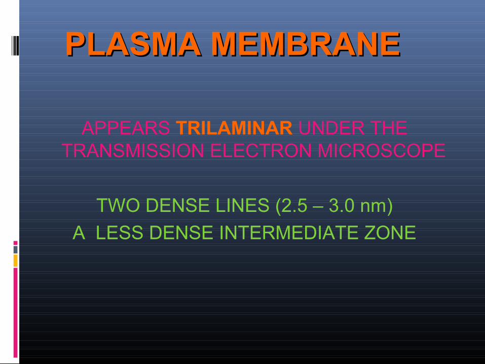
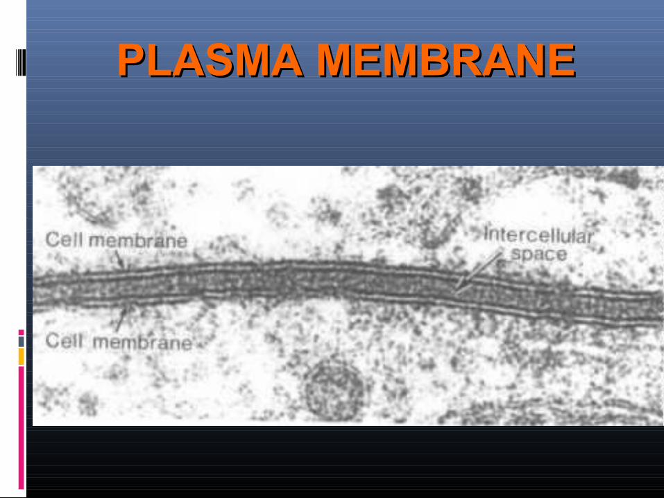
APPEARS TRILAMINAR UNDER THE TRANSMISSION ELECTRON MICROSCOPE
TWO DENSE LINES (2.5 – 3.0 nm)
A LESS DENSE INTERMEDIATE ZONE
PLASMA MEMBRANEPLASMA MEMBRANE
GLYCOCALYXGLYCOCALYX
THE CARBOHYDRATE MOLECULES FORM A THIN OUTER LAYER CALLED THE GLYCOCALYX
PLASMA MEMBRANEPLASMA MEMBRANE
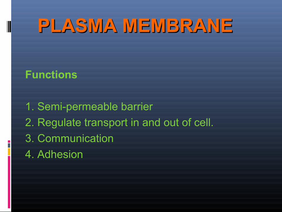
� Functions
� 1. Semi-permeable barrier � 2. Regulate transport in and out of cell. � 3. Communication � 4. Adhesion
PLASMA MEMBRANEPLASMA MEMBRANE
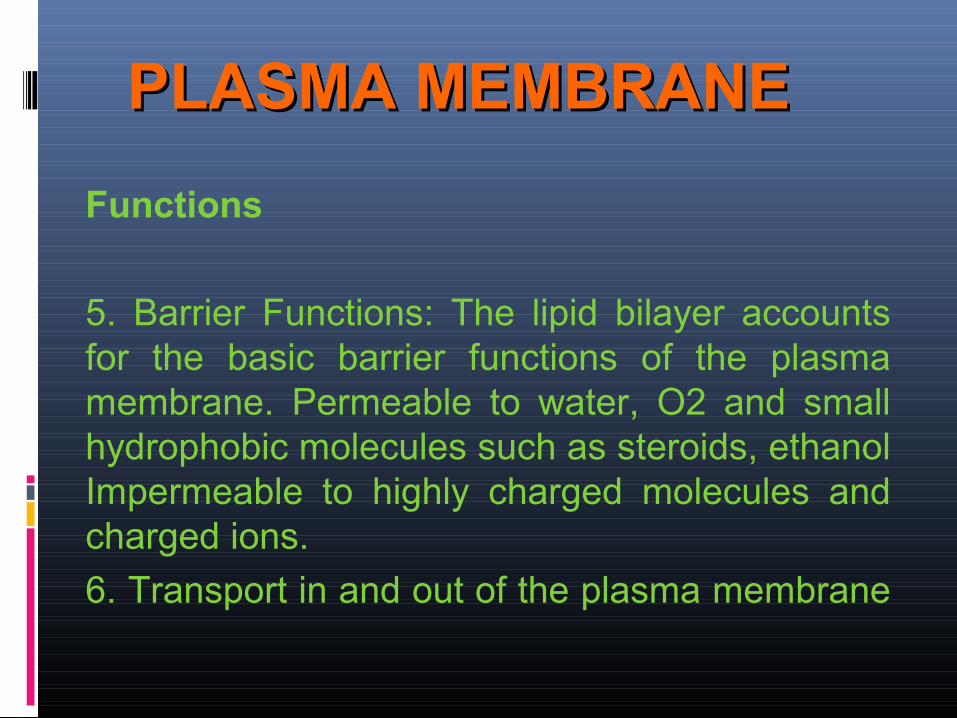
� Functions
� 5. Barrier Functions: The lipid bilayer accounts for the basic barrier functions of the plasma membrane. Permeable to water, O2 and small hydrophobic molecules such as steroids, ethanol Impermeable to highly charged molecules and charged ions.
� 6. Transport in and out of the plasma membrane
NUCLEUSNUCLEUS
NUCLEUSNUCLEUS
The nucleus is the controlling station of The nucleus is the controlling station of eukaryotic cell. eukaryotic cell.
Usually the nucleus is round and is the Usually the nucleus is round and is the largest organelle in the cell. largest organelle in the cell.
NUCLEUSNUCLEUS
It is surrounded by a membrane, called It is surrounded by a membrane, called the nuclear envelope, which is similar to the nuclear envelope, which is similar to the cell membrane that encloses the the cell membrane that encloses the entire cell. entire cell.
NUCLEUSNUCLEUS
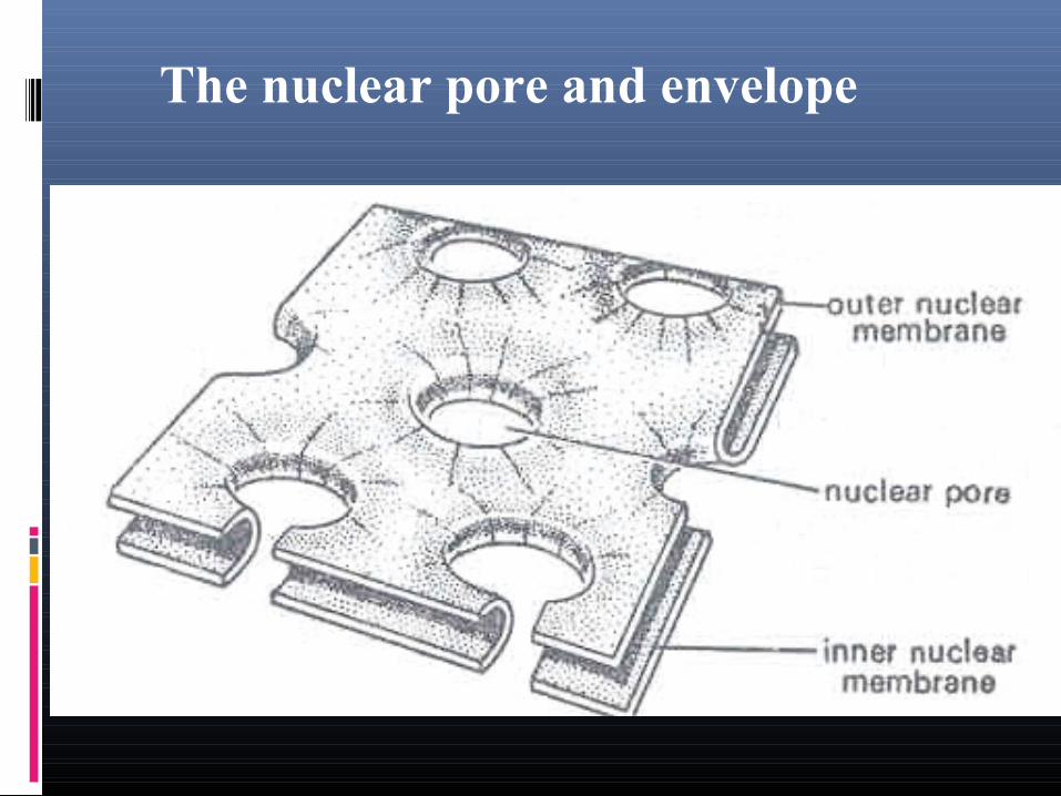
Nuclear membrane possess specific Nuclear membrane possess specific openings called nuclear pores, that allow openings called nuclear pores, that allow specific materials to pass in and out of specific materials to pass in and out of the nucleus.the nucleus.
NUCLEUSNUCLEUS
Attached to the nuclear envelope is the Attached to the nuclear envelope is the endoplasmic reticulum. endoplasmic reticulum.
The nucleus is surrounded by the The nucleus is surrounded by the cytoplasm inside a cell.cytoplasm inside a cell.
NUCLEUSNUCLEUS
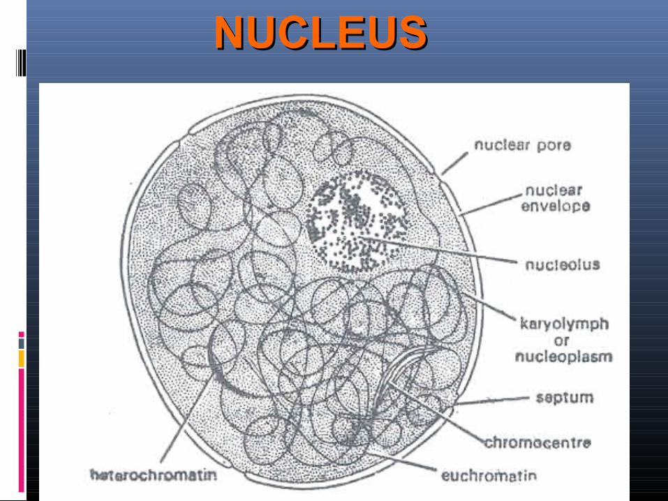
Structurally, the nucleus is Structurally, the nucleus is composed of three main parts, composed of three main parts,
the nucleolus, the nucleolus, the nuclear envelope, and the nuclear envelope, and the chromatin.the chromatin.
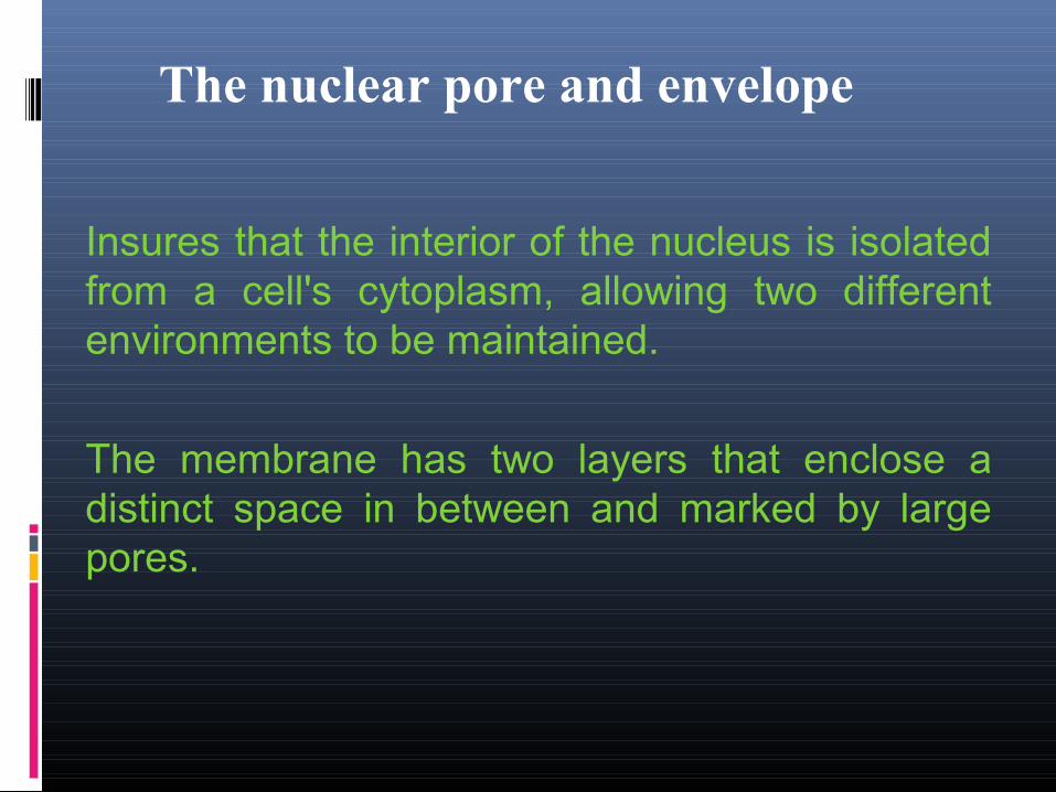
The nuclear pore and envelope
� Insures that the interior of the nucleus is isolated from a cell's cytoplasm, allowing two different environments to be maintained.
� The membrane has two layers that enclose a distinct space in between and marked by large pores.
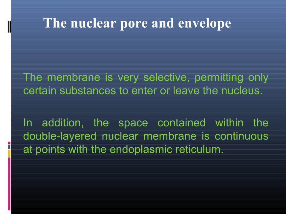
The nuclear pore and envelope
� The membrane is very selective, permitting only certain substances to enter or leave the nucleus.
� In addition, the space contained within the double-layered nuclear membrane is continuous at points with the endoplasmic reticulum.
The nuclear pore and envelope
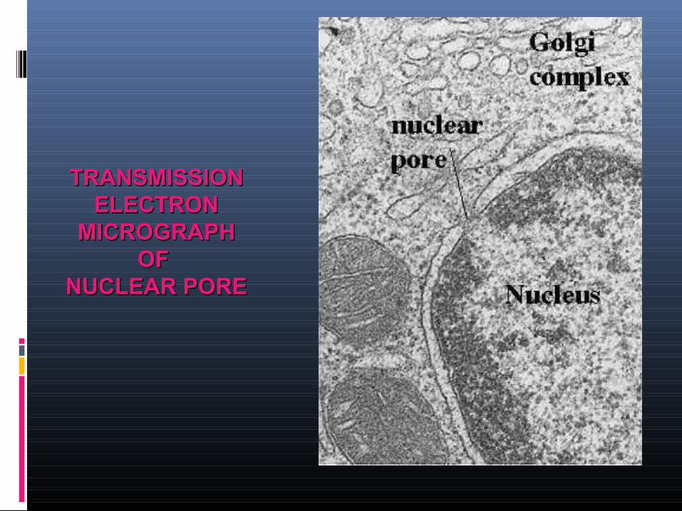
TRANSMISSIONTRANSMISSIONELECTRONELECTRON
MICROGRAPHMICROGRAPHOF OF
NUCLEAR PORENUCLEAR PORE
NUCLEOLUSNUCLEOLUS
� Principal RNA containing structure of the nucleus.
� It is rich in protein � is surrounded by a ring of
heterochromatin which may penetrate into the main body of the nucleolar complex.
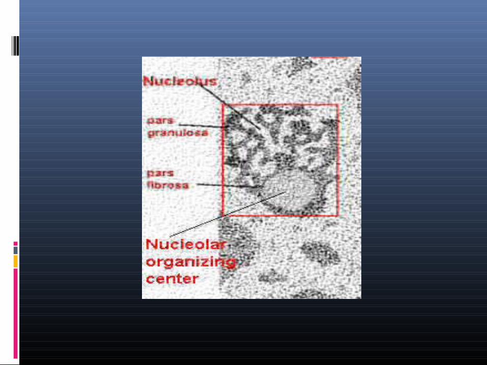
NUCLEOLUSNUCLEOLUS
� The fine structure of the nucleolus reveals the presence of a fibrillar and a granular area, each composed of ribonucleoproeins, and each is related to the biogenesis of ribosomes.
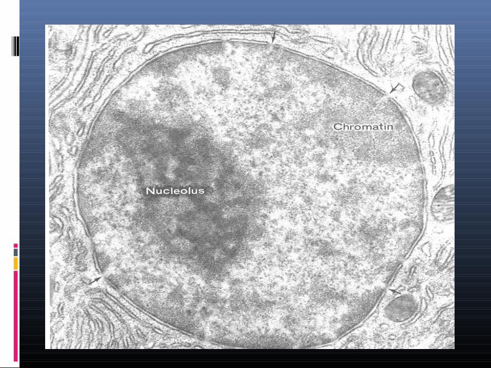
CHROMATINCHROMATIN
� Chromatin is the name that describes nuclear material that contains the genetic code.
� The chromatin (meaning "colored substance") contains DNA and proteins.
� Chromatin gives rise to chromosomes.
CHROMATINCHROMATIN
Two types of chromatin material
Heterochromatin and
Euchromatin.
HETEROCHROMATINHETEROCHROMATIN
The darkly stained, condensed region of the chromatin is known as heterochromatin.
The heterochromatin occurs around the nucleolus and at the periphery.
It is supposed to be metabolically and genetically inert because it contains comparatively small amount of DNA and large amount of RNA.
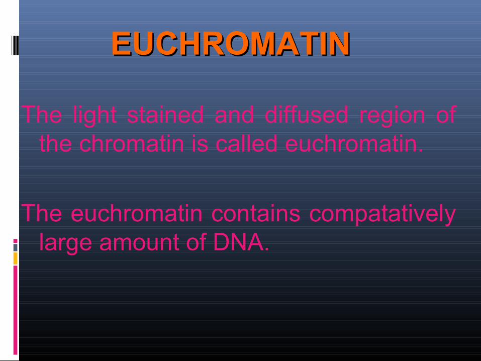
EUCHROMATINEUCHROMATIN
The light stained and diffused region of the chromatin is called euchromatin.
The euchromatin contains compatatively large amount of DNA.
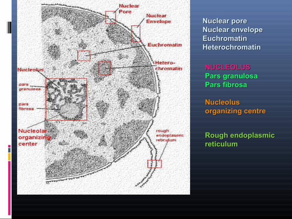
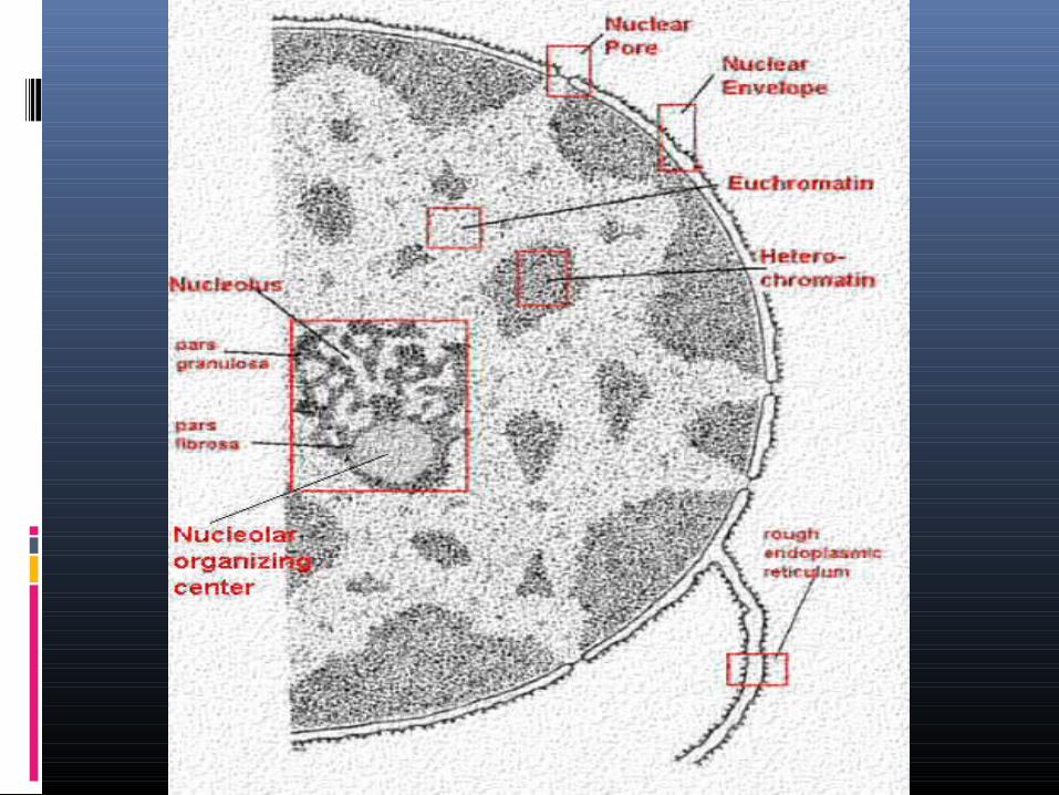
Nuclear poreNuclear poreNuclear envelopeNuclear envelopeEuchromatinEuchromatinHeterochromatinHeterochromatin
NUCLEOLUSNUCLEOLUSPars granulosaPars granulosaPars fibrosaPars fibrosa
Nucleolus Nucleolus organizing centreorganizing centre
Rough endoplasmic Rough endoplasmic reticulumreticulum
GOLGI APPARATUSGOLGI APPARATUS
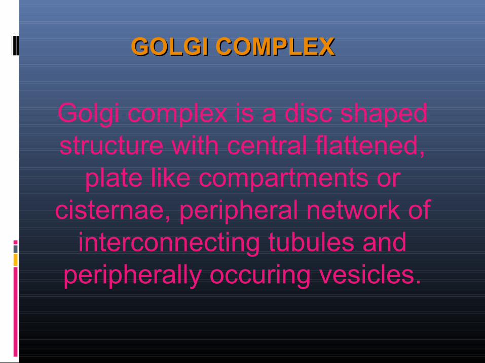
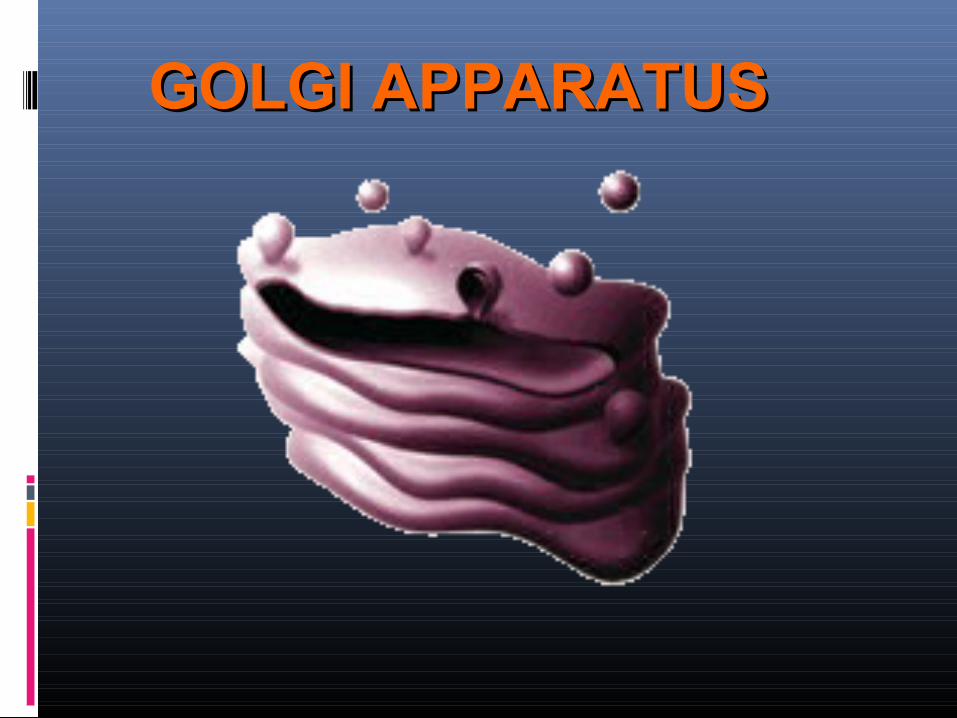
GOLGI COMPLEXGOLGI COMPLEX
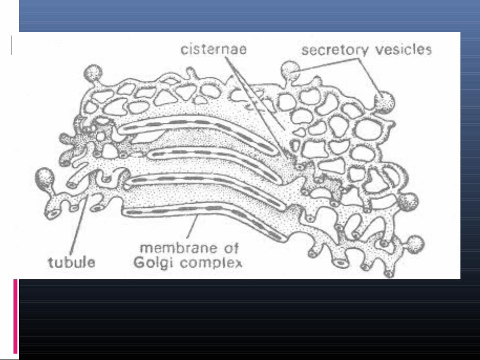
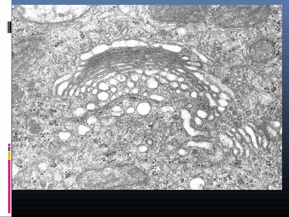
Golgi complex is a disc shaped structure with central flattened,
plate like compartments or cisternae, peripheral network of
interconnecting tubules and peripherally occuring vesicles.
FUNCTIONS OF GOLGI FUNCTIONS OF GOLGI COMPLEXCOMPLEX
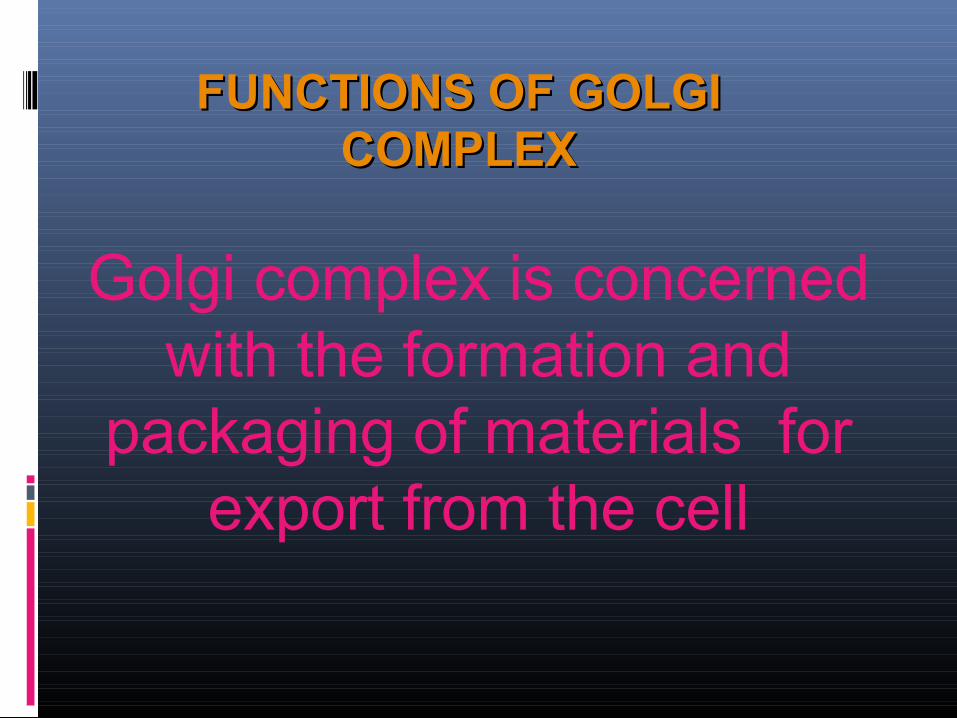
Golgi complex is concerned with the formation and
packaging of materials for export from the cell
ENDOPLASMIC ENDOPLASMIC RETICULUMRETICULUM
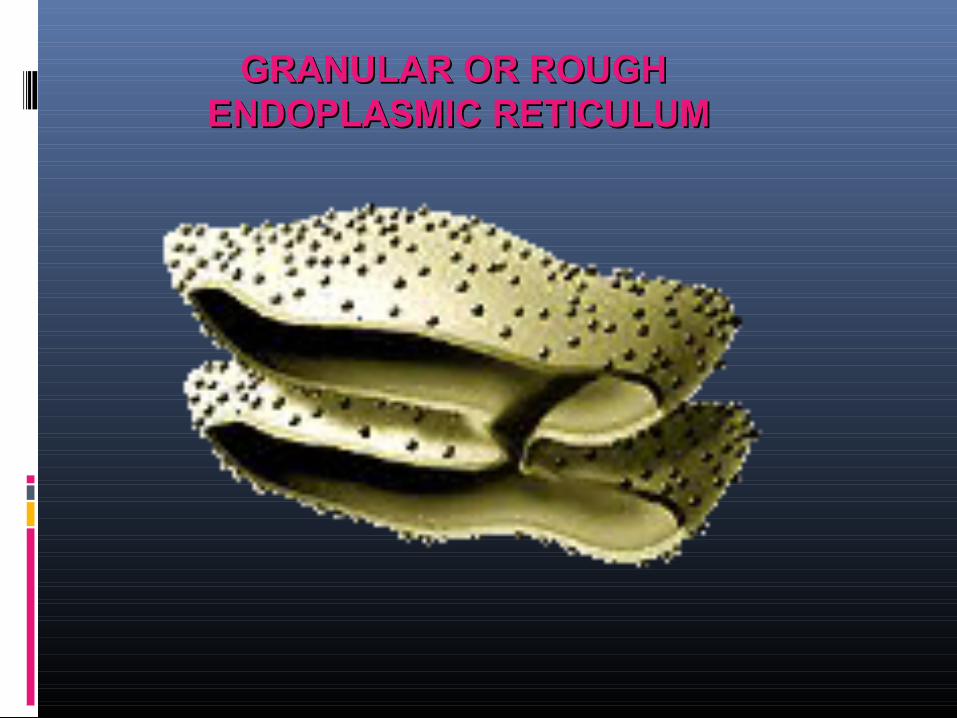
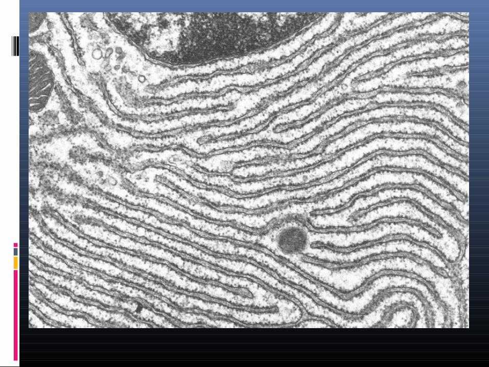
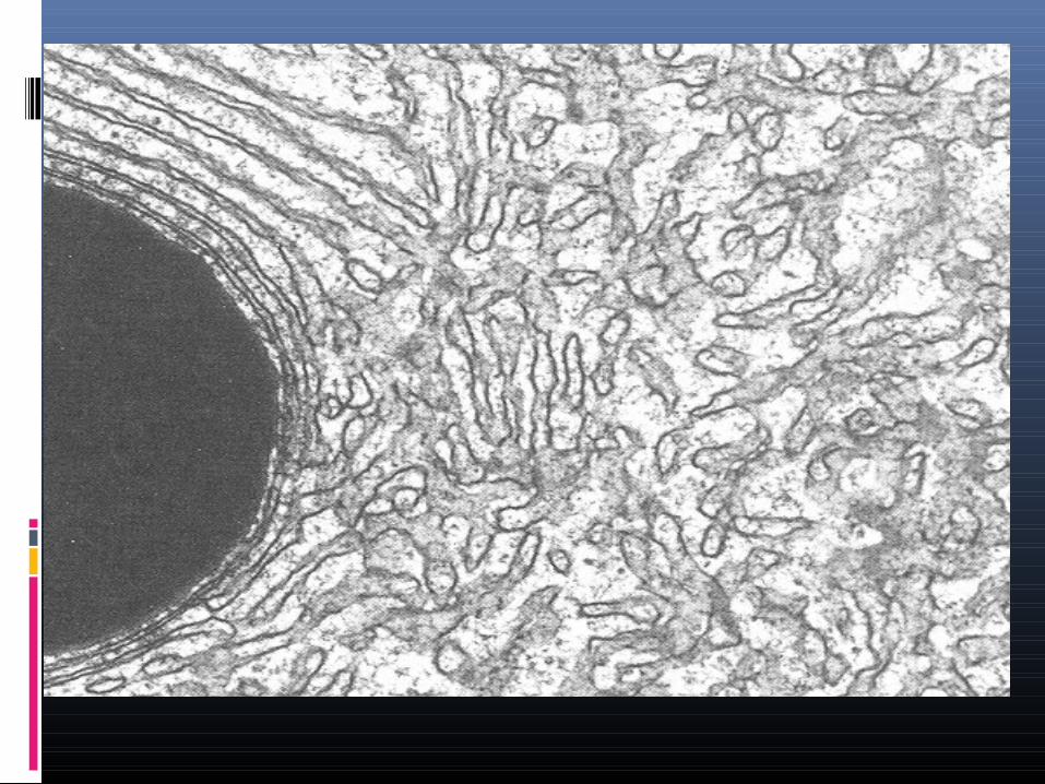
GRANULAR OR ROUGH GRANULAR OR ROUGH ENDOPLASMIC RETICULUMENDOPLASMIC RETICULUM
GRANULAR OR ROUGH GRANULAR OR ROUGH ENDOPLASMIC RETICULUMENDOPLASMIC RETICULUM
It possesses rough walls because the ribosomes remain attached with its membranes.
The granular or rough type of endoplasmic reticulum is found abundantly in those cells which are active in protein synthesis.
FUNCTIONS OF GRANULAR OR ROUGH FUNCTIONS OF GRANULAR OR ROUGH ENDOPLASMIC RETICULUMENDOPLASMIC RETICULUM
Rough ER is involved in protein synthesis.
The endoplasmic reticulum also functions as a transport system.
Protein molecules move from the rough ER into the smooth ER, which then sends them enclosed within the vesicles usually to the Golgi complex.

AGRANULAR OR SMOOTH AGRANULAR OR SMOOTH ENDOPLASMIC RETICULUMENDOPLASMIC RETICULUM
FUNCTIONS OF AGRANULAR OR FUNCTIONS OF AGRANULAR OR
SMOOTHSMOOTHENDOPLASMIC RETICULUMENDOPLASMIC RETICULUM
The smooth type of endoplasmic reticulum occurs mostly in those cells, which have no
active participation in the synthesis of proteins.
It is involved in the synthesis of
LIPIDS, GLYCOGEN AND STEROIDS
RIBOSOMESRIBOSOMES
The ribosomes are small, dense, rounded and granular particles of ribonucleoprotein.
They are found either freely in the matrix of mitochondria, chloroplast and cytoplasm or found attached on the surface of the endoplasmic reticulum and nucleus.
RIBOSOMESRIBOSOMES
Ribosomes are present in most prokaryotic and eukaryotic cells.
They are the sites of protein synthesis where amino acids are assembled to produce the polypeptide chain.
RIBOSOMESRIBOSOMES
Ribosomes are classified into two types, based on the size and the sedimentation coefficient (S).
They are:
1. 70S Ribosomes and2. 80S Ribosomes
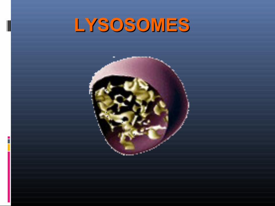
LYSOSOMESLYSOSOMES
The lysosomes are tiny, membrane bounded, vesicular structures of the cytoplasm which perform intracellular digestion of the cell.
The term lysosome means digestive body (Gr., lyso-digestive, soma-body).
LYSOSOMESLYSOSOMES
SUICIDE BAGS OF THE CELL
FUNCTIONS OF FUNCTIONS OF LYSOSOMESLYSOSOMES
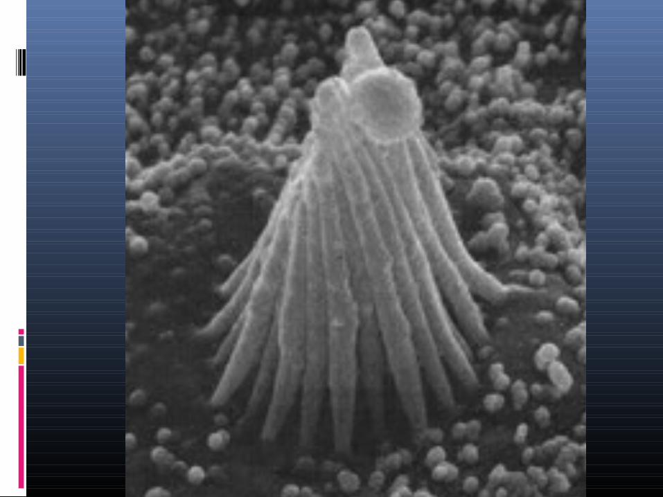
Cytoplasm of some eukaryotic cells contains two cylindrical, rod-shaped, microtubular structures, called centrioles, near the nucleus.
CENTRIOLESCENTRIOLES
Centrioles lack limiting membrane and DNA or RNA.
It gives rise to the mitotic apparatus during mitosis or meiosis.
CENTRIOLESCENTRIOLES
In flagellated or ciliated cells centrioles are found arranged just beneath the plasma membrane to form and bear flagella or cilia.
When a centriole bears a flagellum or cilium, it is called basal body.
BASAL BODIESBASAL BODIES
Peroxisomes are organelles that resemble lysosomes.
They are present in many animal and plant cells.
They contain oxidative enzymes.
PEROXISOMESPEROXISOMES
Peroxisomes are distinguished by a crystalline structure inside a sac which also contains amorphous gray material.
They are self replicating, like the mitochondria.
They also enlarge and bud to produce new peroxisomes.
PEROXISOMESPEROXISOMES
Peroxisomes protect the cells from the action of toxic substances like hydrogen peroxide, or other metabolites.
PEROXISOMESPEROXISOMES
Morphologically they resemble peroxisomes, but the crystalloid core consists of dense rods.
GLYOXISOMESGLYOXISOMES
They have enzymes for fatty acid metabolism and gluconeogenesis.
Glyoxisomes are present in yeast and the oil rich seeds of many plants.
GLYOXISOMESGLYOXISOMES
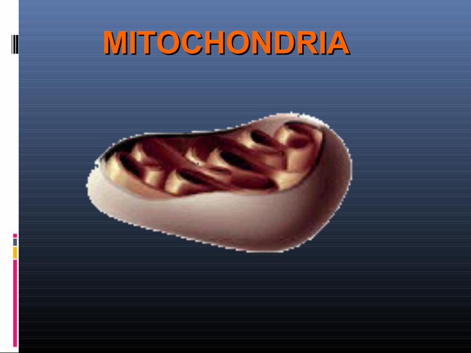
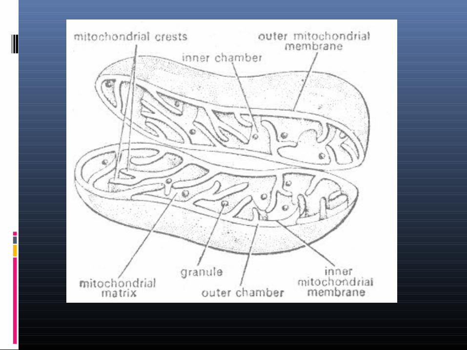
MITOCHONDRIAMITOCHONDRIA
MITOCHONDRIA
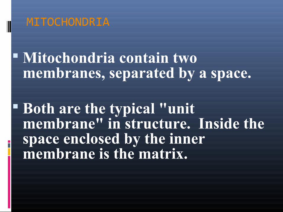
Mitochondria contain two membranes, separated by a space.
Both are the typical "unit membrane" in structure. Inside the space enclosed by the inner membrane is the matrix.
MITOCHONDRIA
The mitochondrial matrix contains lipids, proteins, circular DNA molecules, 70S ribosomes and certain granules which are related to the ability of mitochondria to accumulate ions.
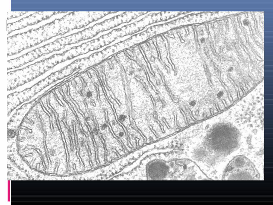
MITOCHONDRIAL CRESTS OR CRISTAE
The inner mitochondrial membrane increases its surface area by giving out
plate-like or tubular invaginations called
MITOCHONDRIAL CRESTS OR CRISTAE
MITOCHONDRIAL CRESTS OR CRISTAE
Mitochondria replicate much like bacterial cells.
They undergo fission. This involves a furrowing of the inner and then the outer membrane of the mitochondrion.
Then the two daughter mitochondria split.
Before the mitochondrion divides, the mitochondrial DNA replication occurs.
Sometimes new mitochondria are synthesized.

"POWER HOUSES" OF THE CELL.
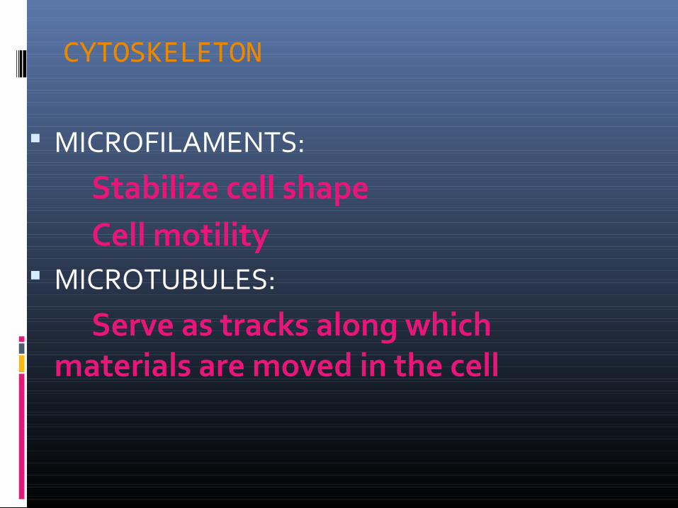
CYTOSKELETON
MICROFILAMENTS:
Stabilize cell shapeCell motility
MICROTUBULES:
Serve as tracks along which materials are moved in the cell
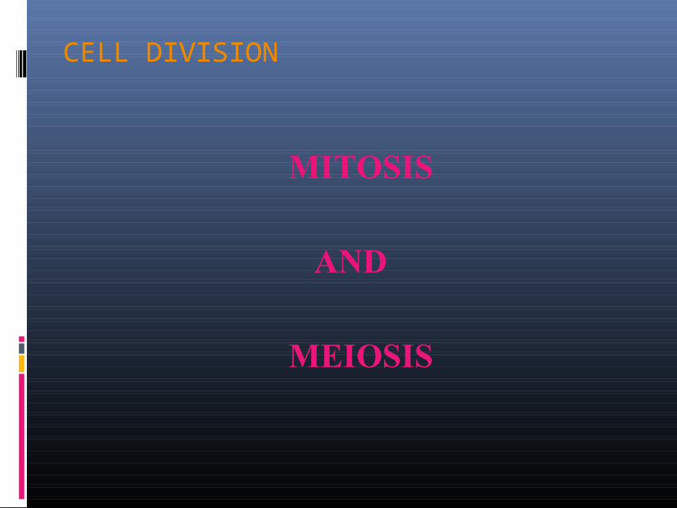
CELL DIVISION
MITOSIS
AND
MEIOSIS
CELL DIVISION
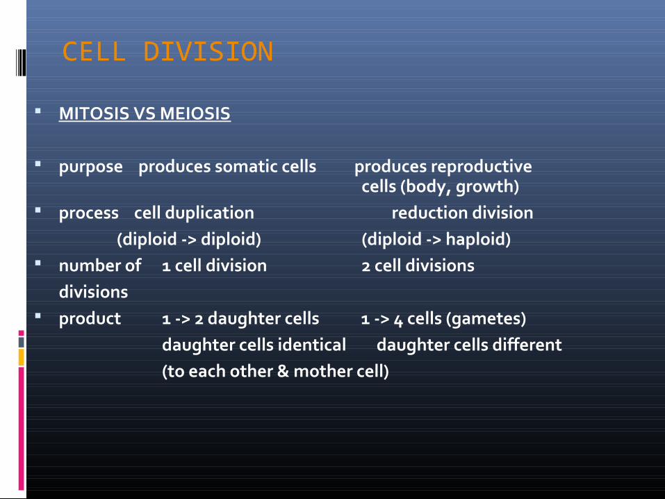
MITOSIS VS MEIOSIS
purpose produces somatic cells produces reproductive cells (body, growth)
process cell duplication reduction division (diploid -> diploid) (diploid -> haploid) number of 1 cell division 2 cell divisions
divisions product 1 -> 2 daughter cells 1 -> 4 cells (gametes)
daughter cells identical daughter cells different(to each other & mother cell)
CELL CYCLE
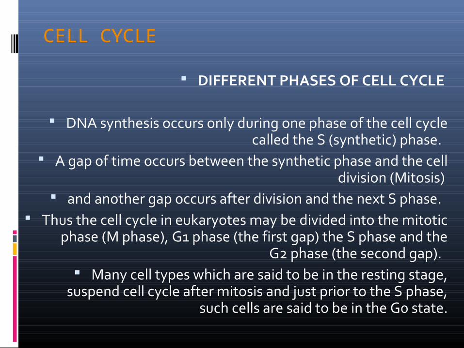
DIFFERENT PHASES OF CELL CYCLE
DNA synthesis occurs only during one phase of the cell cycle called the S (synthetic) phase.
A gap of time occurs between the synthetic phase and the cell division (Mitosis)
and another gap occurs after division and the next S phase. Thus the cell cycle in eukaryotes may be divided into the mitotic
phase (M phase), G1 phase (the first gap) the S phase and the G2 phase (the second gap).
Many cell types which are said to be in the resting stage, suspend cell cycle after mitosis and just prior to the S phase,
such cells are said to be in the G0 state.
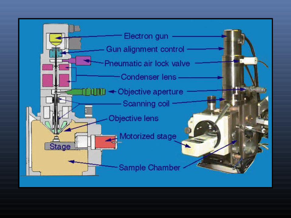
MICROSCOPYMICROSCOPY
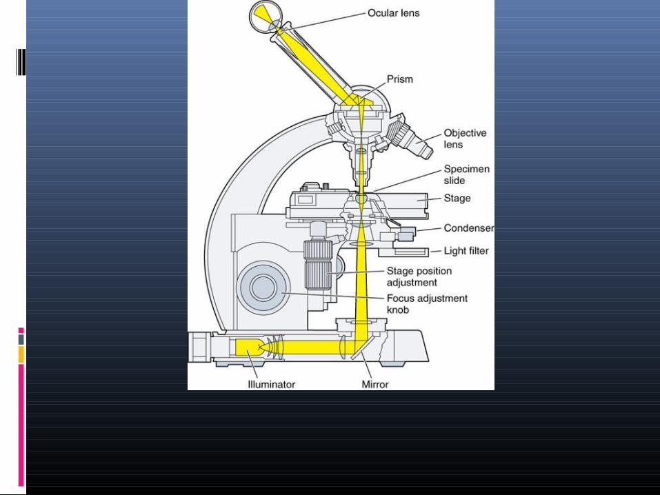
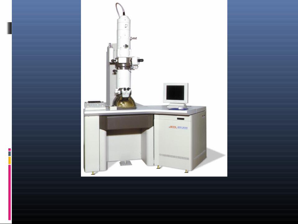
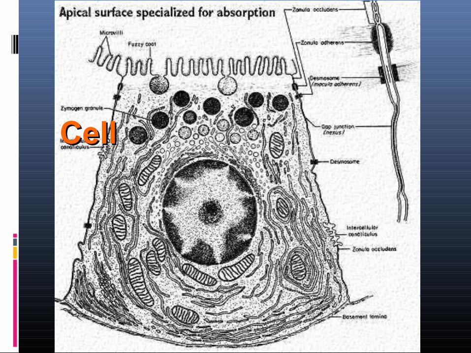
CellCell

IMMUNOHISTOCHEMISTRY

CELL CULTURE
CELL DIVISION- MITOSIS
A. Interphase.
1. This phase includes gap 1, S and gap 2 periods of cell cycle.
2. Since the replication of DNA occurs in the S phase, the nucleus has twice the diploid amount of DNA.
3. As the cell prepares to divide, the chromosome condense and become visible.
CELL DIVISION- MITOSIS
B. Prophase.
1. Chromosomes become visible, Each chromosome consists of a pair of long parallel strands (sister chromatids), which are held together at the centromere.
2. Crossing over between the sister chromatids may occur at this stage.
3. The nuclear membrane and the nucleolus disappears.
4. The centrioles divide and the daughter centrioles migrate towards the opposite poles of the cell.
CELL DIVISION- MITOSIS
C. Metaphase
1. In this stage the chromosomes have reached the maximum level of contraction.
2. The spindle fibers form and chromosomes move to the equatorial plate of the cell.
CELL DIVISION- MITOSIS
D. Anaphase
1. In anaphase the centromere divides and the paired chromatids separate.
2. The spindle fibre contracts, bringing the daughter chromosomes to the two poles of the cell.
CELL DIVISION- MITOSIS
E. Telophase
1. As the daughter chromosomes reach the cell, the cytoplasm divides, and the cell plate forms.
2. The chromosomes start to unwind.
3. The nuclear membrane reforms.
SAMPLE PREPARATION SAMPLE PREPARATION FOR LIGHT FOR LIGHT
MICROSCOPYMICROSCOPY
SAMPLE PREPARATION:i) Fixation: stabilizes cellular constituents (proteins and polymeric nucleic acids)ii) dehydrate/embed: infiltrate tissue with solid material to allow for cutting -dehydrate in ethanol/xylene (removes lipids)-infiltrate with paraffin or plastic-can freeze to solidify tissue for sectioning (cryosectioning)
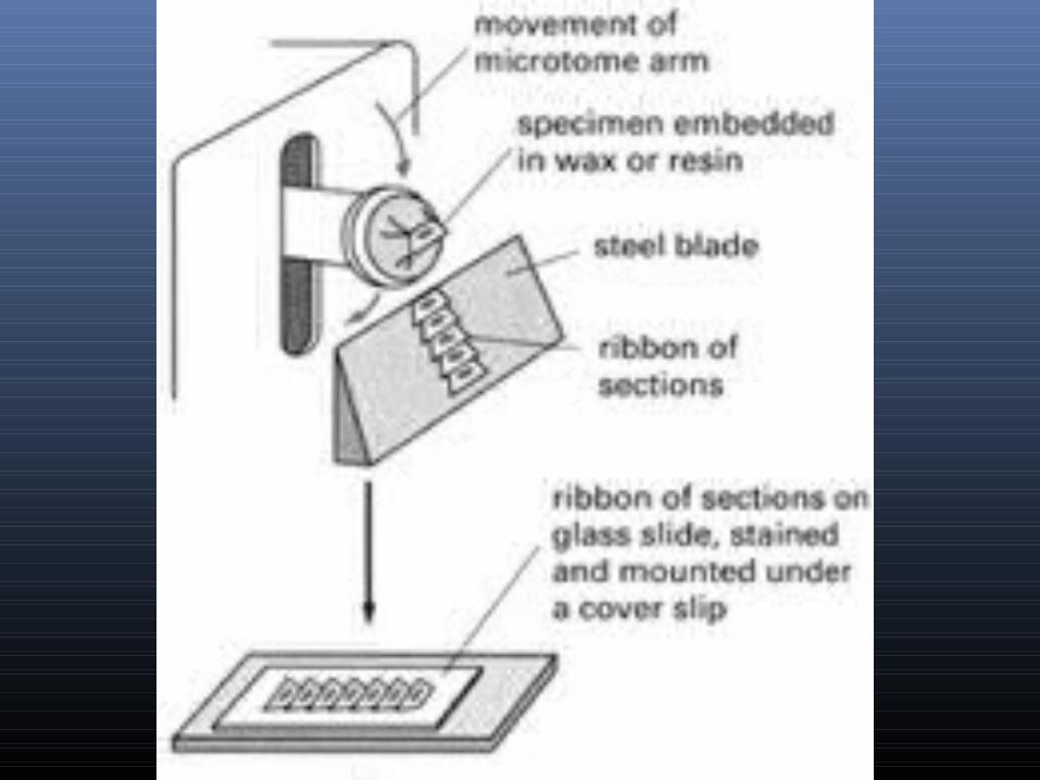
iii) Microtomy: cut tissue very thin -typically 1 - 10 micrometers thick-paraffin/plastic embedded tissue sectioned with micro tome-frozen samples sectioned with cryostat (microtome within a freezer)
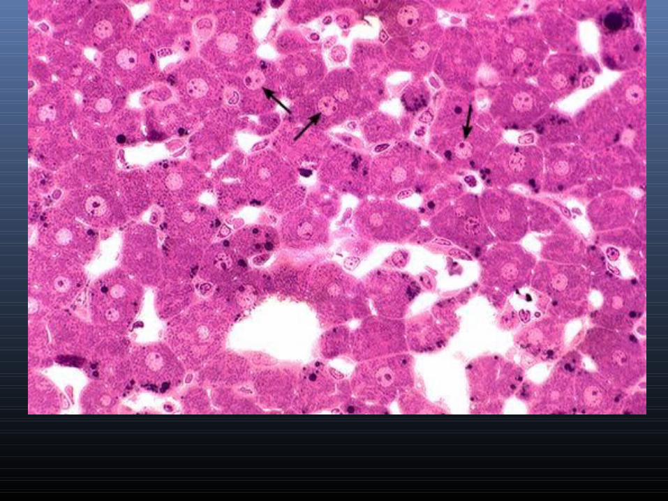
iv) stain specimen with dyes (classical histology): paraffin is removed from section then section is re- hydrated and stainedRoutine stain -hematoxylin: basic dye –STAINS NUCLEUS-eosin: acid dye STAINS CYTOPLASM

THANK YOUTHANK YOU