Embed Size (px)
Citation preview
Hélène BERGESDirector of the Plant Genomic Center
Toward a better understanding of plant genomes structure :
Combining NGS, optical mapping technology and CRISPR-CATCH approach
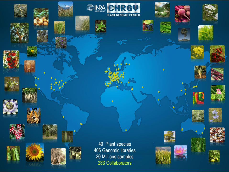
Global warming effects, Population growth, Erosion of genetic progress, Consumer expectations
INNOVATION : Genome’s exploration is one of the strategic
approaches for better understanding plant evolution and plant improvement and adaptation
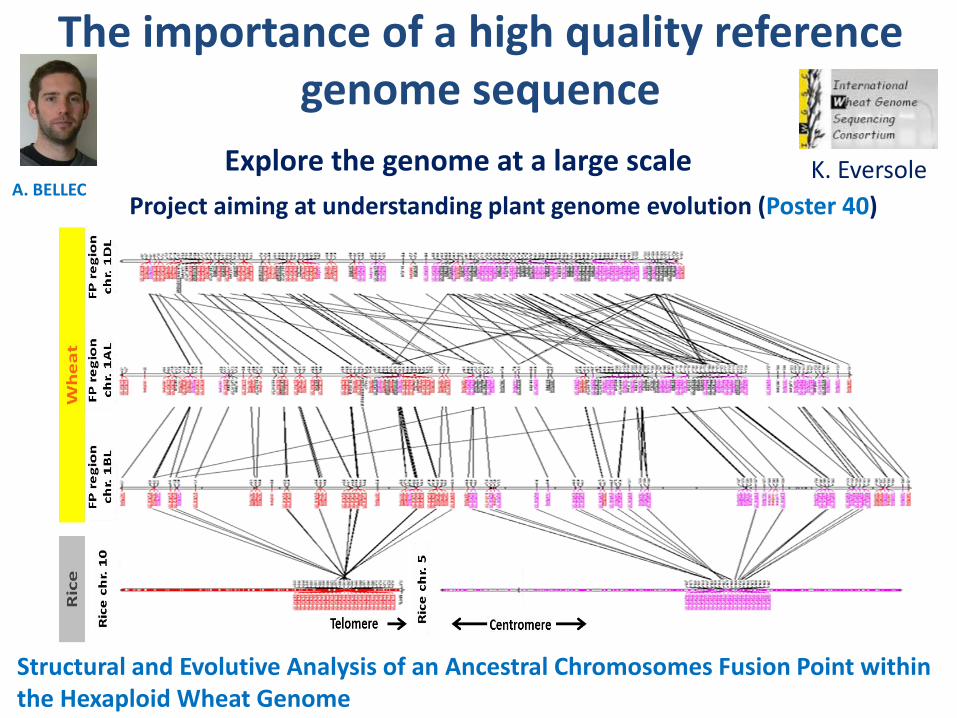
The importance of a high quality reference genome sequence
A. BELLECK. Eversole
Project aiming at understanding plant genome evolution (Poster 40)
Structural and Evolutive Analysis of an Ancestral Chromosomes Fusion Point within the Hexaploid Wheat Genome
Explore the genome at a large scale
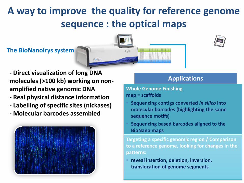
Whole Genome Finishing map = scaffolds
• Sequencing contigs converted in silico into molecular barcodes (highlighting the same sequence motifs)
• Sequencing based barcodes aligned to the BioNano maps
Targeting a specific genomic region / Comparisonto a reference genome, looking for changes in the patterns:
• reveal insertion, deletion, inversion, translocation of genome segments
Applications- Direct visualization of long DNA molecules (>100 kb) working on non-amplified native genomic DNA- Real physical distance information- Labelling of specific sites (nickases)- Molecular barcodes assembled
A way to improve the quality for reference genome sequence : the optical maps
The BioNanoIrys system
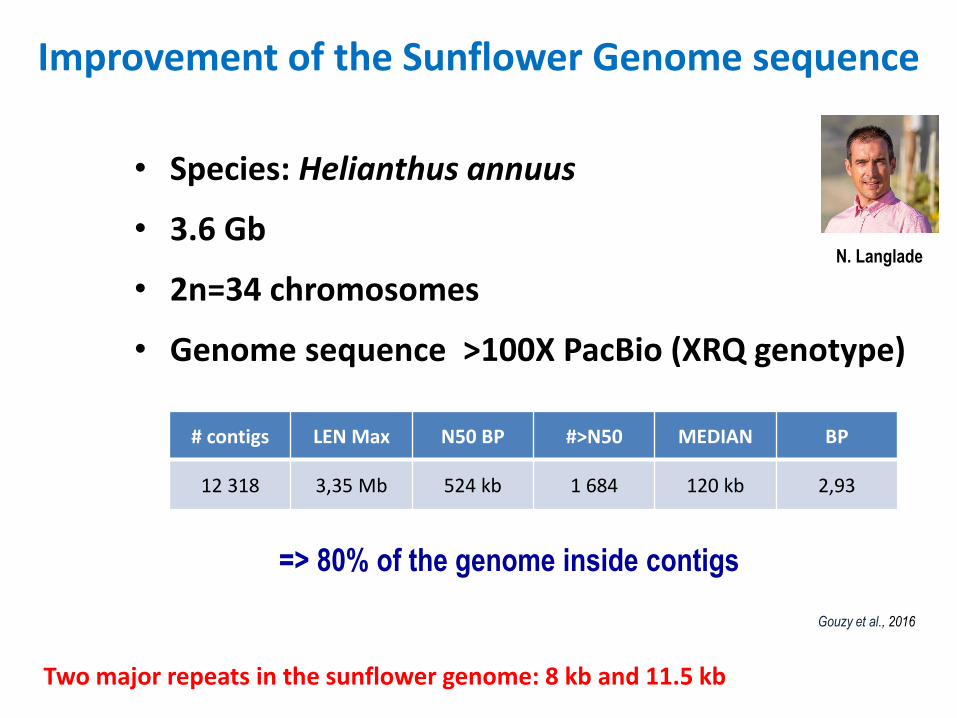
Improvement of the Sunflower Genome sequence
• Species: Helianthus annuus
• 3.6 Gb
• 2n=34 chromosomes
• Genome sequence >100X PacBio (XRQ genotype)
# contigs LEN Max N50 BP #>N50 MEDIAN BP
12 318 3,35 Mb 524 kb 1 684 120 kb 2,93
=> 80% of the genome inside contigs
Gouzy et al., 2016
Two major repeats in the sunflower genome: 8 kb and 11.5 kb
N. Langlade
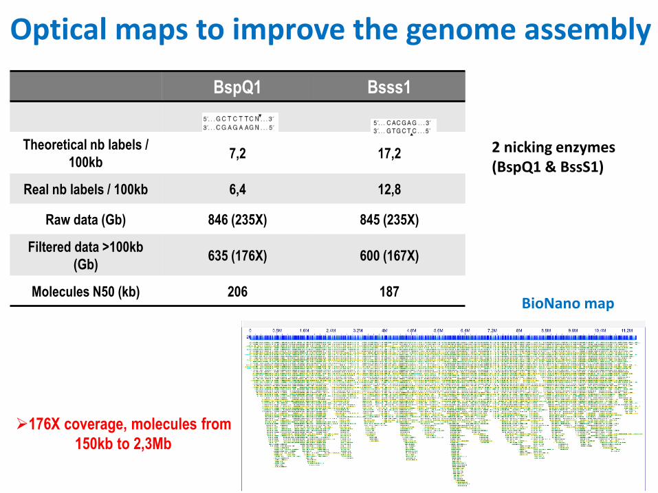
Optical maps to improve the genome assembly
BspQ1 Bsss1
Theoretical nb labels /
100kb7,2 17,2
Real nb labels / 100kb 6,4 12,8
Raw data (Gb) 846 (235X) 845 (235X)
Filtered data >100kb
(Gb)635 (176X) 600 (167X)
Molecules N50 (kb) 206 187
176X coverage, molecules from
150kb to 2,3Mb
BioNano map
2 nicking enzymes (BspQ1 & BssS1)
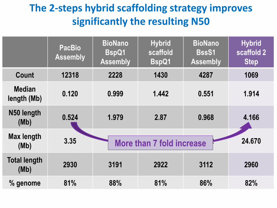
The 2-steps hybrid scaffolding strategy improves significantly the resulting N50
PacBio
Assembly
BioNano
BspQ1
Assembly
Hybrid
scaffold
BspQ1
BioNano
BssS1
Assembly
Hybrid
scaffold 2
Step
Count 12318 2228 1430 4287 1069
Median
length (Mb) 0.120 0.999 1.442 0.551 1.914
N50 length
(Mb) 0.524 1.979 2.87 0.968 4.166
Max length
(Mb) 3.35 11.49 17.45 5.322 24.670
Total length
(Mb) 2930 3191 2922 3112 2960
% genome 81% 88% 81% 86% 82%
Hybrid
scaffold 2
Step
1069
1.914
4.166
24.670
2960
82%
More than 7 fold increase
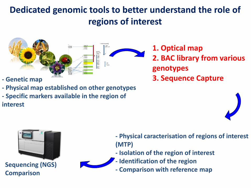
Sequencing (NGS) Comparison
- Genetic map- Physical map established on other genotypes- Specific markers available in the region of interest
1. Optical map2. BAC library from various genotypes3. Sequence Capture
- Physical caracterisation of regions of interest(MTP) - Isolation of the region of interest- Identification of the region- Comparison with reference map
Dedicated genomic tools to better understand the role of regions of interest
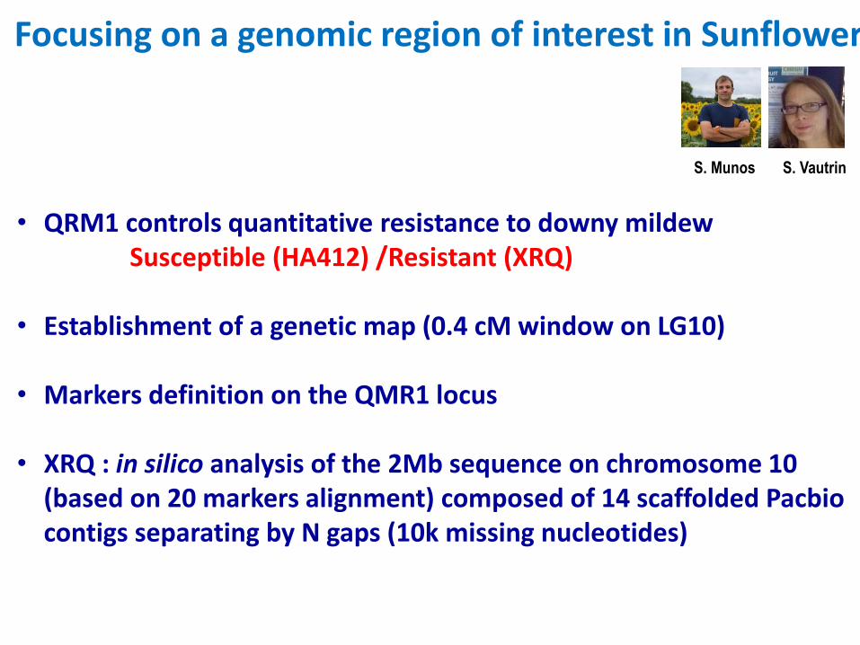
Focusing on a genomic region of interest in Sunflower
• QRM1 controls quantitative resistance to downy mildew Susceptible (HA412) /Resistant (XRQ)
• Establishment of a genetic map (0.4 cM window on LG10)
• Markers definition on the QMR1 locus
• XRQ : in silico analysis of the 2Mb sequence on chromosome 10 (based on 20 markers alignment) composed of 14 scaffolded Pacbiocontigs separating by N gaps (10k missing nucleotides)
S. Munos S. Vautrin
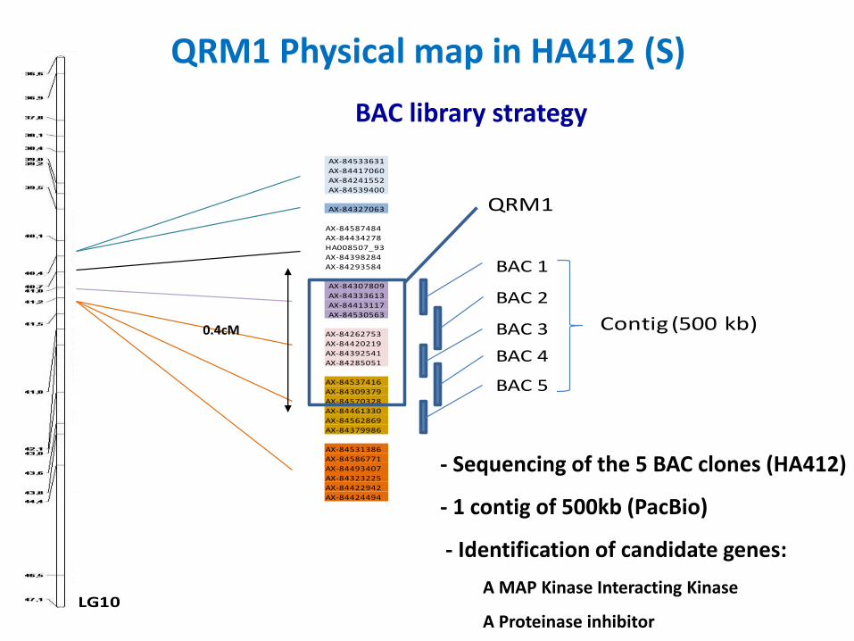
QRM1 Physical map in HA412 (S)
AX-84533631
AX-84417060
AX-84241552
AX-84539400
AX-84327063
AX-84587484
AX-84434278
HA008507_93
AX-84398284
AX-84293584
AX-84307809
AX-84333613
AX-84413117
AX-84530563
AX-84262753
AX-84420219
AX-84392541
AX-84285051
AX-84537416
AX-84309379
AX-84570328
AX-84461330
AX-84562869
AX-84379986
AX-84531386
AX-84586771
AX-84493407
AX-84323225
AX-84422942
AX-84424494
LG10
BAC 1
BAC 2
BAC 3
BAC 4
BAC 5
QRM1
Contig (500 kb) 0.4cM
- Sequencing of the 5 BAC clones (HA412)
- 1 contig of 500kb (PacBio)
- Identification of candidate genes:
A MAP Kinase Interacting Kinase
A Proteinase inhibitor
BAC library strategy
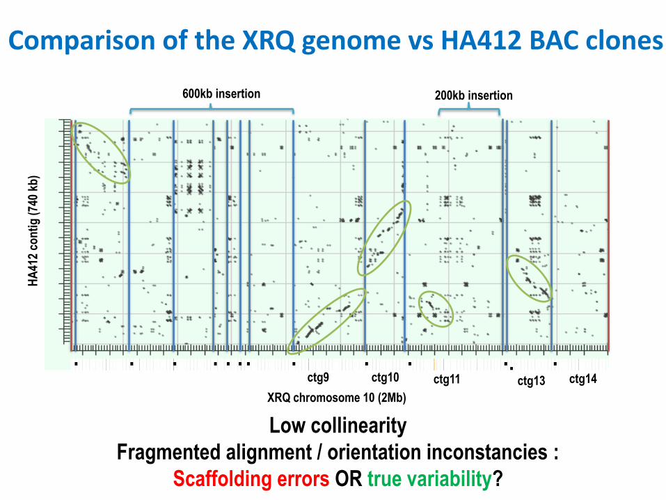
Comparison of the XRQ genome vs HA412 BAC clones H
A41
2 co
nti
g (
740
kb)
XRQ chromosome 10 (2Mb)
ctg9 ctg10 ctg11 ctg13 ctg14
Low collinearity
Fragmented alignment / orientation inconstancies :
Scaffolding errors OR true variability?
600kb insertion 200kb insertion
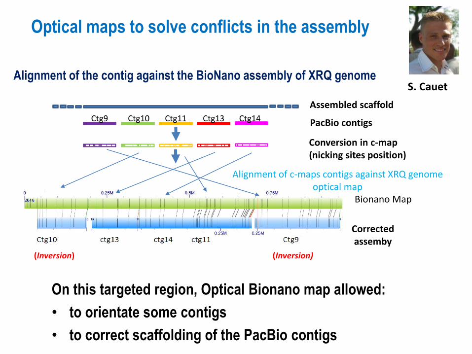
Ctg9 Ctg10 Ctg11 Ctg13 Ctg14 PacBio contigs
Assembled scaffold
Conversion in c-map (nicking sites position)
Alignment of c-maps contigs against XRQ genomeoptical map
(Inversion) (Inversion)
Bionano Map
Correctedassemby
Optical maps to solve conflicts in the assembly
S. Cauet
On this targeted region, Optical Bionano map allowed:
• to orientate some contigs
• to correct scaffolding of the PacBio contigs
Alignment of the contig against the BioNano assembly of XRQ genome
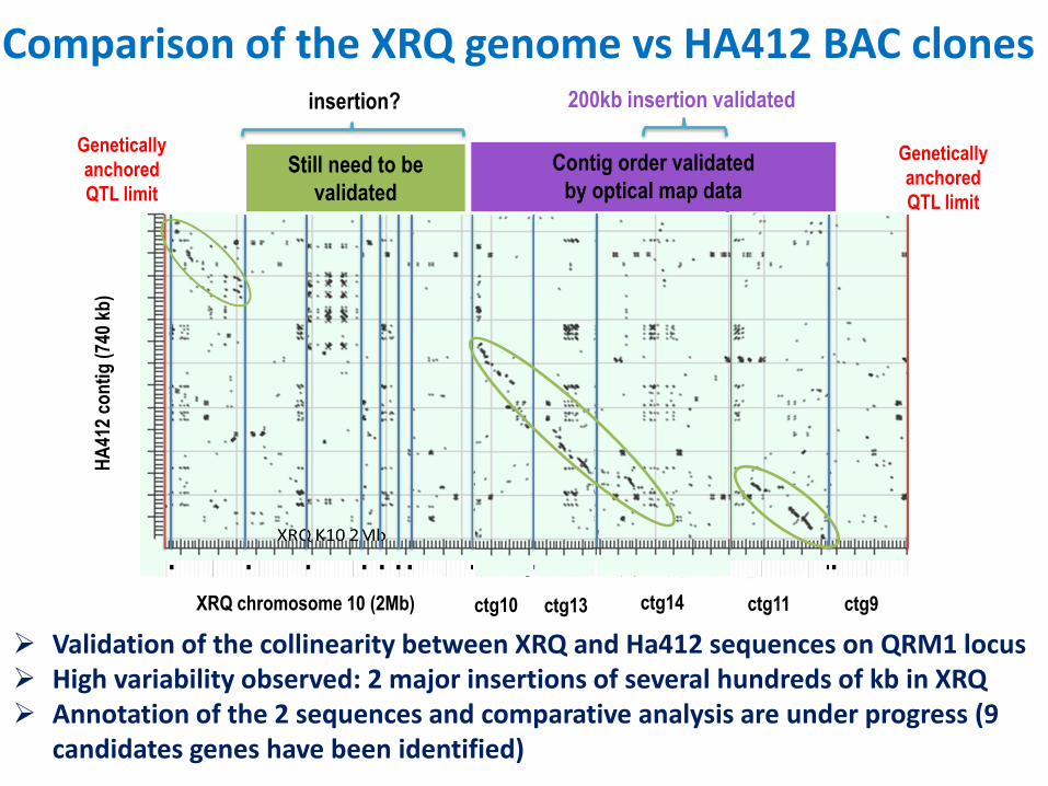
Contig order validated
by optical map dataStill need to be
validated
Genetically
anchored
QTL limit
Genetically
anchored
QTL limit
insertion? 200kb insertion validated
Validation of the collinearity between XRQ and Ha412 sequences on QRM1 locus High variability observed: 2 major insertions of several hundreds of kb in XRQ Annotation of the 2 sequences and comparative analysis are under progress (9
candidates genes have been identified)
HA
412
con
tig
(74
0 kb
)
XRQ chromosome 10 (2Mb)
Comparison of the XRQ genome vs HA412 BAC clones
ctg9ctg10 ctg11ctg13 ctg14
N. RODDE Poster41
Genome assembly improvement will help linking genotype / phenotype
=> How to be more efficient in focusing on genomic regions?
Sunflower proves again to be a highly complex genome, showing very high diversity between genotypes
One reference genome is not enough!
Despite long reads sequencing, assembly (scaffolding) has to be checkedwhen working on reference genomes
The optical map allowed to validate major rearrangements between the 2 genotypes
Proven interest of complementary approaches (NGS – optical map – BAC)
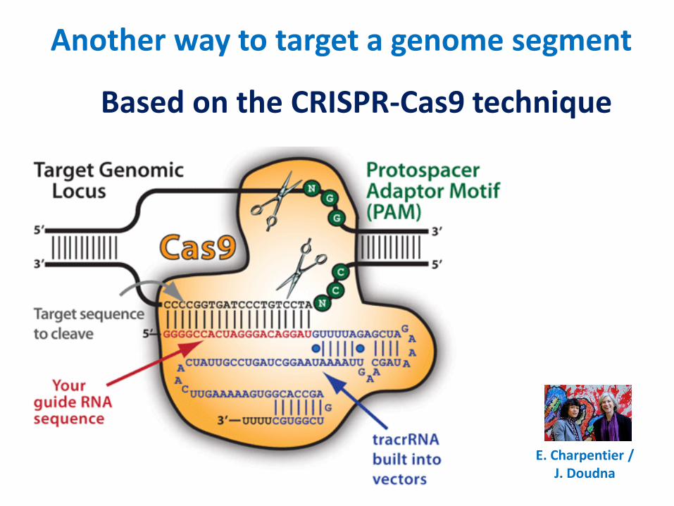
Another way to target a genome segment
E. Charpentier /J. Doudna
Based on the CRISPR-Cas9 technique
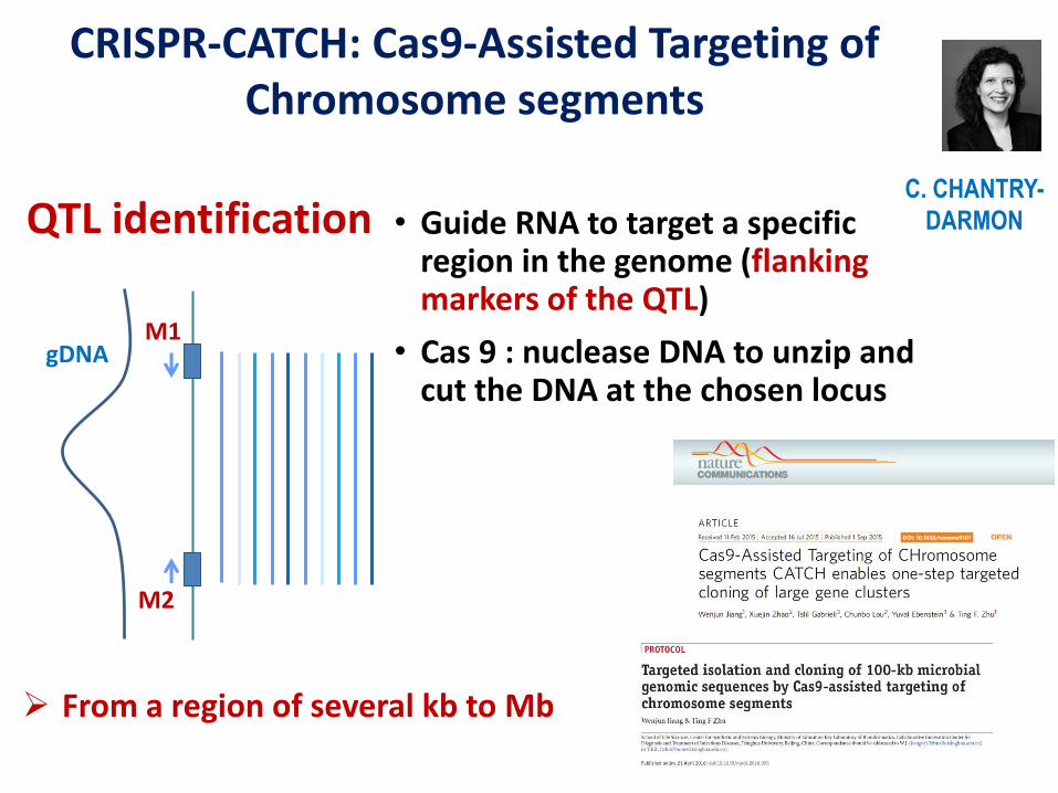
• Guide RNA to target a specific region in the genome (flanking markers of the QTL)
• Cas 9 : nuclease DNA to unzip and cut the DNA at the chosen locus
QTL identification
From a region of several kb to Mb
M1
M2
gDNA
C. CHANTRY-
DARMON
CRISPR-CATCH: Cas9-Assisted Targeting of Chromosome segments
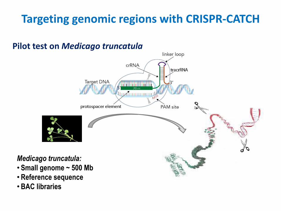
Targeting genomic regions with CRISPR-CATCH
Medicago truncatula:
•Small genome ~ 500 Mb
•Reference sequence
•BAC libraries
Pilot test on Medicago truncatula
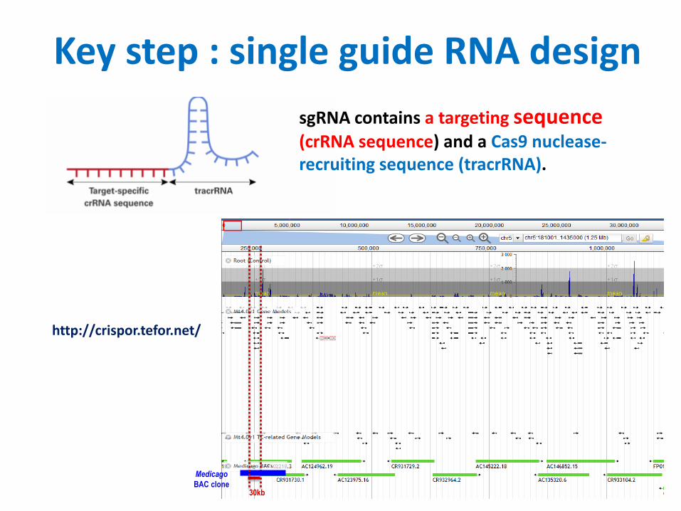
Key step : single guide RNA design
30kb
Medicago
BAC clone
sgRNA contains a targeting sequence(crRNA sequence) and a Cas9 nuclease-recruiting sequence (tracrRNA).
http://crispor.tefor.net/
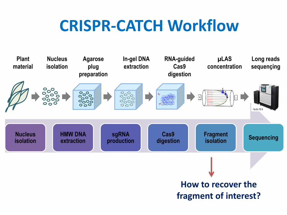
Nucleus isolation
HMW DNA extraction
sgRNAproduction
Cas9 digestion
Fragment isolation
Sequencing
CRISPR-CATCH Workflow
Agarose
plug
preparation
In-gel DNA
extraction
RNA-guided
Cas9
digestion
Plant
material
Nucleus
isolation
Long reads
sequençing
µLAS
concentration
How to recover the fragment of interest?
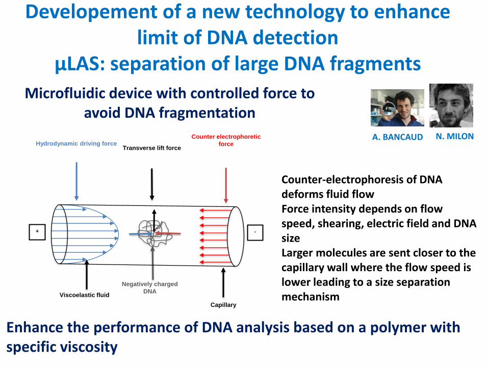
Microfluidic device with controlled force to avoid DNA fragmentation
Negatively charged
DNA
Capillary
Hydrodynamic driving forceTransverse lift force
Counter electrophoretic
force
Viscoelastic fluid
+ -
A. BANCAUD N. MILON
Developement of a new technology to enhancelimit of DNA detection
µLAS: separation of large DNA fragments
Enhance the performance of DNA analysis based on a polymer withspecific viscosity
Counter-electrophoresis of DNA deforms fluid flowForce intensity depends on flow speed, shearing, electric field and DNA sizeLarger molecules are sent closer to the capillary wall where the flow speed is lower leading to a size separation mechanism
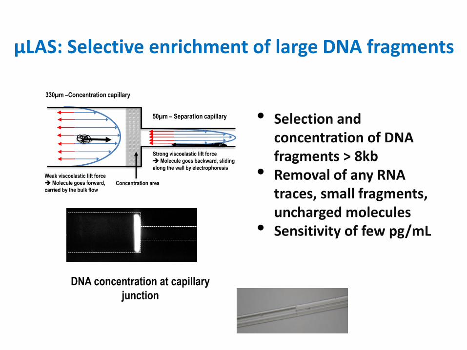
µLAS: Selective enrichment of large DNA fragments
Strong viscoelastic lift force
Molecule goes backward, sliding
along the wall by electrophoresis
Concentration area
Weak viscoelastic lift force
Molecule goes forward,
carried by the bulk flow
330µm –Concentration capillary
50µm – Separation capillary • Selection and concentration of DNA fragments > 8kb
• Removal of any RNA traces, small fragments, uncharged molecules
• Sensitivity of few pg/mL
DNA concentration at capillary
junction
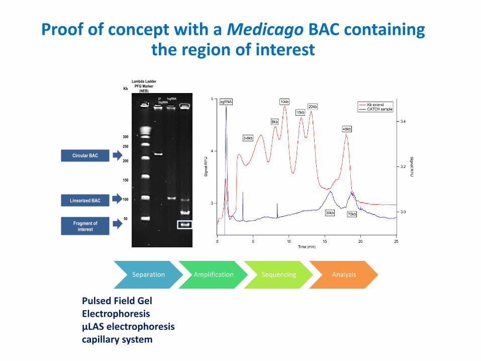
Separation Amplification Sequencing Analyzis
Fragment of
interest
Linearized BAC
Circular BAC
Kb
300
250
200
150
100
50
1sgRNA
2sgRNA
Lambda Ladder
PFG Marker
(NEB)
Pulsed Field Gel Electrophoresis µLAS electrophoresis capillary system
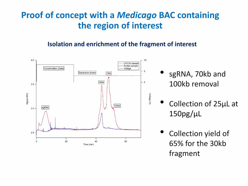
Proof of concept with a Medicago BAC containing the region of interest
1sgRNA
2sgRNA
• sgRNA, 70kb and 100kb removal
• Collection of 25µL at 150pg/µL
• Collection yield of 65% for the 30kb fragment
Isolation and enrichment of the fragment of interest
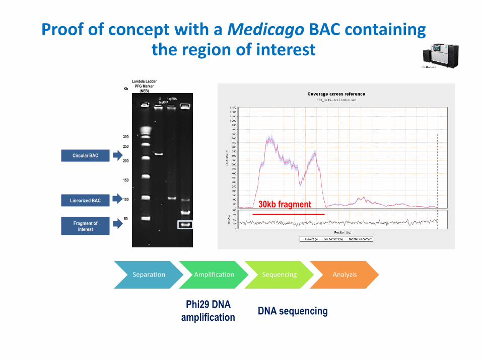
Proof of concept with a Medicago BAC containing the region of interest
Separation Amplification Sequencing Analyzis
Fragment of
interest
Linearized BAC
Circular BAC
Kb
300
250
200
150
100
50
1sgRNA
2sgRNA
Lambda Ladder
PFG Marker
(NEB)
Proof of concept with a Medicago BAC containing the region of interest
30kb fragment
DNA sequencingPhi29 DNA
amplification
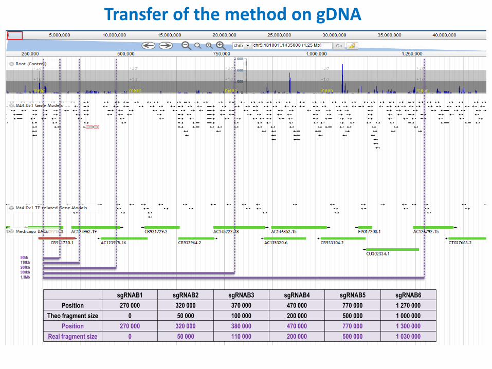
sgRNAB1 sgRNAB2 sgRNAB3 sgRNAB4 sgRNAB5 sgRNAB6
Position 270 000 320 000 370 000 470 000 770 000 1 270 000
Theo fragment size 0 50 000 100 000 200 000 500 000 1 000 000
Position 270 000 320 000 380 000 470 000 770 000 1 300 000
Real fragment size 0 50 000 110 000 200 000 500 000 1 030 000
50kb
110kb
200kb
500kb
1,3Mb
Transfer of the method on gDNA
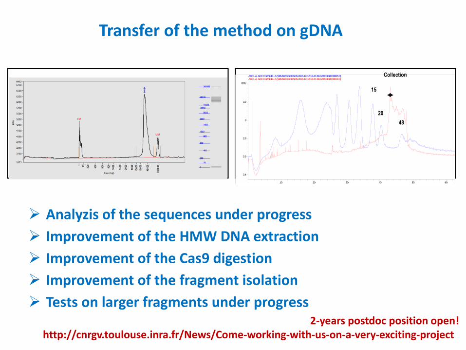
Transfer of the method on gDNA
min10 20 30 40 50 60
RFU
2.4
2.6
2.8
3
3.2
ADC1 A, ADC CHANNEL A (SEM1650\GRDADN 2016-12-12 18-47-31\CATCHG0000005.D) ADC1 A, ADC CHANNEL A (SEM1650\GRDADN 2016-12-12 18-47-31\CATCHG0000013.D)
48
15
20
Collection
Analyzis of the sequences under progress
Improvement of the HMW DNA extraction
Improvement of the Cas9 digestion
Improvement of the fragment isolation
Tests on larger fragments under progress2-years postdoc position open!
http://cnrgv.toulouse.inra.fr/News/Come-working-with-us-on-a-very-exciting-project
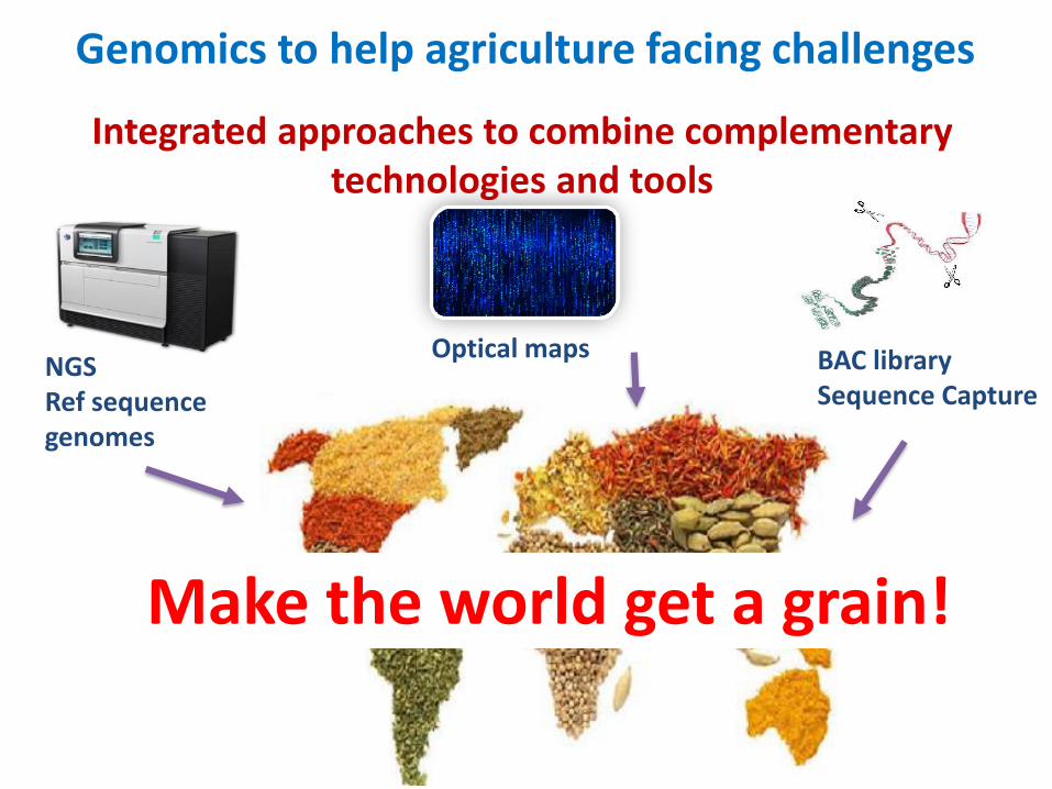
Genomics to help agriculture facing challenges
Make the world get a grain!
Optical mapsNGS Ref sequencegenomes
BAC librarySequence Capture
Integrated approaches to combine complementary technologies and tools
Acknowledgements
Hélène BERGES
Arnaud BELLEC
Sonia VAUTRIN
Céline CHANTRY-DARMON
Nathalie RODDE
Céline JEZIORSKI
William MARANDE
Stéphane CAUET
Nadège ARNAL
Caroline CALLOT
Joëlle FOURMENT
Nadine GAUTIER
Elisa PRAT
David PUJOL
Roseana RODRIGUES
Sandrine ARRIBAT
Laetitia HOARAU
@CNRGV@SUNRISE_France
http://cnrgv.toulouse.inra.fr/
Jérôme GOUZY
Nicolas LANGLADE
Stéphane MUNOS
John BAETEN
Kees-Jan FRANCOIJS
Kellye Eversole
A. BANCAUD
N. Milon
F. Ginot