Embed Size (px)
Citation preview
The effects of Activated CDC-42 Kinase inhibitors on axonal growth of
neurons in vitroMontasir Rahman
Bronx High School of ScienceMentor: Dr. Ijaz Ahmed
Medgar Evers College of CUNY
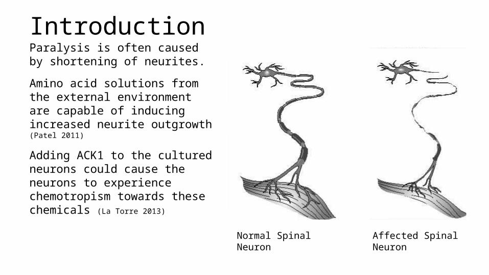
IntroductionParalysis is often caused by shortening of neurites.
Amino acid solutions from the external environment are capable of inducing increased neurite outgrowth (Patel 2011)
Adding ACK1 to the cultured neurons could cause the neurons to experience chemotropism towards these chemicals (La Torre 2013)
Normal Spinal Neuron Affected Spinal Neuron
Why “Inhibitors” for neuronal growth?neuronal apoptosis--programmed cell death--often triggered by inappropriate activation of CDC kinases.
Therefore, inhibitors can prevent the unnecessary activation of these kinases, thereby reducing instances of neuronal apoptosis.
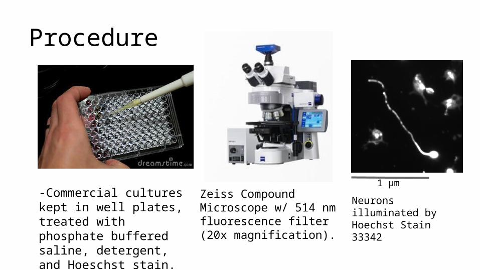
Zeiss Compound Microscope w/ 514 nm fluorescence filter (20x magnification).
Neurons illuminated by Hoechst Stain 33342
1 μm
Procedure
-Commercial cultures kept in well plates, treated with phosphate buffered saline, detergent, and Hoeschst stain. *note: sample image used
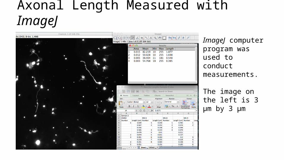
Axonal Length Measured with ImageJImageJ computer program was used to conduct measurements.
The image on the left is 3 μm by 3 μm
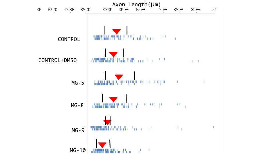
Axon Length(μm)
MG-8
21.8
1.6
1.4
1.2
1.0
0.8
0.6
0.4
0.2
0
CONTROL
CONTROL+DMSO
MG-5
MG-9
MG-10
Conclusion•MG-5 treated neurons showed an increase in neurite length.
•Samples of MG other than that MG-5 will cause a decrease in length.
Future Work
Testing to see why certain neurons will take further advantage of growth space while others do not, possibly due to individual genetics of each neuron, creating a ‘survival of the fittest’ scenario at the molecular level.
Tubulin plays an essential role in the growth mechanism of neurons (Hjorth 2014). Those neurons that had a greater access to the tubulin exposed to them were able to grow at a more rapid rate. We can test if varying concentrations of tubulin can cause increases in axon growth.
ReferencesHjorth, J.; van Pelt, J.; Mansvelder, H. D.; van Ooyen, A. (2014). Competitive Dynamics During Research Driven Neurite Outgrowth. Public Library of Science, 9 (2), 1-10 La Torre, A. (2013), A role for the tyrosine kinase ACK1 in neurotrophin signaling and neuronal extension and branching, Nature, 99, 10-38 Janulevicius, A.; van Pelt, J.; van Ooyen, A. (2006). Compartment Volume Influences Microtubule Dynamic Instability: A Model Study. Biophysical Journal, 90, 788-798 Patel, K. (2011). Analysis of Neurite Outgrowth in Cultured Primary Neurons. Definiens Schmitz, S. K. (2011). Automated Analysis of Neuronal Morphology, Synapse Number and Synaptic Recruitment. Journal of Neuroscience Methods, 195, 185-193

Acknowledgements
Special thanks to Dr. Mohsin Patwary, Dr. Alam Nur-e-Kamal, and Dr. Ijaz Ahmed for their overseeing of this research, and for their provisions of many resources to allow for optimal contribution to the overall project. Further appreciation is due to Mohammad W. Rahman and Muslima J. Khandakar for their parental support throughout the duration of this project, and for providing a means of transportation by which to reach the lab to conduct the research. Much thanks is also due to Dr. Karen Fien and Dr. Robert Muratore for providing guidance over the years in conducting research and formulating an appropriate paper.