1
Level or stage of neural maturation differentially effected susceptibility to neurotoxins. Metabolomic profiling provides a sensitive pathway analysis approach to DNT for both mature neurons, astrocytes and pre-network neural progenitors. Significant alteration in neuronal cellular response in co-cultures with astrocytes suggests need for a more representative multi-cellular DNT assays. Metabolomics and neurite outgrowth as data rich developmental neurotoxicity assays in a pluripotent stem cell derived human neural model Anirban Majumder 1 , Shelley Wallace 1 , Xian Wu 2 , Mayowa Amosu 3 , Kun Lu 3 , Mary A. Smith 2,3 , Steven L. Stice 1,2 1 ArunA Biomedical Inc., Athens GA, 2 Regenerative Bioscience Center, University of Georgia, Athens GA, 3 Environmental Health Sciences, University of Georgia, Athens GA Introduction Methods Neurite HCI assays detect neuronal response at lower Bis-1 doses than viability assays A vast majority of chemical entities, ranging from pesticides to compounds of therapeutic interest, remain untested for potential toxic outcomes on human neural development. Current methods for evaluating developmental neurotoxicity (DNT) rely heavily on animal based testing and non-uniform cell lines, are often prohibitively expensive, and provide suboptimal predictive value. There is thus a need for rapid, cost effective, in vitro methods to identify such chemicals. To fill this critical gap we evaluated a robust, species representative and physiologically relevant cellular system, combined with high content imaging (HCI) and metabolomics, to address human DNT. Scalable and uniform populations of undifferentiated neural progenitor cells (hNP1 TM ), and differentiated neurons (hN2 TM ) and astrocytes (hAstroPro TM ), derived from human embryonic stem cells (hESC) were used to represent a neurodevelopmental continuum and evaluated for toxin induced changes in neurite outgrowth, metabolomic signatures and viability. To address developmental stage specific neurotoxicity, Bis-1, a known neurotoxin, was applied to cultures pre or post neuronal differentiation, as well as in neuron-astrocyte co-cultures to represent complex brain tissue more accurately. Summary Neuron-astrocyte co-culture system Scalable progenitors shorten derivation time reducing cell variability. Cryopreserved differentiated cells can be thawed directly on assay plates for quick assay implementation. Pure neuronal and astrocytic cells allow controlled ratios in co-cultures. Cell culture: Cryopreserved neural prognitors (hNP1 TM ), neurons (hN2 TM ) and astrocytes (hAstroPro TM ) were plated by themselves or as co-cultures. Results Rejected Selected Neurite trace Neurons in Co-culture Neuron only 72 hours treatment: Neurite outgrowth assay Neurite length per field Neurites per field Branch points per field Percent of control Bis-1, uM Co-culture Neuron only Results www.arunabiomedical.com Neurite outgrowth: Neurites were labeled by immunochemistry for MAP2 & Nuclei stained with Hoescht 3342. Neurite outgrowth data was acquired on a Cellomics ArrayScan VTI TM with Neuroprofiler TM software. Neurite tracing parameters were optimized manually. Viability: CellTiter96 TM (Promega) was added to Bis1 treated cultures for last 2 hours and absorbance was read at 490nm. CellTiter96 contains a tetrazolium compound that is bioreduced into a colored foramazan product by NADPH or NADH produced in metabolically active cells. Metabolomics: Metabolomic profiling was done on an Agilent 6890/5973 GC-MS system using DB-5MS GC columns. MS data was collected monitoring mass ranging from 50 to 600 m/z and analyzed using PLS-DA with SIMCA-P+ after picking and aligning the peaks. GC/MS Cells Media MeOH/CHCl 3 extracted Phase separated Derivatized Derivatized Progenitors Neurons Progenitors Neurons Astrocytes Metabolomic profiling detects Bis-1 induced changes progenitors, neurons and astrocytes PLS-DA scores plots from hNP and hN2 cells exposed to Bis1 for 48 hrs. A dose-response is evident along PCA1. Statistical differences were observed at doses ≥1 µM for Bis1 Astrocytes further modulate toxic response and neurite outgrowth in stable neuron-astrocyte co-cultures Neurons per field Bis-1, uM Percent of control Bis-1, uM Bis-1, uM This project is supported by Grant Number 1R43ES023530-01 from the National Institutes of Health (NIH) and EPA SBIR EP-D-13-018 and EPA-G2012-STAR-F1 from the Environmental Protection Agency (EPA). We would like to thank Matthew Henderson (EPA) and members of Dr. Smith’s lab (UGA). CONTROL 3 μM 10 μM 30 μM CONTROL 0.1 μM 1.0 μM 3 μM 10 μM Neural progenitors are more susceptible to Bis-1 than neurons or astrocytes lowest dose with a significant decrease in viability. Progenitors (hNP) Astrocytes (hAstroPro) Neurons (hN2) DIV 0 DIV14 DIV 28 0-14 DIV Exposure(WOS 1) 14-28 DIV Exposure (WOS 2) Early Neuron Late Neuron Progenitor Hu C/D MAP2 Neuron density Neurons at late stages of differentiation are more susceptible to Bis-1 Astrocytes CONTROL 3 μM 10 μM 30 μM

Poster sot 2015_am_final

Embed Size (px)

Citation preview

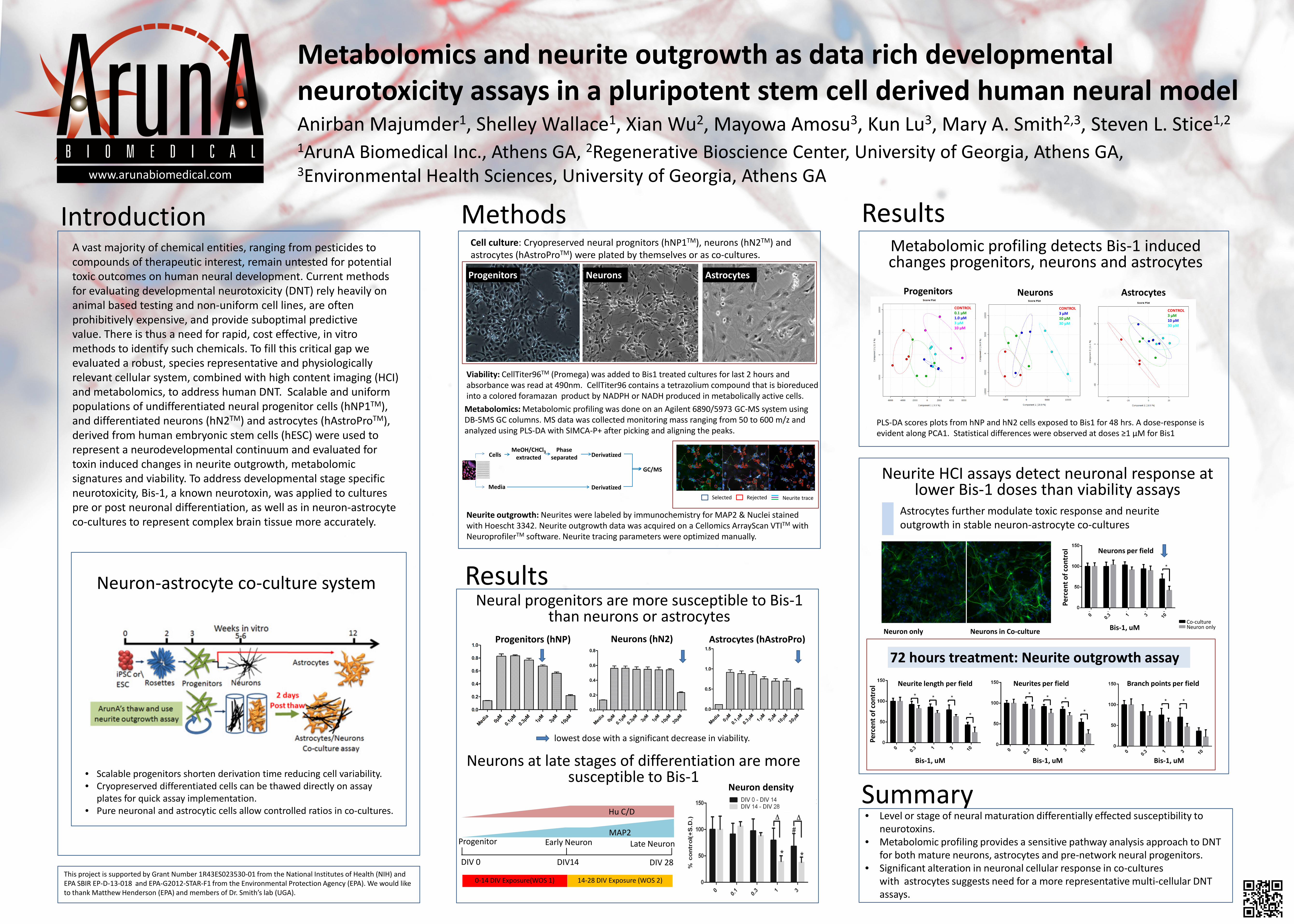
• Level or stage of neural maturation differentially effected susceptibility to neurotoxins.

• Metabolomic profiling provides a sensitive pathway analysis approach to DNT for both mature neurons, astrocytes and pre-network neural progenitors.

• Significant alteration in neuronal cellular response in co-cultures with astrocytes suggests need for a more representative multi-cellular DNT assays.

Metabolomics and neurite outgrowth as data rich developmental neurotoxicity assays in a pluripotent stem cell derived human neural model Anirban Majumder1, Shelley Wallace1, Xian Wu2, Mayowa Amosu3, Kun Lu3, Mary A. Smith2,3, Steven L. Stice1,2 1ArunA Biomedical Inc., Athens GA, 2Regenerative Bioscience Center, University of Georgia, Athens GA, 3Environmental Health Sciences, University of Georgia, Athens GA

Introduction Methods

Neurite HCI assays detect neuronal response at lower Bis-1 doses than viability assays

A vast majority of chemical entities, ranging from pesticides to compounds of therapeutic interest, remain untested for potential toxic outcomes on human neural development. Current methods for evaluating developmental neurotoxicity (DNT) rely heavily on animal based testing and non-uniform cell lines, are often prohibitively expensive, and provide suboptimal predictive value. There is thus a need for rapid, cost effective, in vitro methods to identify such chemicals. To fill this critical gap we evaluated a robust, species representative and physiologically relevant cellular system, combined with high content imaging (HCI) and metabolomics, to address human DNT. Scalable and uniform populations of undifferentiated neural progenitor cells (hNP1TM), and differentiated neurons (hN2TM) and astrocytes (hAstroProTM), derived from human embryonic stem cells (hESC) were used to represent a neurodevelopmental continuum and evaluated for toxin induced changes in neurite outgrowth, metabolomic signatures and viability. To address developmental stage specific neurotoxicity, Bis-1, a known neurotoxin, was applied to cultures pre or post neuronal differentiation, as well as in neuron-astrocyte co-cultures to represent complex brain tissue more accurately.

Summary

Neuron-astrocyte co-culture system

• Scalable progenitors shorten derivation time reducing cell variability. • Cryopreserved differentiated cells can be thawed directly on assay

plates for quick assay implementation. • Pure neuronal and astrocytic cells allow controlled ratios in co-cultures.

Cell culture: Cryopreserved neural prognitors (hNP1TM), neurons (hN2TM) and astrocytes (hAstroProTM) were plated by themselves or as co-cultures.

Results

Rejected Selected Neurite trace

Neurons in Co-culture Neuron only

72 hours treatment: Neurite outgrowth assay Neurite length per field Neurites per field Branch points per field

Perc

ent o

f con

trol

Bis-1, uM

Co-culture Neuron only

Results www.arunabiomedical.com

Neurite outgrowth: Neurites were labeled by immunochemistry for MAP2 & Nuclei stained with Hoescht 3342. Neurite outgrowth data was acquired on a Cellomics ArrayScan VTITM with NeuroprofilerTM software. Neurite tracing parameters were optimized manually.

Viability: CellTiter96TM (Promega) was added to Bis1 treated cultures for last 2 hours and absorbance was read at 490nm. CellTiter96 contains a tetrazolium compound that is bioreduced into a colored foramazan product by NADPH or NADH produced in metabolically active cells. Metabolomics: Metabolomic profiling was done on an Agilent 6890/5973 GC-MS system using DB-5MS GC columns. MS data was collected monitoring mass ranging from 50 to 600 m/z and analyzed using PLS-DA with SIMCA-P+ after picking and aligning the peaks.

GC/MS

Cells

Media

MeOH/CHCl3 extracted

Phase separated Derivatized

Derivatized

Progenitors Neurons

Progenitors Neurons Astrocytes

Metabolomic profiling detects Bis-1 induced changes progenitors, neurons and astrocytes

PLS-DA scores plots from hNP and hN2 cells exposed to Bis1 for 48 hrs. A dose-response is evident along PCA1. Statistical differences were observed at doses ≥1 µM for Bis1

Astrocytes further modulate toxic response and neurite outgrowth in stable neuron-astrocyte co-cultures

Neurons per field

Bis-1, uM

Perc

ent o

f con

trol

Bis-1, uM Bis-1, uM

This project is supported by Grant Number 1R43ES023530-01 from the National Institutes of Health (NIH) and EPA SBIR EP-D-13-018 and EPA-G2012-STAR-F1 from the Environmental Protection Agency (EPA). We would like to thank Matthew Henderson (EPA) and members of Dr. Smith’s lab (UGA).

CONTROL 3 μM 10 μM 30 μM

CONTROL 0.1 μM 1.0 μM 3 μM 10 μM

Neural progenitors are more susceptible to Bis-1 than neurons or astrocytes

lowest dose with a significant decrease in viability.

Progenitors (hNP) Astrocytes (hAstroPro) Neurons (hN2)

DIV 0 DIV14 DIV 28

0-14 DIV Exposure(WOS 1) 14-28 DIV Exposure (WOS 2)

Early Neuron Late Neuron Progenitor

Hu C/D

MAP2

Neuron density

Neurons at late stages of differentiation are more susceptible to Bis-1

Astrocytes

CONTROL 3 μM 10 μM 30 μM