Embed Size (px)
Citation preview

THE ANALYSIS OF THE RELATIONSHIP
BETWEEN MULTIPLE MYELOMA CELLS AND
THEIR MICROENVIRONMENT
NAME : RUCHI RANI ROLL NO. : 19CLASS : M.Sc BIOTECHNOLOGY, IInd SEMESTER
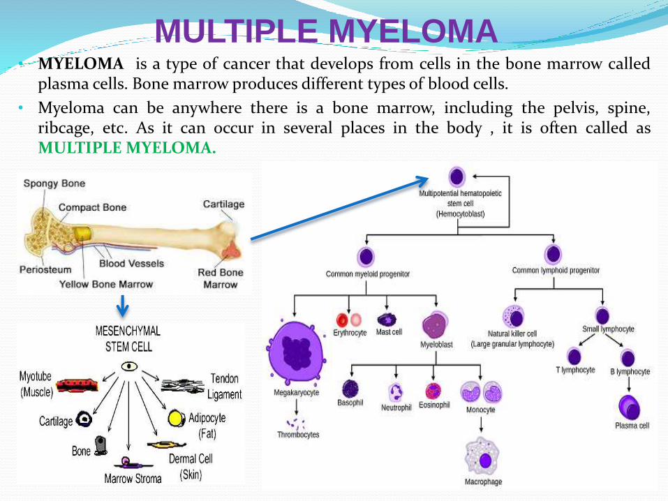
MULTIPLE MYELOMA• MYELOMA is a type of cancer that develops from cells in the bone marrow called
plasma cells. Bone marrow produces different types of blood cells.
• Myeloma can be anywhere there is a bone marrow, including the pelvis, spine,ribcage, etc. As it can occur in several places in the body , it is often called asMULTIPLE MYELOMA.
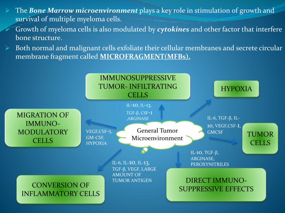
The Bone Marrow microenvironment plays a key role in stimulation of growth and survival of multiple myeloma cells.
Growth of myeloma cells is also modulated by cytokines and other factor that interfere bone structure.
Both normal and malignant cells exfoliate their cellular membranes and secrete circular membrane fragment called MICROFRAGMENT(MFBs).
General Tumor Microenvironment
IMMUNOSUPPRESSIVE TUMOR- INFILTRATING
CELLSHYPOXIA
TUMOR CELLS
MIGRATION OF IMMUNO-
MODULATORY CELLS
CONVERSION OF INFLAMMATORY CELLS
DIRECT IMMUNO-SUPPRESSIVE EFFECTS
IL-10, IL-13,
TGF-β, CSF-1 ,ARGINASE
VEGF,CSF-1,
GM-CSF, HYPOXIA
IL-6, IL-10, IL-13,
TGF-β, VEGF, LARGE AMOUNT OF TUMOR ANTIGEN
IL-6, TGF-β, IL-
10, VEGF,CSF-1,
GMCSF
IL-10, TGF-β,
ARGINASE, PEROXYNITRILES

METHODOLOGY
FOR COMPARASION BETWEEN BM-MSC AND MMBM-MSC
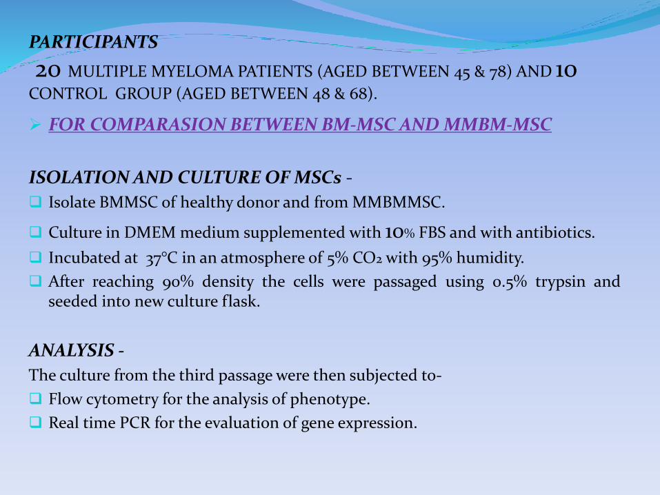
ISOLATION AND CULTURE OF MSCs -
Isolate BMMSC of healthy donor and from MMBMMSC.
Culture in DMEM medium supplemented with 10% FBS and with antibiotics.
Incubated at 37°C in an atmosphere of 5% CO2 with 95% humidity.
After reaching 90% density the cells were passaged using 0.5% trypsin andseeded into new culture flask.
ANALYSIS -
The culture from the third passage were then subjected to-
Flow cytometry for the analysis of phenotype.
Real time PCR for the evaluation of gene expression.
PARTICIPANTS
20 MULTIPLE MYELOMA PATIENTS (AGED BETWEEN 45 & 78) AND 10CONTROL GROUP (AGED BETWEEN 48 & 68).
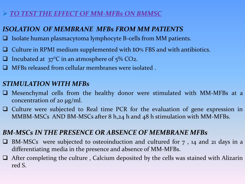
TO TEST THE EFFECT OF MM-MFBs ON BMMSC
ISOLATION OF MEMBRANE MFBs FROM MM PATIENTS
Isolate human plasmacytoma lymphocyte B-cells from MM patients.
Culture in RPMI medium supplemented with 10% FBS and with antibiotics.
Incubated at 37°C in an atmosphere of 5% CO2.
MFBs released from cellular membranes were isolated .
STIMULATION WITH MFBs
Mesenchymal cells from the healthy donor were stimulated with MM-MFBs at aconcentration of 20 μg/ml.
Culture were subjected to Real time PCR for the evaluation of gene expression inMMBM-MSCs AND BM-MSCs after 8 h,24 h and 48 h stimulation with MM-MFBs.
BM-MSCs IN THE PRESENCE OR ABSENCE OF MEMBRANE MFBs
BM-MSCs were subjected to osteoinduction and cultured for 7 , 14 and 21 days in adifferentiating media in the presence and absence of MM-MFBs.
After completing the culture , Calcium deposited by the cells was stained with Alizarinred S.

RESULTS
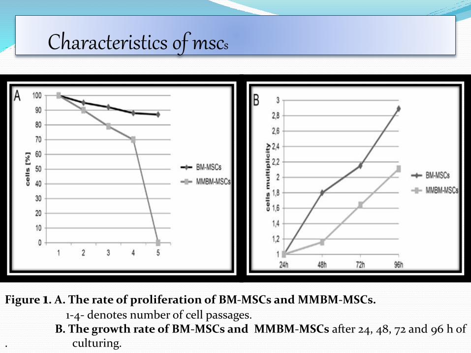
Characteristics of mscs
Figure 1. A. The rate of proliferation of BM-MSCs and MMBM-MSCs.
1-4- denotes number of cell passages. B. The growth rate of BM-MSCs and MMBM-MSCs after 24, 48, 72 and 96 h of
. culturing.
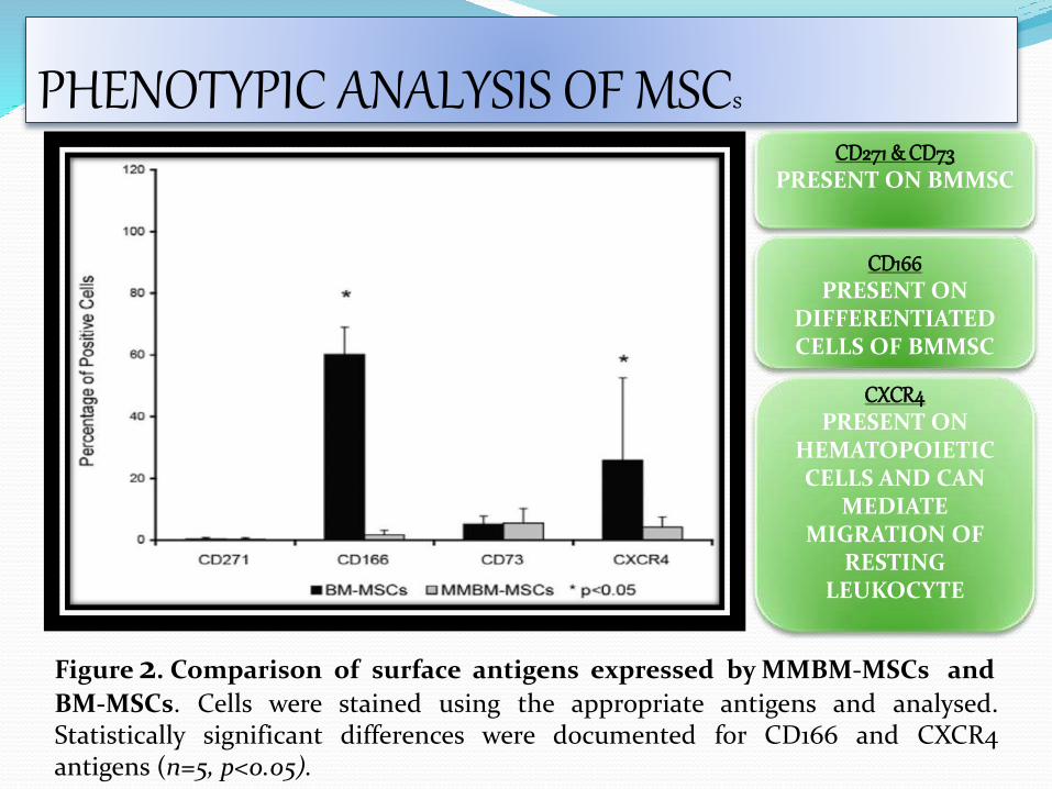
PHENOTYPIC ANALYSIS OF MSCs
Figure 2. Comparison of surface antigens expressed by MMBM-MSCs and
BM-MSCs. Cells were stained using the appropriate antigens and analysed.Statistically significant differences were documented for CD166 and CXCR4antigens (n=5, p<0.05).
CD271 & CD73PRESENT ON BMMSC
CD166PRESENT ON
DIFFERENTIATED CELLS OF BMMSC
CXCR4PRESENT ON
HEMATOPOIETIC CELLS AND CAN
MEDIATE MIGRATION OF
RESTING LEUKOCYTE
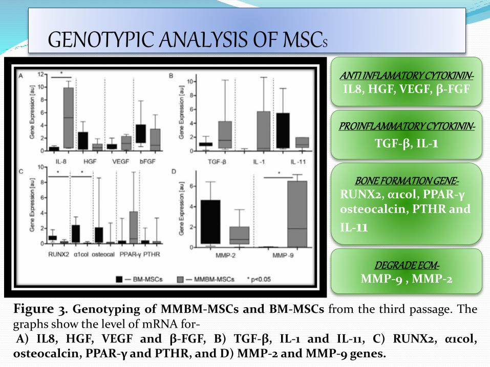
GENOTYPIC ANALYSIS OF MSCS
Figure 3. Genotyping of MMBM-MSCs and BM-MSCs from the third passage. Thegraphs show the level of mRNA for-A) IL8, HGF, VEGF and β-FGF, B) TGF-β, IL-1 and IL-11, C) RUNX2, α1col,
osteocalcin, PPAR-γ and PTHR, and D) MMP-2 and MMP-9 genes.
ANTI INFLAMATORY CYTOKININ-IL8, HGF, VEGF, β-FGF
BONE FORMATION GENE-RUNX2, α1col, PPAR-γ osteocalcin, PTHR and
IL-11
PROINFLAMMATORY CYTOKININ-
TGF-β, IL-1
DEGRADE ECM-MMP-9 , MMP-2
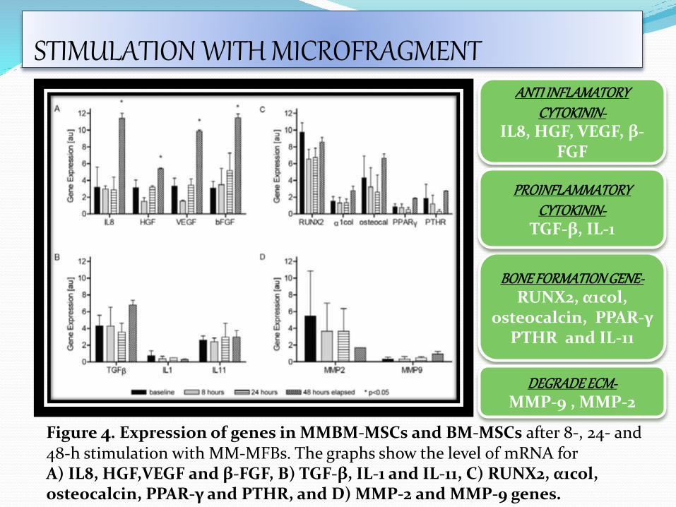
STIMULATION WITH MICROFRAGMENT
Figure 4. Expression of genes in MMBM-MSCs and BM-MSCs after 8-, 24- and 48-h stimulation with MM-MFBs. The graphs show the level of mRNA for A) IL8, HGF,VEGF and β-FGF, B) TGF-β, IL-1 and IL-11, C) RUNX2, α1col, osteocalcin, PPAR-γ and PTHR, and D) MMP-2 and MMP-9 genes.
ANTI INFLAMATORY CYTOKININ-
IL8, HGF, VEGF, β-FGF
BONE FORMATION GENE-RUNX2, α1col,
osteocalcin, PPAR-γ PTHR and IL-11
PROINFLAMMATORY CYTOKININ-
TGF-β, IL-1
DEGRADE ECM-MMP-9 , MMP-2
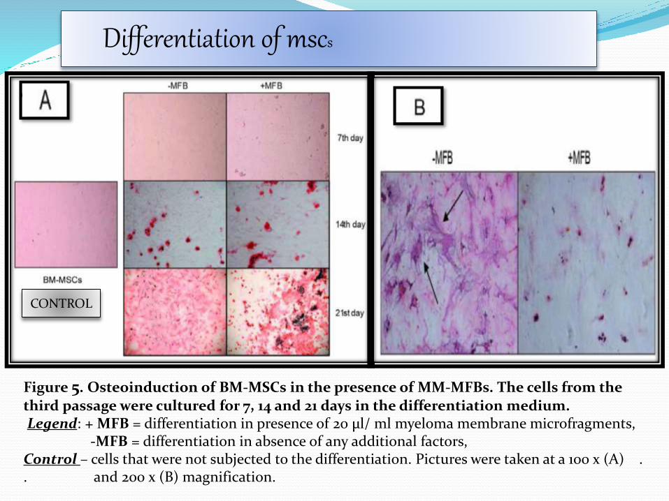
Differentiation of mscs
Figure 5. Osteoinduction of BM-MSCs in the presence of MM-MFBs. The cells from the third passage were cultured for 7, 14 and 21 days in the differentiation medium.Legend: + MFB = differentiation in presence of 20 μl/ ml myeloma membrane microfragments,
-MFB = differentiation in absence of any additional factors, Control – cells that were not subjected to the differentiation. Pictures were taken at a 100 x (A) . . and 200 x (B) magnification.
CONTROL
.
The comparative analysis of mesenchymal cells from MM patients and healthy
volunteers revealed differences in the genetic and phenotypic profiles of these twopopulations, their potential for osteodifferentiation and expression of surface antigens.
Also showed that membrane MFBs alterthe genetic profile of MSCs, leading todisorders of their osteodifferentiation.
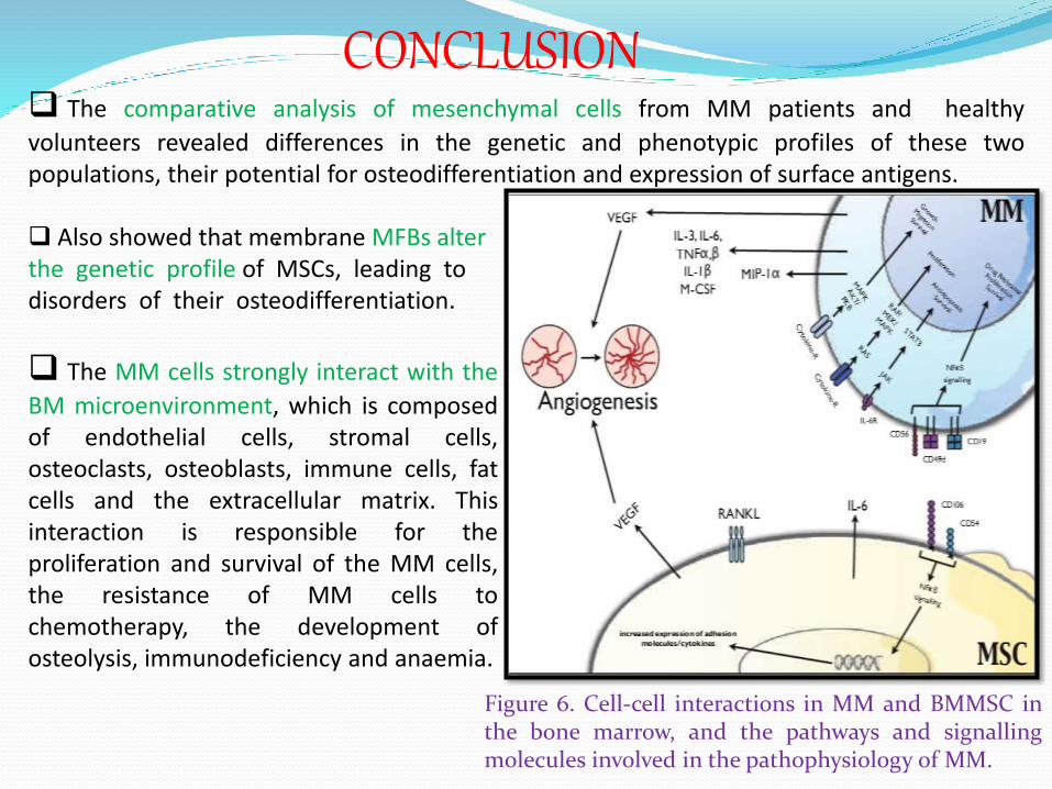
The MM cells strongly interact with the
BM microenvironment, which is composedof endothelial cells, stromal cells,osteoclasts, osteoblasts, immune cells, fatcells and the extracellular matrix. Thisinteraction is responsible for theproliferation and survival of the MM cells,the resistance of MM cells tochemotherapy, the development ofosteolysis, immunodeficiency and anaemia.
CONCLUSION
Figure 6. Cell-cell interactions in MM and BMMSC inthe bone marrow, and the pathways and signallingmolecules involved in the pathophysiology of MM.
FUTURE ASPECTS Modulation of factors (e.g., various growth factors,chemokines, and cytokines) may represent potentialapproaches to myeloma therapy.
In future if we have developed a biomarker signatureusing MFBs, which has potential as a diagnostic andprognostic tool for multiple myeloma then we can find outthe cancer at an early stage.
New therapeutic agents target both the MM, as well asthe interaction MM cell - BM microenvironment.
This is the subject of ongoing research….

REFRENCES
1. Derksen PW, de Gorter DJ, Meijer HP, et al. The hepatocyte growth factor/Met pathway controls proliferation and apoptosis in multiple myeloma.Leukemia. 2003; 17: 764-74.
2. Gahrton G. New therapeutic targets in multiple myeloma. Lancet. 2004; 364:1648-9.
3. Friedenstein AJ, Gorskaja JF, Kulagina NN. Fibroblast precursors in normal and irradiated mouse hematopoietic organs. Exp Hematol. 1976; 4: 267-74.4. De Raeve HR, Vanderkerken K. The role of the bone marrow microenvironment in multiple myeloma. Histol Histopathol. 2005; 20: 1227-50.5. Podar K, Tai YT, Lin BK, et al. Vascular endothelial growth factor-induced migration of multiple myeloma cells is associated with beta 1 integrin- and phosphatidylinositol 3-kinase-dependent PKC alpha activation. J Biol Chem. 2002; 277: 7875-81.6. Jurczyszyn A, Czepiel J, Biesiada G, et al. HGF, sIL-6R and TGF-â1 play a significant role in the progression of multiple myeloma. J Cancer. 2014; 5: 518-24.7. Lesko E, Majka M. The biological role of HGF-MET axis in tumor growth and development of metastasis. Front Biosci. 2008; 13: 1271-80.8. Fevrier B, Raposo G. Exosomes: endosomal-derived vesicles shipping extracellular messages. Curr Opin Cell Biol. 2004; 16: 415-21.9. Hugel B, Martinez MC, Kunzelmann C, et al. Membrane microparticles: two sides of the coin. Physiology. 2005; 20: 22-7.10. Ratajczak J, Wysoczynski M, Hayek F, et al. Membrane-derived microvesicles: important and underappreciated mediators of cell-to-cell communication. Leukemia. 2006; 20: 1487-95.11. Beaudoin AR, Grondin G. Shedding of vesicular material from the cell surface of eukaryotic cells: different cellular phenomena. Biochim Biophys Acta. 1991; 1071: 203-19.12. Ginestra A, Miceli D, Dolo V, et al. Membrane vesicles in ovarian cancer fluids: a new potential marker. Anticancer Res. 1999; 19: 3439-45.
13. Kim CW, Lee HM, Lee TH, et al. Extracellular membrane vesicles from tumor cells promote angiogenesis via sphingomyelin. Cancer Res. 2002; 62: 6312-7.14. Kim HK, Song KS, Park YS, et al. Elevated levels of circulating platelet microparticles, VEGF, IL-6 and RANTES in patients with gastric cancer: possible role of a metastasis predictor. Eur J Cancer. 2003; 39: 184-91.