Embed Size (px)
Citation preview

MEDICAL IMAGE COMPUTING (CAP 5937)
LECTURE 8: Medical Image Segmentation (II)(Region Growing/Merging)
Dr. Ulas BagciHEC 221, Center for Research in Computer Vision (CRCV), University of Central Florida (UCF), Orlando, FL [email protected] or [email protected]
1SPRING 2017

Outline• Region Growing algorithm• Homogeneity Criteria• Split/Merge algorithm• Examples from CT, MRI, PET• Limitations
2

Region Based Segmentation AlgorithmsRegion:A group of connected pixelswith similar properties
Closed boundaries
Computation of regions is based on similarity
Regions may correspond toObjects in a scene or partsof objects
Spatial proximity + similarity
3

Region Growing/Merging• Perhaps, one of the simplest approaches among region
based methods.
4
1. Select a seed point/points2. Define a growth criteria3. Joint all voxels connected to the seed
that follow the growth criteria4. Stop when no adjacent voxel agree with
the growth criteria
4-connectivity 8-connectivity

Example: Fat Segmentation in MRI
5
T1 weighted MRIRed: visceral fatPurple: liverBlue: Subcutaneous fat
Credit: Dougherty, G., Medica Image Processing.

Region-based Segmentation6
3D segmented two nodules are shown.Credit: Dehmeshki, et al. IEEE TMI 2008.
Red: interactive RG, white: manual segm. of lung regions pertaining to rabbits with infectionsCredit: Bagci et al. EJNMMI Research 2013

Simplest RG Pseudo-Code
7

Pictorial Illustration
8
1. Choose the seed pixel
Pictorial Illustration
9
1. Choose the seed pixel2. Check the neighboring pixels and add them to the region if
they are similar to the seed

Pictorial Illustration
10
1. Choose the seed pixel2. Check the neighboring pixels and add them to the region if
they are similar to the seed3. Repeat step 2 for each of the newly added pixels; stop if no
more pixels can be added
|neighboring pixels� seed| < Threshold

Algorithm Illustration
11

Split and Merge Algorithm• Unlike RG, region splitting starts with the whole image as a single region
and subdivides it into subsidiary regions recursively while a condition of homogeneity is not satisfied.
12

Split and Merge Algorithm• Unlike RG, region splitting starts with the whole image as a single region
and subdivides it into subsidiary regions recursively while a condition of homogeneity is not satisfied.
• Region merging is the opposite of splitting, and works as a way of avoiding over-segmentation
13

Split and Merge Algorithm• Unlike RG, region splitting starts with the whole image as a single region
and subdivides it into subsidiary regions recursively while a condition of homogeneity is not satisfied.
• Region merging is the opposite of splitting, and works as a way of avoiding over-segmentation
14

Drawback of Split ?
15

Drawback of Split ?• the final image partition may contain adjacent regions with
identical properties.• Solution?
16

Drawback of Split ?• the final image partition may contain adjacent regions with
identical properties.• Solution?
– REGION MERGING
17

Split and Merge Algorithm• When a homogeneous region is created, its neighboring
regions are checked and the newly created region is merged with an existing one if they have identical properties.
• If the similarity criteria are met by more than one adjacent region, the new region is merged with the most similar one.
18
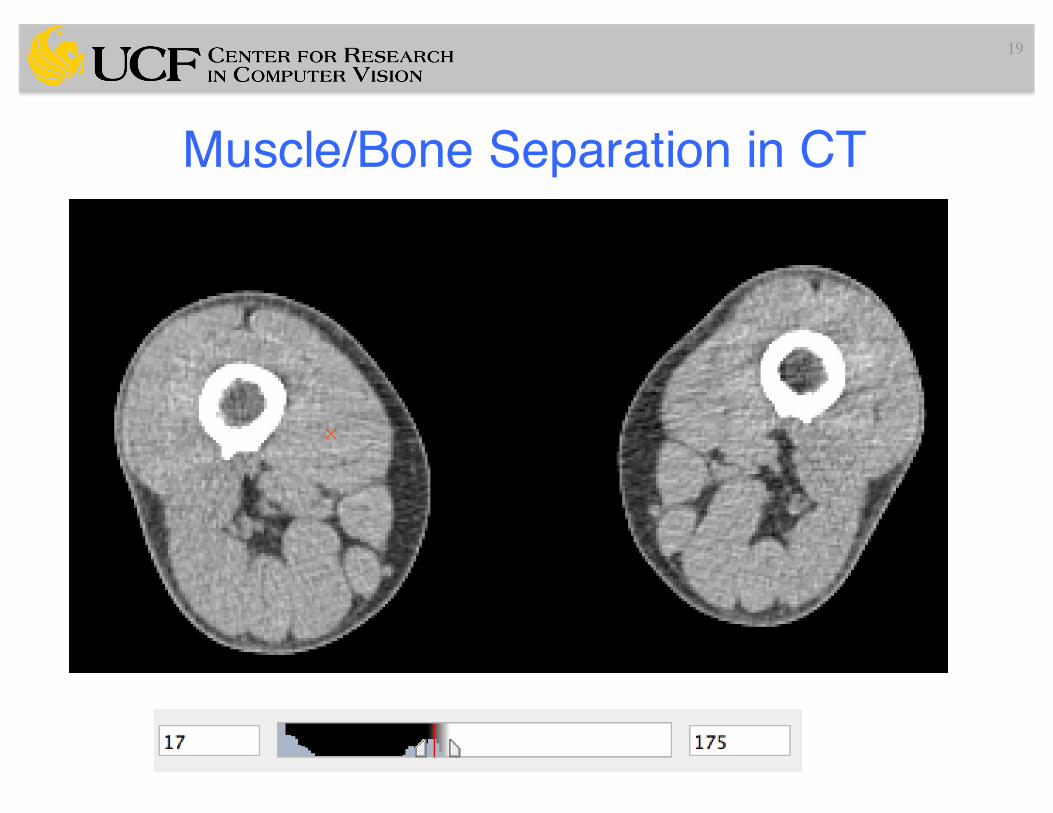
Muscle/Bone Separation in CT
19

Muscle/Bone Separation in CT
20
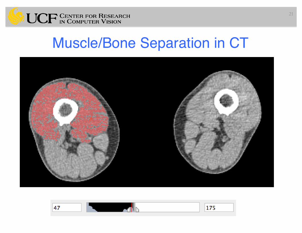
Muscle/Bone Separation in CT
21

Muscle/Bone Separation in CT
22

Muscle/Bone Separation in CT
23

Virtual Endoscopy/Colonoscopy - CT24
(a) Original CT imagethrough the colon. (b) A 3D RG initiated from a seed point. (c) 3D rendering of thesegmented colon.
Credit: P. Seutens
Adaptive Parameterization of RG• Literature is vast!
25

Adaptive Parameterization of RG• Literature is vast!• Ex: Tumor segmentation from PET images
26

Adaptive Parameterization of RG• Literature is vast!• Ex: Tumor segmentation from PET images
T: threshold, mean of R0 à mean intensity of current grown region
27

Adaptive Parameterization of RG
28
Credit: Hua Li et alMedPhys 2008
a. Original imageb. Threshold/Volume curvec. ROId. Thresholdinge. Adaptive RG

Left Ventricular Function in Cardiac CT• The gold standard for the functional evaluation of the left
ventricle (LV) is magnetic resonance imaging (MRI)
29

Left Ventricular Function in Cardiac CT• The gold standard for the functional evaluation of the left
ventricle (LV) is magnetic resonance imaging (MRI)• Contrast-enhanced retrospectively electrocardiogram (ECG)-
gated multi-slice spiral computed tomography (MSCT) has been accepted as an efficient noninvasive tool for the detection of coronary artery stenosis, providing good sensitivity and specificity
30

Left Ventricular Function in Cardiac CT• The gold standard for the functional evaluation of the left
ventricle (LV) is magnetic resonance imaging (MRI)• Contrast-enhanced retrospectively electrocardiogram (ECG)-
gated multi-slice spiral computed tomography (MSCT) has been accepted as an efficient noninvasive tool for the detection of coronary artery stenosis, providing good sensitivity and specificity
• Assessment of the LV volume at end diastole and end systole is used for quantification of function!
31

Left Ventricular Function in Cardiac CT
32
Endocardial contours in systole Endocardial contours in diastole
Credit: muhlenbruch, et al. Eur Rad 2006
Homogenous region and high contrasthelps region growing to be suitable for this case!

CT Lung/Soft Tissue Separation• Seed > -200 HU
33

Airway Segmentation (Basic RG)
34

Slicer Implementation of RG and Airway Segmentation
35

Airway Segmentation (Basic RG)
36
Credit: Nardelli et alBioMedOnline 2015

Bone Vanishing Disease - CT
37

Bone Vanishing Disease - CT
38
(credit: Papadakis, G.Z., NIH

Bone Vanishing Disease - CT
39
(credit: Papadakis, G.Z., NIH
Controlling the homogeneityparameter (similarity criteria) in RGis extremely difficult due to largevariation in bone density shifting towardslower densities
-78 HU
307 HU

Limitations of Region Growing1) Objects in medical images are often non-homogeneous
40

Limitations of Region Growing1) Objects in medical images are often non-homogeneous
2) Poor contrast causes leakages
41

Limitations of Region Growing1) Objects in medical images are often non-homogeneous
2) Poor contrast causes leakages
3) Noise
42

Summary• Region Growing based segmentation algorithms
– Still in use for many applications in radiology• Lung, bone, homogenous (isolated ) tumors, …
– Either terminal use or in-between step– Similarity criteria: homogenous regions
43

Slide Credits and References• Jayaram K. Udupa, MIPG of University of Pennsylvania, PA.• P. Suetens, Fundamentals of Medical Imaging, Cambridge
Univ. Press.
• Adams and Bischof, IEEE PAMI 1994.• Zhu and Yuille, IEEE PAMI 1996.• Hu et al, IEEE TMI 2001.• Dehmeshki et al IEEE TMI 2008.• Nardelli et al BioMedOnline 2015• Dougherty, G., Medical Image Processing• Hua Li et al MedPhys 2008
44