Embed Size (px)
Citation preview

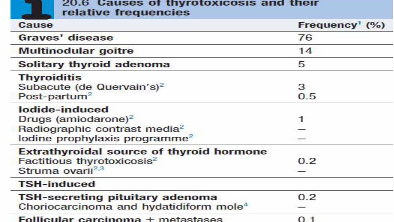
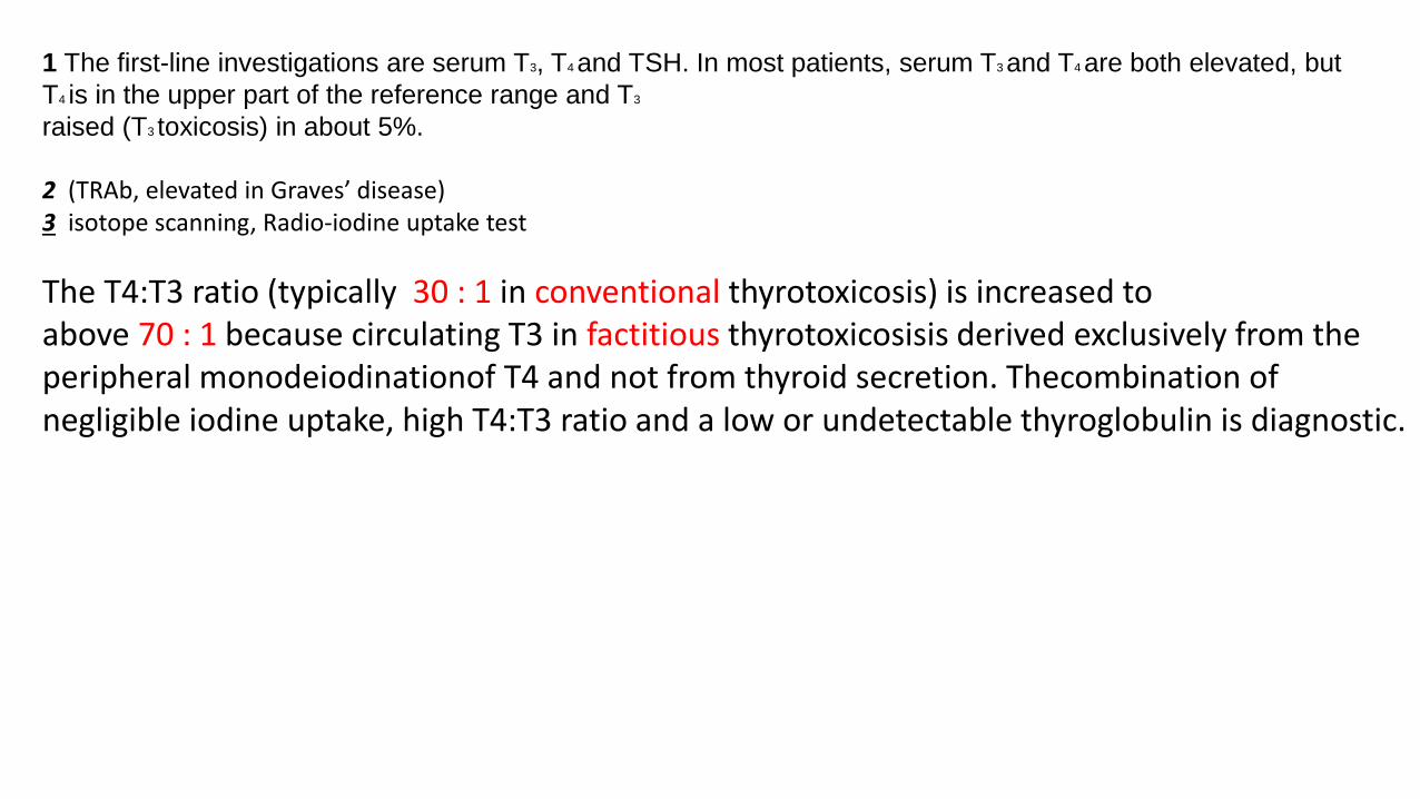
1 The first-line investigations are serum T3, T4 and TSH. In most patients, serum T3 and T4 are both elevated, but
T4 is in the upper part of the reference range and T3
raised (T3 toxicosis) in about 5%.
2 (TRAb, elevated in Graves’ disease) 3 isotope scanning, Radio-iodine uptake test
The T4:T3 ratio (typically 30 : 1 in conventional thyrotoxicosis) is increased toabove 70 : 1 because circulating T3 in factitious thyrotoxicosisis derived exclusively from the peripheral monodeiodinationof T4 and not from thyroid secretion. Thecombination of negligible iodine uptake, high T4:T3 ratio and a low or undetectable thyroglobulin is diagnostic.

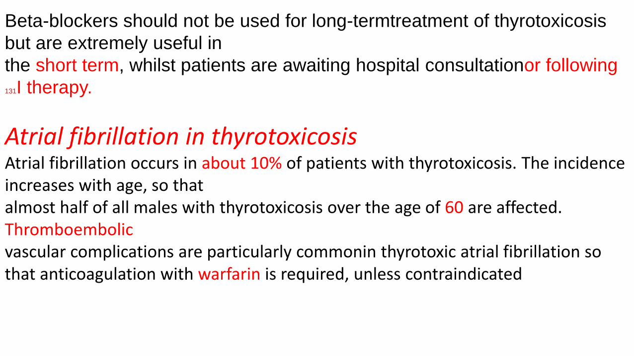
Beta-blockers should not be used for long-termtreatment of thyrotoxicosis
but are extremely useful in
the short term, whilst patients are awaiting hospital consultationor following
131I therapy.
Atrial fibrillation in thyrotoxicosisAtrial fibrillation occurs in about 10% of patients with thyrotoxicosis. The incidence increases with age, so thatalmost half of all males with thyrotoxicosis over the age of 60 are affected.Thromboembolicvascular complications are particularly commonin thyrotoxic atrial fibrillation so that anticoagulation with warfarin is required, unless contraindicated
Thyrotoxic crisis (‘thyroid storm} This is a rare but life-threatening complication of thyrotoxicosis. The most prominent
signs are fever, agitation, confusion, tachycardia or atrialfibrillation and, in the older
patient, cardiac failure. It is a medical emergency,
which has a mortality of 10% despite early recognitionand treatment. Thyrotoxic crisis
is most commonly precipitated by infection in a patient with previously
unrecognised
or inadequately treated thyrotoxicosisIt mayalso develop shortly after subtotal
thyroidectomy in anill-prepared patient or within a few days of 131I therapy, when acute
irradiation damage may lead to a transientrise in serum thyroid hormone levels.
Patients should be
rehydrated and given propranolol, either orally (80 mg 4 times daily) or
intravenously(1–5 mg 4 times daily). Sodium ipodate (500 mg perday orally) will
restore serum T3 levels to normal
in
This is a radiographic contrast mediumwhich not only inhibits the
release of thyroid hormones, but also reduces the conversion of T4 to T3 and
is, therefore, more effective than potassium iodide or Lugol’ssolution
.
Dexamethasone (2 mg 4 times daily) and amiodarone
have similar effects. Oral carbimazole 40–60 mg
daily should be given to inhibit the synthesis of
new thyroid hormone. If the patient is unconscious or
uncooperative, carbimazole can be administered rectally
with good effect, but no preparation is available for
parenteral use.
After 10–14 days the patient can usually
be maintained on carbimazole alone.

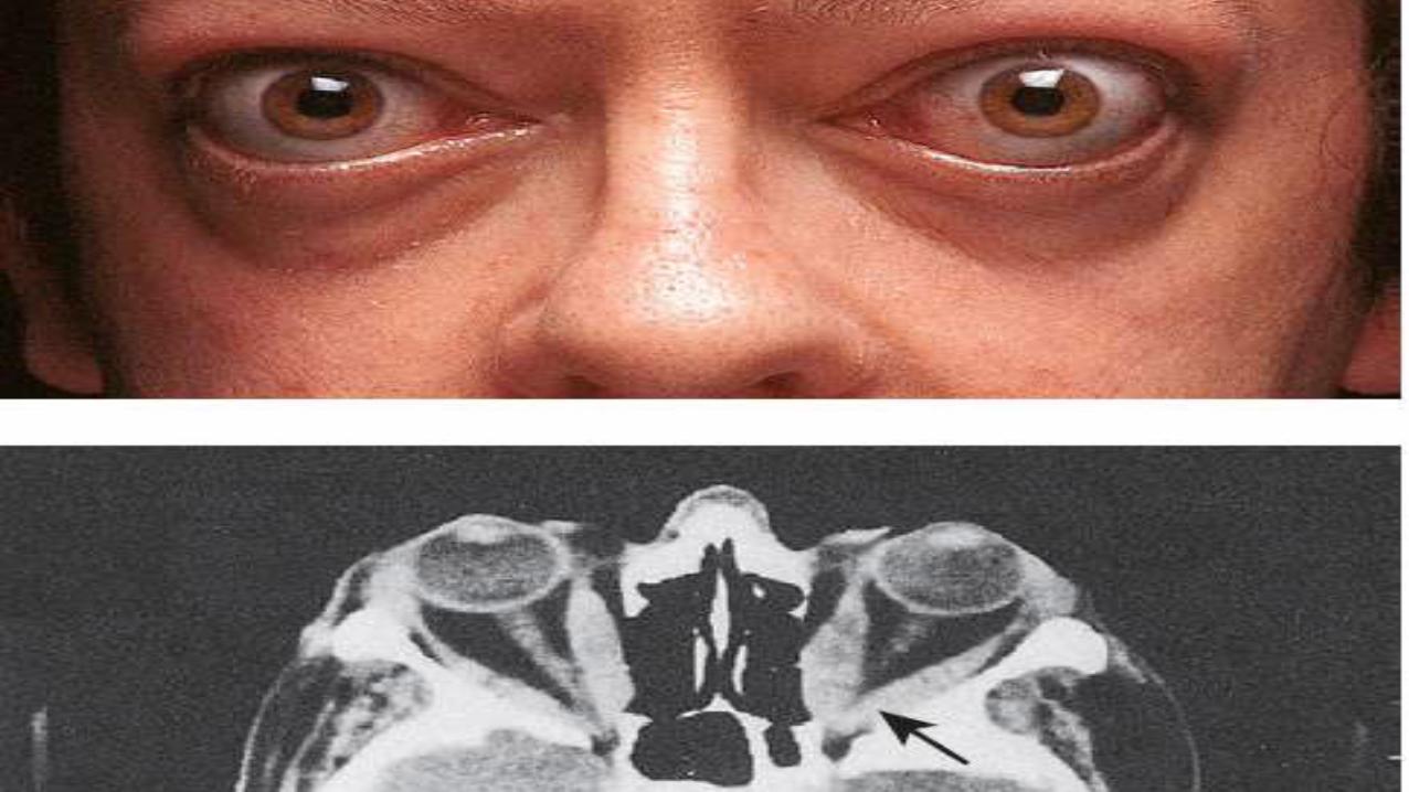
Graves’ diseaseGraves’ disease can occur at any age but is unusuall before puberty and most commonly affects women
aged 30–50 years. The most common manifestationis thyrotoxicosis with or without a diffuse goitre
clinical features and differential diagnosis are describedon Graves’ disease also causes ophthalmopathy
and, rarely, pretibial myxoedema These extrathyroidal features usually occur in thyrotoxic
patients, but can occur in the absence of thyroid dysfunction.
Graves’ thyrotoxicosis
PathophysiologyThe thyrotoxicosis results from the production of IgG
antibodies directed against the TSH receptor on the
thyroid follicular cell, which stimulate thyroid hormone
production and proliferation of follicular cells, leading
to goitre in the majority of patients.
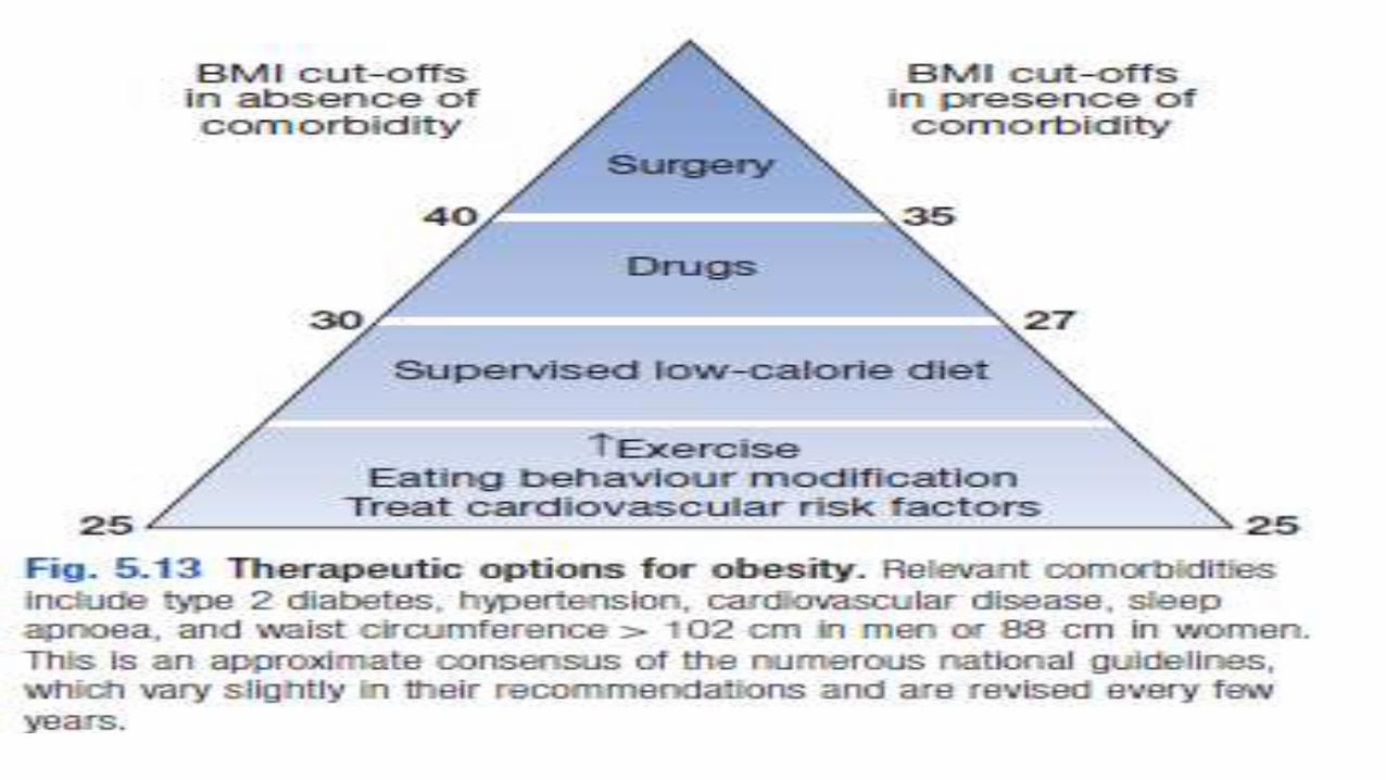
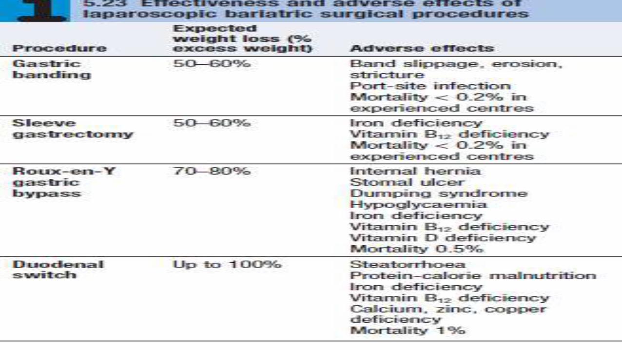
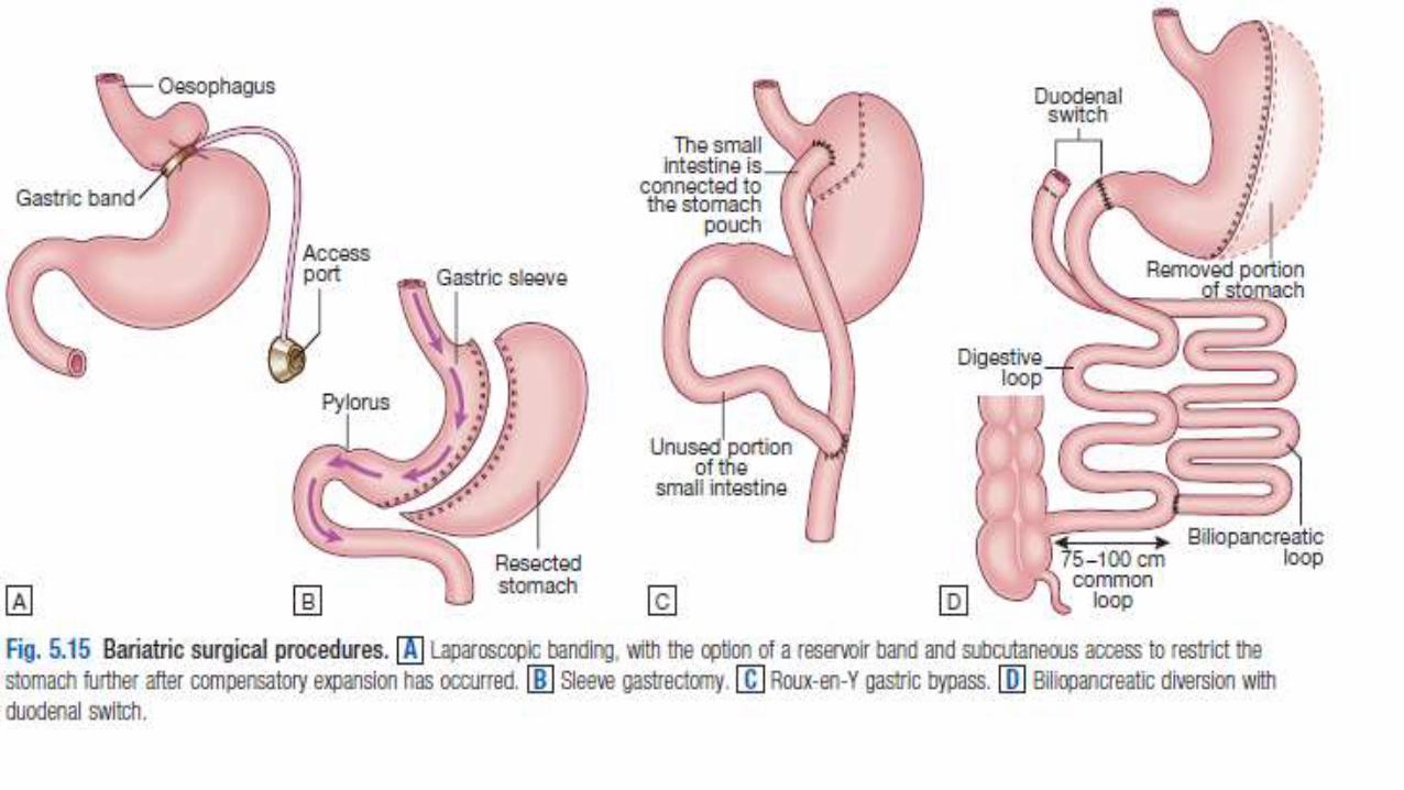
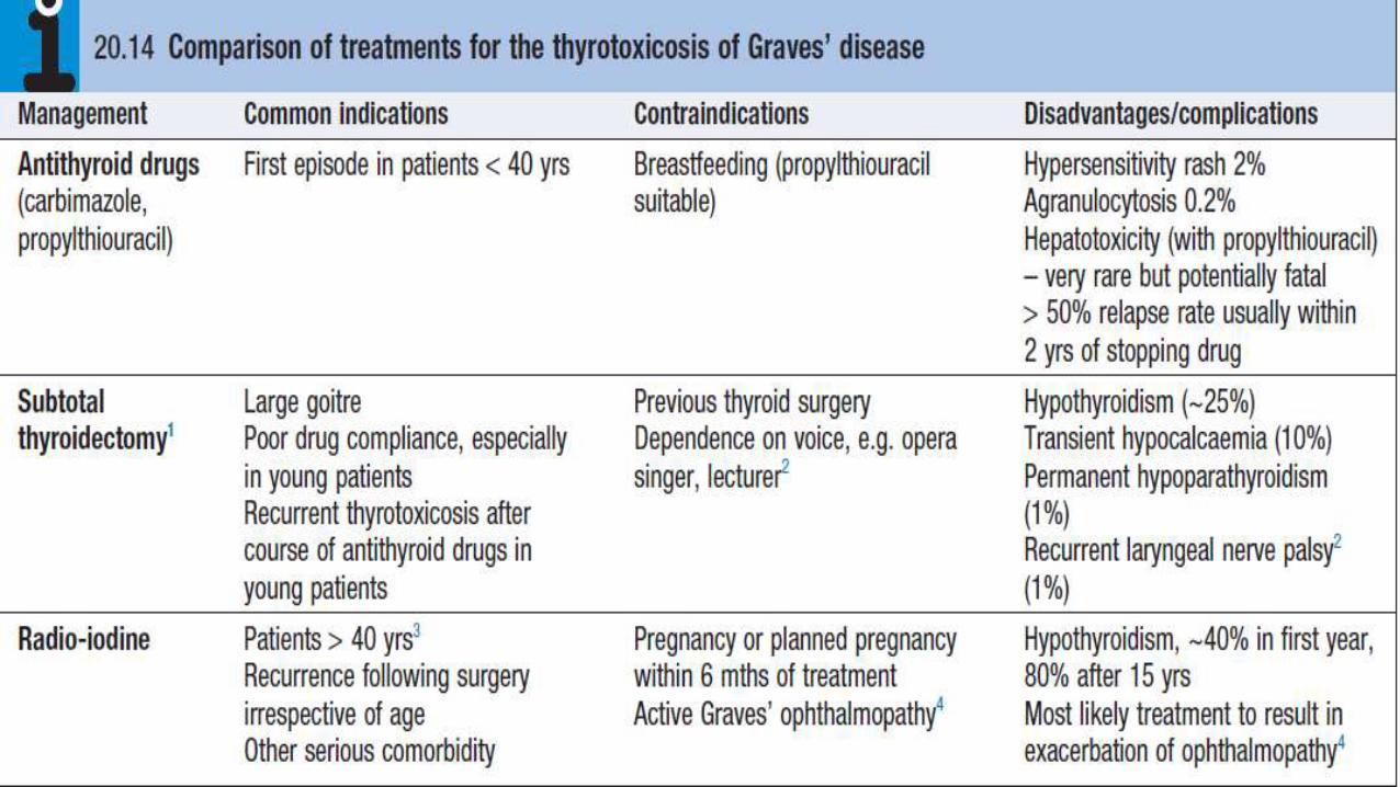
ManagementSymptoms of thyrotoxicosis respond to β-blocker but definitive treatment
requires control of thyroid hormone secretion. The different options are
compared in .
patients under 40 years of age, most clinicians adopt the empirical
approach ofprescribing a course of carbimazole and recommending
surgery if relapse occurs, while 131I is employed as first or second-line
treatment in those aged over 40.
131I with increased incidence of some malignancies, particularly
of the thyroid and gastrointestinal tract,
Antithyroid drugs.
The most commonly used are carbimazoleand its active metabolite, methimazole (notavailable in the UK). Propylthiouracil is equally effective.
These drugs reduce the synthesis of new thyroid hormones by inhibiting the iodination of tyrosine
. Carbimazole also has an immunosuppressive action, leading to a reduction in serum TRAb
concentrations, but this is not enough to influence the natural history of the thyrotoxicosis significantly.
Antithyroid drugs should be introduced at high doses (carbimazole 40–60 mg daily or propylthiouracil
400–600 mg daily).Usually, this results in subjective improvement within 10–14 days and renders the patient
clinically and biochemically euthyroid at 3–4 weeks. Atthis point, the dose can be reduced and titrated to maintain
T4 and TSH within their reference range. In most patients, carbimazole is continued at 5–20 mg per day
for 12–18 months in the hope that remission will occur.
Patients with thyrotoxicosis relapse in at least 50% of cases, usually within 2 years of stopping treatment.
Rarely, T4 and TSH levels fluctuate between those of thyrotoxicosis and hypothyroidism at successive review
appointments, despite good drug compliance, presumably due to rapidly changing concentrations of TRAb. Inthese
patients, satisfactory control can be achieved by
blocking thyroid hormone synthesis with carbimazole 30–40 mg daily and adding levothyroxine 100–150 μg
daily as replacement
Thyroid surgery.Patients should be rendered euthyroid with antithyroid drugs before operation.
Potassium iodide, 60 mg 3 times daily orally, is often added for 2 weeks before
surgery to inhibit thyroid hormone release and reduce the size and vascularity of the
gland, making surgery technically easier.
‘subtotal’ thyroidectomy is performed, in which a portionof one lobe of the
thyroid is left in situ, with the aim of rendering the patient euthyroid post-operatively.
While complications of surgery are rare and 80% of patients are euthyroid, 15% are
permanently hypothyroid and 5% remain thyrotoxic.,
many endocrine surgeons now opt to perform a ‘near total’ thyroidectomy, leaving
behind only a small portion
of gland adjacent to the recurrent laryngeal nerves. This strategy invariably results in
permanent hypothyroidism
and is probably associated with a higher risk
Radioactive iodine. 131I 131I is administered orally as a single dose400 MBq (10 mCi) is given orally.
, and is trapped and organified in the thyroid Although 131I decays within a few weeks, it has long-lasting
inhibitory effects on survival and replication of follicular cells. This regimen is effective in 75% of patients within
4–12 weeks.
During the lag period, symptoms can be controlled by a β-blocker or, in more severe cases, by
carbimazole. However, reduces the efficacy of 131I therapy because it prevents organification of 131I in the gland,
and so should be carbimazole avoided until 48 hours after radio-iodine administration.
If thyrotoxicosis persists after 6 months, a further dose of 131I can be given.
The disadvantage of 131I treatment 1 hypothyroidism.
2 131I isusually avoided in patients with Graves’ ophthalmopathy It can be administered with caution in those with mild
or ‘burnt-out’ eye disease, when it is customary to cover the treatment with a 6-week tapering course of oral
prednisolone. 3 In women of reproductive age, pregnancy must be excluded before administration of 131I and avoided
for 6 months thereafter; men are also advised against fathering children for 6 months after receiving 131I


HypothyroidismHypothyroidism is a common condition with various
causes (but autoimmune disease(Hashimoto’s
thyroiditis) and thyroid failure following131I or
surgical treatment of thyrotoxicosis account for
over 90% of cases, except in areas where iodine
deficiency is endemic. Women are affected
approximately six times more frequently than men

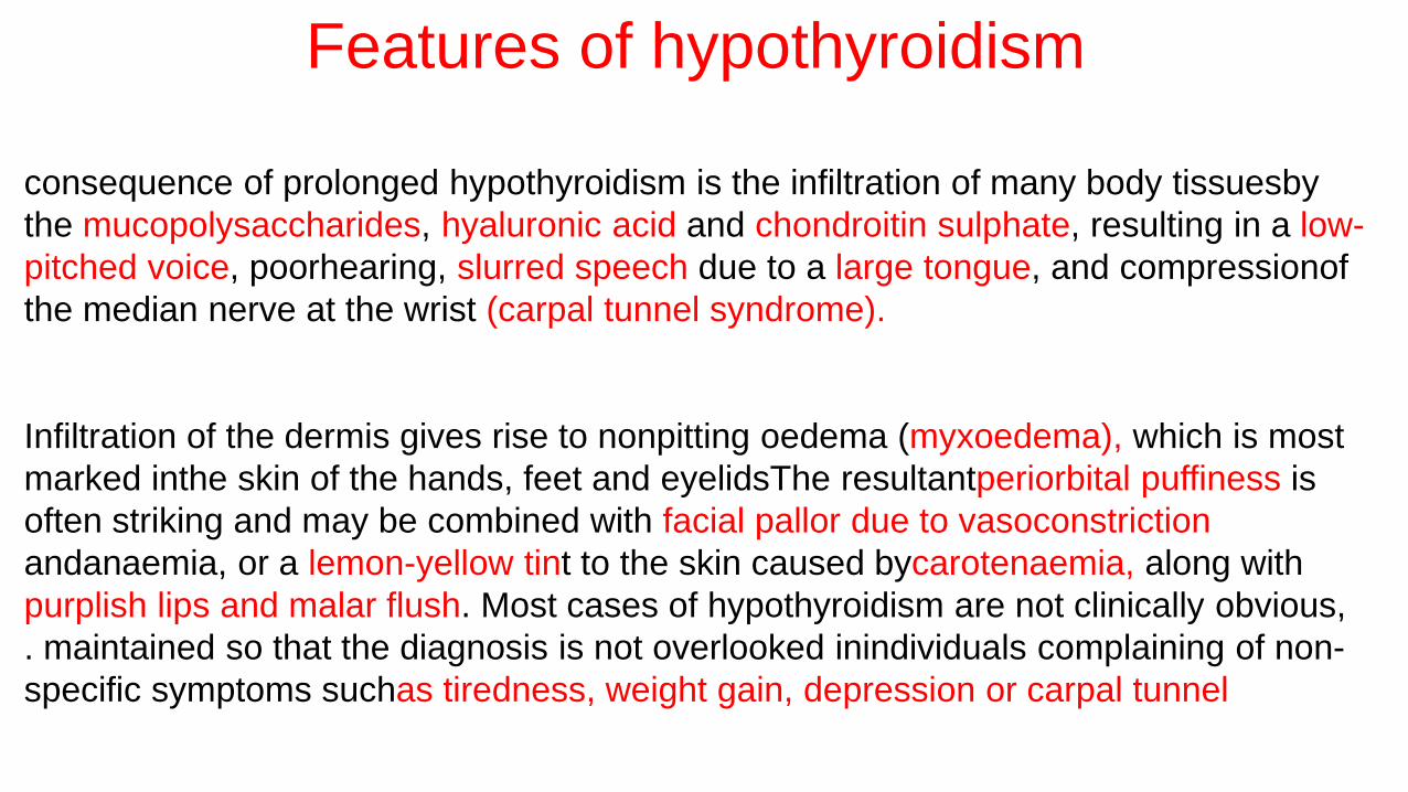
Features of hypothyroidism
consequence of prolonged hypothyroidism is the infiltration of many body tissuesby
the mucopolysaccharides, hyaluronic acid and chondroitin sulphate, resulting in a low-
pitched voice, poorhearing, slurred speech due to a large tongue, and compressionof
the median nerve at the wrist (carpal tunnel syndrome).
Infiltration of the dermis gives rise to nonpitting oedema (myxoedema), which is most
marked inthe skin of the hands, feet and eyelidsThe resultantperiorbital puffiness is
often striking and may be combined with facial pallor due to vasoconstriction
andanaemia, or a lemon-yellow tint to the skin caused bycarotenaemia, along with
purplish lips and malar flush. Most cases of hypothyroidism are not clinically obvious,
. maintained so that the diagnosis is not overlooked inindividuals complaining of non-
specific symptoms suchas tiredness, weight gain, depression or carpal tunnel

Management
Treatment is with levothyroxine replacement. It is customary
to start with a low dose of 50 μg per day for3 weeks, increasing thereafter
to 100 μg per day for afurther 3 weeks and finally to a maintenance dose
of100–150 μg per day.
In younger patients, it is safe toinitiate levothyroxine at a higher dose
to allow a more rapid normalisation of thyroid hormone levels.
Levothyroxine has a half-life of 7 days so it should always be taken as a
single daily dose and at least 6 weeks should pass before repeating
thyroid function tests and adjusting the dose, usually
by 25 μg per day. Patients feel better within 2–3 weeks.
Reduction in weight and periorbital puffiness occursquickly, but the restoration of skin and hair texture may take 3–6 months It is
important to measure thyroid function every1–2 years
Levothyroxine replacement in ischaemic heart disease
exacerbation of myocardial ischaemia, infarction and
sudden death are recognised complications of levothyroxine replacement, even using doses as low as 25 μgper day. In replacement should be introduced at low dose and increased very slowly under specialistsupervision. It has been suggested that T3 has an advantage over T4, since T3 has a shorter half-life and anyadverse effect will reverse more quickly, but the moredistinct peak in hormone levels after each dose of T3 isadisadvantage. Coronary angioplasty or bypass surgerymay be required if angina is exacerbated by levothyroxine
Hypothyroidism in pregnancyMost pregnant women with primary hypothyroidismrequire an increase in the dose of
levothyroxine of
approximately 25–50 μg daily to maintain normal TSH levels. This may reflect increased
metabolism of thyroxine
by the placenta and increased serum thyroxinebindingglobulin during pregnancy,
resulting in an increase in the total thyroid hormone pool to maintain the same free T4
and T3 concentrations
. Inadequate maternal T4 therapy may be associated with impaired cognitive
development in an unborn child and so women are usually advised to increase their
daily levothyroxine dose by 25 μg when pregnancy is confirmed. Serum TSH
and free T4 should be measured during each trimester and the dose of
levothyroxine adjusted to maintain a normal TSH

Myxoedema comadepressed level of consciousness,
usually in an elderly patient who appears myxoedematous. Body temperature may be as low as 25°C, convulsions
are not uncommon and cerebrospinal fluid (CSF) pressure and protein content are raised. The mortalityrate is 50%
and survival depends on early recognitionand treatment of hypothyroidism and other factors
contributing to the altered consciousness level, as medication, cardiac failure, pneumonia, dilutional
hyponatraemia and respiratory failure.
Myxoedema coma is a medical emergency and treatmentmust begin before biochemical confirmation of the
diagnosis. Suspected cases should be treated with anintravenous injection of 20 μg triiodothyronine, followed
by further injections of 20 μg 3 times daily unti lthere is sustained clinical improvement. In survivors,
there is a rise in body temperature within 24 hours
after 48–72 hours, it is usually possible to switch patientsto oral levothyroxine in a dose of 50 μg daily. Unlessit is
apparent that the patient has primary hypothyroidism, the thyroid failure should also be assumed to be secondary to
hypothalamic or pituitary disease
and treatment given with hydrocortisone 100 mg IM
3 times daily, pending the results of T4, TSH and cortisol
measurement (p. 787). Other measures include slow
rewarming (p. 105), cautious use of intravenous fluids,
broad-spectrum antibiotics and high-flow oxygen. Occasionally,
assisted ventilation may be necessary
Symptoms of hypothyroidism with normal thyroid function tests
The classic symptoms of hypothyroidism are, by theirvery nature, non-specific There is a wide
differential diagnosis for symptoms such as ‘fatigue’, weight gain’ and ‘low mood’. As has been noted,
outside the context of pituitary and hypothalamicdisease, serum TSH is an excellent measure of an individual’s
thyroid hormone status. However, some individualsbelieve that they have hypothyroidism despite
normal serum TSH concentrations. There are a largenumber of websites which claim that serum TSH is not
a good measure of thyroid hormone status and suggestthat other factors, such as abnormalities of T4 to T3 conversion,
may lead to low tissue levels of active thyroidhormones. Such websites often advocate a variety of
tests of thyroid function of dubious scientific validity, including measurement of serum reverse T3, 24-hour
urine T3, basal body temperature, skin iodine absorption, and levels of selenium in blood and urine. Individuals
who believe they have hypothyroidism, despitenormal conventional tests of thyroid function, can be
difficult to manage. They require reassurance that theirsymptoms are being taken seriously and that organic
disease has been carefully considered; if their symptomspersist, then referral to a team specialising in medically
unexplained symptoms should be considered.
‘


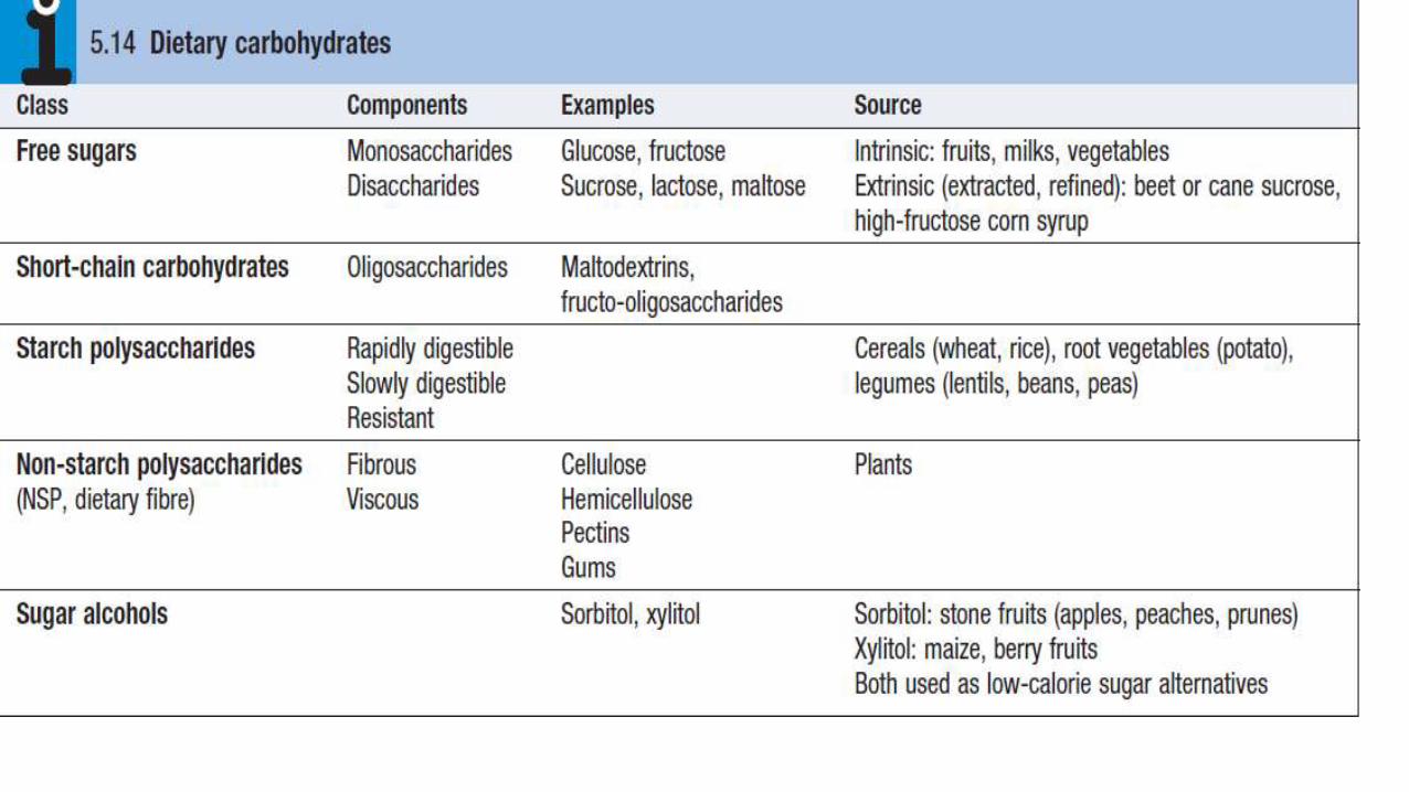
Nutrients
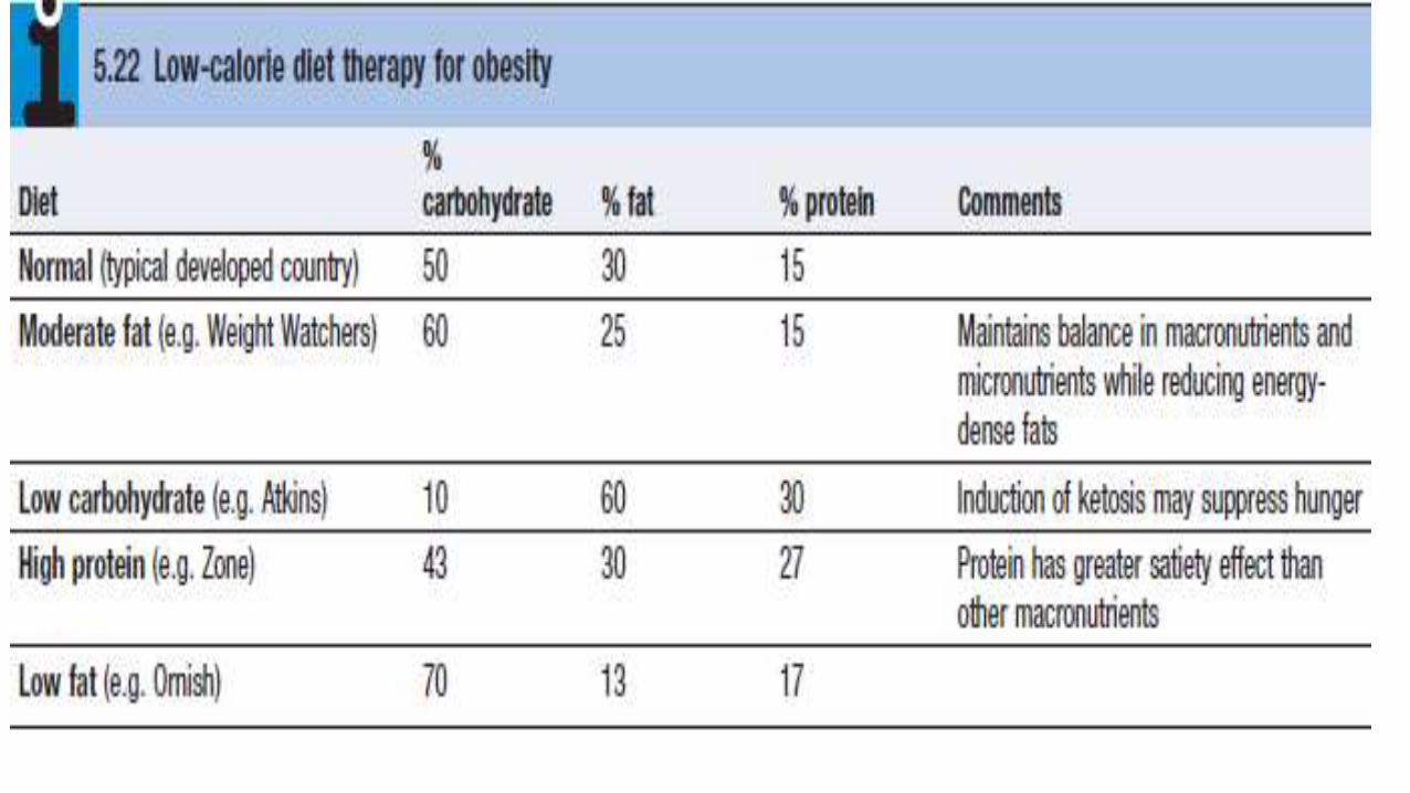
the diet can be classified into ‘macronutrients’,
which are eaten in relatively large amounts to
provide fuel for energy, and ‘micronutrients’ (e.g.
vitaminsand minerals), which do not contribute to
energy balance but are required in small amounts
because they are not synthesised in the body.
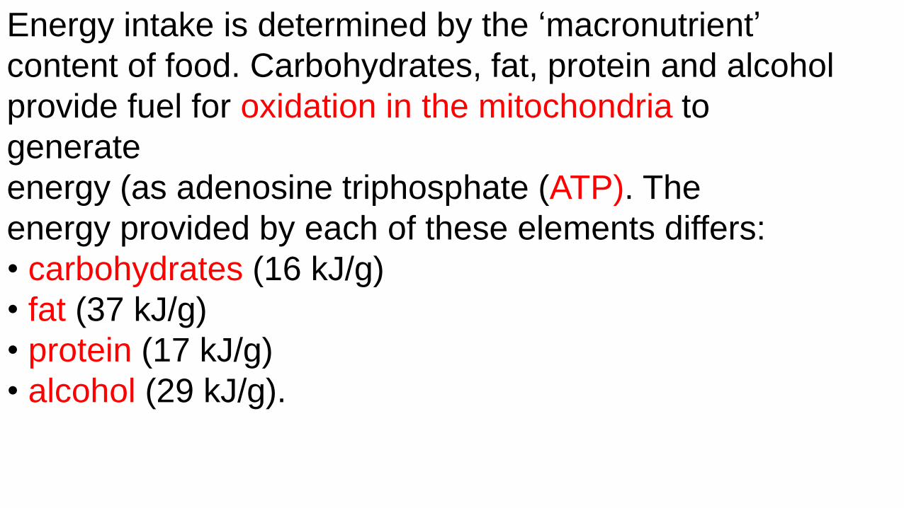
Energy intake is determined by the ‘macronutrient’
content of food. Carbohydrates, fat, protein and alcohol
provide fuel for oxidation in the mitochondria to
generate
energy (as adenosine triphosphate (ATP). The
energy provided by each of these elements differs:
• carbohydrates (16 kJ/g)
• fat (37 kJ/g)
• protein (17 kJ/g)
• alcohol (29 kJ/g).


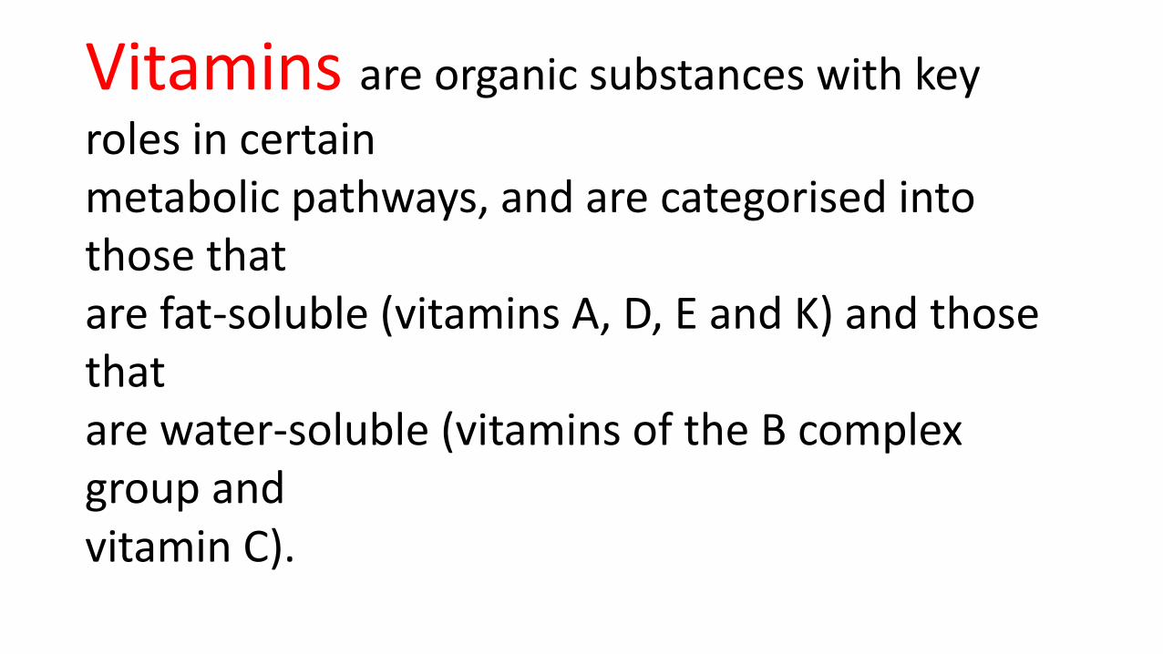
Vitamins are organic substances with key
roles in certainmetabolic pathways, and are categorised into those thatare fat-soluble (vitamins A, D, E and K) and those thatare water-soluble (vitamins of the B complex group andvitamin C).


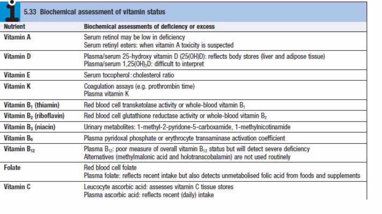

Vitamin A (retinol)Pre-formed retinol is found only in foods of animalorigin. Vitamin A can also be derived from carotenes,which are present in green and coloured vegetables and some fruits. Carotenes provide most of the totalvitamin A in the UK, and constitute the only supply invegans. Retinol is converted to several other importantmolecules:
• 11-cis retinaldehyde is part of the photoreceptor complex in rods of the retina.• Retinoic acid induces differentiation of epithelialcells by binding to specific nuclear receptors, whichinduce responsive genes. In vitamin A deficiency,mucus-secreting cells are replaced by keratinproducingcells.• Retinoids are necessary for normal growth, fetaldevelopment, fertility, haematopoiesis and immunefunction.
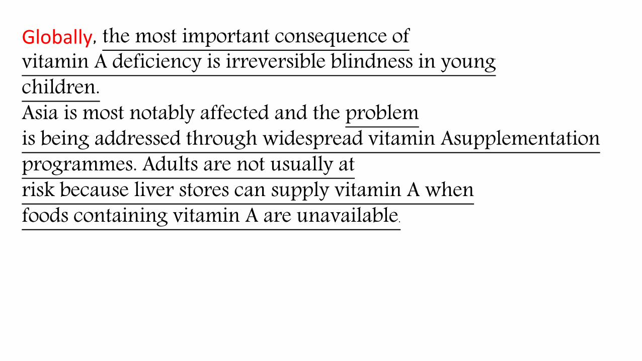
Globally, the most important consequence ofvitamin A deficiency is irreversible blindness in youngchildren.Asia is most notably affected and the problemis being addressed through widespread vitamin Asupplementationprogrammes. Adults are not usually atrisk because liver stores can supply vitamin A whenfoods containing vitamin A are unavailable.
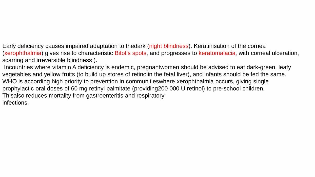
Early deficiency causes impaired adaptation to thedark (night blindness). Keratinisation of the cornea
(xerophthalmia) gives rise to characteristic Bitot’s spots, and progresses to keratomalacia, with corneal ulceration,
scarring and irreversible blindness ).
Incountries where vitamin A deficiency is endemic, pregnantwomen should be advised to eat dark-green, leafy
vegetables and yellow fruits (to build up stores of retinolin the fetal liver), and infants should be fed the same.
WHO is according high priority to prevention in communitieswhere xerophthalmia occurs, giving single
prophylactic oral doses of 60 mg retinyl palmitate (providing200 000 U retinol) to pre-school children.
Thisalso reduces mortality from gastroenteritis and respiratory
infections.

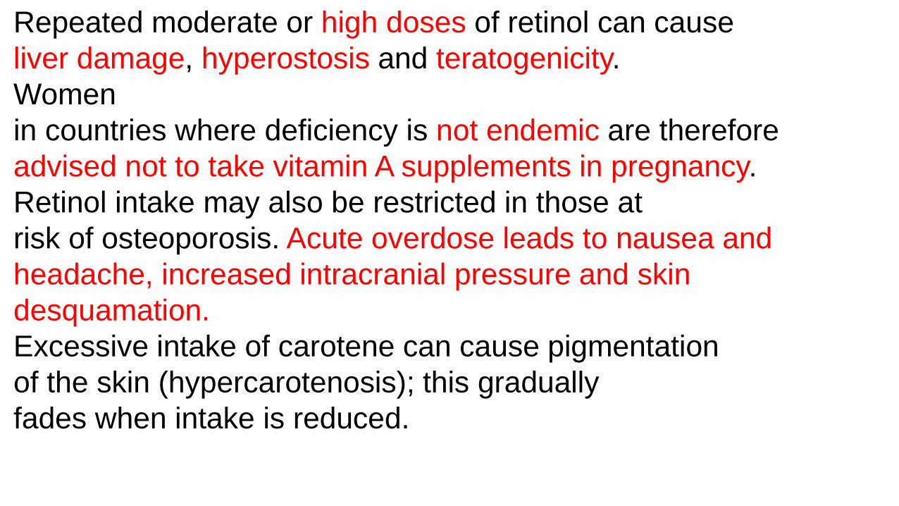
Repeated moderate or high doses of retinol can cause
liver damage, hyperostosis and teratogenicity.
Women
in countries where deficiency is not endemic are therefore
advised not to take vitamin A supplements in pregnancy.
Retinol intake may also be restricted in those at
risk of osteoporosis. Acute overdose leads to nausea and
headache, increased intracranial pressure and skin
desquamation.
Excessive intake of carotene can cause pigmentation
of the skin (hypercarotenosis); this gradually
fades when intake is reduced.
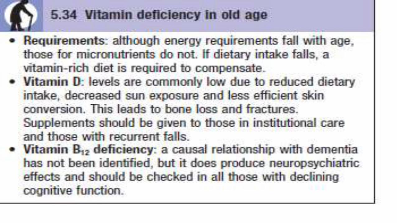
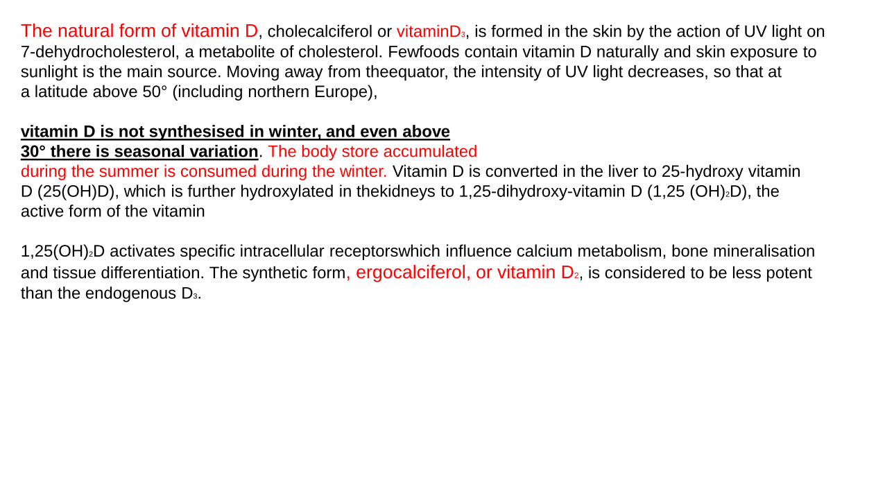
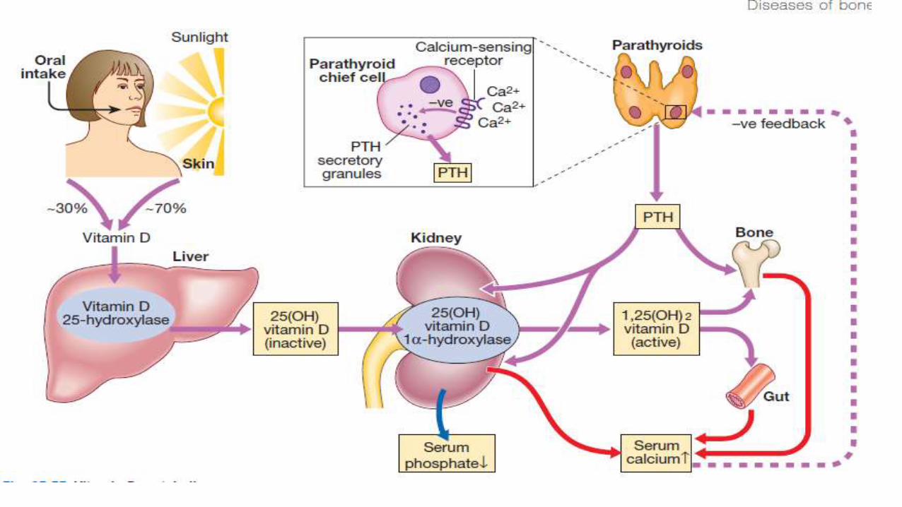
The natural form of vitamin D, cholecalciferol or vitaminD3, is formed in the skin by the action of UV light on
7-dehydrocholesterol, a metabolite of cholesterol. Fewfoods contain vitamin D naturally and skin exposure to
sunlight is the main source. Moving away from theequator, the intensity of UV light decreases, so that at
a latitude above 50° (including northern Europe),
vitamin D is not synthesised in winter, and even above
30° there is seasonal variation. The body store accumulated
during the summer is consumed during the winter. Vitamin D is converted in the liver to 25-hydroxy vitamin
D (25(OH)D), which is further hydroxylated in thekidneys to 1,25-dihydroxy-vitamin D (1,25 (OH)2D), the
active form of the vitamin
1,25(OH)2D activates specific intracellular receptorswhich influence calcium metabolism, bone mineralisation
and tissue differentiation. The synthetic form, ergocalciferol, or vitamin D2, is considered to be less potent
than the endogenous D3.

The effects of vitamin D deficiency (calcium
deficiency,
rickets and osteomalacia) are described on An
analogue of vitamin D (calcipotriol) is used for
treatment of skin conditions such as psoriasis.
Excessive
doses of cholecalciferol, ergocalciferol or the
hydroxylated
metabolites cause hypercalcaemia

-Vitamihasn E αtocopherol. many direct metabolicactions:• It prevents oxidation of polyunsaturated fatty acidsin cell membranes by free radicals.1 helps maintain cell membrane structure.2 DNA synthesis and cell signalling.3 anti-inflammatory and immunesystems.

Human deficiency is rare and has only been
described
in premature infants and in malabsorption. It can
cause a mild haemolytic anaemia, ataxia and
scotomas. visual
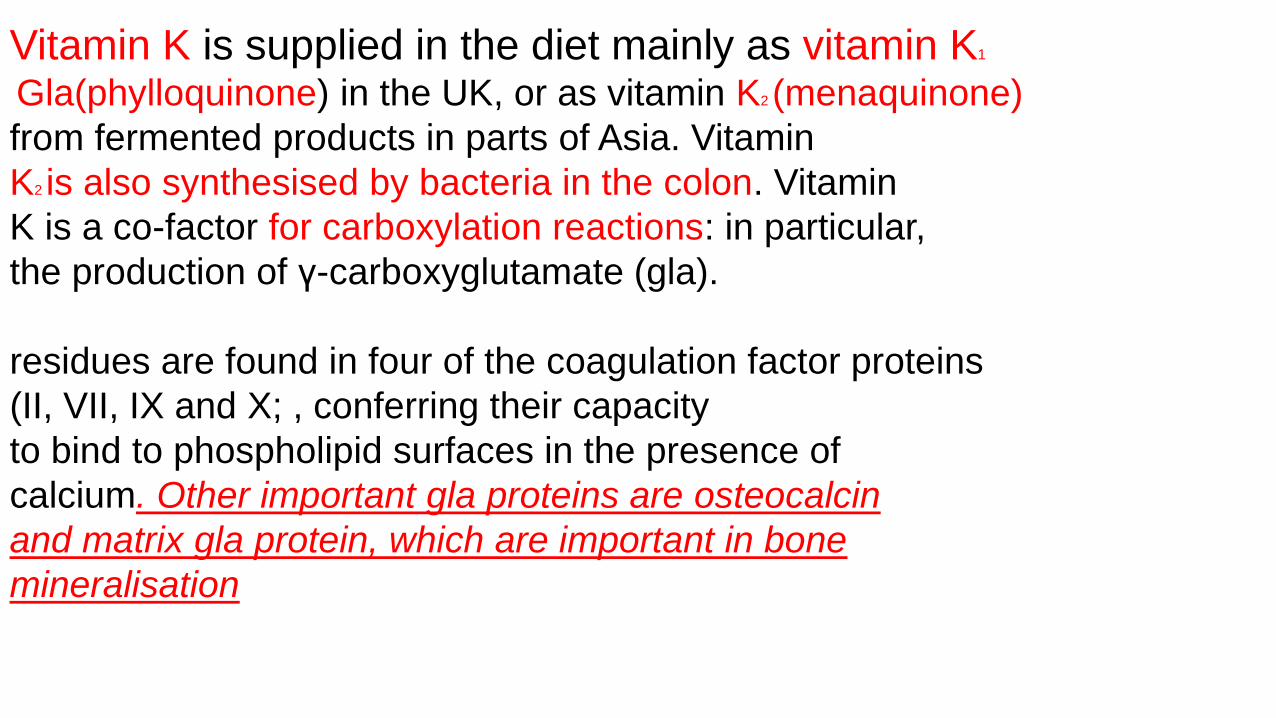
Vitamin K is supplied in the diet mainly as vitamin K1
Gla(phylloquinone) in the UK, or as vitamin K2 (menaquinone)
from fermented products in parts of Asia. Vitamin
K2 is also synthesised by bacteria in the colon. Vitamin
K is a co-factor for carboxylation reactions: in particular,
the production of γ-carboxyglutamate (gla).
residues are found in four of the coagulation factor proteins
(II, VII, IX and X; , conferring their capacity
to bind to phospholipid surfaces in the presence of
calcium. Other important gla proteins are osteocalcin
and matrix gla protein, which are important in bone
mineralisation

Vitamin K deficiency leads to delayed coagulation
and bleeding.
In obstructive jaundice, dietary vitamin K
is not absorbed and it is essential to administer the
vitamin in parenteral form before surgery.
Warfarin andrelated anticoagulants act by antagonising
vitamin K. Vitamin K is given routinely to newborn
babies to prevent haemorrhagic disease. Symptoms of
excess have been reported only in infants, with synthetic
preparations linked to haemolysis and liver damage

Water-soluble vitamins
Thiamin (vitamin B1 )Thiamin is widely distributed in foods of both vegetableandanimal origin. Thiamin pyrophosphate (TPP) isa co-factor for enzyme reactions involved in themetabolism of macronutrients (carbohydrate, fat andalcohol), including• decarboxylation of pyruvate to acetyl-co-enzyme A,which bridges between glycolysis and thetricarboxylic acid (Krebs) cycle• transketolase activity in the hexose monophosphateshunt pathway• decarboxylation of α-ketoglutarate to succinate inthe Krebs cycle.
In thiamin deficiency, cells cannot metabolise
glucoseaerobically to generate energy as
ATP. Neuronal cellsare most vulnerable,
since they depend almost exclusivelyon
glucose for energy requirements. Impaired
glucose oxidation also causes an
accumulation ofpyruvic and lactic acids,
which produce vasodilatationand increased
cardiac output.

Deficiency – beri-beri
In the developed world, thiamin deficiency is
mainlyencountered in chronic alcoholics. Poor diet,
impairedabsorption, storage and phosphorylation of
thiamin inthe liver, and the increased requirements for
thiamin to metabolise ethanol all contribute. In the
developingworld, deficiency usually arises as a
consequence of adiet based on polished rice. The body
has very limitedstores of thiamin, so deficiency is manifest
after only 1month on a thiamin-free diet. There are two
forms of thedisease in adults:
• Dry (or neurological) beri-beri manifests with chronic
peripheral neuropathy and with wrist and/or foot
drop, and may cause Korsakoff’s psychosis and
Wernicke’s encephalopathy
• Wet (or cardiac) beri-beri causes generalised oedema
due to biventricular heart failure with pulmonary
congestion.
In dry beri-beri, response to thiamin administration
is not uniformly good. However, multivitamin therapy
seems to produce some improvement, suggesting
that other vitamin deficiencies may be involved.
Wernicke’s encephalopathy and wet beri-beri should
be treated without delay with intravenous vitamin B
and C mixture (‘Pabrinex’,
Korsakoff’s psychosis is irreversible and does not respond to thiamin
A rare but important effect of chronic alcohol misuse
is the Wernicke–Korsakoff syndrome. This organic brain
disorder results from damage to the mamillary bodies,
dorsomedial nuclei of the thalamus and adjacent areas
of periventricular grey matter caused by a deficiency of
thiamin (vitamin B1), which most commonly results
from long-standing heavy drinking and an inadequate
diet. It can also arise from malabsorption or even protracted
vomiting. Without prompt treatment (see below),
the acute presentation of Wernicke’s encephalopathy
(nystagmus, ophthalmoplegia, ataxia and confusion)
can progress to the irreversible deficits of Korsakoff’s
syndrome (severe short-term memory deficits and
confabulation, and also reduced red blood cell transketolase).
In those who die in the acute stage, microscopic
examination of the brain shows hyperaemia,
petechial haemorrhages and astrocytic prolifera
Riboflavin (vitamin B2 )
Riboflavin is required for the flavin co-factors involved
in oxidation–reduction reactions. It is widely
distributed
in animal and vegetable foods. Levels are low in staple
cereals but germination increases its content. It is
destroyed under alkaline conditions by heat and by
exposure to sunlight.
Deficiency is rare in developed countries. It mainly
affects the tongue and lips and manifests as glossitis,
angular stomatitis and cheilosis. The genitals may be
affected, as well as the skin areas rich in sebaceous
glands, causing nasolabial or facial dyssebacea.
Rapid
Niacin (vitamin B3 )Niacin encompasses nicotinic acid and nicotinamide. Nicotinamide is an essential part of the two
pyridinenucleotides, nicotinamide adenine dinucleotide (NAD) and
nicotinamide adenine dinucleotide phosphate
(NADP), which play a key role as hydrogen acceptors and donors for many enzymes. Niacin can be synthesised
in the body in limited amounts from the aminoacid tryptophan
.
Deficiency – pellagra
Pellagra was formerly endemic among poor people whosubsisted chiefly on maize, which contains niacytin, a
form of niacin that the body is unable to utilise. Pellagracan develop in only 8 weeks in individuals eating
diets
that are very deficient in niacin and tryptophanremains a problem in parts of Africa, and is occasionally
seen in alcoholics and in patients with chronic smallintestinal disease in developed countries.
Pellagra canoccur in Hartnup’s disease, a genetic disorder characterisedby impaired absorption of several
amino acids, including tryptophan.
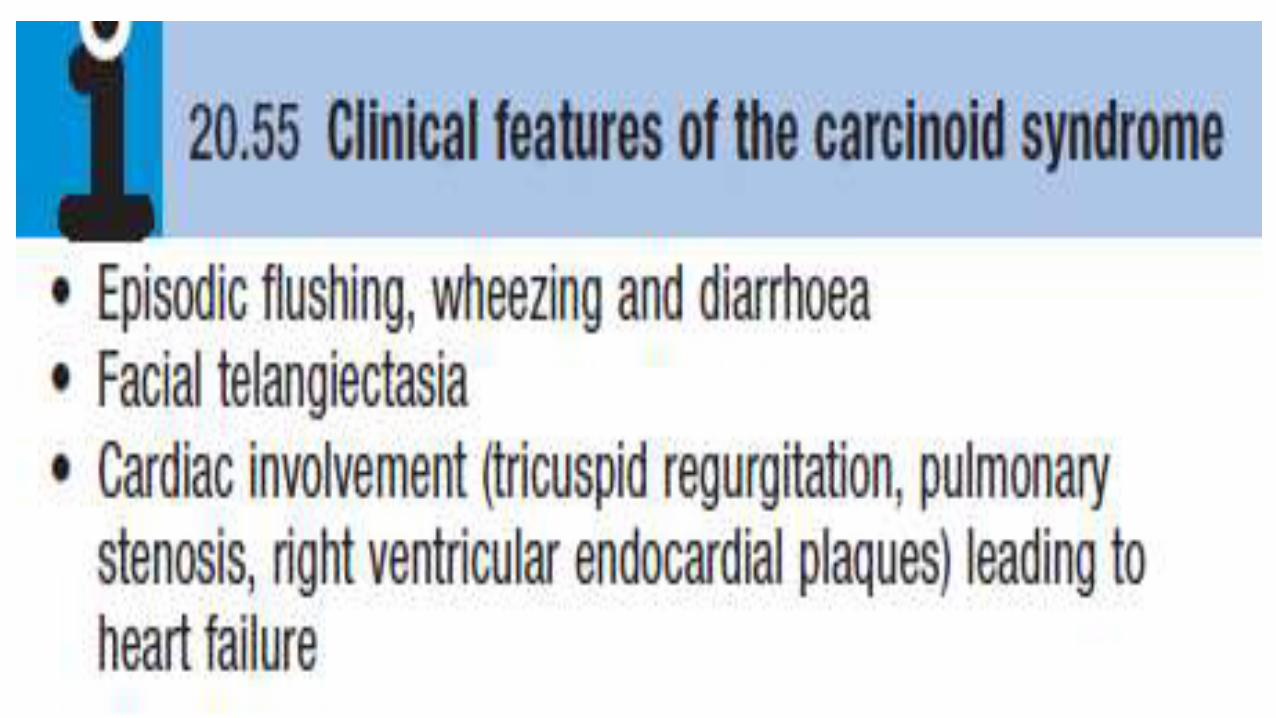
It is also seen occasionally in carcinoid syndrome , when tryptophan is consumed
in the excessive production of 5-hydroxytryptamine(5-HT). Pellagra has been called the disease of the
three D

• Dermatitis. Characteristically, there is erythemaresembling severe
sunburn, appearingsymmetrically over the parts of the body exposed to
sunlight, particularly the limbs and especiallyon the neck, but not the
face (Casal’s necklace,
The skin lesions may progress tovesiculation, cracking, exudation and
secondaryinfection.
• Diarrhoea. This is often associated with anorexia, nausea, glossitis
and dysphagia, reflecting thepresence of a non-infective inflammation
thatextends throughout the gastrointestinal tract.
• Dementia. In severe deficiency, delirium occursacutely and dementia
develops in chronic cases.
ToxicityExcessive intakes of niacin may lead to reversiblehepatotoxicity.Nicotinic acid is a lipid-lowering agent,but at doses above 200 mg a day gives rise to vasodilatorysymptoms (‘flushing’ and/orhypotension)
Pyridoxine (vitamin B6 )Pyridoxine, pyridoxal and pyridoxamine are differentforms of vitamin B6 that undergo phosphorylation to
produce pyridoxal 5-phosphate (PLP). PLP is theco-factor for a large number of enzymes involved in the
metabolism of amino acids. Vitamin B6 is available inmost foods.
Deficiency is rare, although certain drugs, such asisoniazid and penicillamine, act as chemical antagonists
to pyridoxine.
Pyridoxine administration is effectivein isoniazid-induced peripheral neuropathy and some
cases of sideroblastic anaemia.
Large doses of vitaminB6 have an antiemetic effect in radiotherapy-induced nausea
. Although vitamin B6 supplements have become popular in the treatment of nausea in pregnancy, carpal
tunnel syndrome and premenstrual syndrome, there is no convincing evidence of benefit.
Very high doses ofvitamin B6 taken for several months can cause a sensorypolyneuropathy.
BiotinBiotin is a co-enzyme in the synthesis of fatty
acids, isoleucine and valine and is also involved
in gluconeogenesis.
Deficiency results from consuming very large
quantities of raw egg whites (> 30% energy
intake)
because the avidin they contain binds to and
inactivates
biotin in the intestine. It may also be seen after
long
periods of total parenteral nutrition. The clinical
features
Folate (folic acid)Folates exist in many forms. The main circulating formis 5-
methyltetrahydrofolate. Folic acid is the stable synthetic form. Folate works
as a methyl donor for cellular methylationand protein synthesis. It is
directly involved in DNAand RNA synthesis, and requirements increase
during embryonic development
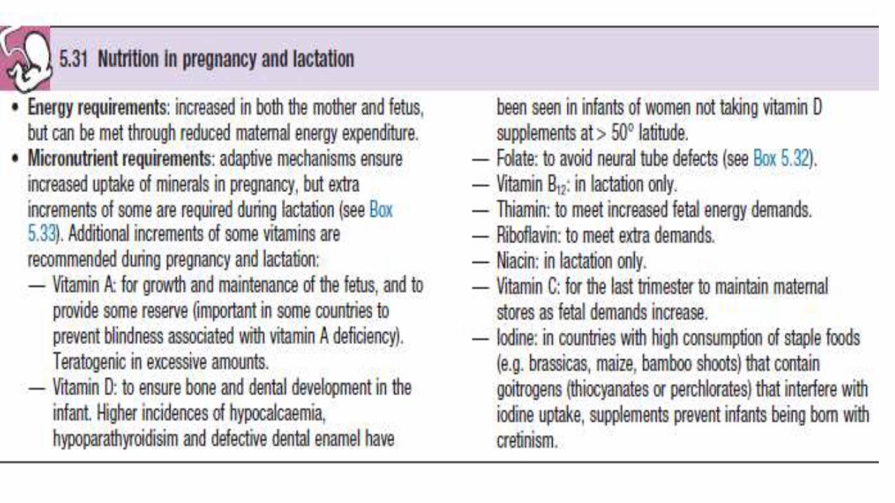
Folate deficiency may cause three major birth defects
(spina bifida, anencephaly and encephalocele) resulting
from imperfect closure of the neural tube, which takesplace 3–4 weeks after
conception.
Departmentof Health advises that women who have experienced a
pregnancy affected by a neural tube defect should take5 mg of folic acid daily
from before conception andthroughout the first trimester
All
women planning a pregnancy are advised to include good sources of
folate in their diet, and to take folate supplements throughout the first
trimester. Liver is the richest source of folate but an alternative source
(e.g. leafy vegetables) is advised in early pregnancy because of the
high vitamin A content of liver
Folate deficiency also has been associated with heart disease, dementia and cancer.. There are now concerns that this may contribute to the increased incidence of colon cancer through promotion of the growth of polyps.

Hydroxycobalamin (vitamin B12 )Vitamin B12 is a co-factor in folate co-enzyme recyclingand nerve myelination. Vitamin B12 and
folate are particularlyimportant in DNA synthesis in red blood cells
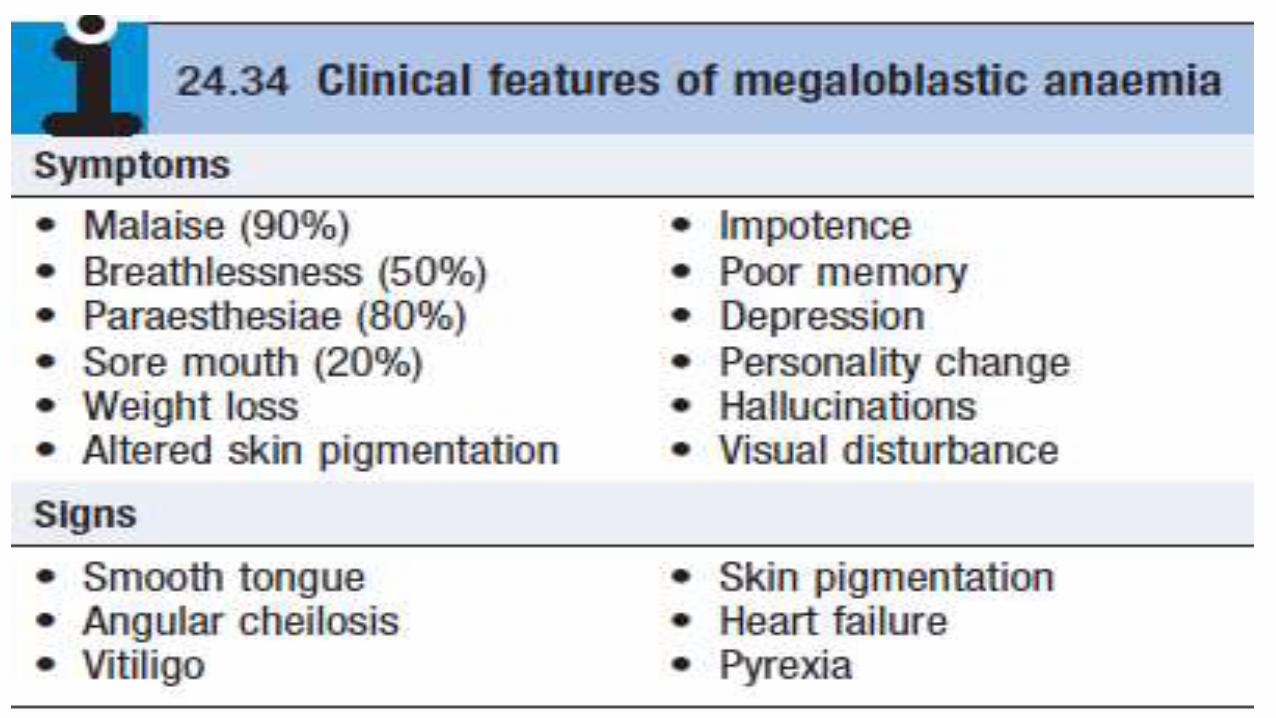
The haematological disorders (macrocytic ormegaloblastic anaemias) due to their deficiency
are discussedon . Vitamin B12, but not folate, is needed for the integrity of myelin, so that
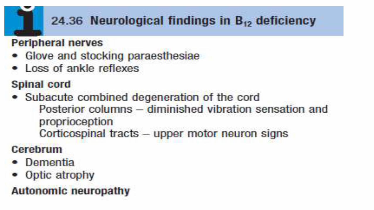
vitamin B12deficiency is also associated with neurological disease
Neurological consequences of vitamin B12 deficiency. In older people and chronic alcoholics, vitamin B12 deficiencyarises from
insufficient intake and/or frommalabsorption. Several drugs, including
neomycin, canrender vitamin B12 inactive.
Adequate intake of folatemaintains erythropoiesis and there is a concern
that fortification of foods with folate may mask underlyingvitamin B12
deficiency. In severe deficiency there is insidious, diffuse and uneven
demyelination. It may be clinically manifest as peripheral neuropathy or
spinalcord degeneration affecting both posterior and lateral columns
(‘subacute combined degeneration of the spinal
cord’ or there may be cerebral
manifestations(resembling dementia) or optic
atrophy. Vitamin B12therapy improves symptoms in most cases

The average daily diet contains 5–30 μg of vitamin B12,
mainly in meat, fish, eggs and milk – well in excess of
the 1 μg daily requirement. In the stomach, gastric
enzymes release vitamin B12 from food and at gastric pH
it binds to a carrier protein termed R protein. The gastric
parietal cells produce intrinsic factor, a vitamin B12-
binding protein which optimally binds vitamin B12 at pH
8. As gastric emptying occurs, pancreatic secretion raises
the pH and vitamin B12 released from the diet switches
from the R protein to intrinsic factor. Bile also contains
vitamin B12 which is available for reabsorption in the
intestine. The vitamin B12–intrinsic factor complex binds
to specific receptors in the terminal ileum, and vitamin
B12 is actively transported by the enterocytes to plasma

where it binds to transcobalamin II, a transport proteinproduced by the liver, which carries it to the tissues forutilisation. The liver stores enough vitamin B12 for3 years and this, together with the enterohepaticcirculation,.Blood levels of vitamin B12 provide a reasonable indicationof tissue stores and are usually diagnostic of deficiency. Levels of cobalamins fall in normal pregnancy.Reference ranges vary between laboratories but levelsbelow 150 ng/L are common and, in the last trimester,. Spuriouslylow B12 values occur in women using the oralcontraceptive pill and in patients with myeloma,.