Embed Size (px)
Citation preview
Copyright © 2009 Pearson Education, Inc.
4.0 Crossing over
Prepared by Pratheep SandrasaigaranLecturer at Manipal International University
Copyright © 2009 Pearson Education, Inc.
By the end of this chapter you should be able to:
• Overview of Mitosis and Meiosis• Crossing over
• Holliday model
Prepared by Pratheep Sandrasaigaran
Copyright © 2009 Pearson Education, Inc.
4.1 Overview of Mitosis and Meiosis
Prepared by Pratheep Sandrasaigaran
Copyright © 2009 Pearson Education, Inc.
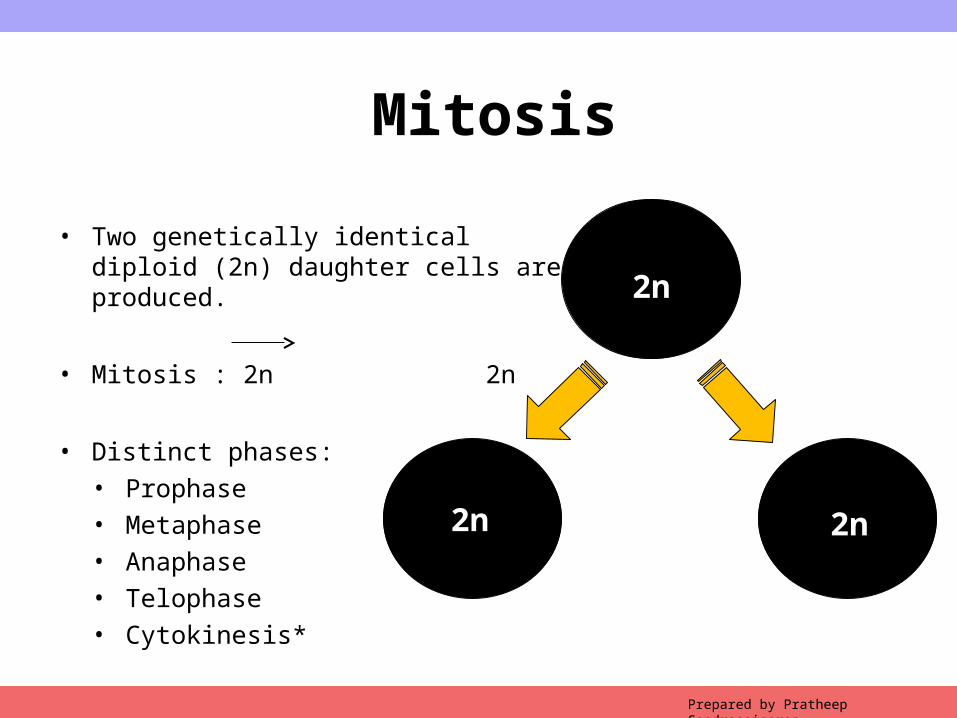
Mitosis
• Two genetically identical diploid (2n) daughter cells are produced.
• Mitosis : 2n 2n
• Distinct phases:• Prophase• Metaphase• Anaphase• Telophase• Cytokinesis*
Prepared by Pratheep Sandrasaigaran
2n
2n 2n
Copyright © 2009 Pearson Education, Inc.
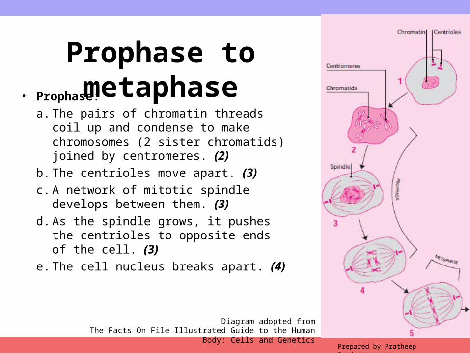
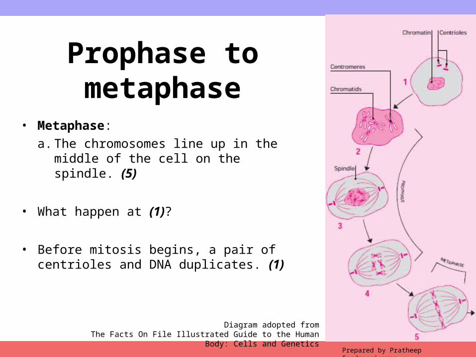
Prophase to metaphase• Prophase:
a. The pairs of chromatin threads coil up and condense to make chromosomes (2 sister chromatids) joined by centromeres. (2)
b. The centrioles move apart. (3)c. A network of mitotic spindle develops
between them. (3)d. As the spindle grows, it pushes the
centrioles to opposite ends of the cell. (3)e. The cell nucleus breaks apart. (4)
Prepared by Pratheep Sandrasaigaran
Diagram adopted fromThe Facts On File Illustrated Guide to the Human Body: Cells and Genetics
Copyright © 2009 Pearson Education, Inc.
Prophase to metaphase
• Metaphase:a. The chromosomes line up in the middle of
the cell on the spindle. (5)
• What happen at (1)?
• Before mitosis begins, a pair of centrioles and DNA duplicates. (1)
Prepared by Pratheep Sandrasaigaran
Diagram adopted fromThe Facts On File Illustrated Guide to the Human Body: Cells and Genetics
Copyright © 2009 Pearson Education, Inc.
Diagram adopted fromThe Facts On File Illustrated Guide to the Human Body: Cells and Genetics
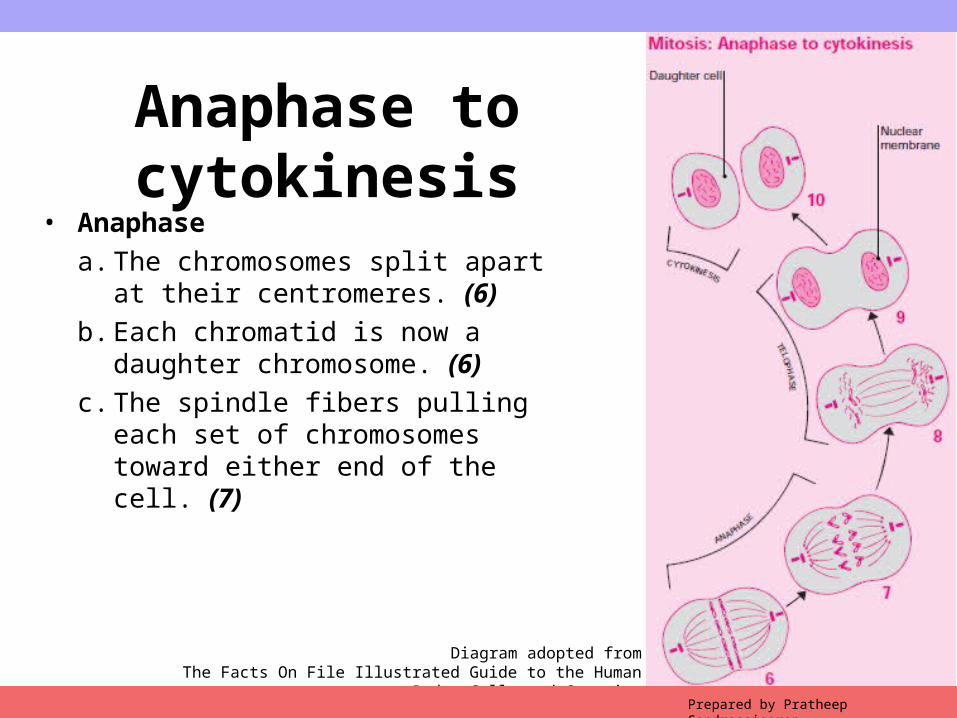
Anaphase to cytokinesis
• Anaphasea. The chromosomes split apart at their
centromeres. (6) b. Each chromatid is now a daughter
chromosome. (6) c. The spindle fibers pulling each set of
chromosomes toward either end of the cell. (7)
Prepared by Pratheep Sandrasaigaran
Copyright © 2009 Pearson Education, Inc.
Diagram adopted fromThe Facts On File Illustrated Guide to the Human Body: Cells and Genetics
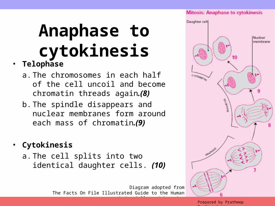
Anaphase to cytokinesis
• Telophasea. The chromosomes in each half of the cell
uncoil and become chromatin threads again.(8)
b. The spindle disappears and nuclear membranes form around each mass of chromatin.(9)
• Cytokinesisa. The cell splits into two identical daughter
cells. (10)
Prepared by Pratheep Sandrasaigaran
Copyright © 2009 Pearson Education, Inc.
Meiosis• Meiosis is the process by which reproductive or sex
cells are produced.
• Egg and sperm cells have only 23 chromosomes—half the normal amount.
• The zygote has the correct number of 46 chromosomes.
• The parent cell shown in example here has only four chromosomes, rather than the actual 46 found in human body cells
Prepared by Pratheep Sandrasaigaran
Copyright © 2009 Pearson Education, Inc.
Before meiosis begins• The centriole duplicates itself, so that there are two just
before cell division. (1)
• The chromosomes in the nucleus duplicate themselves, too.
• First meiotic division: This division is split into four phases• Prophase 1• Metaphase 1• Anaphase 1• Telophase1
• The division ends with cytokinesis
Prepared by Pratheep Sandrasaigaran
Copyright © 2009 Pearson Education, Inc.
Diagram adopted fromThe Facts On File Illustrated Guide to the Human Body: Cells and Genetics
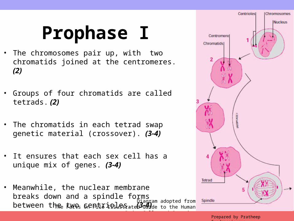
Prophase I• The chromosomes pair up, with two chromatids
joined at the centromeres. (2)
• Groups of four chromatids are called tetrads. (2)
• The chromatids in each tetrad swap genetic material (crossover). (3-4)
• It ensures that each sex cell has a unique mix of genes. (3-4)
• Meanwhile, the nuclear membrane breaks down and a spindle forms between the two centrioles. (3-4)
Prepared by Pratheep Sandrasaigaran
Copyright © 2009 Pearson Education, Inc.
Diagram adopted fromThe Facts On File Illustrated Guide to the Human Body: Cells and Genetics
Prepared by Pratheep Sandrasaigaran
Metaphase 1 to cytokinesis
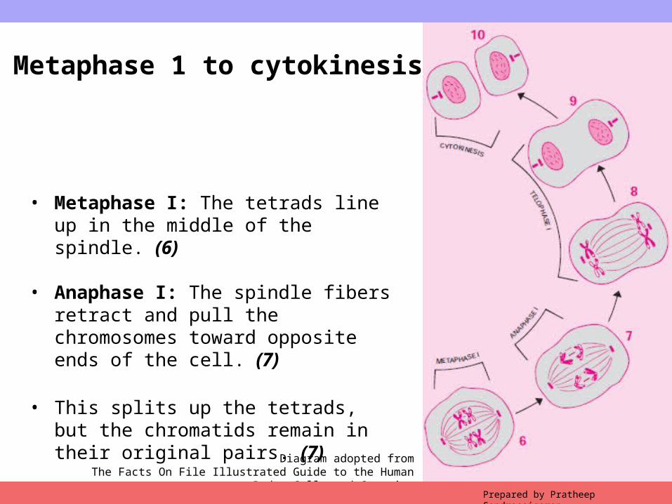
• Metaphase I: The tetrads line up in the middle of the spindle. (6)
• Anaphase I: The spindle fibers retract and pull the chromosomes toward opposite ends of the cell. (7)
• This splits up the tetrads, but the chromatids remain in their original pairs. (7)
Copyright © 2009 Pearson Education, Inc.
Diagram adopted fromThe Facts On File Illustrated Guide to the Human Body: Cells and Genetics
Prepared by Pratheep Sandrasaigaran
Metaphase 1 to cytokinesis
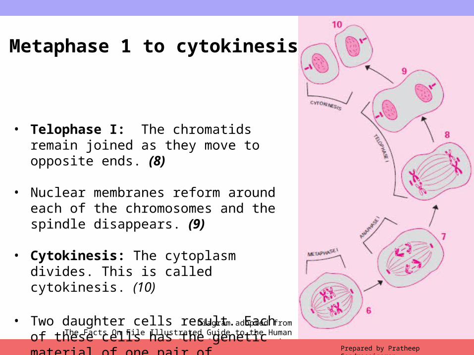
• Telophase I: The chromatids remain joined as they move to opposite ends. (8)
• Nuclear membranes reform around each of the chromosomes and the spindle disappears. (9)
• Cytokinesis: The cytoplasm divides. This is called cytokinesis. (10)
• Two daughter cells result. Each of these cells has the genetic material of one pair of chromatids from each tetrad. (10)
Copyright © 2009 Pearson Education, Inc.Prepared by Pratheep Sandrasaigaran
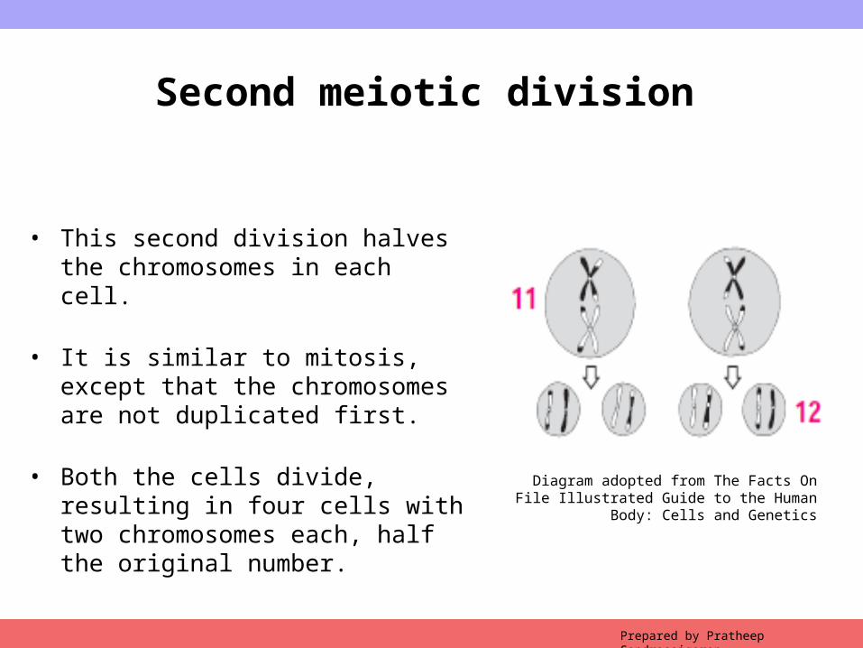
Second meiotic division
• This second division halves the chromosomes in each cell.
• It is similar to mitosis, except that the chromosomes are not duplicated first.
• Both the cells divide, resulting in four cells with two chromosomes each, half the original number. Diagram adopted from The Facts On File Illustrated
Guide to the Human Body: Cells and Genetics
Copyright © 2009 Pearson Education, Inc.
TEST YOUR KNOWLEDGE 1
Prepared by Pratheep Sandrasaigaran
Copyright © 2009 Pearson Education, Inc.Prepared by Pratheep Sandrasaigaran

Copyright © 2009 Pearson Education, Inc.Prepared by Pratheep Sandrasaigaran

Copyright © 2009 Pearson Education, Inc.Prepared by Pratheep Sandrasaigaran

Copyright © 2009 Pearson Education, Inc.Prepared by Pratheep Sandrasaigaran
Copyright © 2009 Pearson Education, Inc.Prepared by Pratheep Sandrasaigaran
Copyright © 2009 Pearson Education, Inc.
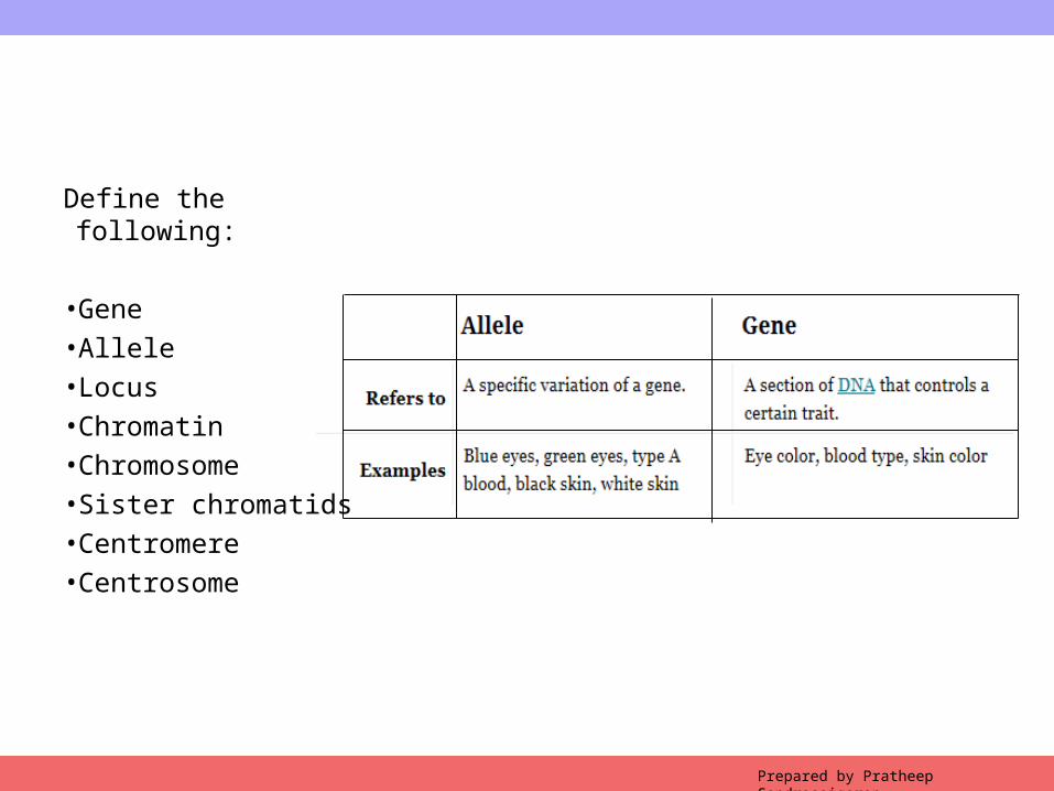
Define the following:
•Gene•Allele•Locus•Chromatin•Chromosome•Sister chromatids•Centromere•Centrosome
Prepared by Pratheep Sandrasaigaran
Copyright © 2009 Pearson Education, Inc.
4.2 Crossing over
Prepared by Pratheep Sandrasaigaran
Copyright © 2009 Pearson Education, Inc.Prepared by Pratheep Sandrasaigaran
Crossing over
• The process of crossing over (recombination) between homologs depends on the breakage and rejoining of the DNA strands.
• This results in the exchange of genetic information between DNA molecules to increase genetic diversity.
• The genetic exchange may happen between any two homologous double stranded DNA molecules• Virus chromosomes • Bacterial chromosomes • Eukaryotic homologs during meiosis
Copyright © 2009 Pearson Education, Inc.
4.2.1 Crossing overHolliday model
Prepared by Pratheep Sandrasaigaran
Copyright © 2009 Pearson Education, Inc.Prepared by Pratheep Sandrasaigaran
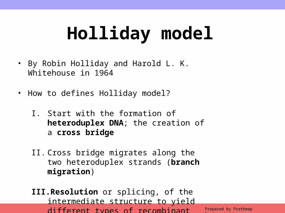
Holliday model• By Robin Holliday and Harold L. K. Whitehouse in 1964
• How to defines Holliday model?
I. Start with the formation of heteroduplex DNA; the creation of a cross bridge
II. Cross bridge migrates along the two heteroduplex strands (branch migration)
III. Resolution or splicing, of the intermediate structure to yield different types of recombinant molecules.*
Copyright © 2009 Pearson Education, Inc.Prepared by Pratheep Sandrasaigaran
Holliday model
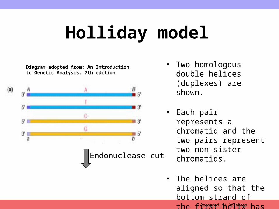
• Two homologous double helices (duplexes) are shown.
• Each pair represents a chromatid and the two pairs represent two non-sister chromatids.
• The helices are aligned so that the bottom strand of the first helix has the same polarity as the top strand of the second helix.
Endonuclease cut
Diagram adopted from: An Introduction to Genetic Analysis. 7th edition
Copyright © 2009 Pearson Education, Inc.Prepared by Pratheep Sandrasaigaran
Holliday model
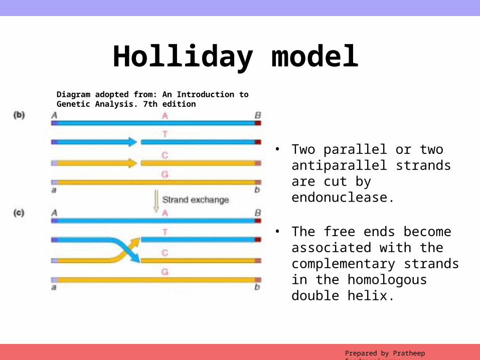
• Two parallel or two antiparallel strands are cut by endonuclease.
• The free ends become associated with the complementary strands in the homologous double helix.
Diagram adopted from: An Introduction to Genetic Analysis. 7th edition
Copyright © 2009 Pearson Education, Inc.Prepared by Pratheep Sandrasaigaran
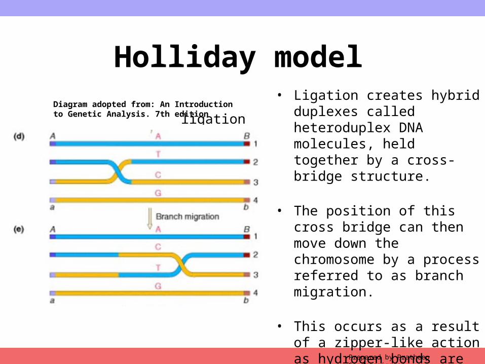
Holliday model• Ligation creates hybrid duplexes
called heteroduplex DNA molecules, held together by a cross-bridge structure.
• The position of this cross bridge can then move down the chromosome by a process referred to as branch migration.
• This occurs as a result of a zipper-like action as hydrogen bonds are broken and then re-formed between complementary bases of each duplex.
ligationDiagram adopted from: An Introduction to Genetic Analysis. 7th edition
Copyright © 2009 Pearson Education, Inc.Prepared by Pratheep Sandrasaigaran
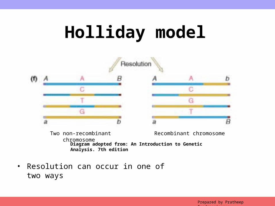
Holliday model
• Resolution can occur in one of two ways
Diagram adopted from: An Introduction to Genetic Analysis. 7th edition
Two non-recombinant chromosome Recombinant chromosome
Copyright © 2009 Pearson Education, Inc.Prepared by Pratheep Sandrasaigaran
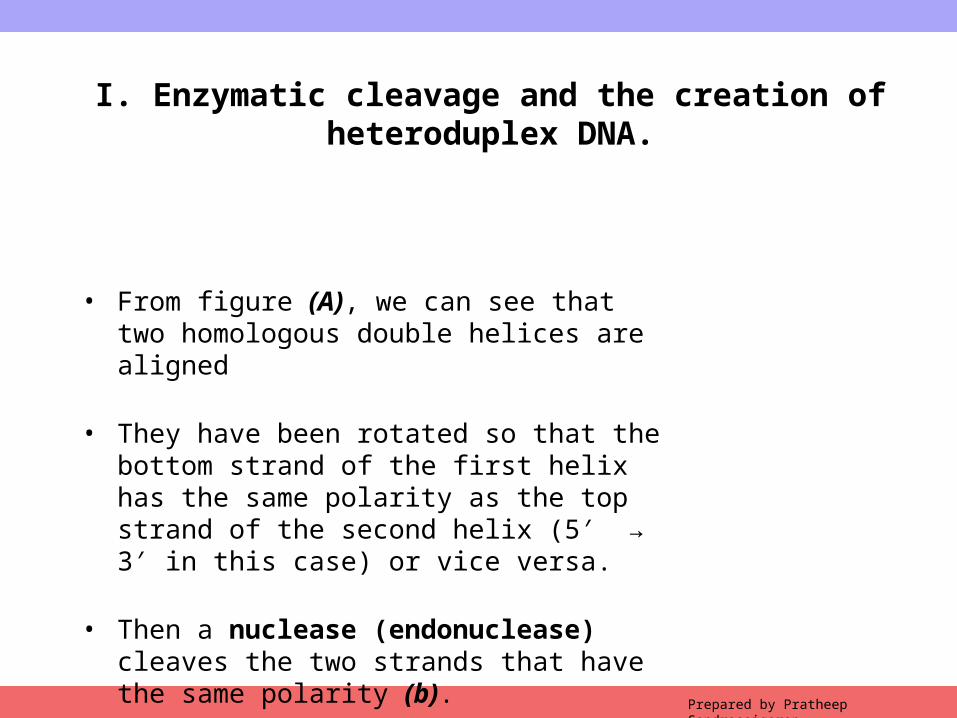
I. Enzymatic cleavage and the creation of heteroduplex DNA.
• From figure (A), we can see that two homologous double helices are aligned
• They have been rotated so that the bottom strand of the first helix has the same polarity as the top strand of the second helix (5 → 3 in this case) or ′ ′vice versa.
• Then a nuclease (endonuclease) cleaves the two strands that have the same polarity (b).
Copyright © 2009 Pearson Education, Inc.Prepared by Pratheep Sandrasaigaran
I. Enzymatic cleavage and the creation of heteroduplex DNA.
• The free ends leave their original complementary strands and undergo hydrogen bonding with the complementary strands in the homologous double helix (C).
• Ligation produces the structure shown in Figure D.
• This partially heteroduplex double helix is a crucial intermediate in recombination, and has been termed the Holliday structure.
Copyright © 2009 Pearson Education, Inc.Prepared by Pratheep Sandrasaigaran
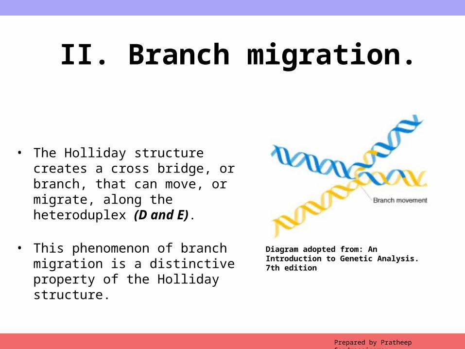
II. Branch migration.
• The Holliday structure creates a cross bridge, or branch, that can move, or migrate, along the heteroduplex (D and E).
• This phenomenon of branch migration is a distinctive property of the Holliday structure. Diagram adopted from: An Introduction to Genetic
Analysis. 7th edition
Copyright © 2009 Pearson Education, Inc.Prepared by Pratheep Sandrasaigaran
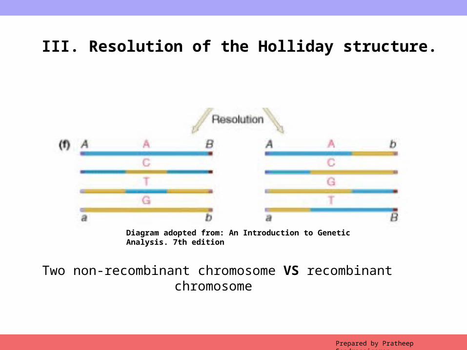
III. Resolution of the Holliday structure.
Two non-recombinant chromosome VS recombinant chromosome
Diagram adopted from: An Introduction to Genetic Analysis. 7th edition
Copyright © 2009 Pearson Education, Inc.Prepared by Pratheep Sandrasaigaran
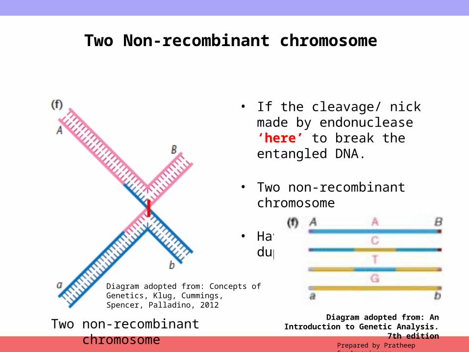
Two Non-recombinant chromosome
• If the cleavage/ nick made by endonuclease ‘here’ to break the entangled DNA.
• Two non-recombinant chromosome
• Having short hetero-duplex regions.
Two non-recombinant chromosome Diagram adopted from: An Introduction to
Genetic Analysis. 7th edition
Diagram adopted from: Concepts of Genetics, Klug, Cummings, Spencer, Palladino, 2012
Copyright © 2009 Pearson Education, Inc.Prepared by Pratheep Sandrasaigaran
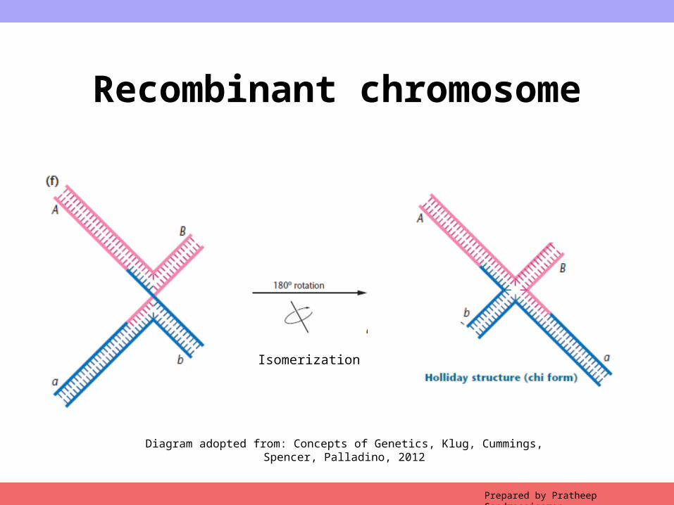
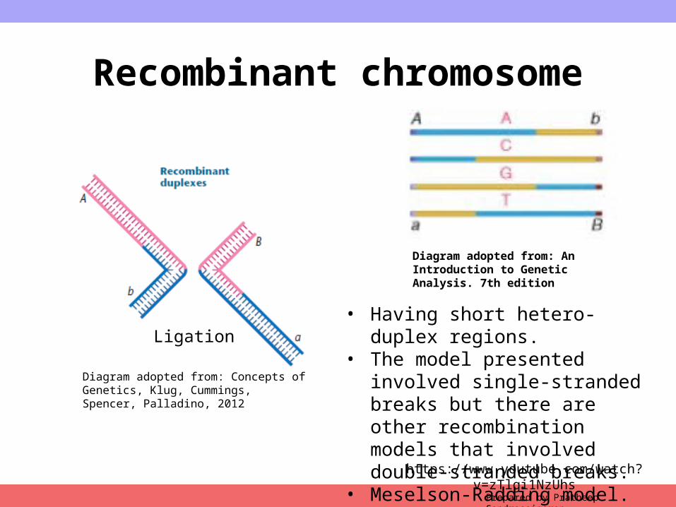
Recombinant chromosome
Diagram adopted from: Concepts of Genetics, Klug, Cummings, Spencer, Palladino, 2012
Isomerization
Copyright © 2009 Pearson Education, Inc.Prepared by Pratheep Sandrasaigaran
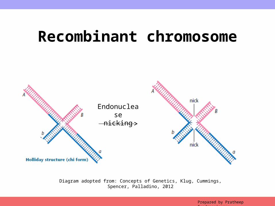
Recombinant chromosome
Endonucleasenicking
Diagram adopted from: Concepts of Genetics, Klug, Cummings, Spencer, Palladino, 2012
Copyright © 2009 Pearson Education, Inc.Prepared by Pratheep Sandrasaigaran
Recombinant chromosome
Ligation
Diagram adopted from: An Introduction to Genetic Analysis. 7th edition
Diagram adopted from: Concepts of Genetics, Klug, Cummings, Spencer, Palladino, 2012
• Having short hetero-duplex regions.• The model presented involved single-
stranded breaks but there are other recombination models that involved double-stranded breaks.
• Meselson-Radding model.
https://www.youtube.com/watch?v=zTlgi1NzUhs

Copyright © 2009 Pearson Education, Inc.
TEST YOUR KNOWLEDGE 2
Prepared by Pratheep Sandrasaigaran
Copyright © 2009 Pearson Education, Inc.
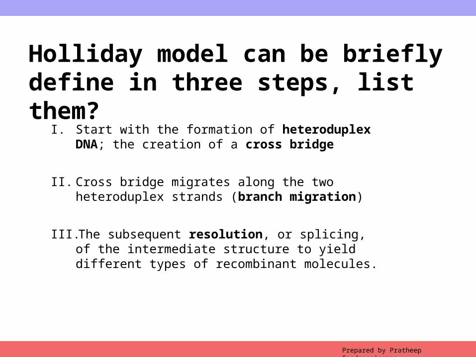
Holliday model can be briefly define in three steps, list them?
I. Start with the formation of heteroduplex DNA; the creation of a cross bridge
II. Cross bridge migrates along the two heteroduplex strands (branch migration)
III. The subsequent resolution, or splicing, of the intermediate structure to yield different types of recombinant molecules.
Prepared by Pratheep Sandrasaigaran
Copyright © 2009 Pearson Education, Inc.
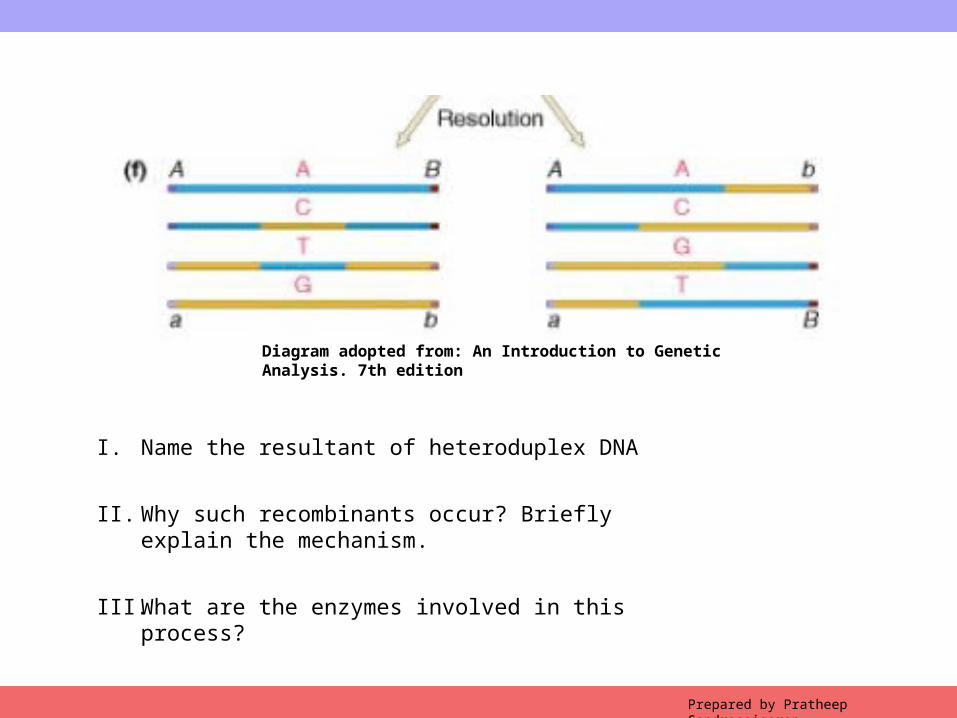
Diagram adopted from: An Introduction to Genetic Analysis. 7th edition
Prepared by Pratheep Sandrasaigaran
I. Name the resultant of heteroduplex DNA
II. Why such recombinants occur? Briefly explain the mechanism.
III. What are the enzymes involved in this process?