Embed Size (px)
Citation preview

Microbial physiologyBacterial Permeation Unit
3
1
PROKARYOTIC CELL MEMBRANE
STRUCTURE
2

• ProProkkaryotic cells are smaller and generally less complex aryotic cells are smaller and generally less complex than euthan eukkaryotic cells, with one exception: the cell envelope aryotic cells, with one exception: the cell envelope is more complex.is more complex.
• The primary distinguishing characteristics of the The primary distinguishing characteristics of the proprokkaryotes are their relatively small size, usually of 1 aryotes are their relatively small size, usually of 1 micrometer in diameter, and the absence of a nuclear micrometer in diameter, and the absence of a nuclear membrane. membrane.
• The DNA of almost all bacteria is a circle with a length of The DNA of almost all bacteria is a circle with a length of about 1 mm: this is the prokaryotic chromosome. The about 1 mm: this is the prokaryotic chromosome. The specialised region of the cell containing DNA is termed the specialised region of the cell containing DNA is termed the nucleoid and can be observed by electron microscopy. nucleoid and can be observed by electron microscopy.
3

Cell wall • The layers of the cell envelope lying between the cytoplasmic
membrane and the capsule are referred to collectively as the “cell wall“.
• The cell wall in prokaryotic cells is extremely complex. This rigid structure protects the cell from rupture caused by the high osmotic pressure inside the bacterial cell. The internal osmotic pressure of most bacteria ranges from 5 to 20 atmospheres.
• Additionally, the cell wall is the site of many of the antigenic determinants that characterize and differentiate microorganisms. Endotoxin activity associated with certain groups of bacteria is also associated with the cell wall.
4

Cell wall • Bacteria have historically been subdivided Bacteria have historically been subdivided
by their reaction with the Gram stain. by their reaction with the Gram stain.
• Although both gram-positive and gram-Although both gram-positive and gram-negative bacteria have cell walls, their negative bacteria have cell walls, their differential staining properties are in large differential staining properties are in large part attributed to the structure of the cell part attributed to the structure of the cell wall.wall.
5

Cell wall • The basic structure of the cell wall of gram-The basic structure of the cell wall of gram-
positive bacteria is a thick (15-80 nm) positive bacteria is a thick (15-80 nm) peptidoglycan layer composed of chains of peptidoglycan layer composed of chains of alterating subunits of A-acetylglucoseamine and alterating subunits of A-acetylglucoseamine and A-acetylmuramic acid. A-acetylmuramic acid.
• All gram-positive cell walls also contain teichoic All gram-positive cell walls also contain teichoic acid bound to the cytoplamic membrane. acid bound to the cytoplamic membrane.
6

Cell wall • The structure of the cell wall of gram-negative bacteria is The structure of the cell wall of gram-negative bacteria is
more complex. more complex.
• The peptidoglycan layer is thinner, only 1 to 2 nm. Outside The peptidoglycan layer is thinner, only 1 to 2 nm. Outside the peptidoglycan layer is phospholipid outer membrane the peptidoglycan layer is phospholipid outer membrane (absent in gram-positive bacteria). (absent in gram-positive bacteria).
• The area between the outer meThe area between the outer memmbrane and the cytoplasmic brane and the cytoplasmic membrane is called the periplasmic space. The outer membrane is called the periplasmic space. The outer membrane prevents loss omembrane prevents loss off periplasmatic proteins and forms periplasmatic proteins and forms a protective barrier preventing exposure of bacteria to a protective barrier preventing exposure of bacteria to hydrolytic enzymes and toxic substances such as bile in the hydrolytic enzymes and toxic substances such as bile in the gastrointestinal tractgastrointestinal tract..
7

The Plasma MembraneThe Plasma Membrane -
8Gateway to the CellGateway to the Cell
1

Photograph of a Cell MembranePhotograph of a Cell Membrane
92

Cell MembraneCell Membrane
The cell membrane is flexibleflexible and allows a unicellular organism to move
10

Functions of Plasma MembraneFunctions of Plasma Membrane
11
Protective barrierProtective barrier Regulate transport in & out of cell Regulate transport in & out of cell (selectively permeable)(selectively permeable) Allow cell recognitionAllow cell recognition Provide anchoring sites for filaments Provide anchoring sites for filaments of cytoskeletonof cytoskeleton

Functions of Plasma MembraneFunctions of Plasma Membrane
12
Provide a binding site for enzymesProvide a binding site for enzymes Interlocking surfaces bind cells Interlocking surfaces bind cells together (junctions)together (junctions)Contains the cytoplasm (fluid in cell) Contains the cytoplasm (fluid in cell)

HomeostasisHomeostasis
• Balanced internal condition of cells• Also called equilibrium• Maintained by plasma membrane
controlling what enters & leaves the cell
13

Structure of the Cell Structure of the Cell MembraneMembrane
14
3

15
PhospholipidsPhospholipidsCholesterolCholesterol
Proteins(peripheral and integral)Carbohydrates (glucose)
Membrane ComponentsMembrane Components
4

16
5

PhospholipidsPhospholipids
Make up the cell membrane
17
Contains 2 fatty acid chains that are nonpolar
Head is polar & contains a –PO4 group & glycerol
6

Early Membrane Model• Lipid and lipid soluble materials enter cells more
rapidly than substances that are insoluble in lipids.– Deduction: Membranes are made of lipids.– Deduction: Fat-soluble substance move through
the membrane by dissolving in it ("like dissolves like").
– Amphipathic = Condition where a molecule has both a hydrophilic region and a hydrophobic region.
18

The Davson/Danielli Model
• Danielli and Davson (1935) proposed a phospholipid interior with protein coats on both sides.
• It is also called Bimolecular Leaflet Model
197

Trilaminar Model (Unit membrane Model)
• This model was proposed by Robertson in 1959. According to this model, the plasma membrane is formed of three layers. The three layers are an outer protein layer, a middle lipid layer and an inner protein layer.
20
8

FLUID- because individual phospholipids and proteins can FLUID- because individual phospholipids and proteins can move side-to-side within the layer, like it’s a liquid.move side-to-side within the layer, like it’s a liquid.
MOSAIC- because of the pattern produced by the scattered MOSAIC- because of the pattern produced by the scattered protein molecules when the membrane is viewed from protein molecules when the membrane is viewed from above.above.
21
FLUID MOSAIC MODELFLUID MOSAIC MODEL
9

22
Cell MembraneCell Membrane
Polar heads are hydrophilichydrophilic “water loving”Nonpolar tails are hydrophobichydrophobic “water fearing”
Makes membrane “Selective” in what crosses
10

23
11

24
Cell MembraneCell Membrane
Hydrophobic molecules pass easily; hydrophilic DO NOT
The cell membrane is made of 2 layers of phospholipidphospholipids called the lipid bilayerbilayer
12

SolubilitySolubility
• Materials that are soluble in lipids can pass through the cell membrane easily
25
13
26
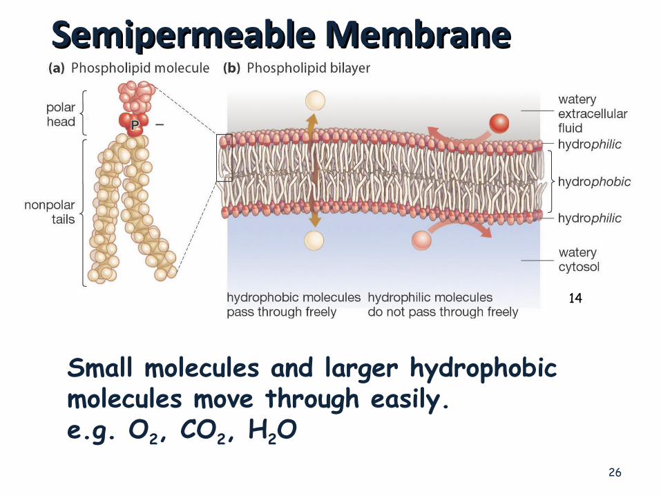
Semipermeable MembraneSemipermeable Membrane
Small molecules and larger hydrophobic molecules move through easily.e.g. O2, CO2, H2O
14

27
Ions, hydrophilic molecules larger than water, and large molecules such as proteins do not move through the membrane on their own.
Semipermeable MembraneSemipermeable Membrane

28
• Cholesterol molecules have several functions in the membrane:
• They immobilize the first few hydrocarbon groups of the phospholipid molecules. This makes the lipid bilayer less deformable and decreases its permeability to small water-soluble molecules. Without cholesterol (such as in a bacterium) a cell would need a cell wall.
• Cholesterol prevents crystallization of hydrocarbons and phase shifts in the membrane.

Types of Transport Across Types of Transport Across Cell MembranesCell Membranes
29
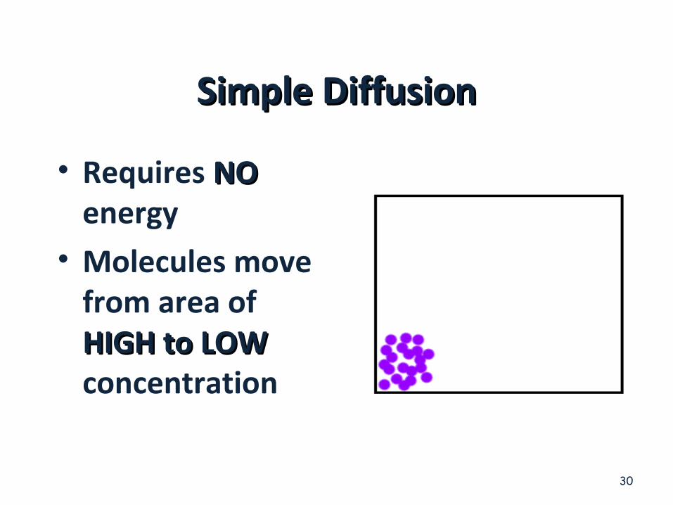
Simple DiffusionSimple Diffusion
• Requires NONO energy• Molecules move
from area of HIGH to LOWHIGH to LOW concentration
30

DIFFUSIONDIFFUSION
Diffusion is a PASSIVEPASSIVE process which means no energy is used to make the molecules move, they have a natural KINETIC ENERGY
31
15
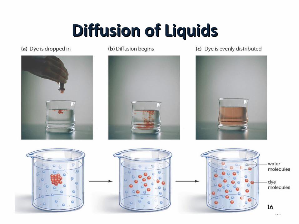
Diffusion of LiquidsDiffusion of Liquids
3216

Diffusion through a MembraneDiffusion through a Membrane
33
Cell membrane
Solute moves DOWN concentration gradient (HIGH to LOW)
17

OsmosisOsmosis• Diffusion of water Diffusion of water
across a membraneacross a membrane• Moves from HIGH Moves from HIGH
water potential (low water potential (low solute) to LOW water solute) to LOW water potential (high solute)potential (high solute)
34
Diffusion across a membrane
Semipermeable membrane

Diffusion of HDiffusion of H22O Across A O Across A MembraneMembrane
35
High H2O potentialLow solute concentration
Low H2O potentialHigh solute concentration

AquaporinsAquaporins• Water Channels• Protein pores used during OSMOSIS
36
WATERMOLECULES
18

Cell in Isotonic SolutionCell in Isotonic Solution
37
CELLCELL
10% NaCL90% H2O
10% NaCL90% H2O
What is the direction of water movement?
The cell is at _______________.equilibrium
ENVIRONMENTENVIRONMENT
NO NET NO NET MOVEMENTMOVEMENT
19

Cell in Hypotonic SolutionCell in Hypotonic Solution
38
CELLCELL
10% NaCL90% H2O
20% NaCL80% H2O
What is the direction of water movement?
20

Cell in Hypertonic SolutionCell in Hypertonic Solution
39
CELLCELL
15% NaCL85% H2O
5% NaCL95% H2O
What is the direction of water movement?
ENVIRONMENTENVIRONMENT
21

Cells in SolutionsCells in Solutions
40
22

41
Isotonic Solution
NO NET MOVEMENT OF
H2O (equal amounts entering & leaving)
Hypotonic Solution
CYTOLYSIS
Hypertonic Solution
PLASMOLYSIS
23

Cytolysis & PlasmolysisCytolysis & Plasmolysis
42Cytolysis Plasmolysis
24

Osmosis in Red Blood CellsOsmosis in Red Blood Cells
43
IsotonicIsotonic Hypotonic Hypertonic25

What Happens to Blood Cells?
44
26

45
hypotonic hypertonic isotonic
hypertonic isotonic hypotonic 27

46
Three Forms of Transport Across the MembraneThree Forms of Transport Across the Membrane
28

47
Passive TransportPassive Transport
Simple DiffusionSimple Diffusion Doesn’t require energyDoesn’t require energy Moves high to low Moves high to low concentrationconcentration Example: Oxygen or Example: Oxygen or water diffusing into a water diffusing into a cell and carbon dioxide cell and carbon dioxide diffusing outdiffusing out.29
48
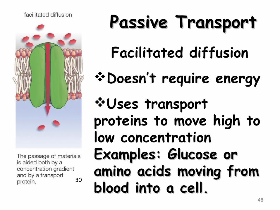
Passive TransportPassive TransportFacilitated diffusion
Doesn’t require energyUses transport proteins to move high to low concentrationExamples: Glucose or Examples: Glucose or amino acids moving from amino acids moving from blood into a cell.blood into a cell.
30

Permeability• The permeability of a membrane is the rate of
passive diffusion of molecules through the membrane. These molecules are known as permeant molecules. Permeability depends mainly on the electric charge and polarity of the molecule and to a lesser extent the molar mass of the molecule. Due to the cell membrane's hydrophobic nature, small electrically neutral molecules pass through the membrane more easily than charged, large ones.
49

Permeases
• Any of the proteins that mediate the transport of various molecules across biological membranes.
50

The membrane of E. coli.
51
The topographical features of the membrane are: 1. lactose transport system 2. The flagellar motor coupled to the hook and filament 3. Na+ transport (export) system 4. Ca++ transport (export) system 5. electron transport system 6. ATPase enzyme 7. Proline transport system.
31

Different Permeases In E. coli.
52
•The operation ot the electron transport system during respiration produces the H+ charge on the membrane (pmf). The pmf ( H+) is used by the transport systems to move molecules from one side of the membrane to the other; by the flagellar motor ring to rotate the flagellar filament; and by the ATPase enzyme to synthesize ATP.•E. coli needs to make two proteins to utilize lactose, as shown below. In addition to β-galactosidase, it must also make a permease that allows lactose to pass through the plasma membrane.
32

53
• Group translocation systems (GT), more commonly known as the phosphotransferase system (PTS) in E. coli, are used primarily for the transport of sugars. Like binding protein-dependent transport systems, they are composed of several distinct components. However, PTS systems specific for one sugar may share some of their components with other group transport systems. In E. coli, glucose may be transported by a group translocation process that involves the phosphotransferase system. The actual carrier in the membrane is a protein channel fairly specific for glucose. Glucose specifically enters the channel from the outside, but in order to exit into the cytoplasm, it must first be phosphorylated by the phosphotransferase system. The PTS derives energy from the metabolic intermediate phosphoenol pyruvate (PEP). PEP is hydrolyzed to pyruvate and glucose is phosphorylated to form glucose-phosphate during the process. Thus, by the expenditure of a single molecule of high energy phosphate, glucose is transported and changed to glucose-phosphate.

54
Proteins Are Critical to Proteins Are Critical to Membrane FunctionMembrane Function
33

Types of Transport ProteinsTypes of Transport Proteins• Channel proteins are embedded in the
cell membrane & have a pore for materials to cross• Carrier proteins can change shape to
move material from one side of the membrane to the other
55

Facilitated DiffusionFacilitated Diffusion
56
Molecules will randomly move through Molecules will randomly move through the pores in Channel Proteins.the pores in Channel Proteins.
34

Facilitated DiffusionFacilitated Diffusion
• Some Carrier Some Carrier proteins do not proteins do not extend through the extend through the membrane.membrane.
• They bond and drag They bond and drag molecules through molecules through the lipid bilayer and the lipid bilayer and release them on the release them on the opposite side. opposite side.
57
35

Carrier ProteinsCarrier Proteins
• Other carrier Other carrier proteins change proteins change shape to move shape to move materials across materials across the cell the cell membranemembrane
58

59
Active TransportActive TransportRequires energy or ATPMoves materials from LOW to HIGH concentrationAGAINST concentration gradient
37

• Important terminology for active transport mechanisms:
• Antiport - moves one or more molecules in as it moves one or more molecules out
• Synport - moves all molecules in same direction• Uniport - transport one ion in only one direction.
60
38

Primary active transport• Primary active transport involves using energy
(usually through ATP hydrolysis) at the membrane protein itself to cause a conformational change that results in the transport of the molecule through the protein. The most well-known example of this is the Na+-K+ pump. The Na+-K+ pump is an antiport, it transports K+ into the cell and Na+ out of the cell at the same time, with the expenditure of ATP.
61

Secondary active transport• Secondary active transport involves using energy to establish a gradient
across the cell membrane, and then utilizing that gradient to transport a molecule of interest up its concentration gradient. An example of this mechanism is as follows: E. coli establishes a proton (H+) gradient across the cell membrane by using energy to pump protons out of the cell. Then those protons are coupled to lactose at the lactose permease transmembrane protein. The lactose permease uses the energy of the proton moving down its concentration gradient to transport lactose into the cell. This coupled transport in the same direction across the cell membrane is known as a symport. E. coli uses similar proton driven symports to transport ribose, arabinose, and several amino acids.
62

Active transportActive transport
Examples: Pumping Na+ (sodium ions) out and K+ (potassium ions) in against strong concentration gradients.
Called Na+-K+ Pump
63

The Na+-glucose secondary transport mechanism
• Another secondary active transport system uses the Na+-K+ pump as the first step, generating a strong Na+ gradient across the cell membrane. Then the glucose-Na+ symport protein uses that Na+ gradient to transport glucose into the cell.
• This system is used in a novel way in human gut epithelial cells. These cells take in glucose and Na+ from the intestines and transport them through to the blood stream using the concerted actions of Na+-glucose symports, glucose permeases (a glucose facilitated diffusion protein), and Na+-K+ pumps.
64

65
Sodium-Potassium PumpSodium-Potassium Pump
3 Na+ pumped in for every 2 K+ pumped out; creates a membrane potential
39
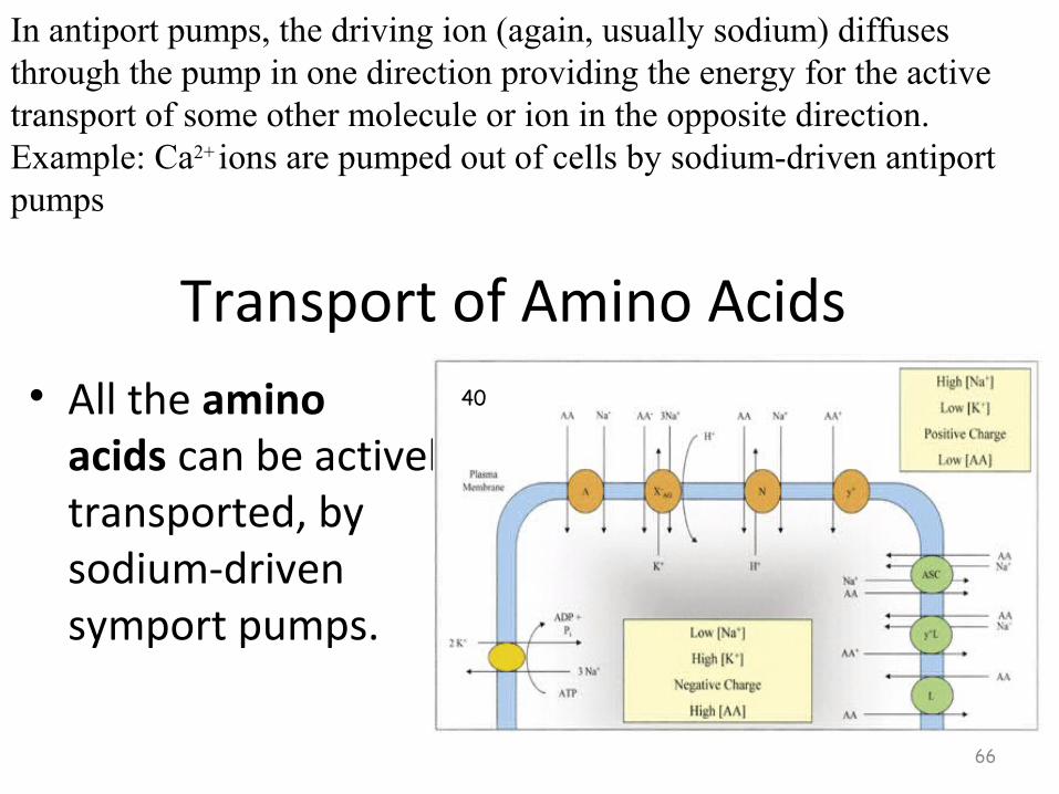
Transport of Amino Acids • All the amino
acids can be actively transported, by sodium-driven symport pumps.
66
In antiport pumps, the driving ion (again, usually sodium) diffuses through the pump in one direction providing the energy for the active transport of some other molecule or ion in the opposite direction.Example: Ca2+
ions are pumped out of cells by sodium-driven antiport pumps
40

67
• Chemiosmosis is the movement of ions across a selectively permeable membrane, down their electrochemical gradient. More specifically, it relates to the generation of ATP by the movement of hydrogen ions across a membrane during cellular respiration.
• An Ion gradient has potential energy and can be used to power chemical reactions when the ions pass through a channel (red).
• Hydrogen ions (protons) will diffuse from an area of high proton concentration to an area of lower proton concentration. Peter Mitchell proposed that an electrochemical concentration gradient of protons across a membrane could be harnessed to make ATP. He linked this process to osmosis, the diffusion of water across a membrane, which is why it is called chemiosmosis.
41

The proton-motive force
• The movement of ions across the membrane depends on a combination of two factors:
• Diffusion force caused by concentration gradient - all particles including ions tend to diffuse from higher concentration to lower.
• Electrostatic force caused by electrical potential gradient - cations like protons H+ tend to diffuse down the electrical potential, anions in the opposite direction.
• These two gradients taken together can be expressed as an electrochemical gradient.
68

• The term proton-motive force (PMF), derived from the electrochemical gradient can be described as the measure of the potential energy stored as a combination of proton and voltage gradients across a membrane (differences in proton concentration and electrical potential). The electrical gradient is a consequence of the charge separation across the membrane (when the protons H+ move without a counterion, such as chloride Cl-).
69
42

70
• Ion pumps - Directly couple ATP hydrolysis to transport. A well-studied example is the sodium-potassium pump of the plasma membrane. Note that in one turn of the multistep cycle, two potassiums are pumped in, three sodiums are pumped out, and one ATP is cleaved. The pump can be blocked by ouabain which, in the heart, stimulates contraction because sodium concentration increases and stimulates the sodium-calcium pump to remove sodium and import calcium. Increasing calcium leads to stronger muscular contraction.
• Cotransport Systems - The sodium-glucose cotransport system relies on the concentration gradient built up by the sodium-potassium pump to drive the import of glucose into cells. In this case, sodium outside the cell binds to the receptor and, upon binding of a glucose molecule, the sodium concentration gradient drives the sodium inward and glucose is carried with it.
• Transport by Modification - This system relies upon covalently modifying a molecule during (or shortly after) passive or facilitated transport so that it can no longer pass back through the membrane. For example, the phosphotransferase system (PTS) of E. coli uses ATP to phosphorylate sugars as they are transported into the cell. The phosphorylated sugars cannot pass back out.

71
Moving the “Big Stuff”Moving the “Big Stuff”
Molecules are moved out of the cell by vesicles that fuse Molecules are moved out of the cell by vesicles that fuse with the plasma membrane.with the plasma membrane.
ExocytosisExocytosis- moving things out.
This is how many hormones are secreted and how nerve This is how many hormones are secreted and how nerve cells communicate with one anothercells communicate with one another.
43

72
ExocytosisExocytosisExocytic Exocytic vesicle vesicle immediately immediately after fusion after fusion with plasma with plasma membrane.membrane.
44

73
Moving the “Big Stuff”Moving the “Big Stuff”Large molecules move materials into the cell by Large molecules move materials into the cell by
one of three forms of endocytosisone of three forms of endocytosis.
45

74
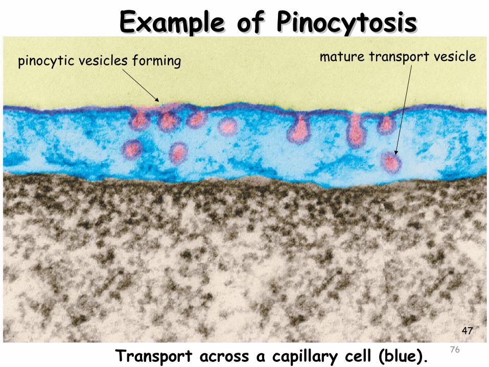
PinocytosisPinocytosis
Most common form of endocytosisMost common form of endocytosis. Takes in dissolved molecules as a vesicleTakes in dissolved molecules as a vesicle.
46

PinocytosisPinocytosis
• Cell forms an Cell forms an invaginationinvagination
• Materials dissolve Materials dissolve in water to be in water to be brought into cellbrought into cell
• Called “Cell Called “Cell Drinking”Drinking”
75
76
Example of PinocytosisExample of Pinocytosispinocytic vesicles forming mature transport vesicle
Transport across a capillary cell (blue).47

77
Receptor-Mediated EndocytosisReceptor-Mediated Endocytosis
Some integral proteins have receptors Some integral proteins have receptors on their surface to recognize & take in on their surface to recognize & take in hormones, cholesterol, etc.hormones, cholesterol, etc.
48

78
Receptor-Mediated EndocytosisReceptor-Mediated Endocytosis
49

79
50

80
Endocytosis – Phagocytosis Endocytosis – Phagocytosis
Used to engulf large particles such as Used to engulf large particles such as food, bacteria, etc. into vesiclesfood, bacteria, etc. into vesicles
Called “Cell Eating”Called “Cell Eating”
51

81
52

82
Phagocytosis About to OccurPhagocytosis About to Occur
53

83
PhagocytosisPhagocytosis - Capture of a Yeast Cell (yellow) by Membrane Extensions of an Immune System Cell (blue) 54

84
ExocytosisExocytosis The opposite of endocytosis is exocytosis. Large The opposite of endocytosis is exocytosis. Large molecules that are manufactured in the cell are molecules that are manufactured in the cell are
released through the cell membranereleased through the cell membrane..
Inside Cell Cell environment
55

References
• Reading• Brock biology of
microorgamism (13th edition) by Madigan, Martinko, Stahl, Clark
• Microbiology (10th edition) by Tortora, Funke and Case
• Microbiology (5th edition) by Prescott
• Images 1 to 55• Microbiology (5th
edition) by Prescott