Embed Size (px)
Citation preview
Upper Gastrointestinal Bleeding – what to do…
Dr Chris RoseveareConsultant PhysicianSouthampton University Hospitals NHS Trust
What to do…. Resuscitate Endoscope If all else fails – call a surgeon
Introduction 7-8% mortality overall 26-44% mortality if already hospitalised
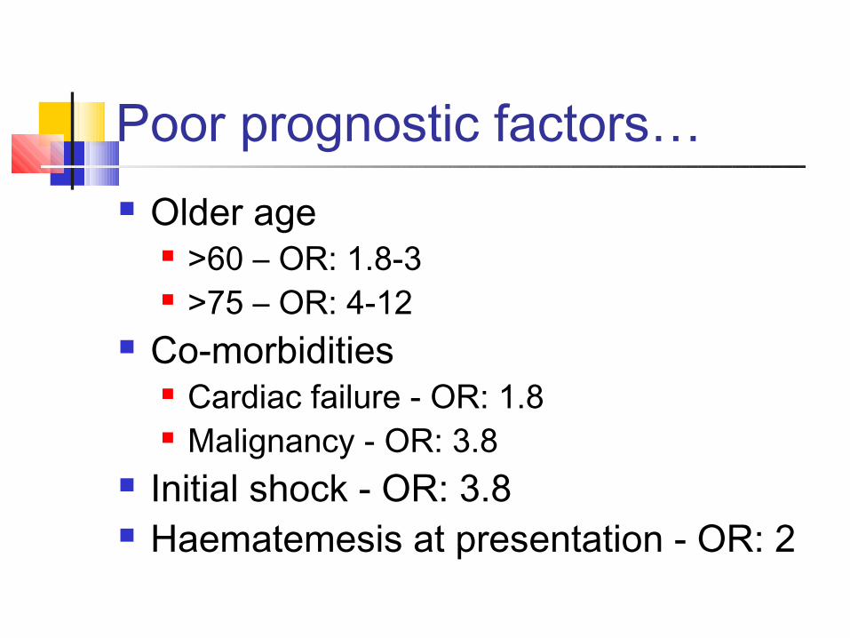
Poor prognostic factors… Older age
>60 – OR: 1.8-3 >75 – OR: 4-12
Co-morbidities Cardiac failure - OR: 1.8 Malignancy - OR: 3.8
Initial shock - OR: 3.8 Haematemesis at presentation - OR: 2
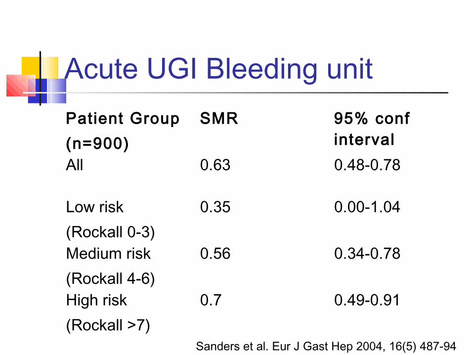
Acute UGI Bleeding unitPatient Group(n=900)
SMR 95% conf interval
All 0.63 0.48-0.78
Low risk(Rockall 0-3)
0.35 0.00-1.04
Medium risk(Rockall 4-6)
0.56 0.34-0.78
High risk(Rockall >7)
0.7 0.49-0.91
Sanders et al. Eur J Gast Hep 2004, 16(5) 487-94
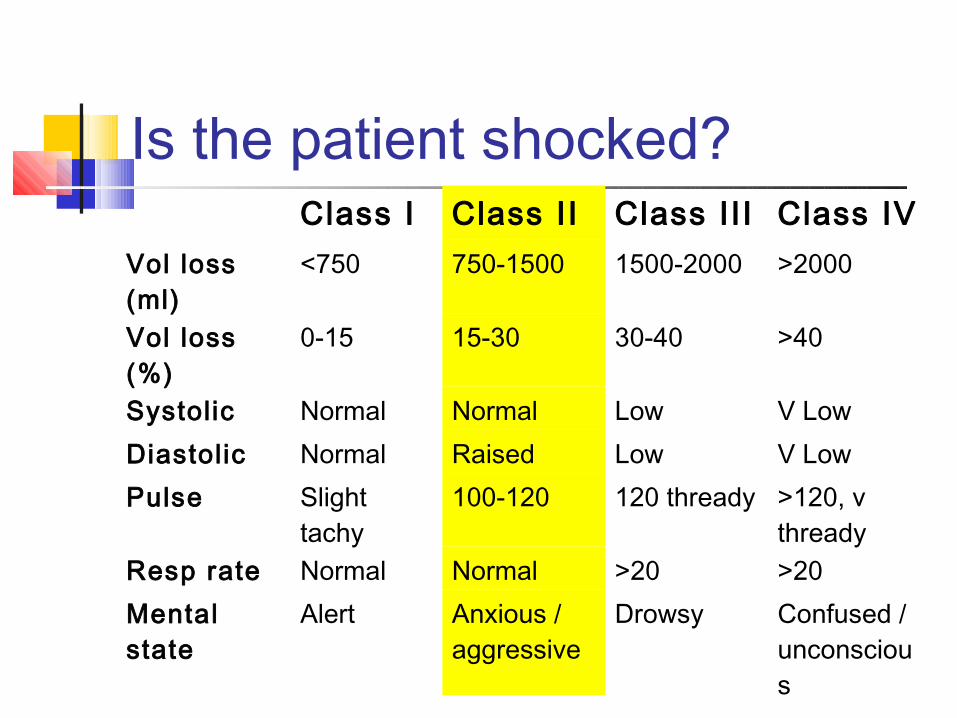
Is the patient shocked?Class I Class II Class II I Class IV
Vol loss (ml)
<750 750-1500 1500-2000 >2000
Vol loss (%)
0-15 15-30 30-40 >40
Systolic Normal Normal Low V LowDiastolic Normal Raised Low V LowPulse Slight
tachy100-120 120 thready >120, v
threadyResp rate Normal Normal >20 >20Mental state
Alert Anxious / aggressive
Drowsy Confused / unconscious
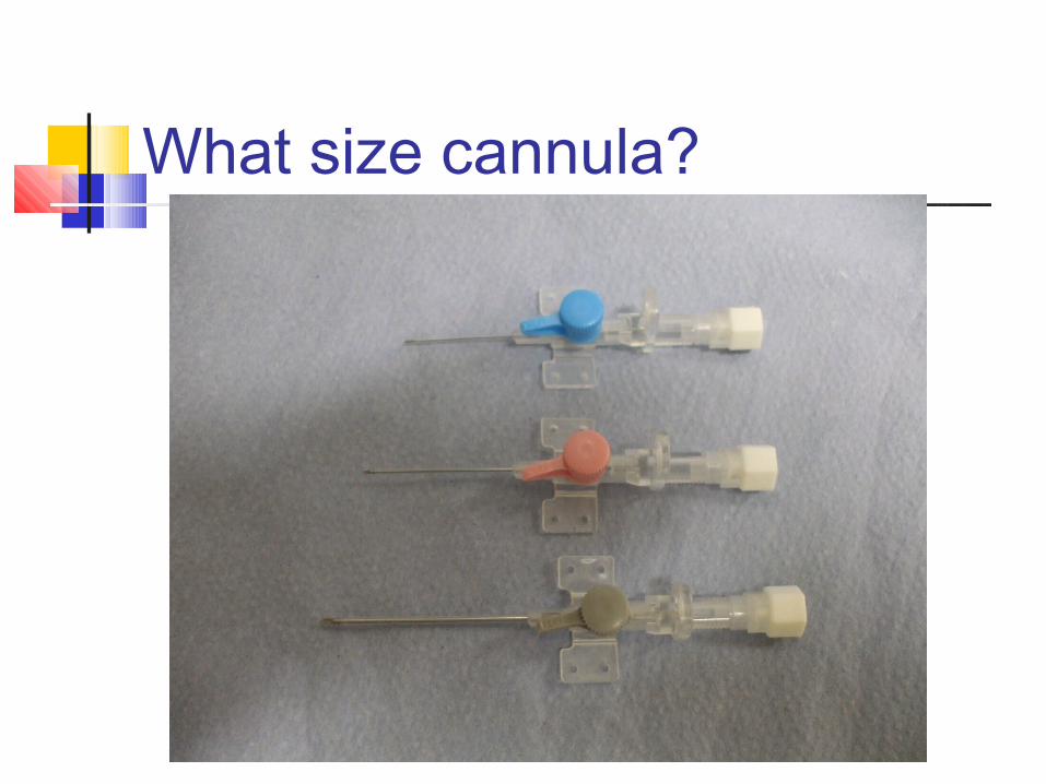
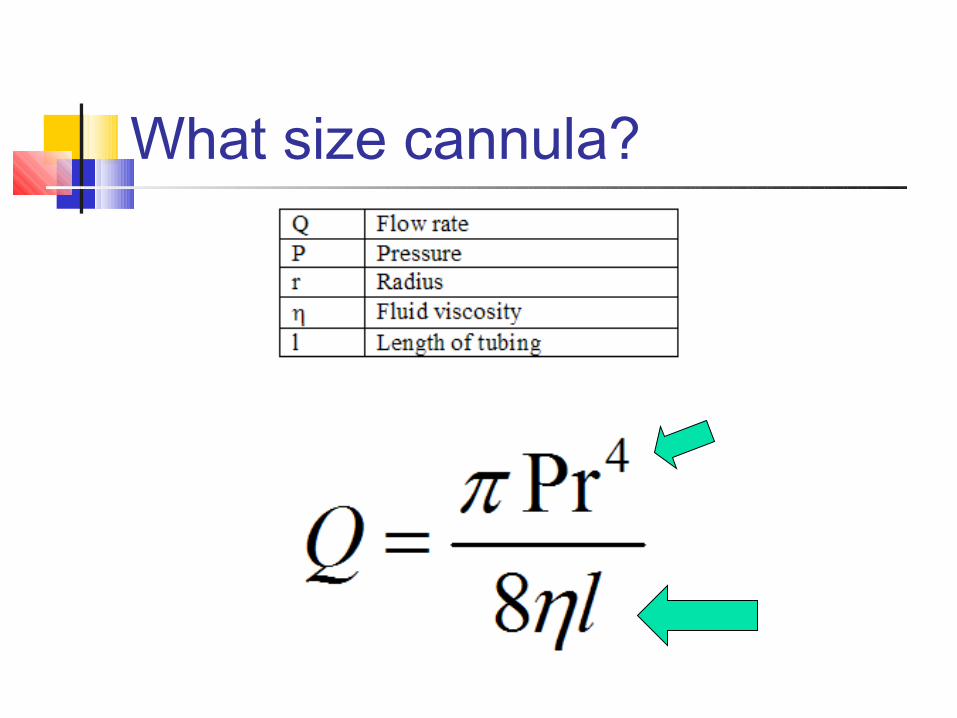
What size cannula?
Jean Louis Poiseuille (1799 - 1869)
What size cannula?
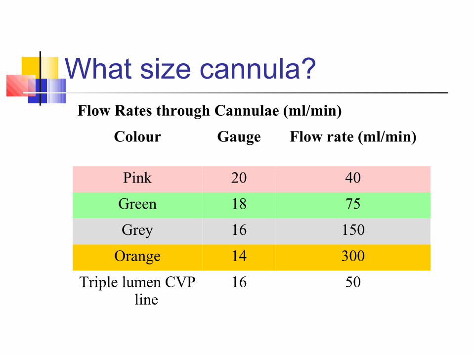
Flow Rates through Cannulae (ml/min)
Colour Gauge Flow rate (ml/min)
Pink 20 40
Green 18 75
Grey 16 150
Orange 14 300
Triple lumen CVP line
16 50
What size cannula?
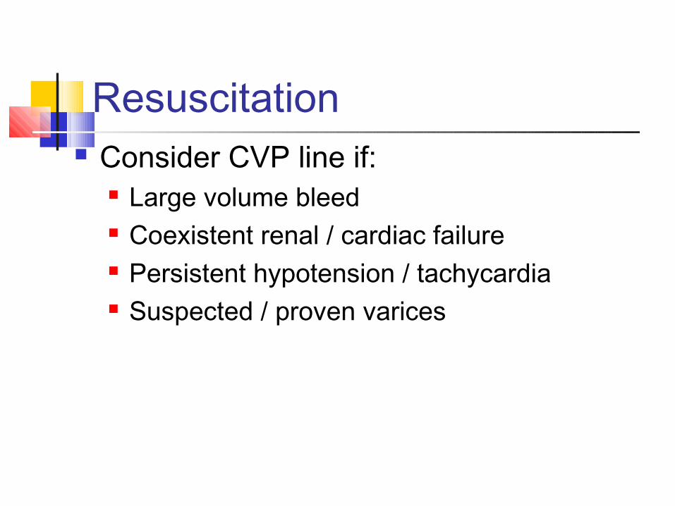
Consider CVP line if: Large volume bleed Coexistent renal / cardiac failure Persistent hypotension / tachycardia Suspected / proven varices
Resuscitation
What fluid replacement? Blood if >30% volume loss
?O-negative ?Group specific ?Cross-matched
Crystalloid or colloid? No comparative studies in UGIB Probably makes no difference
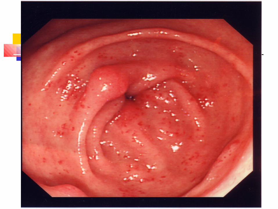
Could it be varices? Any upper GI bleed with:
Previous history of varices / variceal bleed Clinical evidence of chronic liver disease or
portal hypertension
NB: most ‘alcoholics’ with GI bleeds do not have chronic liver disease (or varices)
Could it be varices? Yes……….
Consider airway protection
High risk of aspiration with high mortality
Could it be varices? Yes……….
Reconsider CVP line (if not already)
Avoid over-transfusion
Could it be varices? Yes……….
Correct clotting and platelets
Could it be varices? Yes……….
Commence Terlipressin 2mg 6 hourly
Superior to endoscopic sclerotherapy inBleeding control (Cochrane 2002)
20% reduction in 5 day bleeding control When combined with endoscopic therapy
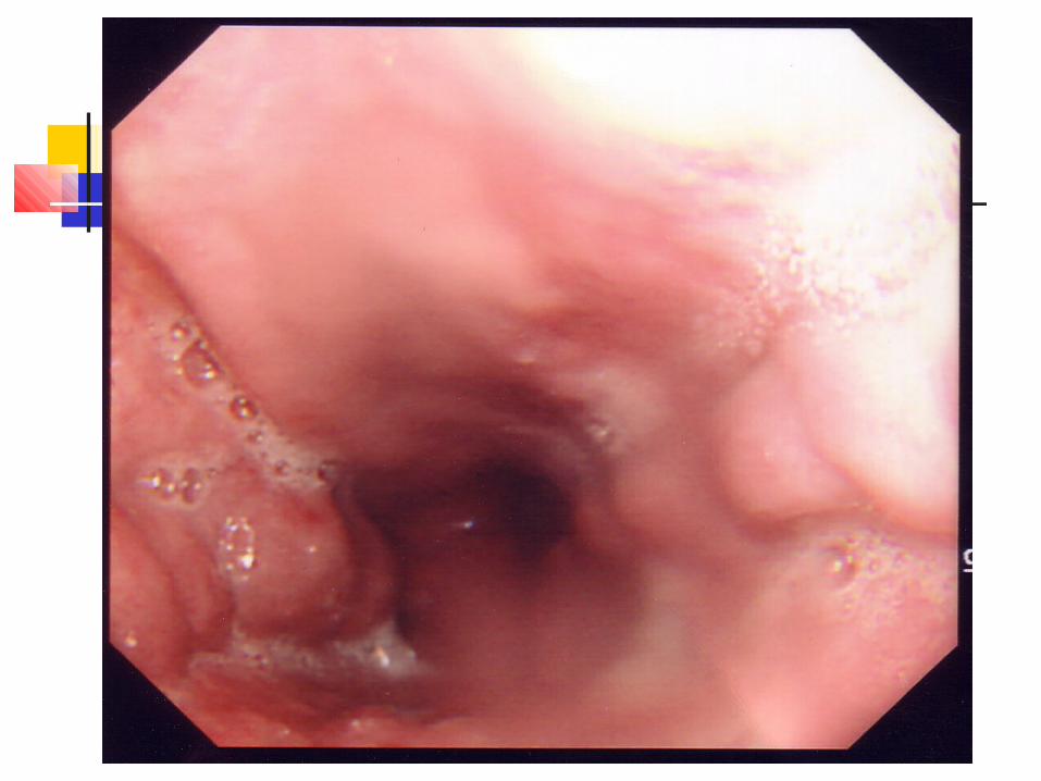
Could it be varices? Yes……….
Endoscopy at earliest opportunity
It’s not always varices
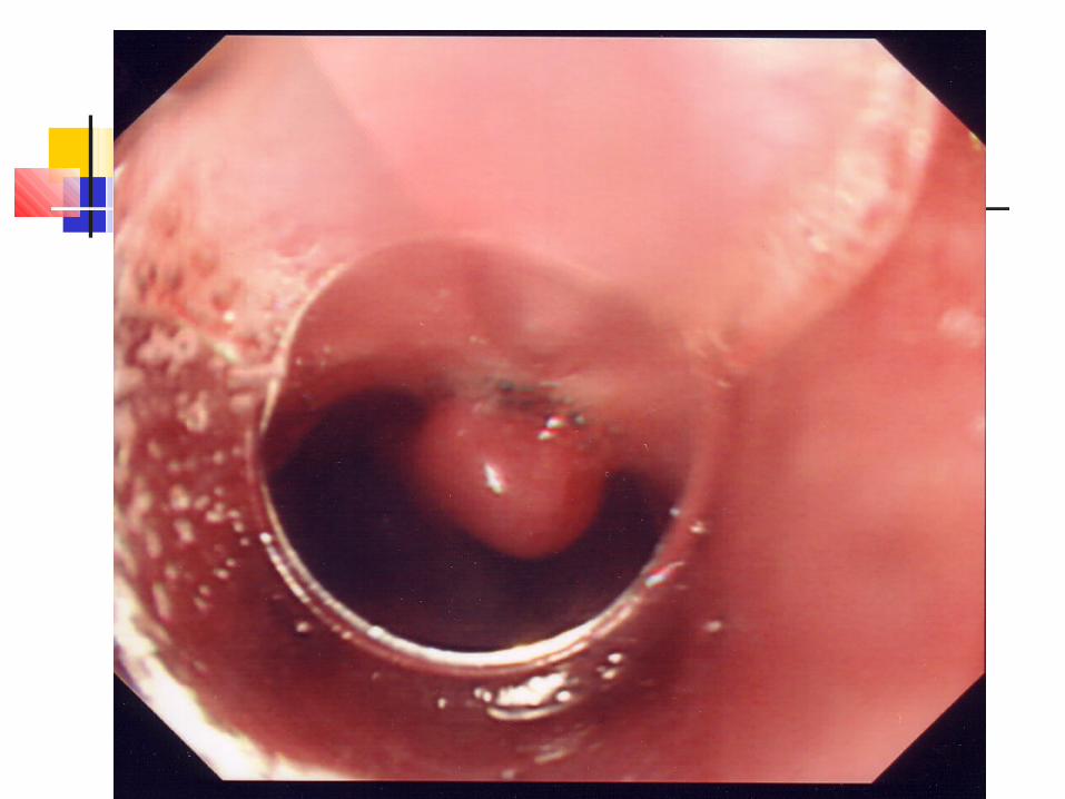
Could it be varices? Yes……….
Endoscopy at earliest opportunity
Enables endoscopic therapy
Could it be varices? Yes……….
Consider Sengstaken-Blakemore tube
Reconsider airway protection
May be safer to transfuse and await endoscopist
What’s the diagnosis?
An 80 year-old woman is brought to hospital having collapsed in her home. On arrival of the ambulance she was hypotensive, grey and sweaty. The ambulance crew reported ‘coffee ground vomit’ while en-route.
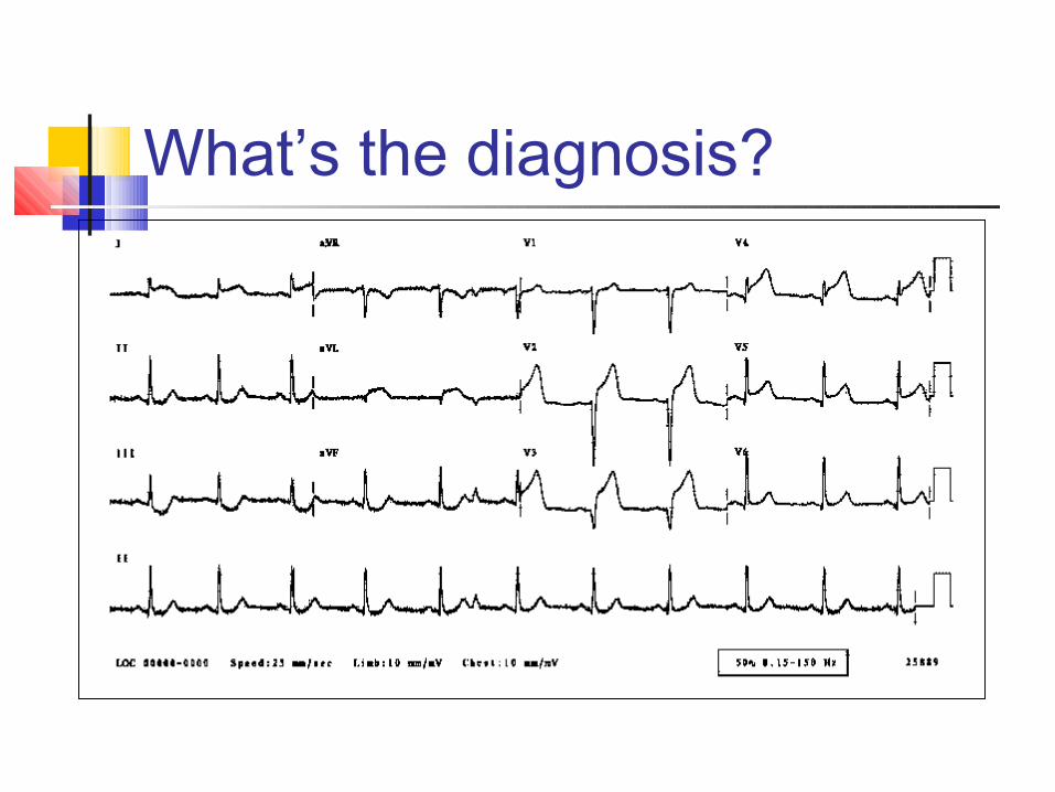
What’s the diagnosis?
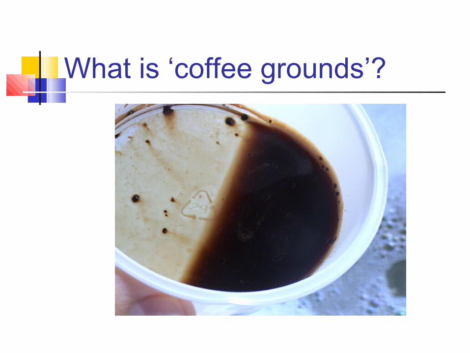
What is ‘coffee grounds’?

A B
C D
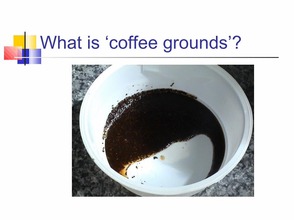
What is ‘coffee grounds’?
Think…. Is the degree of haemodynamic
compromise consistent with volume of reported blood loss?
Beware a shocked patient with ‘dark’ vomit look for an alternative explanation for
hypotension.

Why endoscope? Diagnose Treat Risk stratification
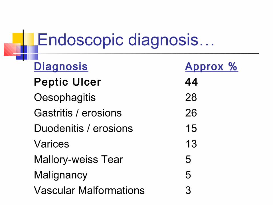
Endoscopic diagnosis…Diagnosis Approx %Peptic Ulcer 44Oesophagitis 28Gastritis / erosions 26Duodenitis / erosions 15Varices 13Mallory-weiss Tear 5Malignancy 5Vascular Malformations 3
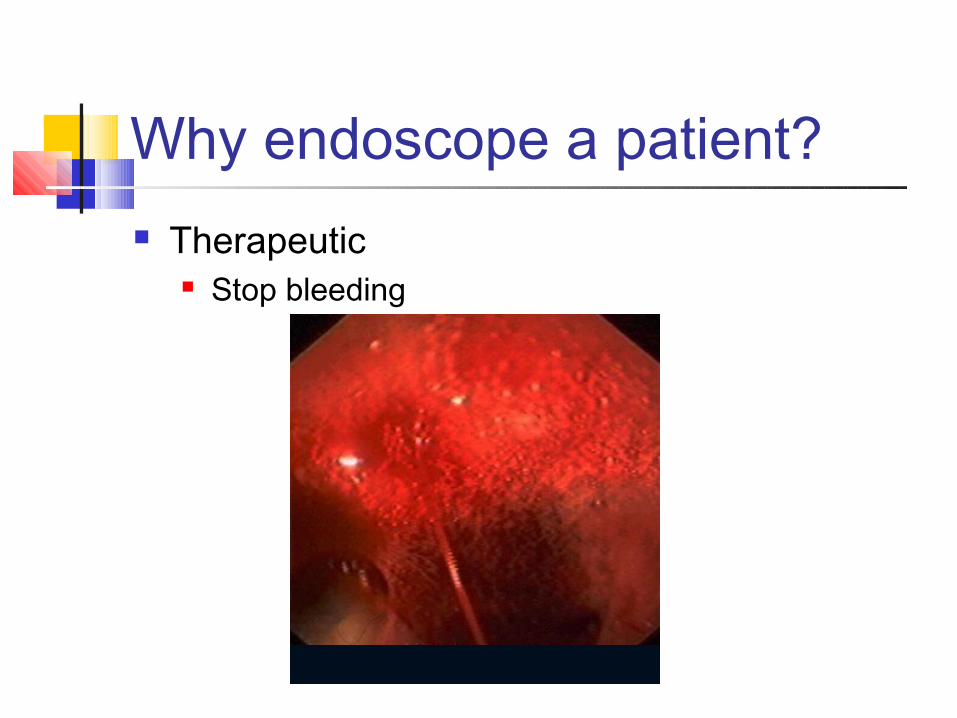
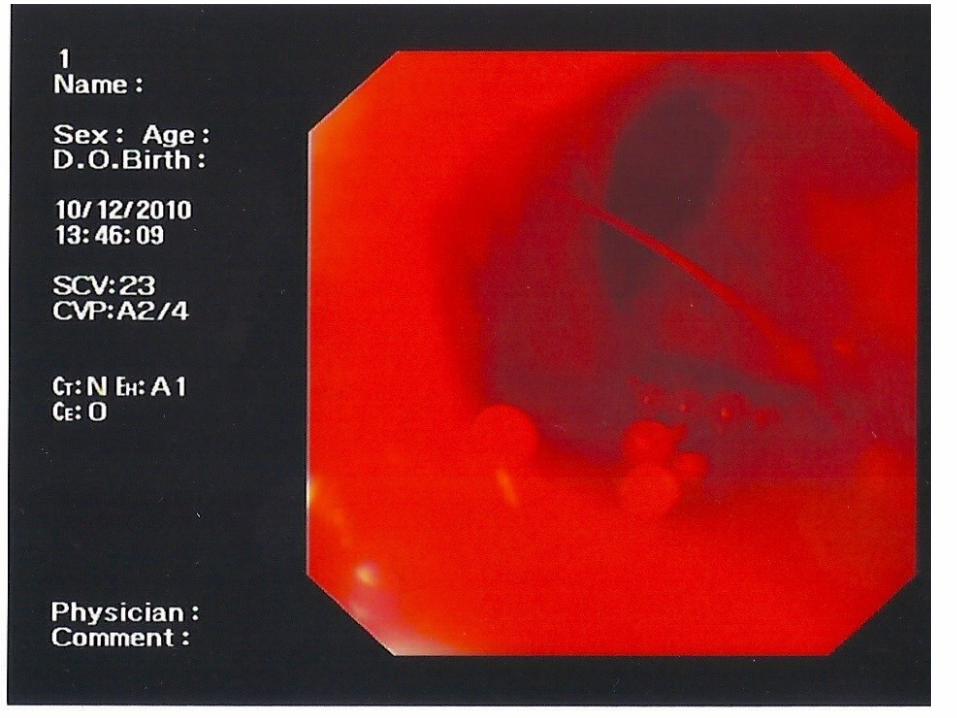
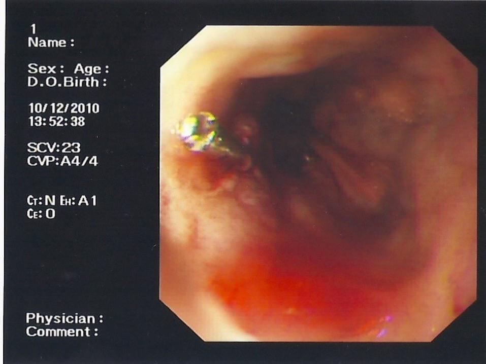
Why endoscope a patient? Therapeutic
Stop bleeding
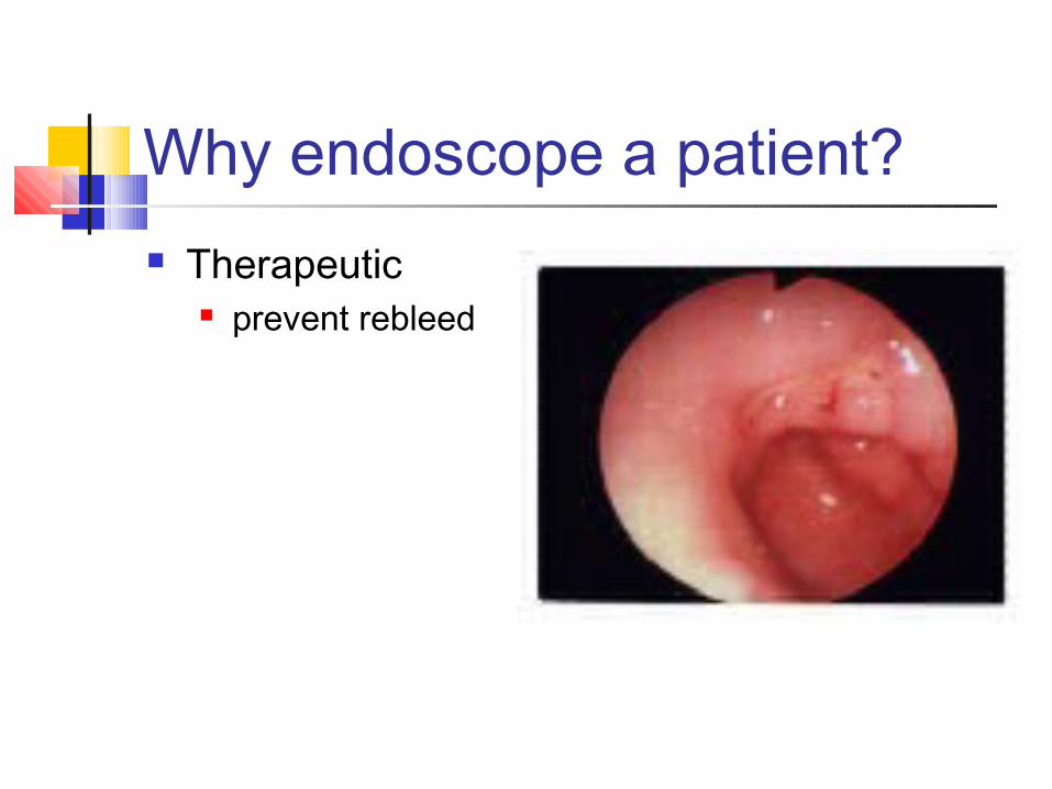
Why endoscope a patient? Therapeutic
prevent rebleed
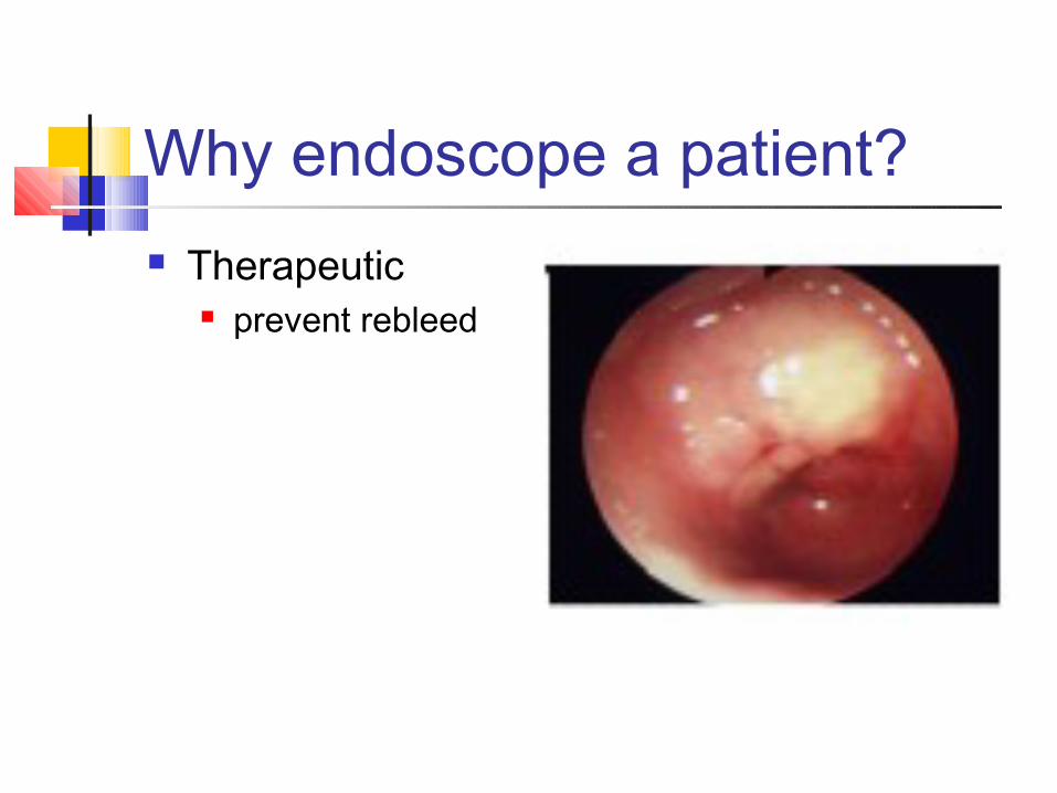
Why endoscope a patient? Therapeutic
prevent rebleed
Risk Stratification Calculation of full Rockall Score
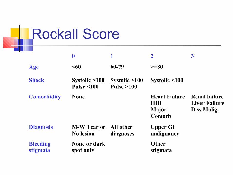
Rockall Score0 1 2 3
Age <60 60-79 >=80
Shock Systolic >100Pulse <100
Systolic >100Pulse >100
Systolic <100
Comorbidity None Heart FailureIHDMajor Comorb
Renal failureLiver FailureDiss Malig.
Diagnosis M-W Tear orNo lesion
All other diagnoses
Upper GI malignancy
Bleeding stigmata
None or dark spot only
Other stigmata
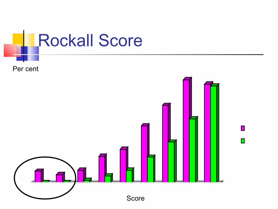
Rockall Score
0
5
1015
20
25
30
35
40
45
0 1 2 3 4 5 6 7 8+
Rebleed
Mortality
Score
Per cent
When to endoscope? Too soon?
Inadequate resuscitation: higher risk Poor views (blood in the way) Aspiration (stomach full of blood)
When to endoscope? Too late?
Ongoing bleeding Rebleeding Delay to surgery
When to endoscope? ASAP if:
Evidence of ongoing bleeding Suspected varices Suspected early rebleed
Otherwise within 12 hrs is usually OK
What about IV PPI? Clots more stable when pH >4 Clot lysis occurs when pH <4
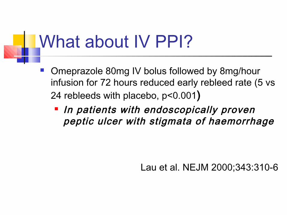
What about IV PPI? Omeprazole 80mg IV bolus followed by 8mg/hour
infusion for 72 hours reduced early rebleed rate (5 vs 24 rebleeds with placebo, p<0.001) In patients with endoscopically proven
peptic ulcer with st igmata of haemorrhage
Lau et al. NEJM 2000;343:310-6
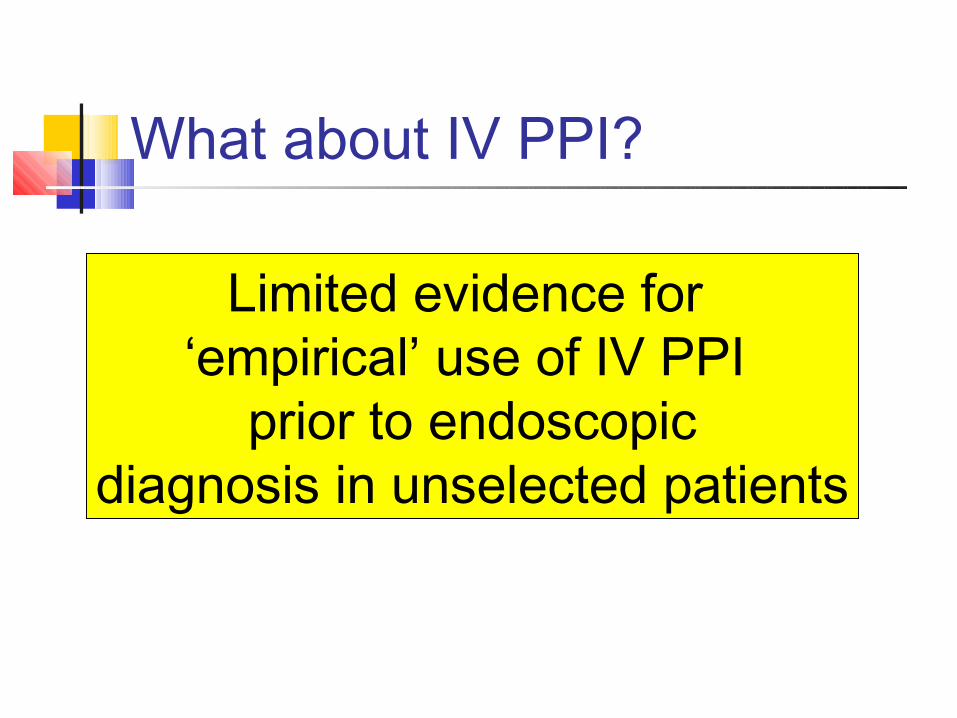
What about IV PPI?
Limited evidence for ‘empirical’ use of IV PPI
prior to endoscopicdiagnosis in unselected patients
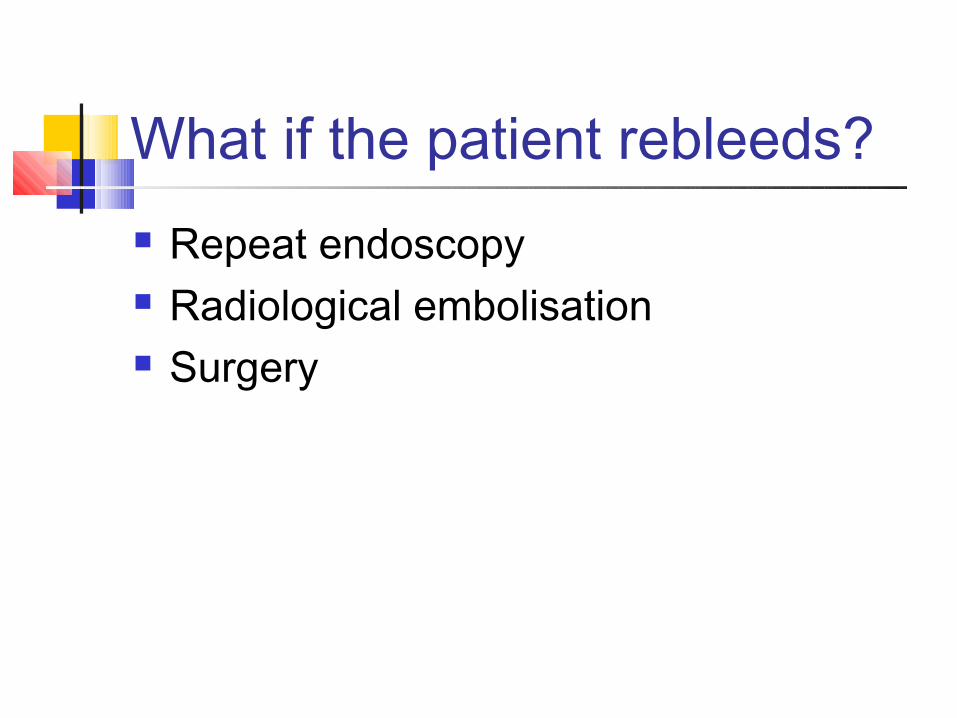
What if the patient rebleeds? Repeat endoscopy Radiological embolisation Surgery

Repeat Endoscopy No difference in bleeding control between
surgery and second endoscopic treatment 30 day mortality and transfusion requirements
similar More complications in group randomised to
surgery
Lau et al N Engl J Med 1999;340:751-756
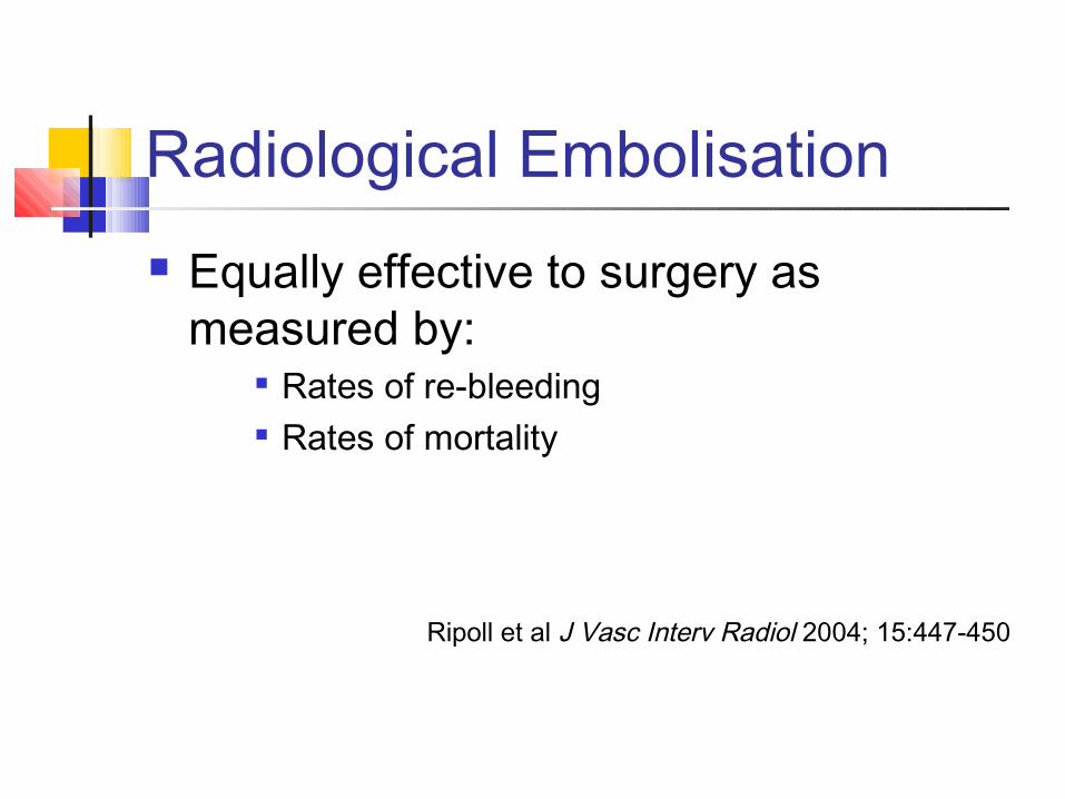
Radiological Embolisation Equally effective to surgery as
measured by: Rates of re-bleeding Rates of mortality
Ripoll et al J Vasc Interv Radiol 2004; 15:447-450
What about surgery? >65 with one ‘rebleed’ or > 4 units blood
required for fluid resuscitation <65 with 2 rebleeds or >8 units blood
required for fluid resuscitation
What about Surgery? Dependent on:
Type of lesion Site of lesion Co-morbidities Likelihood of continued bleeding
Summary Resuscitate adequately Exclude varices (and non-GI source of
shock) Endoscopy within 12 hours if non-
variceal Intravenous PPI infusion if peptic ulcer
bleed with stigmata of haemorrhage