Embed Size (px)
Citation preview
Non Incisional, Non Laser Refractive Surgery
Dr. Ankit Gupta
2 CATEGORIESA CORNEAL Epikeratophakia Intracorneal rings
B LENTICULAR Clear lens extraction Phakic IOLs
NON INCISIONAL NON LASER REFRACTIVE SURGERY
KERATOPHAKIA In keratophakia, a plus-powered lens is
placed intrastromally to increase the curvature of the anterior cornea to correct hyperopia and presbyopia.
A central lamellar keratectomy is performed with a microkeratome or femtosecond laser, the flap is lifted, the lenticule is placed onto the host bed, and the flap is replaced and adheres without sutures.
KERATOPHAKIA
Kaufmann , werblin in 1980 To eliminate the complexity of the lamellar
dissection Epikeratoplasty involved suturing a preformed
homoplastic lenticule directly onto the Bowman layer of the host cornea.
Graft rejection did not occur because no viable cells existed in the donor tissue.
EPIKERATOPHAKIA
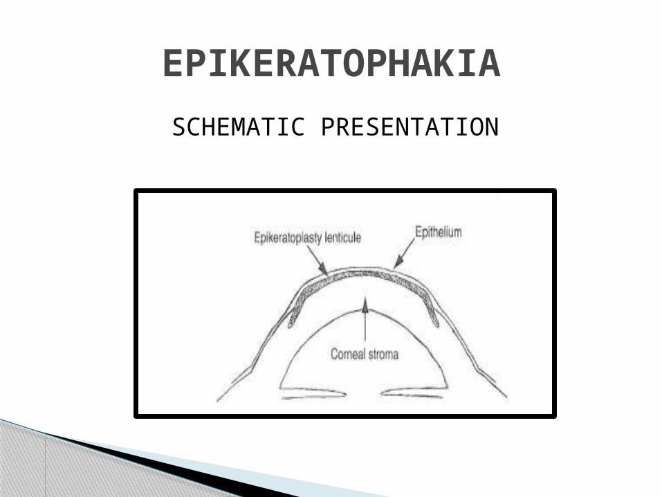
SCHEMATIC PRESENTATION
EPIKERATOPHAKIA
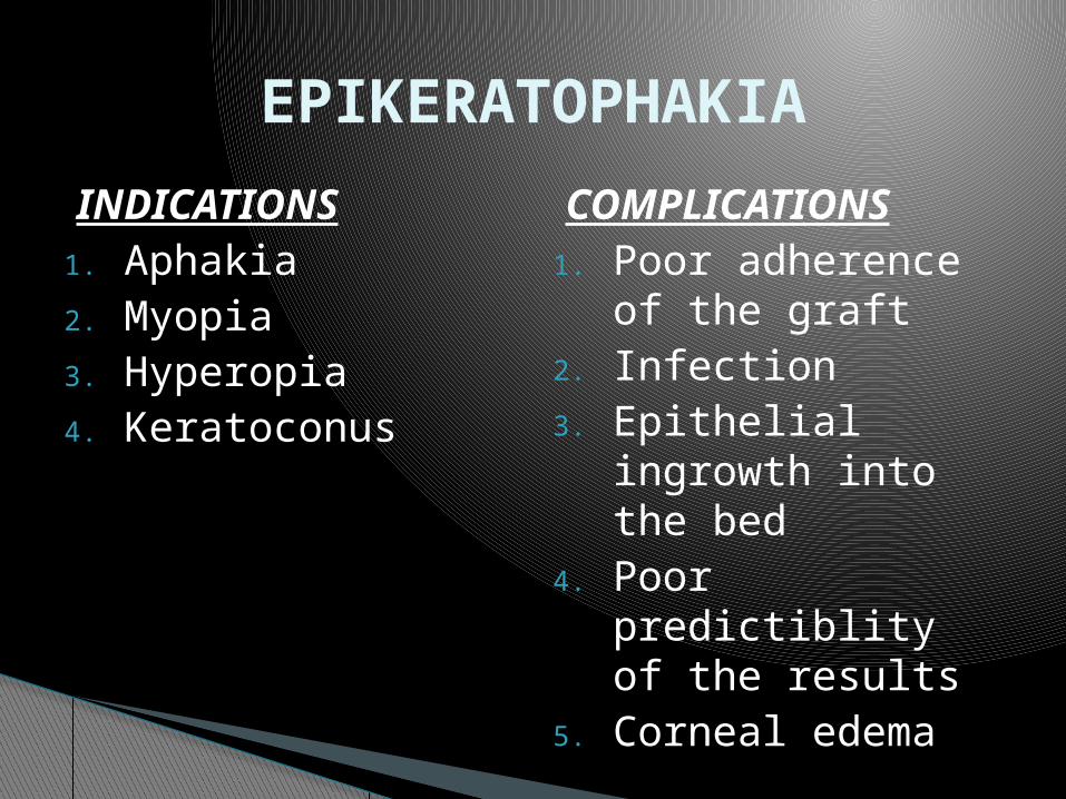
INDICATIONS1. Aphakia2. Myopia3. Hyperopia4. Keratoconus
COMPLICATIONS1. Poor adherence of
the graft2. Infection3. Epithelial ingrowth
into the bed4. Poor predictiblity
of the results5. Corneal edema
EPIKERATOPHAKIA
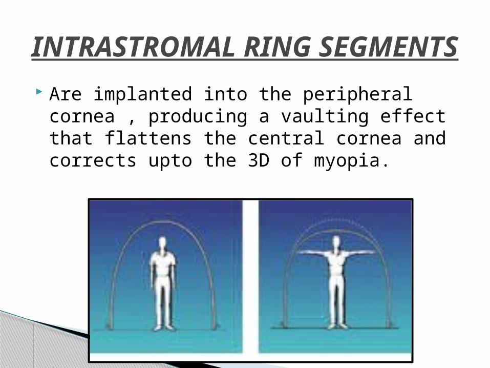
Are implanted into the peripheral cornea , producing a vaulting effect that flattens the central cornea and corrects upto the 3D of myopia.
INTRASTROMAL RING SEGMENTS
HOW INTACS WORK

Made up of PMMA Outter diameter 8.1mm Inner diameter 6.7mm Arc length 150 ° Positioning hole diameter 0.28mm Ring cross sectional – hexagonal Each package consists of two rings Available in 11 thickness from 0.210mm to
0.450mm
FEATURES OF ICRS
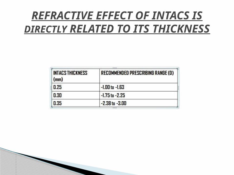
REFRACTIVE EFFECT OF INTACS IS DIRECTLY RELATED TO ITS THICKNESS
Myopia upto -3D Keratoconus Pellucid marginal degeneration Post LASIK corneal ectasias.
INDICATIONS OF ICRS
Patient should have central clear cornea. Thickness of cornea should be greater than
450micron at the incision site.
PREREQUISITES FOR ICRS

Collagen vascular disease Autoimmune, or immunodeficiency diseases Pregnant or breastfeeding women Presence of ocular conditions such as
recurrent corneal erosion syndrome Corneal dystrophy that may predispose the
patient to future complications.
CONTRAINDICATIONS OF ICRS
The procedure involves creating a lamellar channel at approximately 68%-70% stromal depth, followed by insertion of the ring segments.
Pachymeter is used to measure the thickness of the cornea over the entry mark
TECHNIQUE FOR ICRS
The geometric center of the cornea is marked with a blunt hook.
A diamond knife is set to 68%-70% of the stromal depth and then used to create a 1.0-mm radial incision.( also by femtosecond laser)
Corneal tunnels are then created at approximately 2/3rd of stromal depth using pocketing hook.
Intacs are then implanted. Tissue glue or 10-0 nylon sutures may be used
to close the radial incision at the corneal incision.
TECHNIQUE FOR ICRS
INTACS INSERTION
Removal or exchange rate – 3 % to 15 % Most common reason for exchange is
residual myopia. Removal of ring is done usually because of
disabling vision symptoms such as glare , double vision and photophobia.
REMOVAL OR EXCHANGE OF ICRS
Adverse events (defined as events that, if left untreated, could be serious or result in permanent sequelae) occur in approximately 1% of patients.
1. Anterior chamber perforation2. Microbial keratitis3. Implant extrusion4. Shallow ring segment placement5. Corneal thinning over Intacs
ADVERSE EVENTS OF ICRS

EXTRUSION
TIP EXTRUISON
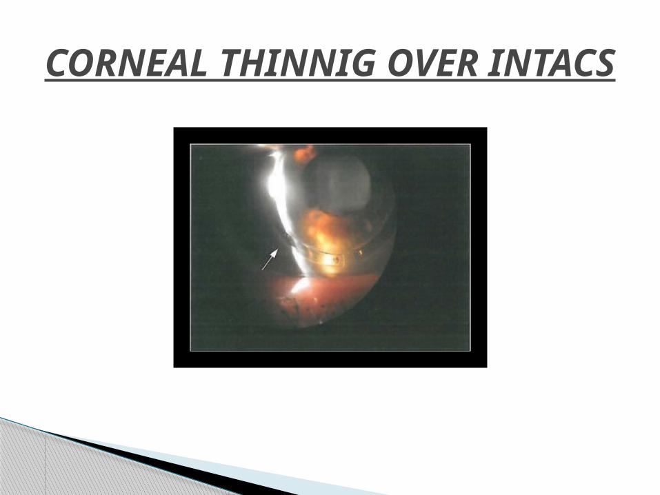
CORNEAL THINNIG OVER INTACS
Ocular complications defined as clinically significant events that do not result in permanent sequelae occurs in approximately 11% of patients.
1. Reduced corneal sensitivity.2. Induced astigmatism between 1.00 and
2.00 D.3. Deep neovascularization at the incision site.4. Persistent epithelial defect.5. Iritis/uveitis.
COMPLICATIONS OF ICRS
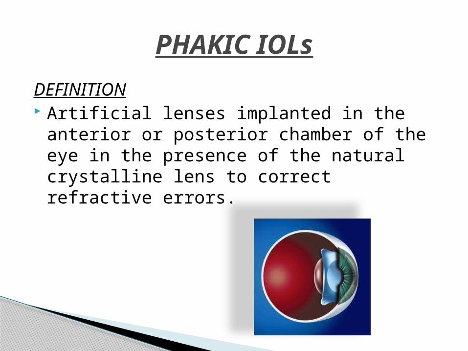
DEFINITION Artificial lenses implanted in the anterior or
posterior chamber of the eye in the presence of the natural crystalline lens to correct refractive errors.
PHAKIC IOLs

1950s - correcting myopia by inserting a
concave lens into the phakic eye 1988 - Baikoff : anterior chamber angle-fixed
PIOL Mid 1980s - Posterior chamber phakic IOLs :
Fyodorov 1991 - Artisan-Worst iris claw lens
HISTORY OF PHAKIC IOLs
Can treat a larger range of refractive errors Allows the crystalline lens to retain its
function preserving accommodation (as compared with refractive lens exchange)
Removable and exchangeable. Less expansive Lower risk of endophthalmitis and post op
retinal detachment because the barrier of crystalline lens is present.
ADVANTAGES OF PHAKIC IOLs
Potential risks of an intraocular procedure Nonfoldable models require large incision that
may result in high postoperative astigmatism.
PC PHAKIC IOLs have a higher incidence of cataract formation.
At the time of cataract surgery posterior PHAKIC IOL has to be removed possibly through a larger than usual wound.
AC PHAKIC IOLs may damage corneal endothelium.
DISADVANTAGES OF PHAKIC IOLs
Anterior chamber –angle supported PIOL AC iris fixated Posterior chamber PIOL
TYPES OF PHAKIC IOLs
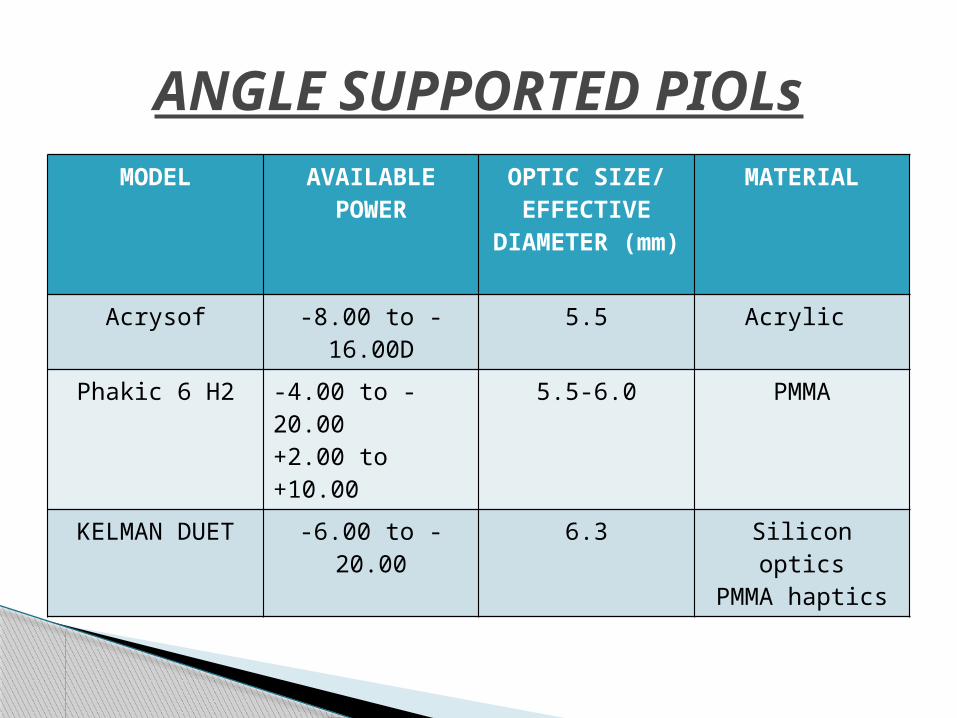
MODEL AVAILABLE POWER
OPTIC SIZE/EFFECTIVE DIAMETER
(mm)
MATERIAL
Acrysof -8.00 to -16.00D
5.5 Acrylic
Phakic 6 H2 -4.00 to -20.00+2.00 to +10.00
5.5-6.0 PMMA
KELMAN DUET -6.00 to -20.00 6.3 Silicon opticsPMMA haptics
ANGLE SUPPORTED PIOLs
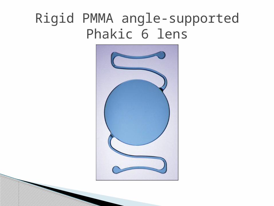
Rigid PMMA angle-supported Phakic 6 lens
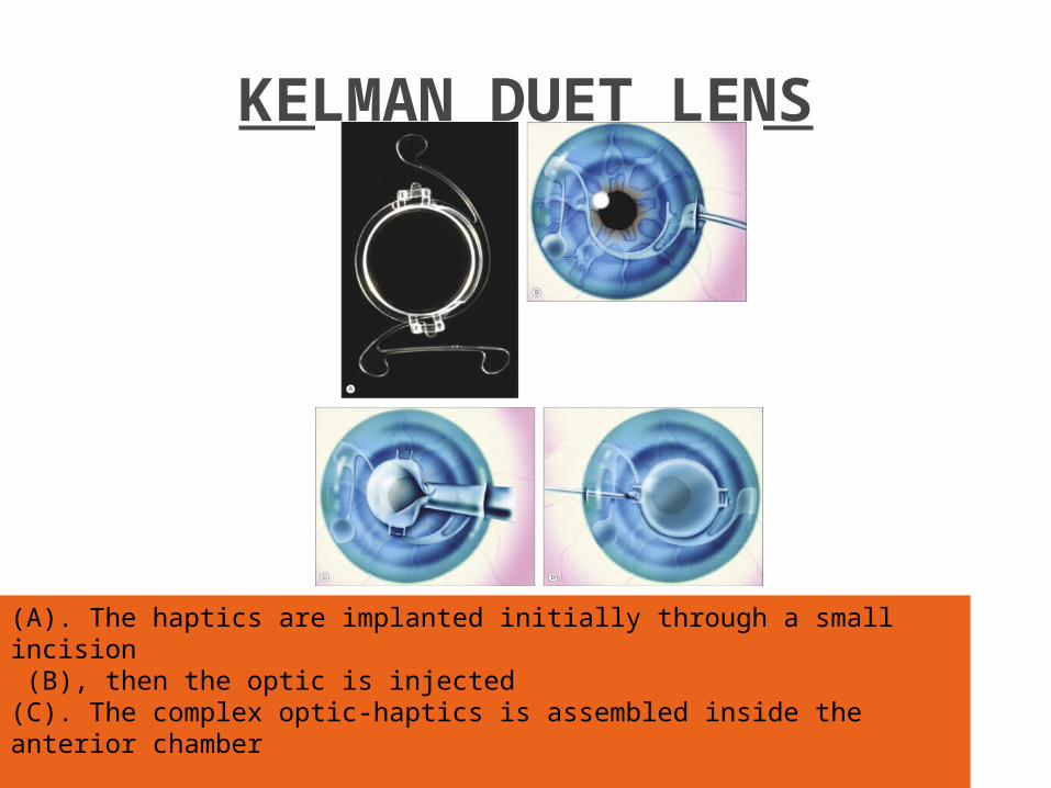
KELMAN DUET LENS
(A). The haptics are implanted initially through a small incision (B), then the optic is injected (C). The complex optic-haptics is assembled inside the anterior chamber
MODEL AVAILABLE POWER
OPTIC SIZE/EFFECTIVE DIAMETER
(mm)
MATERIAL
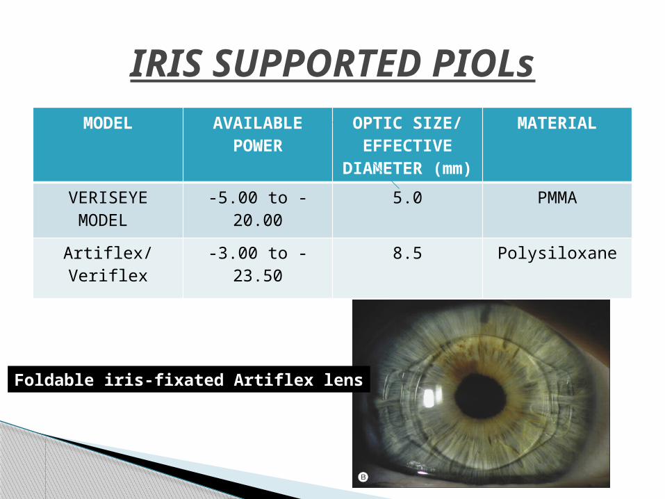
VERISEYE MODEL
-5.00 to -20.00 5.0 PMMA
Artiflex/ Veriflex -3.00 to -23.50 8.5 Polysiloxane
IRIS SUPPORTED PIOLs
Foldable iris-fixated Artiflex lens
MODEL EFFECTIVE POWER
OPTIC SIZE/EFFECTIVE DIAMETER
(mm)
MATERIAL
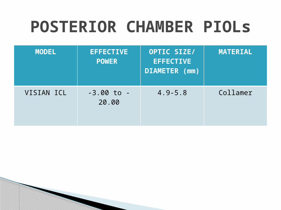
VISIAN ICL -3.00 to -20.00 4.9-5.8 Collamer
POSTERIOR CHAMBER PIOLs
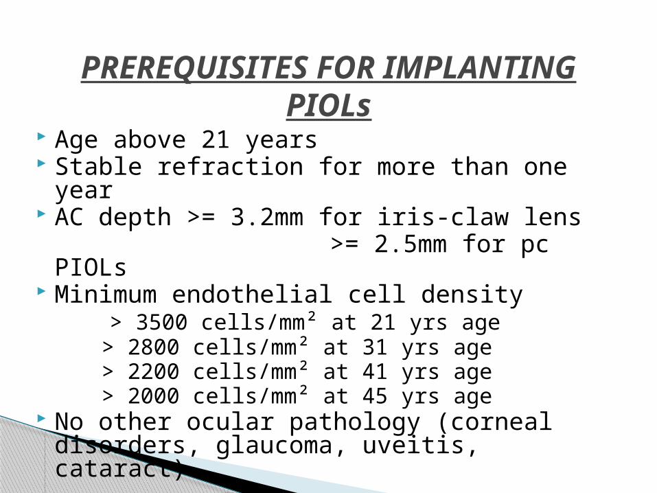
Age above 21 years Stable refraction for more than one year AC depth >= 3.2mm for iris-claw lens >= 2.5mm for pc PIOLs Minimum endothelial cell density > 3500 cells/mm² at 21 yrs age > 2800 cells/mm² at 31 yrs age > 2200 cells/mm² at 41 yrs age > 2000 cells/mm² at 45 yrs age No other ocular pathology (corneal disorders,
glaucoma, uveitis, cataract)
PREREQUISITES FOR IMPLANTING PIOLs
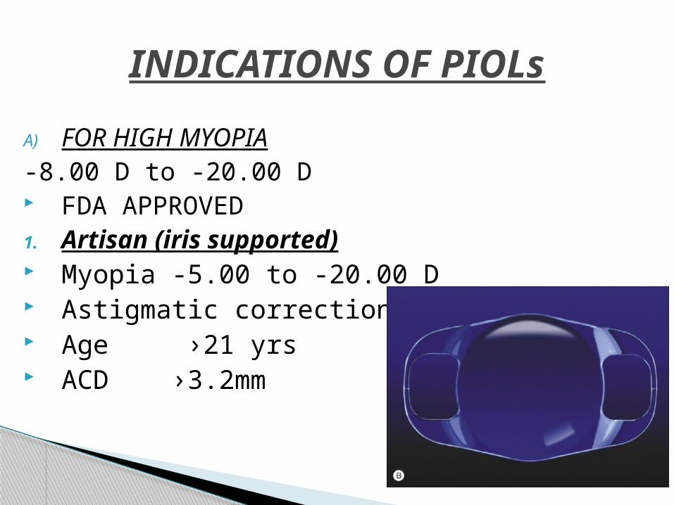
A) FOR HIGH MYOPIA-8.00 D to -20.00 D FDA APPROVED1. Artisan (iris supported) Myopia -5.00 to -20.00 D Astigmatic correction of 2.5D Age ›21 yrs ACD ›3.2mm
INDICATIONS OF PIOLs
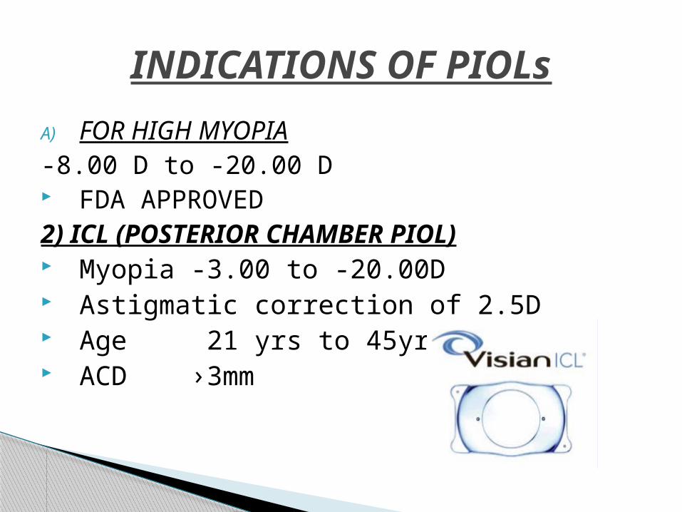
A) FOR HIGH MYOPIA-8.00 D to -20.00 D FDA APPROVED2) ICL (POSTERIOR CHAMBER PIOL) Myopia -3.00 to -20.00D Astigmatic correction of 2.5D Age 21 yrs to 45yrs ACD ›3mm
INDICATIONS OF PIOLs
B) FOR HYPERMETROPIAAvailable power in
1. ICL upto +20.00 D2. ARTISAN upto +12 D
INDICATIONS OF PIOLs
C) FOR ASTIGMATISM PIOLs are available upto 6D But treatment of choice is laser ablation
upto 4 to 5 D
INDICATIONS OF PIOLs

Preexisting ocular diseases – 1. Compromised corneal endothelium2. Iritis3. Rubeosis iridis4. Cataract5. Glaucoma AC depth AC diameter Pupil size
CONTRAINDICATIONS OF PIOLs
Must be appropriate
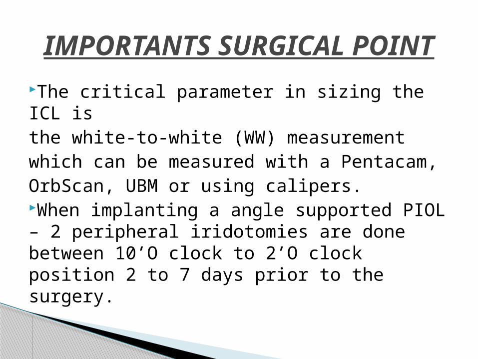
The critical parameter in sizing the ICL isthe white-to-white (WW) measurementwhich can be measured with a Pentacam,OrbScan, UBM or using calipers.When implanting a angle supported PIOL – 2 peripheral iridotomies are done between 10’O clock to 2’O clock position 2 to 7 days prior to the surgery.
IMPORTANTS SURGICAL POINT
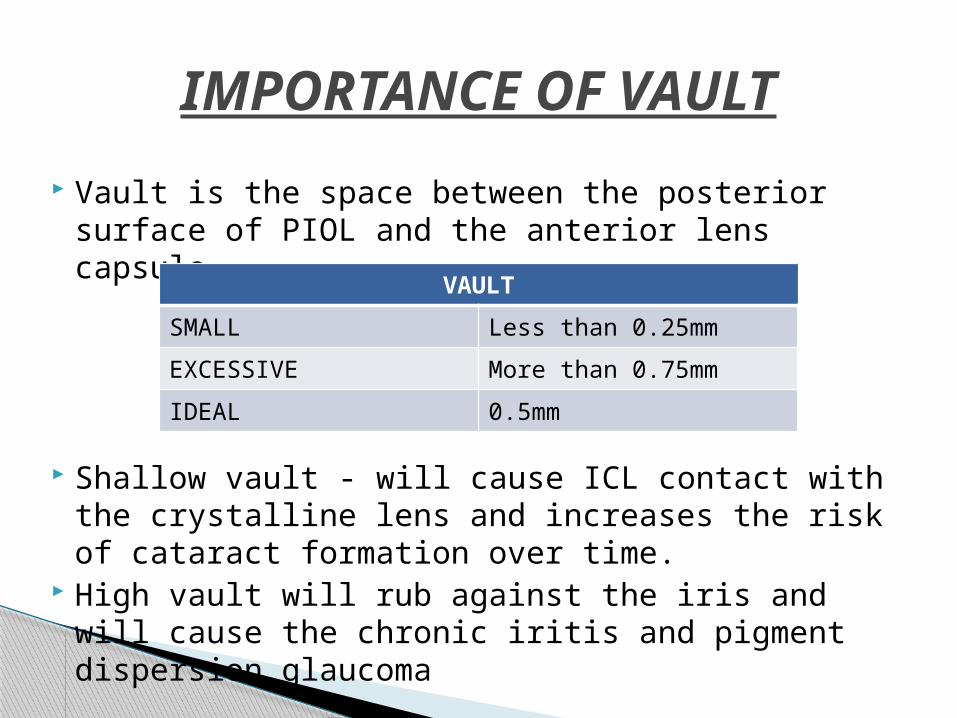
Vault is the space between the posterior surface of PIOL and the anterior lens capsule.
Shallow vault - will cause ICL contact with the crystalline lens and increases the risk of cataract formation over time.
High vault will rub against the iris and will cause the chronic iritis and pigment dispersion glaucoma
IMPORTANCE OF VAULT
VAULTSMALL Less than 0.25mmEXCESSIVE More than 0.75mmIDEAL 0.5mm

ICL IMPLANTATION

ICL REMOVAL
Changes in vaulting with accommodation. Loss of Corneal Endothelial Cells. Pupil Ovalization / Iris Retraction. Glare,Halos. Pigment dispersion glaucoma. Chronic Inflammation or Uveitis. Pupillary Block Glaucoma. Phakic Intraocular Lens Rotation. Cataractogenesis. Retinal Detachment.
COMPLICATIONS OF PIOLs
The combination of phakic IOL implantation followed by LASIK in patients with extreme myopia or hypermetropia and high levels of astigmatism.
When an anterior chamber phakic IOL is planned to be combined with LASIK, the corneal flap can be created just prior to the insertion of the lens; then, at a later time, usually after 1month, the flap is lifted for laser correction of the residual ametropia. This two-step technique was called adjustable refractive surgery (ARS) by Guell.
The rationale in performing the flap first is to avoid any possibility of contact between the endothelium and the IOL during the suction and cut for the LASIK procedure.
BIOPTICS


![Dynamic OCT measurement of corneal deformation by an air ... · A new technique is presented for the non-invasive imaging of the ... Incisional refractive surgery [8 9], now of limited](https://img.pdfslide.us/doc/110x75/5f7928d431584c2dc057368d/dynamic-oct-measurement-of-corneal-deformation-by-an-air-a-new-technique-is.jpg)