Embed Size (px)
Citation preview
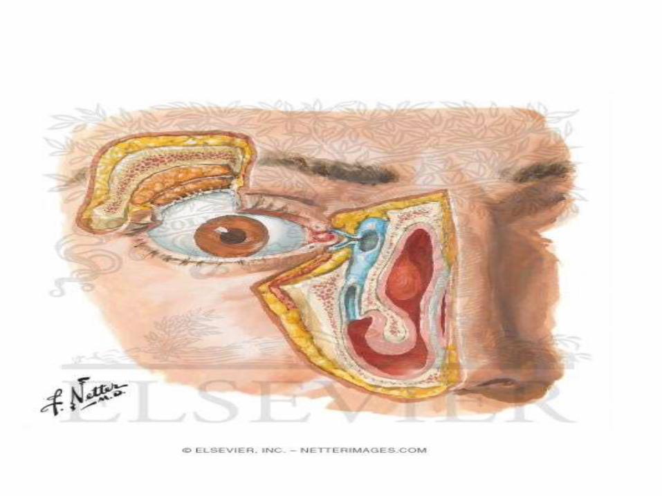
Lacrimal Apparatus
DR.Chandra Philip.

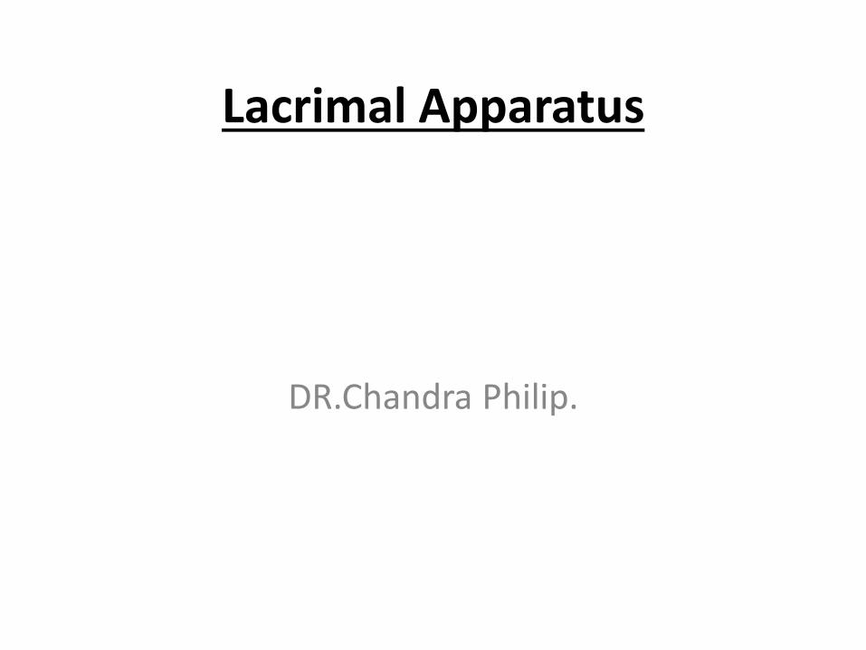
Lacrimalapparatus- It is concerned with production and drainage of lacrimal fluid
Components
1) Lacrimalgland
2) Conjunctivalsac
3) Lacrimal punctawith canaliculi
4) Lacrimal sac
5) Nasolacrimalduct
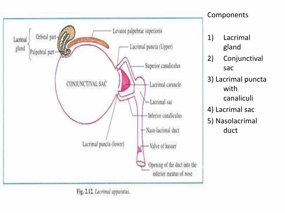
Lacrimal gland
Parts-
1) Upper part or orbital part
2) Lower part or palpebral part
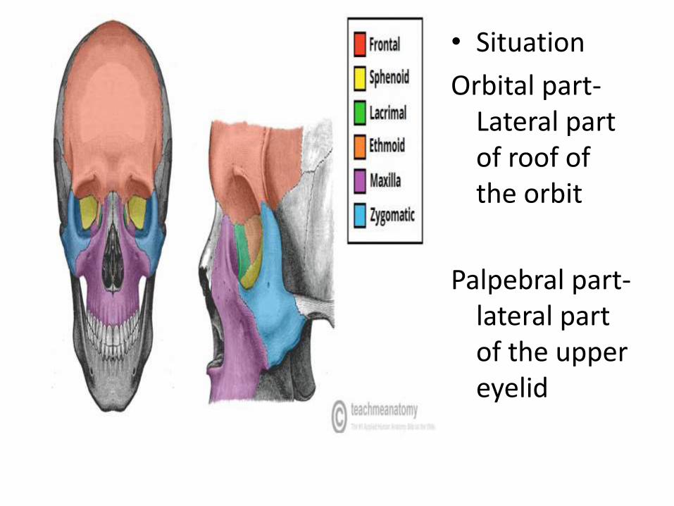
• Situation
Orbital part-Lateral part of roof of the orbit
Palpebral part-lateral part of the upper eyelid
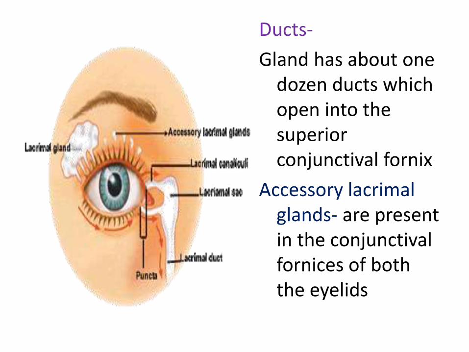
Ducts-
Gland has about one dozen ducts which open into the superior conjunctival fornix
Accessory lacrimalglands- are present in the conjunctivalfornices of both the eyelids
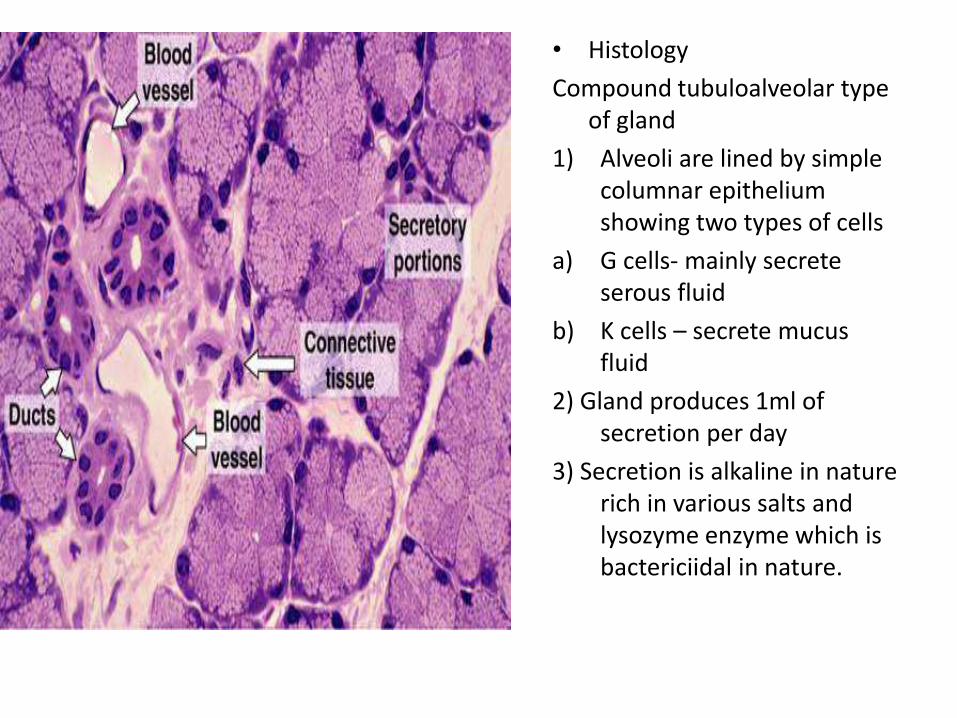
• Histology
Compound tubuloalveolar type of gland
1) Alveoli are lined by simple columnar epithelium showing two types of cells
a) G cells- mainly secrete serous fluid
b) K cells – secrete mucus fluid
2) Gland produces 1ml of secretion per day
3) Secretion is alkaline in nature rich in various salts and lysozyme enzyme which is bactericiidal in nature.
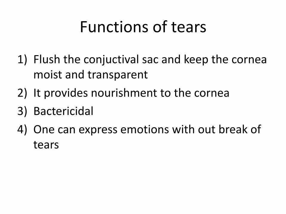
Functions of tears
1) Flush the conjuctival sac and keep the cornea moist and transparent
2) It provides nourishment to the cornea
3) Bactericidal
4) One can express emotions with out break of tears
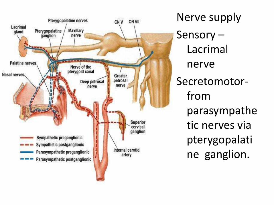
Nerve supply
Sensory –Lacrimalnerve
Secretomotor-from parasympathetic nerves via pterygopalatine ganglion.
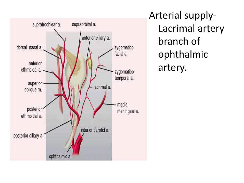
Arterial supply-Lacrimal artery branch of ophthalmic artery.
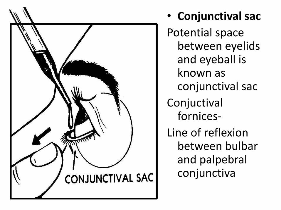
• Conjunctival sac
Potential space between eyelids and eyeball is known as conjunctival sac
Conjuctivalfornices-
Line of reflexionbetween bulbar and palpebralconjunctiva

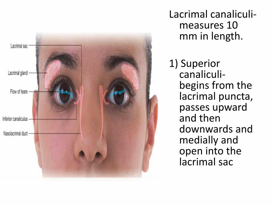
Lacrimal canaliculi-measures 10 mm in length.
1) Superior canaliculi-begins from the lacrimal puncta, passes upward and then downwards and medially and open into the lacrimal sac
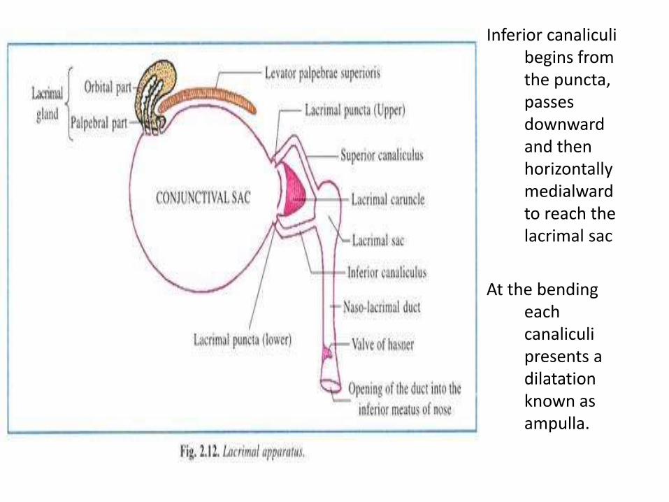
Inferior canaliculibegins from the puncta, passes downward and then horizontally medialwardto reach the lacrimal sac
At the bending each canaliculipresents a dilatation known as ampulla.
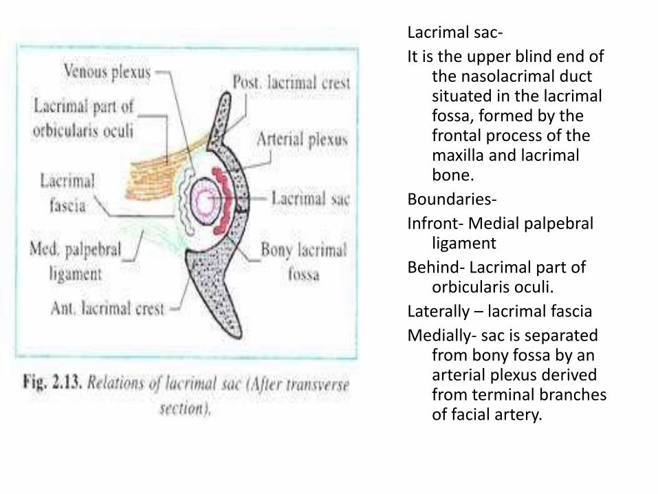
Lacrimal sac-
It is the upper blind end of the nasolacrimal duct situated in the lacrimalfossa, formed by the frontal process of the maxilla and lacrimalbone.
Boundaries-
Infront- Medial palpebralligament
Behind- Lacrimal part of orbicularis oculi.
Laterally – lacrimal fascia
Medially- sac is separated from bony fossa by an arterial plexus derived from terminal branches of facial artery.
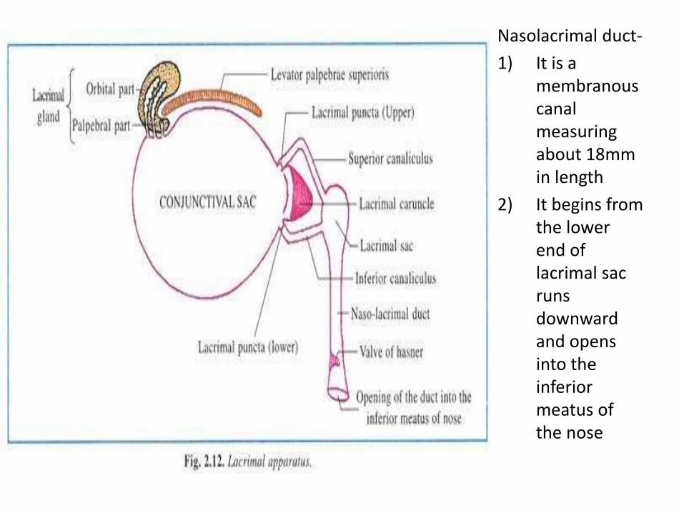
Nasolacrimal duct-
1) It is a membranous canal measuring about 18mm in length
2) It begins from the lower end of lacrimal sac runs downward and opens into the inferior meatus of the nose
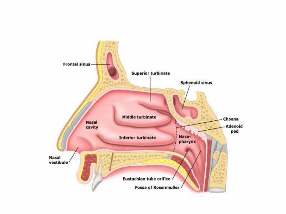
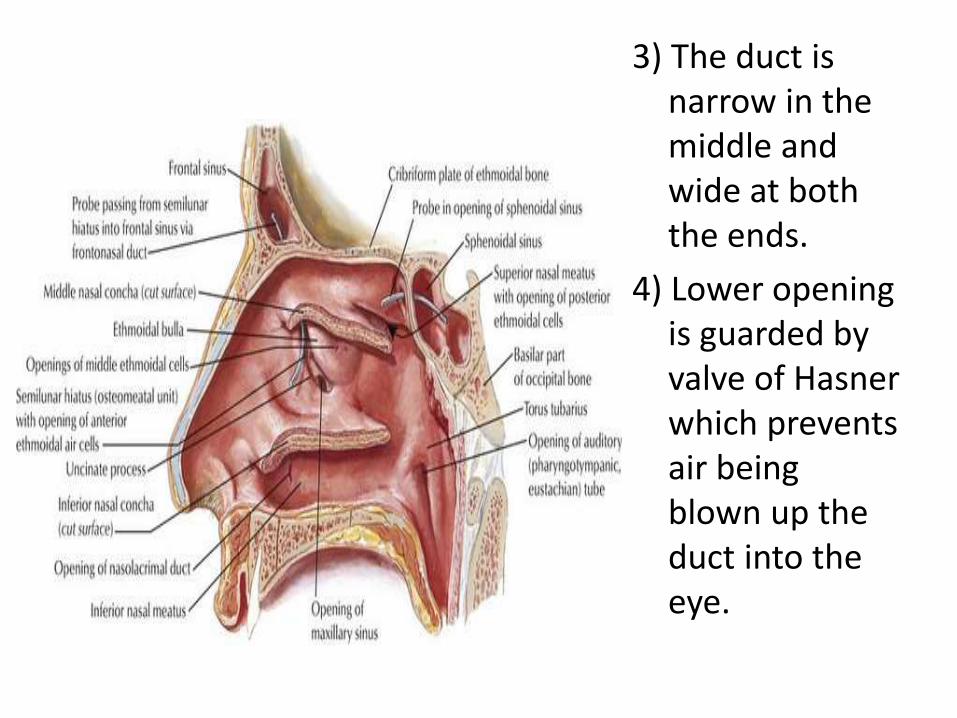
3) The duct is narrow in the middle and wide at both the ends.
4) Lower opening is guarded by valve of Hasnerwhich prevents air being blown up the duct into the eye.

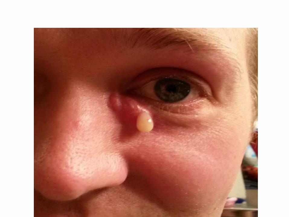
• Inflammation of the lacrimal sac due to obstruction of the nasolacrimal duct, leading to retention of mucus and tears in the sac .


![[PPT]Osteon (Haversian) System - Lone Star College – Start … · Web viewLacrimal Apparatus Lacrimal gland Canaliculi Lacrimal sac Conjunctiva Cornea Anterior cavity w/ Aqueous](https://img.pdfslide.us/doc/110x75/5ae7f9f47f8b9acc268f6a98/pptosteon-haversian-system-lone-star-college-start-viewlacrimal-apparatus.jpg)