Embed Size (px)
Citation preview
ISBN 978-92-2-124541-4
9 789221 245414
GUIDELINES FOR THE USE OF THE ILO INTERNATIONAL CLASSIFICATION OF RADIOGRAPHS OF PNEUMOCONIOSES
(REVISED EDITION 2011)
22Occupational Safety and Health
Series
GUIDELINES FOR THE USE OF THE ILO INTERNATIONAL CLASSIFICATION OF RADIOGRAPHS OF PNEUMOCONIOSES
(REVISED EDITION 2011)
In the continuing struggle to protect the health of workers occupationally exposed to airborne dusts, the ILO has for many years sought to improve the understanding of pneumoconiosis problems. The Guidelines for the use of the ILO International Classifi cation of Radiographs of Pneumoconioses is the latest version of a well-established publication designed to standardize classifi cation methods and facilitate international comparisons of pneumoconiosis statistics and research reports.
This revised edition of the Guidelines supplements the preceding 2000 edition with an entirely new Chapter 6. This chapter extends the applicability of the ILO scheme to classifi cations of results from digital radiographic images of the chest. The ILO Standard Digital Images (ILO 2011-D), which derive from the ILO (2000) standard radiographs, have been produced for this purpose.
The new text in Chapter 6 identifi es principles for viewing digitally acquired images of the chest and covers effective acquisition, display and storage of digital images. The Foreword to this revised edition defi nes the nomenclature used to distinguish different types of chest images.
The earlier (2000) Guidelines for classifi cation of conventionally acquired “fi lm-screen” radiography remain applicable. The relevant text from the earlier edition is reproduced in this edition, and the associated sets of standard radiographs remain available from the ILO.
OCCUPATIONAL SAFETY AND HEALTH SERIES No. 22 (Rev. 2011)
GUIDELINES FOR THE USEOF THE ILO INTERNATIONAL
CLASSIFICATION OF RADIOGRAPHS OF PNEUMOCONIOSES
Revised edition 2011
INTERNATIONAL LABOUR OFFICE · GENEVA
Copyright © International Labour Organization 2011First published 2011
Publications of the International Labour Offi ce enjoy copyright under Protocol 2 of the Universal Copyright Convention. Nevertheless, short excerpts from them may be reproduced without authorization, on condition that the source is indicated. For rights of reproduction or translation, application should be made to ILO Publica-tions (Rights and Permissions), International Labour Offi ce, CH-1211 Geneva 22, Switzerland, or by email: [email protected]. The International Labour Offi ce welcomes such applications.
Libraries, institutions and other users registered with reproduction rights organizations may make copies in ac-cordance with the licences issued to them for this purpose. Visit www.ifrro.org to fi nd the reproduction rights organization in your country.
ILOGuidelines for the use of the ILO International Classification of Radiographs of PneumoconiosesRevised edition 2011Geneva, International Labour Office, 2011
pneumoconiosis/medical examination/standardization
15.04.2
1 v. (Occupational safety and health series; 22 (Rev.2011))
ISBN 978-92-2-124541-4 (print)ISBN 978-92-2-124542-1 (pdf)ISBN 978-92-2-125049-4 (complete set)
ILO Cataloguing in Publication Data
The designations employed in ILO publications, which are in conformity with United Nations practice, and the presentation of material therein do not imply the expression of any opinion whatsoever on the part of the International Labour Offi ce concerning the legal status of any country, area or territory or of its authorities, or concerning the delimitation of its frontiers.
The responsibility for opinions expressed in signed articles, studies and other contributions rests solely with their authors, and publication does not constitute an endorsement by the International Labour Offi ce of the opinions expressed in them.
Reference to names of fi rms and commercial products and processes does not imply their endorsement by the International Labour Offi ce, and any failure to mention a particular fi rm, commercial product or process is not a sign of disapproval.
ILO publications and electronic products can be obtained through major booksellers or ILO local offi ces in many countries, or direct from ILO Publications, International Labour Offi ce, CH-1211 Geneva 22, Switzer-land. Catalogues or lists of new publications are available free of charge from the above address, or by email: [email protected]
Visit our website: www.ilo.org/publns
Photocomposed in Switzerland BRIPrinted in Switzerland ART
Contents
Foreword to the 2011 revised edition vii
Foreword to the 2000 revised edition ix
1. Introduction 1
2. General instructions 2
3. Specific instructions for use of the Complete Classification 3 3.1. Technical quality 3
3.2. Parenchymal abnormalities 3
3.3. Pleural abnormalities 6
3.4. Symbols 8
3.5. Comments 9
4. Specific instructions for the use of the Abbreviated Classification 10
5. Using the ILO Classification 12
6. Using the ILO Classification to classify digital radiographic images of the chest 14
7. Appendices 18 A. A note on technical quality for chest radiographs of dust-exposed workers 19
B. Reading sheets 21
C. Description of standard radiographs 27
D. Diagrams 35
E. Summary of details of the ILO (2000) International Classification of Radiographs of Pneumoconioses 39
F. Participants in ILO-convened meetings leading to the revised (2000 and 2011) editions of the Classification 43
v
vii
FOREWORD
Foreword to the Revised Edition (2011)
This revised (2011) edition of the Guidelines for the use of the ILO International Classification of Radiographs of Pneumoconioses extends the applicability of the Classification to digital radiographic images of the chest, as described in a new chapter 6 (page 14).
Chapters 1 through 5 are identical to those that appeared in the preceding (2000) edition of the Guidelines. That text remains applicable as written for classifying conven-tional film-screen radiographs and the associated sets of ILO standard radiographs remain available from the ILO.
Note that in chapter 6:• the word “image” refers to both film-based and soft copy images;• “ILO standard chest image” refers to both electronic and film-based versions
of standard images provided by the ILO;• “ILO 2000” or “ILO standard radiograph” refer to standard films distributed
by the ILO since the year 2000;• “ILO 2011-D” or “ILO Standard Digital Image” refer to images derived from
the ILO (2000) standard radiographs that have been distributed by the ILO in electronic format since the year 2011.
The ILO is grateful to 14 experts from seven countries who participated in a two-day scientific meeting in Rockville, Maryland, United States, on 13 and 14 March 2008 (see Appendix F). Their enthusiastic and intensive work during the meeting and in preceding months enabled publication of this revised (2011) edition of the Guidelines for the use of the ILO International Classification of Radiographs of Pneumoconioses.
Foreword to the Revised Edition (2000)
Over the last seven decades the International Labour Office (ILO) has promoted discussion and published a series of guidelines on how to classify chest radiographs of persons with pneumoconioses. The goals have been to standardize classification methods and facilitate international comparisons of data on pneumoconioses, epidemiological investigations and research reports. This revised edition of the ILO’s International Clas-sification of Radiographs of Pneumoconioses is a further effort towards these objectives. Based on the principles that governed the development of earlier editions of the Classifica-tion (those of 1950, 1958, 1968, 1971 and 1980), it refers to radiological appearances seen in all types of pneumoconioses. The description of the scheme in this revision of the Guidelines is more concise than previously. Some ambiguities in earlier editions have been clarified further, and the conventions for classifying pleural abnormalities have been revised. The changes are based on a comprehensive review of experience in using the preceding (1980) edition of the Classification.
The ILO initiated the review process in November 1989 at a meeting of 11 experts from seven countries. Participants were asked to advise on the kind of changes to the scheme that might be desirable, and to reconsider the suitability of the standard radiographs that accompanied the 1980 edition. Some parts of the Guidelines were identified as requiring revision, but the importance of continuity in the Classification was re-emphasized. With this in mind, it was agreed that the set of standard radiographs that were distributed with the 1980 edition should be retained, although it was recognized that the technical quality of many of them was inferior to that available with modern equipment and techniques. Participants in the meeting also suggested that the number of radiographs included in the complete set of standards (22) might be usefully reduced by reproducing critical parts from some of them onto quadrant sections of full-size radiographs. It was agreed, however, that it was necessary to verify that such a reform would not, in itself, result in a change in the way that radiographs of persons exposed to dust were classified. A controlled trial was therefore arranged by the ILO and the Division of Respiratory Disease Studies of the United States National Institute for Occupational Safety and Health (NIOSH). This involved 40 physicians, working at specialized clinical and research centres in ten coun-tries (see Appendix F).
Results from the trial showed that the proposed modification to the ILO standard radiographs, involving reproduction of sections from 15 of the ILO (1980) standards onto five new “ quadrant ” radiographs, would not increase variability between readers, and might improve the reproducibility of small-opacity profusion classification in some respects, but could also reduce slightly the frequency with which some readers identify large opacities. Use of the standards containing the quadrant radiographs was associated with an increase in the frequency with which some readers described the shapes of the small opacities that they saw as predominantly irregular, rather than rounded. It was
ix
concluded, however, that the effects found were unlikely to be distinguishable from inter- and intra-reader variability in most occupational health survey situations. 1
In October 1997 more than 200 participants in the Ninth International Confer-ence on Occupational Respiratory Diseases in Kyoto, Japan, attended an ILO-convened Working Group on the Classification. That meeting recommended further work on the development of quadrant or sectional composite radiographs and improved techniques for standard radiograph reproduction prior to the introduction of revised standard radiographs. A smaller group of experts attending the same conference then considered in detail a draft revised text of the Guidelines to the Classification. Discussion of this draft continued at a further meeting in March 1998 at the offices of the American College of Radiology (ACR) in Reston, Virginia, and was concluded on 26 October 2000 at the ILO Branch Office in Washington, DC. Participants in the latter meeting also compared two types of new copies of several sets of ILO (1980) standard radiographs, of sectional quadrant radiographs that had been used in the international trial, and of a newly prepared composite radiograph illustrating pleural abnormalities. The new copies that were under review were produced from earlier copies, both by standard film copying methods and by improved techniques from digitized versions of the earlier copies. The experts preferred the copies made from the digitized versions, and they recommended the use of this technology and the associ-ated reproduction process for producing future copies of ILO standard radiographs. The individuals who attended the various ILO-convened meetings concerned with the revision of the Classification are listed in Appendix F.
The ILO (2000) International Classification of Radiographs of Pneumoconioses is accompanied by two sets of standard radiographs, as described in Appendix C. Both sets are available from the ILO. The first (“ Complete ”) Set consists of 22 radiographs. Twenty of them are new copies from digitized full-size standard radiographs distributed previously with the 1980 edition of the ILO Classification. A further radiograph illustrates u/u-sized irregular opacities. Three quadrants of this radiograph reproduce the sections of the composite radiograph that was used in 1980 to depict increasing profusion of u/u-sized irregular opacities ; the fourth quadrant illustrates subcategory 0/0. A new composite radiograph is provided to illustrate pleural abnormalities.
The “ Quad ” Set consists of 14 radiographs. Nine of them are the most commonly used standards from the Complete Set. The other five reproduce (quadrant) sections of the remaining radiographs in the Complete Set.
The development of this revised (2000) edition of the Guidelines for the Use of the ILO International Classification of Radiographs of Pneumoconioses has been made possible by virtue of intensive and sustained activity on the part of many individuals and organizations. Some of them are named in Appendix F. Others, too numerous to list, provided valuable comments and suggestions in writing and by contributing to discussions at various scientific meetings, including four ILO international conferences on pneumo-conioses and occupational lung diseases (Bochum, Germany, 1983 ; Pittsburgh, Pennsyl-vania, 1987 ; Prague, 1992 ; and Kyoto, 1997). The ILO wishes to express its sincere thanks to all concerned, and to acknowledge gratefully the active assistance from the Committee on Pneumoconiosis (previously the Task Force on Pneumoconiosis) of the American College of Radiology (ACR), the United States National Institute for Occupa-tional Safety and Health (NIOSH), the Rosai Hospital for Silicosis in Japan, the WHO
GUIDELINES FOR THE USE OF RADIOGRAPHS OF PNEUMOCONIOSES
x
1 A trial of additional composite standard radiographs for use with the ILO International Classifica-tion of Radiographs of Pneumoconioses, NIOSH Report No. HETA 93-0340, July 1997, available from National Technical Information Service (NTIS), 5825 Port Royal Road, Springfield, Virginia 2216, United States. A shorter report has been published : “ New composite (“ Quadrant ”) standard films for classifying radiographs of pneumoconioses ”, in Industrial Health, Vol. 36, No. 4, Oct. 1998, pp. 380–383.

Collaborating Centre for Radiological Education in Sweden, the Finnish Institute of Occupational Health, the German Committee for Diagnostic Radiology of Occupational and Environmental Diseases, and the Institute for Occupational and Social Medicine of the University of Cologne. Continuing use of the ILO International Classification of Radiographs of Pneumoconioses will contribute further to the protection of the health of workers in dusty occupations.
FOREWORD
xi

1
Introduction
Scope of the Classification
The Classification provides a means for describing and recording systematically the radiographic abnormalities in the chest provoked by the inhalation of dusts. It is used to describe radiographic abnormalities that occur in any type of pneumoconiosis and is designed for classifying only the appearances seen on postero-anterior chest radiographs. Other views and imaging techniques may be required for clinical assessment of individuals, but the ILO International Classification has not been designed to code such findings.
Object of the Classification
The object of the Classification is to codify the radiographic abnormalities of the pneumoconioses in a simple, reproducible manner. The Classification neither defines pathological entities nor takes into account working capacity. It does not imply legal defin itions of pneumoconioses for compensation purposes and does not set or imply a level at which compensation is payable.
Uses of the Classification
The Classification is used internationally for epidemiological research, for screening and surveillance of those in dusty occupations, and for clinical purposes. Use of the scheme may lead to better international comparability of data concerning the pneumoconioses.
Standard radiographs and written definitions
The Classification consists of a set of standard radiographs and this text, with the accompanying footnotes. These footnotes are intended to reduce ambiguity and are based on experience with earlier editions of the ILO Classification. In some parts of the scheme, the standard radiographs take precedence over the written definitions. The text makes it clear when this is so.
1
2
General instructions
No radiographic features are pathognomonic of dust exposure. Some radio-graphic features that are unrelated to inhaled dust may mimic those caused by dust. Readers may differ about the interpretation of such appearances.
In epidemiological studies, therefore, the study protocol will usually require that all appearances described in these Guidelines and seen on the standard radiographs are to be classified. Symbols must always be used and appropriate Comments must be reported. 1
When the Classification is used for some clinical purposes, the protocol may require that medical readers classify only those appearances which the reader believes or suspects to be pneumoconiotic in origin. Symbols must always be used and appropriate Comments must be reported. 1
2
1 See sections 3.4 and 3.5.
3
Specific instructions for use of the Complete Classification
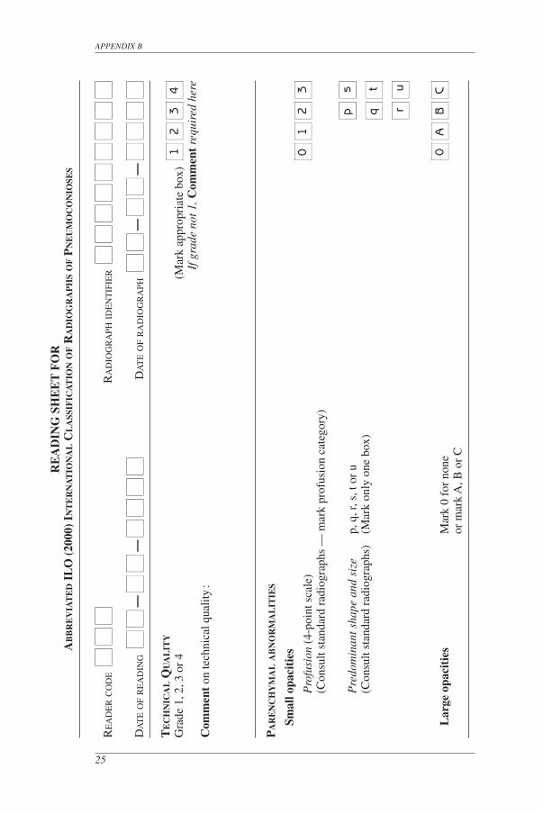
3.1. Technical quality 2, 3
Four grades of technical quality are used :1. Good.2. Acceptable, with no technical defect likely to impair classification of the
radio graph for pneumoconiosis.3. Acceptable, with some technical defect but still adequate for classification
purposes.4. Unacceptable for classification purposes.
If technical quality is not grade 1, a Comment must be made about the technical defects.
3.2. Parenchymal abnormalities
Parenchymal abnormalities include both small opacities and large opacities.
Small opacitiesSmall opacities are described by profusion, affected zones of the lung, shape
(rounded or irregular) and size. The order of identifying and recording the presence or absence of these findings while classifying a radiograph is left to the reader’s preference.
ProfusionThe profusion of small opacities refers to the concentration of small opacities in
affected zones of the lung. The category of profusion is based on comparisons with the standard radiographs. For profusion the written descriptions are a guide, but the standard
3
2 Appendix A emphasizes the importance of good radiographic quality for the interpretation of chest radiographs. It is essential to produce radiographs that show clearly both the parenchyma and the pleural char-acteristics. For clinical purposes, special views or techniques may also be required. When it is not possible to replace a grade 3 radiograph by a better one, more details about technical defects should be recorded.
3 The standard radiographs are not to be considered in determining technical quality of the subject radiographs. The standard radiographs were chosen to demonstrate the radiographic features of the pneumo-conioses, rather than to demonstrate technical quality.
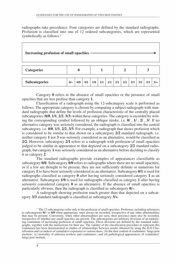
radiographs take precedence. Four categories are defined by the standard radiographs. Profusion is classified into one of 12 ordered subcategories, which are represented symbolically as follows. 4
Increasing profusion of small opacities
Categories 0 1 2 3
Subcategories 0/– 0/0 0/1 1/0 1/1 1/2 2/1 2/2 2/3 3/2 3/3 3/+
Category 0 refers to the absence of small opacities or the presence of small opacities that are less profuse than category 1.
Classification of a radiograph using the 12-subcategory scale is performed as follows. The appropriate category is chosen by comparing a subject radiograph with stan-dard radiographs that define the levels of profusion characteristic of the centrally placed subcategories (0/0, 1/1, 2/2, 3/3) within these categories. The category is recorded by writ-ing the corresponding symbol followed by an oblique stroke, i.e. 0/ , 1/ , 2/ , 3/. If no alternative category was seriously considered, the radiograph is classified into the central subcategory, i.e. 0/0, 1/1, 2/2, 3/3. For example, a radiograph that shows profusion which is considered to be similar to that shown on a subcategory 2/2 standard radiograph, i.e. neither category 1 nor 3 was seriously considered as an alternative, would be classified as 2/2. However, subcategory 2/1 refers to a radiograph with profusion of small opacities judged to be similar in appearance to that depicted on a subcategory 2/2 standard radio-graph, but category 1 was seriously considered as an alternative before deciding to classify it as category 2.
The standard radiographs provide examples of appearances classifiable as subcategory 0/0. Subcategory 0/0 refers to radiographs where there are no small opacities, or if a few are thought to be present, they are not sufficiently definite or numerous for category 1 to have been seriously considered as an alternative. Subcategory 0/1 is used for radiographs classified as category 0 after having seriously considered category 1 as an alternative. Subcategory 1/0 is used for radiographs classified as category 1 after having seriously considered category 0 as an alternative. If the absence of small opacities is particularly obvious, then the radiograph is classified as subcategory 0/–.
A radiograph showing profusion much greater than that depicted on a subcat-egory 3/3 standard radiograph is classified as subcategory 3/+.
GUIDELINES FOR THE USE OF RADIOGRAPHS OF PNEUMOCONIOSES
4
4 The 12 subcategories refer only to the profusion of small opacities. Profusion, including references to subcategories 0/– or 0/0 when appropriate, must always be recorded, irrespective of any other abnormalities that may be present. Conversely, when other abnormalities are seen, their presence must also be recorded, ir respective of whether any small opacities are present. The subcategories are arbitrary divisions of an underly-ing continuum of increasing profusion of small opacities. Those divisions are defined by the standard radio-graphs, together with the instructions for their use. The validity of the classification procedure to represent this continuum has been demonstrated in studies of relationships between results obtained by using the ILO Clas-sification and (a) indices of cumulative exposures to various dusts ; (b) the dust content of coalminers’ lungs post mortem ; (c) mortality of asbestos workers and coalminers ; and (d) pathological appearances of coalminers’ lungs post mortem.
Affected zonesThe zones in which the opacities are seen are recorded. Each lung field is divided
into three zones (upper, middle, lower) by horizontal lines drawn at approximately one-third and two-thirds of the vertical distance between the lung apices and the domes of the diaphragm.
The overall profusion of small opacities is determined by considering the profu-sion as a whole over affected zones of the lungs. When there is a marked (three subcat-egories or more) difference in profusion in different zones of the lungs, then the zone or zones showing the marked lesser degree of profusion is/are ignored for the purpose of classifying the overall profusion. 5
Shape and sizeFor shape and size, the written definitions are a guide, and the standard radio-
graphs take precedence. The shape and size of small opacities are recorded. Two kinds of shape are recognized : rounded and irregular. In each case, three sizes are differentiated.
For small rounded opacities, the three size ranges are denoted by the letters p, q and r, and are defined by the appearances of the small opacities on the corresponding standard radiographs. These illustrate :
p-opacities with diameters up to about 1.5 mm ;q-opacities with diameters exceeding about 1.5 mm and up to about 3 mm ;r-opacities with diameters exceeding about 3 mm and up to about 10 mm.The three size ranges of small irregular opacities are denoted by the letters s, t
and u, and are defined by the appearances of the small opacities on the corresponding standard radiographs. These illustrate :
s-opacities with widths up to about 1.5 mm ;t-opacities with widths exceeding about 1.5 mm and up to about 3 mm ;u-opacities with widths exceeding about 3 mm and up to about 10 mm.
INSTRUCTIONS FOR USE OF THE COMPLETE CLASSIFICATION
5
5 A “ marked (three subcategories or more) difference ” in profusion in different zones of the lung is present when there are two or more subcategories of profusion between the zone (or zones) of the lowest profu-sion and the zone (or zones) of the highest profusion. For example, if a subject radiograph displays zones with profusion levels 1/1, 1/2, 2/1 and 2/2, the overall profusion is determined by ignoring the zone with profusion level 1/1, since two or more subcategories (1/2, 2/1) are between that zone and the zone of the highest profusion (2/2). The overall profusion, therefore, is determined by considering only the affected zones showing profusion levels 1/2, 2/1 and 2/2, since there is only one subcategory of profusion (2/1) between profusion levels 1/2 and 2/2.Example 1Only one intervening subcategory between the zones of lowest (1/2) and highest (2/2) profusion ; use all three to determine overall profusion. ▼ ▼1/1 1/2 2/1 2/2 ▲ ▲There are two intervening subcategories between the zones of lowest (1/1) and highest (2/2) profusion ; ignore 1/1 to determine overall profusion.Example 2Only one intervening subcategory between the zones of lowest (2/1) and highest (2/3) profusion ; use all three to determine overall profusion. ▼ ▼1/1 1/2 2/1 2/2 2/3▲ ▲There are three intervening subcategories between the zones of lowest (1/1) and highest (2/3) profusion ; ignore 1/1 and 1/2 ; use 2/1, 2/2, 2/3 to determine overall profusion since there is only one subcategory between 2/1 and 2/3.All zones in which opacities are seen are recorded, irrespective of whether some are later ignored in determining overall profusion.
Two letters must be used to record shape and size. Thus, if the reader considers that all, or virtually all, opacities seen are of one shape and size, then this is noted by recording the letter twice, separated by an oblique stroke (for example q/q). If, however, significant numbers of another shape or size are seen, then this is recorded by writing a different letter after the oblique stroke (for example q/t) ; q/t would mean that the predom-inant small opacities are rounded and of size q, but that there are significant numbers of small irregular opacities present of size t. In this way, any combination of small opacities may be recorded. 6 When small opacities of different shapes and/or size are seen, the letter for the predominant shape and size (primary) is recorded before the oblique stroke, while the letter for the less frequently occurring shape and size (secondary) is recorded after the oblique stroke.
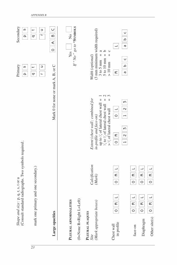
Large opacitiesA large opacity is defined as an opacity having the longest dimension exceeding
10 mm. Categories of large opacities are defined below. These definitions take precedence over the examples of large opacities illustrated on standard radiographs.Category A One large opacity having the longest dimension up to about 50 mm, or
several large opacities with the sum of their longest dimensions not exceed-ing about 50 mm.
Category B One large opacity having the longest dimension exceeding 50 mm but not exceeding the equivalent area of the right upper zone, or several large opac-ities with the sum of their longest dimensions exceeding 50 mm but not exceeding the equivalent area of the right upper zone.
Category C One large opacity which exceeds the equivalent area of the right upper zone, or several large opacities which, when combined, exceed the equivalent area of the right upper zone.
3.3. Pleural abnormalities
Pleural abnormalities are divided into pleural plaques (localized pleural thicken-ing), costophrenic angle obliteration and diffuse pleural thickening.
Pleural plaques (localized pleural thickening)Pleural plaques represent localized pleural thickening, generally of the parietal
pleura. Pleural plaques may be seen on the diaphragm, on the chest wall (in-profile or face-on), and at other sites. At times, they are recognized only by their calcification. Pleu-ral plaques are recorded as absent or present. If present on the chest wall, they are recorded as in-profile or face-on, and separately for the right and left sides. A minimum width of about 3 mm is required for an in-profile plaque to be recorded as present. 7, 8
GUIDELINES FOR THE USE OF RADIOGRAPHS OF PNEUMOCONIOSES
6
6 See Appendix E for possible combinations.7 The measurement of width is made from the innermost margin of the rib to the innermost sharp
margin of the plaque at the pleural-parenchymal boundary.8 If more detailed measurement of width is required for a particular study, three categories may be used :
a – about 3 mm up to about 5 mm ;b – about 5 mm up to about 10 mm ;c – over about 10 mm.
Site, calcification and extent of pleural plaques are recorded separately for the right and for the left side of the chest. The written guidelines describing these features take precedence over the examples provided on the standard radiograph.
SiteThe sites (locations) of pleural plaques include chest wall, diaphragm and other
sites. Other sites include the mediastinal pleura in the para-spinal or para-cardiac loca-tions. The presence or absence of pleural plaques is recorded for all sites, and separately for the right and for the left sides.
CalcificationRadiographic images of pleural plaques may include recognizable areas of calci-
fication. The presence or absence of calcification is recorded for all plaques, and separ-ately for the right and for the left sides. When calcification is seen, a plaque is also recorded as present at that site.
ExtentExtent is not recorded for plaques on the diaphragm or at other sites. It is recorded
only for plaques along the chest wall, and is combined for both in-profile and face-on varieties. Extent is defined in terms of the total length of involvement with respect to the projection of the lateral chest wall (from the apex to the costophrenic angle) on the poste-ro–anterior chest radiograph :
1 = total length up to one-quarter of the projection of the lateral chest wall ;2 = total length exceeding one-quarter and up to one-half of the projection of
the lateral chest wall ;3 = total length exceeding one-half of the projection of the lateral chest wall.
Costophrenic angle obliterationCostophrenic angle obliteration is recorded as either present or absent, sepa-
rately for the right and for the left side. The lower limit for recording costophrenic angle obliteration is defined by the standard radiograph showing profusion subcategory 1/1 t/t. If the pleural thickening extends up the lateral chest wall from the obliterated costophrenic angle, the thickening should be classified as diffuse pleural thickening. Costophrenic angle obliteration may occur without diffuse pleural thickening.
Diffuse pleural thickeningDiffuse pleural thickening historically has referred to thickening of the visceral
pleura. The radiological distinction between parietal and visceral pleural thickening is not always possible on a postero–anterior radiograph.
For the purpose of the ILO (2000) Classification, diffuse pleural thickening extending up the lateral chest wall is recorded only in the presence of, and in continuity with, an obliterated costophrenic angle. Diffuse pleural thickening is recorded as absent or present along the chest wall. If present, it is recorded as in-profile or face-on, and sep arately for the right and the left side. Its extent is recorded in the same manner as for pleural plaques. A minimum width of about 3 mm is required for in-profile diffuse pleural
INSTRUCTIONS FOR USE OF THE COMPLETE CLASSIFICATION
7
thickening to be recorded as present. If detailed measurement of its width is required for a particular study, see the comment provided in footnote 8.
Calcification and extent of diffuse pleural thickening on the chest wall are recorded separately for the right and for the left side (see guidelines for pleural plaques). The pleura may often be seen at the apex of the lung and should not be recorded as part of diffuse pleural thickening of the chest wall.
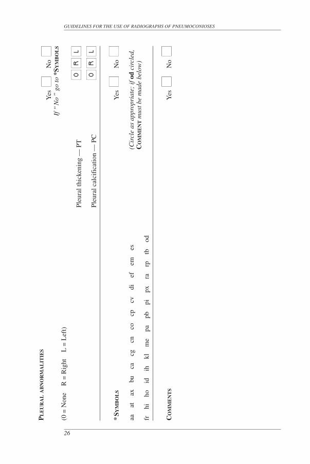
3.4. Symbols
Symbols to record radiographic features of importance are listed below. Their use is relevant because they describe additional features related to dust exposure and other aetiologies. Use of these symbols is obligatory. 9
Some of the symbols imply interpretations, rather than just descriptions, of what is seen on the radiograph. A postero–anterior chest radiograph on its own may not be suf ficient to justify definitive interpretation ; therefore, each of the following definitions of symbols assumes an introductory qualifying word or phrase such as “ changes indica-tive of ”, or “ opacities suggestive of ”, or “ suspect ”.
The symbols are :aa atherosclerotic aorta at significant apical pleural thickening (see Appendix D)ax coalescence of small opacities 10
bu bulla(e)ca cancer : thoracic malignancies excluding mesothelioma cg calcified non-pneumoconiotic nodules (e.g. granuloma) or nodescn calcification in small pneumoconiotic opacitiesco abnormality of cardiac size or shapecp cor pulmonalecv cavitydi marked distortion of an intrathoracic structureef pleural effusionem emphysemaes eggshell calcification of hilar or mediastinal lymph nodesfr fractured rib(s) (acute or healed)hi enlargement of non-calcified hilar or mediastinal lymph nodesho honeycomb lungid ill-defined diaphragm border 11
ih ill-defined heart border 12
kl septal (Kerley) linesme mesothelioma
GUIDELINES FOR THE USE OF RADIOGRAPHS OF PNEUMOCONIOSES
8
9 Inclusion of this information in statistical analyses of results may help to explain otherwise in explicable variation between readers in their classifications of the same radiographs.
10 The symbol ax represents coalescence of small opacities with margins of the small opacities remaining visible, whereas a large opacity demonstrates a homogeneous opaque appearance.The symbol ax (coalescence of small opacities) may be recorded either in the presence or in the absence of large opacities.
11 The symbol id (ill-defined diaphragm border) should be recorded only if more than one-third of one hemidiaphragm is affected.
12 The symbol ih (ill-defined heart border) should be recorded only if the length of the heart border affected, whether on the right or on the left side, is more than one-third of the length of the left heart border.
pa plate atelectasispb parenchymal bands 13
pi pleural thickening of an interlobar fissure 14
px pneumothoraxra rounded atelectasisrp rheumatoid pneumoconiosis 15
tb tuberculosis 16
od other disease or significant abnormality 17
3.5. Comments
If the technical quality of the radiograph is not recorded as 1 (good), then a Comment on why this is so should be made at that time, before proceeding with the clas-sification.
Comments are also required if the symbol od (other disease) is recorded, and to identify any part of the reading of a chest radiograph which is believed by a reader to be probably or certainly not dust related.
Comments should also be recorded to provide other relevant information.
INSTRUCTIONS FOR USE OF THE COMPLETE CLASSIFICATION
9
13 Significant parenchymal fibrotic strands in continuity with the pleura.14 Illustrated on the 3/3 s/s standard radiograph.15 Illustrated on the 1/1 p/p standard radiograph.16 The symbol tb should be used for either suspect active or suspect inactive tuberculosis. The
symbol tb should not be used for the calcified granuloma of tuberculosis or other granulomatous processes, e.g. histoplasmosis. Such appearances should be recorded as cg.
17 If the symbol od is used, then an explanatory Comment must be made.
4
Specific instructions for the use of the Abbreviated Classification
The Abbreviated Classification, described below, is a simplified version of the Complete Classification and includes its major components.
Technical quality
The recording of the technical quality of the radiograph is the same as for the Complete Classification (see section 3.1).
Small opacities
Profusion is determined by comparison with standard radiographs and recorded as one of the categories : 0, 1, 2 or 3 (see section 3.2).
Shape and size are determined by comparison with standard radiographs. The predominant shape and size are recorded using only one of the following letters : p, q, r, s, t or u (see section 3.2).
Large opacities
Large opacities are recorded as size A, B or C, in the same way as for the Complete Classification (see section 3.2).
Pleural abnormalities
All types of pleural thickening are recorded by the letters PT.All types of pleural calcifications are recorded by the letters PC.
10

Symbols
Symbols are recorded as for the Complete Classification (see section 3.4).
Comments
Comments are recorded as for the Complete Classification (see section 3.5).
INSTRUCTIONS FOR USE OF THE ABBREVIATED CLASSIFICATION
11
5
Using the ILO Classification
Efficient use of the ILO Classification requires good viewing and recording conditions. The following recommendations are particularly important for epidemiological studies.
Viewing
The illuminated boxes for viewing the radiographs to be classified and the stan-dard radiographs must be close enough for the observer to see opacities only 1 mm in diameter, that is, a distance of about 250 mm. It is also essential to view the entire radiograph. The observer should be seated comfortably.
The minimum number of viewing spaces is two, allowing comparisons between the subject radiograph and the standard radiographs. However, it is recommended that three viewing spaces be used, so that the subject radiograph can be placed between the appropriate standard radiographs to assess profusion. It is important to make it easy to select and put up the standard radiographs for comparison, which is mandatory.
The viewing surfaces must be clean and the intensity of illumination should be uniform over all surfaces. The general illumination in the room should be low, without direct daylight. The room should be quiet, comfortable and free from distractions.
Epidemiological reading protocols
When classifying radiographs for epidemiological purposes, it is essential that the reader does not consider any other information about the individuals being studied. Awareness of supplementary details specific to individuals can introduce bias into results. If the epidemiological objective is to make comparisons between two or more groups, then the radiographs from all groups should be mixed and presented to the reader in random order. Failure to observe these principles may invalidate conclusions from the study.
Recording
Recording of results should be standardized and systematic. It is important to make provision for recording explicitly the presence or absence of all features to be evaluated for a particular study. Clerical help for recording results is valuable when
12
classifying large numbers of radiographs. The clerical assistant should be asked to remind the reader of failure to report the presence or absence of any features to be analysed in the study.
Reading rates
The number of radiographs classifiable per unit of time can vary greatly. Factors influencing reading rates include the technical quality of the radiographs, the prevalence of abnormalities on the radiographs, the experience of the reader, the purpose of the read-ing exercise and the length of the reading session.
Number of readers
It is recognized that there is considerable variation in multiple readings of some radiographs, not only from reader to reader (inter-observer variation), but also between readings by the same reader (intra-observer variation). It is recommended that, in epide-miological studies, at least two, but preferably more, readers each classify all radiographs independently.
When many radiographs are being read, intra-observer variation, i.e. variation in repeated readings by the same reader, should be assessed.
USING THE ILO CLASSIFICATION
13
14
6
Using the ILO Classification to classify digital radiographic images of the chest
Purpose
The purpose of this chapter is to extend the applicability of the ILO International Classification of Radiographs of Pneumoconioses beyond conventional chest radiographs to digital radiographic images of the chest.
Introduction
Earlier editions of these Guidelines referred to postero-anterior chest radiographs and to comparisons of a subject’s radiograph with ILO standard radiographs. Both subject and standard radiographs were obtained using film-screen radiography (FSR)1. (Readers who have the film versions of the radiographs should note that the present revised Guide-lines apply equally to the film and digital versions.) The revised (2000) edition of the Guidelines recognized that other imaging techniques may be required for clinical purposes, but noted that the ILO Classification was not designed to code findings from the applica-tion of such methods. As anticipated at that time, methods for imaging the chest for lung diseases have continued to evolve. Major advances during recent years have included widespread application of digital techniques which, under appropriately controlled condi-tions, allow classification of chest abnormalities in a manner consistent with classifica-tions using FSR.
The remainder of this chapter provides guidance on how to classify digital images of the chest in a way that maintains continuity and consistency with the ILO (2000) Classification and its associated standard radiographs. The following paragraphs supplement earlier parts of this monograph which remain in effect for digital images, except as specifically noted below. Valid application of the ILO Classification to digital chest images requires users to adhere to all recommendations in this chapter, and to remain alert to future developments in digital chest imaging techniques.
1 FSR – Film-screen-radiography. (Synonyms include chest radiograph, chest x-ray, conventional film-screen radiography. See also the Foreword to the present edition of these Guidelines (page vii) for notes on the nomenclature used in this chapter.)
15
USING THE ILO CLASSIFICATION TO CLASSIFY DIGITAL RADIOGRAPHIC IMAGES OF THE CHEST
ILO Standard Digital Images
Standard digital images corresponding to the ILO (2000) standard radiographs are now available from the ILO on electronic storage media. These ILO Standard Digital Images (ILO 2011 -D) define and illustrate the shapes, sizes and profusion of small opac-ities visible on digital images. The corresponding text in Chapter 3, above, provides supplementary information, but the appearances of the standard digital images take prece-dence for classification purposes. On the other hand, the standard digital images of large opacities and of pleural abnormalities represent examples of those features. Their size, location and other characteristics are defined in Chapter 3, and for classification purposes that text takes precedence.
Viewing principles
When viewing either the film-screen radiographs or hard copies2 of digital images, valid classifications consistent with the ILO system require use of the ILO (2000) standard radiographs, as detailed in Chapters 3 through 5. Hard copies of digital images should not be reduced below two-thirds of a standard-sized image (14" x 17", or 35 cm x 43 cm).
When viewing and classifying a subject’s soft copy3 digital image, the ILO (2011-D) standard digital images must be used. Subject and standard digital images should be displayed on medical-grade flat-panel monitors designed for diagnostic radiology. The diagonal display should be at least 21" (54 cm) per image, with a ratio of maximum to minimum luminance at least 50; a maximum luminance of at least 250 candelas per m2; pixel pitch no greater than 210 μm; and with resolution at least 2.5 line-pairs per mm.4 The subject and standard images should be displayed simultaneously, similarly sized, side-by-side.
Examples of approaches not recommended for viewing soft copies include:• Displaying the images on a personal computer screen rather than on a medi-
cal-grade flat-panel monitor designed for diagnostic radiology;• Comparing the subject digital image to ILO (2000) standard radiographs
displayed on a view box;
2 Hard copy – A hard copy is a digital image printed on transparent film in an attempt to reproduce the x-ray image in its original quality.
3 Soft copy – A soft copy is a digital image displayed on a monitor. For classification purposes, digital images should be displayed on medical-grade flat-panel monitors designed for diagnostic radiology.
4 Definitions and further explanations of technical terms, as well as other information relevant to digital radiography, can be found in publications by professional associations, standard-setting organizations and other agencies, for instance:(a) American College of Radiology. ACR Technical Standard for Electronic Practice of Medical Imaging, Res.
13-2007. In ACR Practice Guidelines and Technical Standards, pp. 1053–1067. ACR, Reston, VA, 2007. (b) American College of Radiology. Practice Guidelines for Digital Radiography, Res. 42-2007. In ACR Practice
Guidelines and Technical Standards, pp. 23–57. ACR, Reston, VA, 2007.(c) European Guidelines on Quality Criteria for Diagnostic Radiographic Images. Report EUR 16260. Euro-
pean Commission. Luxembourg, Office for Official Publications of the European Communities, 1996.(d) The 1991 CEC Trial on quality criteria for diagnostic radiographic images: Detailed results and findings.
Report EUR 16635. European Commission. Luxembourg, Office for Official Publications of the European Communities, 1999.
(e) American Association of Physicists in Medicine. Assessment of Display Performance for Medical Imaging Systems. AAPM On-Line Report No. 03, Task Group 18. College Park, MD. April 2005.
GUIDELINES FOR THE USE OF RADIOGRAPHS OF PNEUMOCONIOSES
16
• Viewing the subject digital image, or the ILO (2011-D) standard digital images (or both) in formats reduced to less than two-thirds of their full size; and
• Using images printed on paper for classification.
As with all radiographic viewing activities, procedures should be followed to ensure an appropriate environment, including restriction of ambient light sources and other distracting factors. (See page 12 of these Guidelines.)
Acquisition, display, and storage of digital chest images
Radiographic systems for collecting, displaying, and storing chest images for classification of pneumoconiosis should adhere to the most recent version of the Digital Imaging and Communications in Medicine (DICOM) standards or other comparable stan-dards (for example, MEDICOM EN12052). The DICOM standards (http://medical.nema.org) are widely accepted and used internationally for radiographic imaging. They specify a common format for storage and transfer of digital images, and for brightness, contrast levels, and a consistent greyscale when presenting images on display monitors and printing digital images. Care should be taken to maintain image quality and consistency when producing hard copies of digital images.
Additional notes and recommendations5
Imaging equipmentHardware for production of digital chest images is manufactured and distributed
by many commercial entities with varying approaches to image capture. A group of systems based on storage-phosphor technology is described as computed radiography (CR). A second group of systems, known as digital radiography (DR), has become avail-able more recently. Both are acceptable for application of the ILO Classification, with appropriate attention to image quality.
Image acquisition and processingDigital radiographic systems vary in the approaches taken to address the display
quality of digital chest images. Image processing software continues to evolve. No specific recommendations are made by the ILO regarding the selection of digital systems. Perfor-mance testing and monitoring should be used to evaluate the ability to produce quality images for any combination of hardware, exposure parameters and software. Facilities providing images for classification should employ a program for continual quality assur-ance consistent with national practices and standards. Staff at facilities that perform digital chest radiography for pneumoconiosis classification should review each image to ensure optimal quality.
5 See footnote 4.
17
USING THE ILO CLASSIFICATION TO CLASSIFY DIGITAL RADIOGRAPHIC IMAGES OF THE CHEST
Image displayGood image quality is essential for accurate classification of digital chest radio-
graphs. Maintenance, assessment, and optimisation of the image display monitors and all other components of the digital radiographic systems should be undertaken periodically, as recommended or specified by manufacturers, professional organizations, or govern-mental agencies.6 While classifying digital images, the ILO (2011-D) Standard Digital Images should be displayed as provided, without alteration.7
Data recording, storage, and securityDigital images should be securely archived and transferred in a manner that
permits retrieval of their original appearance, in compliance with national practices and standards. Standard measures to prevent unauthorized access to data should be employed, for instance by password-protected access and rigorous security precautions for transfers through data networks.
6 For example, see (a) and (c) in footnote 4.7 The standard digital radiographs were chosen to demonstrate the radiographic features of the pneu-
moconioses, rather than technical quality.
7
Appendices
The appendices have been prepared by individual experts to assist understanding of the principles and development of the ILO International Classification. They are not part of the text of the ILO (2000) International Classification of Radiographs of Pneumo-conioses. The ILO wishes to express its gratitude to Dr. Kurt G. Hering, Dr. Yutaka Hosoda, Dr. Michael Jacobsen, Dr. Yukinori Kusaka, Mr. Otha W. Linton, Dr. John E. Parker, Dr. Anthony V. Proto, Dr. Hisao Shida, Dr. Gregory R.Wagner, Dr. Jerome F. Wiot and Dr. Anders Zitting for the preparation of the appendices.
18
Appendix A – A note on technical quality for chest radiographs of dust-exposed workers
It has long been recognized that the technique and equipment used for chest radiographic imaging of dust-exposed workers affect the radiographic appearance of pneumoconiotic lesions, and that this can influence the classification of a radiograph for pneumoconiosis. Both clinical interpretations of chest radiographs, and the use of the ILO Classification for medical screening, public health surveillance and epidemiological research, require good-quality radiographs. Consequently, readers may find it difficult to use the ILO Classification if the quality of chest radiographs is suboptimal. In some cases, it may be impossible to classify such a radiograph. Provision has been made for this contingency in section 3.1 of these Guidelines by the definition of technical quality grade 4 (“ unacceptable for classification purposes ”).
Common quality faults include underexposure (often associated with a tendency to read more profusion than would be recognized on an optimally produced radiograph) and overexposure (associated with the converse tendency). Experienced readers may sometimes adjust their assessments of such radiographs to compensate, to some extent, for these perceived technical faults. Nevertheless, physicians and radiographers should strive always to obtain good-quality radiographs.
An optimal radiographic technique for the assessment of pneumoconiosis should reveal the fine detail of parenchymal markings, demonstrate clearly the costal–pleural junctions and show vascular markings through the cardiac shadow. It should be noted, however, that good contrast, required to evaluate the pulmonary parenchyma, may be suboptimal for assessment of mediastinal structures.
Methods for imaging the chest for dust-related lung diseases continue to evolve as new technologies are introduced. In view of these ongoing developments, it would be inappropriate here to attempt to provide detailed technical advice on radiographic tech-nique and equipment. Authoritative information on these topics may be found in a number of specialist publications. A select bibliography is provided at the end of this appendix.
These Guidelines require that a decision on whether a radiograph is of good, or at least of acceptable, technical quality rests ultimately with the physician who classifies the radiograph. Therefore, a key general principle must be the establishment and main-tenance of good communication between the physician and the radiographer, so that high-quality images, providing an adequate view of the pulmonary parenchyma and pleura, are obtained. The radiographer must be well trained and supervised, and must work in a climate that invites dialogue with the physician/reader. The physician must provide feed-back to the radiographer to ensure improvement of any suboptimal images, and should be prepared to advise on quality control for the production of chest radiographs of dust-exposed workers. Physicians and radiographers should take cognizance of local regulations.
19
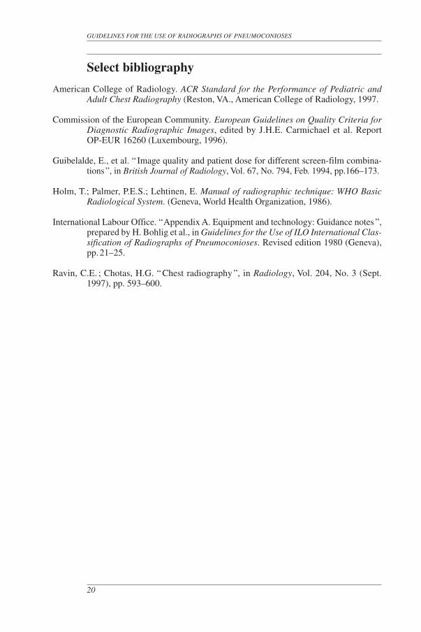
Select bibliography
American College of Radiology. ACR Standard for the Performance of Pediatric and Adult Chest Radiography (Reston, VA., American College of Radiology, 1997.
Commission of the European Community. European Guidelines on Quality Criteria for Diagnostic Radiographic Images, edited by J.H.E. Carmichael et al. Report OP-EUR 16260 (Luxembourg, 1996).
Guibelalde, E., et al. “ Image quality and patient dose for different screen-film combina-tions ”, in British Journal of Radiology, Vol. 67, No. 794, Feb. 1994, pp.166–173.
Holm, T.; Palmer, P.E.S.; Lehtinen, E. Manual of radiographic technique: WHO Basic Radiological System. (Geneva, World Health Organization, 1986).
International Labour Office. “ Appendix A. Equipment and technology: Guidance notes ”, prepared by H. Bohlig et al., in Guidelines for the Use of ILO International Clas-sification of Radiographs of Pneumoconioses. Revised edition 1980 (Geneva), pp. 21–25.
Ravin, C.E. ; Chotas, H.G. “ Chest radiography ”, in Radiology, Vol. 204, No. 3 (Sept. 1997), pp. 593–600.
GUIDELINES FOR THE USE OF RADIOGRAPHS OF PNEUMOCONIOSES
20
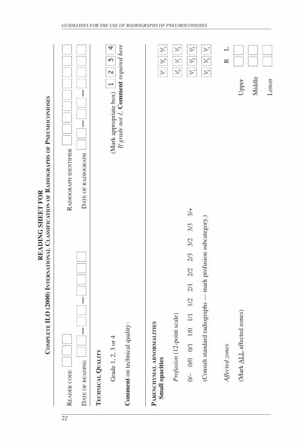
Appendix B – Reading sheets
The reading sheets on the following pages are examples of what may be used with the ILO (2000) International Classification of Radiographs of Pneumoconioses. In some situations, clinical or epidemiological, other designs may be preferred for specific uses. The sheets illustrated here make provision for recording all features described in the Complete Classification and the Abbreviated Classification. However, they are not a formal part of the ILO International Classification.
21
RE
AD
ING
SH
EE
T F
OR
CO
MP
LE
TE
IL
O (
2000
) IN
TE
RN
AT
ION
AL
CL
ASS
IFIC
AT
ION
OF R
AD
IOG
RA
PH
S O
F P
NE
UM
OC
ON
IOSE
S
RE
AD
ER
CO
DE ■
■■
■■
■
RA
DIO
GR
APH
IDE
NT
IFIE
R ■
■■
■■
■■
■■
■■
■■
■■
■■
■
DA
TE O
F R
EA
DIN
G ■
■■
■–
■■
■■
–■
■■
■■
■■
■
DA
TE O
F R
AD
IOG
RA
PH ■
■■
■–
■■
■■
–■
■■
■■
■■
■T
EC
HN
ICA
L Q
UA
LIT
Y
G
rade
1, 2
, 3 o
r 4
(Mar
k ap
prop
riat
e bo
x) ■
■■
■■
■■
■
If g
rade
not
1, C
omm
ent
requ
ired
her
eC
omm
ent
on te
chni
cal q
ualit
y :
PA
RE
NC
HY
MA
L A
BN
OR
MA
LIT
IES
Sm
all o
paci
ties
P
rofu
sion
(12
-poi
nt s
cale
)
0/
– 0
/0
0/1
1/0
1/
1 1
/2
2/1
2/2
2/
3 3
/2
3/3
3/+
(C
onsu
lt st
anda
rd r
adio
grap
hs —
mar
k pr
ofus
ion
subc
ateg
ory.
)
A
ffect
ed z
ones
R
L
(M
ark
AL
L a
ffec
ted
zone
s)
Upp
er
■■
■■
Mid
dle
■■
■■
Low
er
■■
■■
GUIDELINES FOR THE USE OF RADIOGRAPHS OF PNEUMOCONIOSES
22
1
2
3
4
■■
■■
■■
■■
■■
■■
■■
■■
■■
■■
■■
■■
0⁄ –
0⁄ 0
0⁄ 1
1⁄ 0
1⁄ 1
1⁄ 2
2⁄ 1
2⁄ 2
2⁄ 3
3⁄ 2
3⁄ 3
3⁄ +
Sh
ape
and
size
: p,
q, r
, s, t
or
u Pr
imar
y Se
cond
ary
(C
onsu
lt st
anda
rd r
adio
grap
hs. T
wo
sym
bols
req
uire
d ;
■■
■■
■
■■
■
mar
k on
e pr
imar
y an
d on
e se
cond
ary.
) ■
■■
■
■■
■■
■■
■■
■
■■
■
L
arge
opa
citi
es
Mar
k 0
for
none
or
mar
k A
, B, o
r C
■
■■
■■
■■
■
PL
EU
RA
L A
BN
OR
MA
LIT
IES
Yes
■■
No ■■
If
“ N
o ”
go
to *
SYM
BO
LS
(0=
Non
e R
=R
ight
L=
Lef
t)
PL
EU
RA
L P
LA
QU
ES
Site
C
alci
ficat
ion
Ext
ent (
ches
t wal
l ; c
ombi
ned
for
Wid
th (
opti
onal
)(M
ark
appr
opri
ate
boxe
s)
(Mar
k)
in-p
rofil
e an
d fa
ce-o
n)
(3
mm
min
imum
wid
th r
equi
red)
up
to 1 ⁄ 4
of
late
ral c
hest
wal
l =
1
3
to 5
mm
=
a
1 ⁄ 4 to
1 ⁄ 2 o
f la
tera
l che
st w
all
= 2
5 to
10
mm
= b
>
1 ⁄ 2 o
f la
tera
l che
st w
all
= 3
> 1
0 m
m
= c
Che
st w
all
in
pro
file
■
■■
■
■■
■
■
■■
■
■
■■
■
■■
■
■■
■
■
■
■
■■
■
■■
■
■■
■
■■
■■
■■
■
■■
■
■■
■
■■
fa
ce-o
n ■
■■
■
■■
■■
■
■■
■
Dia
phra
gm
■■
■
■■
■■
■■
■
■■
O
ther
site
(s)
■■
■
■■
■■
■■
■
■■
APPENDIX B
23
p
s
p
s
q
t
q
t
r
u
r
u
0
A
B
C
0
R
L
0
R
L 0
R
0
L
R
L
1
2
3
1
2
3
a
b
c a
b
c
0
R
L
0
R
L
0
R
L
0
R
L
0
R
L
0
R
L
CO
STO
PH
RE
NIC
AN
GL
E O
BL
ITE
RA
TIO
N ■
■■
■■
■
DIF
FU
SE P
LE
UR
AL
C
alci
ficat
ion
Ext
ent (
ches
t wal
l ; c
ombi
ned
for
Wid
th (
opti
onal
)T
HIC
KE
NIN
G
(Mar
k)
in-p
rofil
e an
d fa
ce-o
n)
(3
mm
min
imum
wid
th r
equi
red)
(Mar
k ap
prop
riat
e bo
xes)
up
to 1 ⁄ 4
of
late
ral c
hest
wal
l =
1
3
to 5
mm
=
a
1 ⁄ 4 to
1 ⁄ 2 o
f la
tera
l che
st w
all
= 2
5 to
10
mm
= b
>
1 ⁄ 2 o
f la
tera
l che
st w
all
= 3
> 1
0 m
m
= c
Che
st w
all
in
pro
file
■
■■
■
■■
■
■
■■
■
■
■■
■
■■
■
■■
■
■
■
■
■■
■
■■
■
■■
■
■■
■■
■■
■
■■
■
■■
■
■■
fa
ce-o
n ■
■■
■
■■
■■
■
■■
■
* SY
MB
OL
S Y
es ■■
No ■■
aa
at
ax
bu
ca
cg
cn
co
cp
cv
di
ef
em
es
(Cir
cle
as a
ppro
pria
te;
if o
d ci
rcle
d,
C
OM
ME
NT
mus
t be
mad
e be
low
)fr
hi
ho
id
ih
kl
m
e p
a p
b p
i p
x r
a r
p t
b o
d
CO
MM
EN
TS
Yes
■■
N
o ■■
GUIDELINES FOR THE USE OF RADIOGRAPHS OF PNEUMOCONIOSES
24
0
R
L 0
R
L
0
R
0
L R
L
1
2
3
1
2
3
a
b
c a
b
c
0
R
L 0
R
L
0
R
L
RE
AD
ING
SH
EE
T F
OR
AB
BR
EV
IAT
ED
IL
O (
2000
) IN
TE
RN
AT
ION
AL
CL
ASS
IFIC
AT
ION
OF R
AD
IOG
RA
PH
S O
F P
NE
UM
OC
ON
IOSE
S
RE
AD
ER
CO
DE ■
■■
■■
■
RA
DIO
GR
APH
IDE
NT
IFIE
R ■
■■
■■
■■
■■
■■
■■
■■
■■
■
DA
TE O
F R
EA
DIN
G ■
■■
■–
■■
■■
–■
■■
■■
■■
■
DA
TE O
F R
AD
IOG
RA
PH ■
■■
■–
■■
■■
–■
■■
■■
■■
■T
EC
HN
ICA
L Q
UA
LIT
Y
Gra
de 1
, 2, 3
or
4 (M
ark
appr
opri
ate
box)
■■
■■
■■
■■
If
gra
de n
ot 1
, Com
men
t re
quir
ed h
ere
Com
men
t on
tech
nica
l qua
lity
:
PA
RE
NC
HY
MA
L A
BN
OR
MA
LIT
IES
Sm
all o
paci
ties
P
rofu
sion
(4-
poin
t sca
le)
■■
■■
■■
■■
(C
onsu
lt st
anda
rd r
adio
grap
hs —
mar
k pr
ofus
ion
cate
gory
)
P
redo
min
ant s
hape
and
siz
e p,
q, r
, s, t
or
u ■
■■
■
(Con
sult
stan
dard
rad
iogr
aphs
) (M
ark
only
one
box
) ■
■■
■
■■
■■
L
arge
opa
citi
es
Mar
k 0
for
none
■
■■
■■
■■
■
or
mar
k A
, B o
r C
APPENDIX B
25
1
2
3
4
0
1
2
3
0
A
B
C
p
s
q
t
r
u
PL
EU
RA
L A
BN
OR
MA
LIT
IES
Y
es ■■
N
o ■■
If
“ N
o ”
go
to *
SYM
BO
LS
(0 =
Non
e R
= R
ight
L
= L
eft)
Pl
eura
l thi
cken
ing
— P
T
■■
■■
■■
Pl
eura
l cal
cifi
catio
n —
PC
■
■■
■■
■
* SY
MB
OL
S Y
es ■■
N
o ■■
aa
at
ax
bu
ca
cg
cn
co
cp
cv
di
ef
em
es
(Cir
cle
as a
ppro
pria
te;
if o
d ci
rcle
d,
CO
MM
EN
T m
ust b
e m
ade
belo
w)
fr
hi
ho
id
ih
kl
me
pa
pb
pi
px
ra
rp
tb
od
CO
MM
EN
TS
Yes
■■
No ■■
GUIDELINES FOR THE USE OF RADIOGRAPHS OF PNEUMOCONIOSES
26
0
R
L
0
R
L
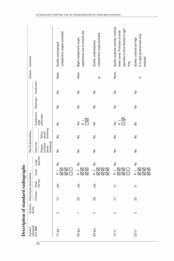
Appendix C – Description of standard radiographs
The Complete Set (22 radiographs)
The ILO (2000) International Classification of Radiographs of Pneumoconioses is accompanied by 22 standard radiographs. Two of them illustrate category 0/0 profusion of small opacities. Fifteen others define small-opacity profusion categories (1/1, 2/2 and 3/3), and some of the shapes and sizes of these opacities (p, q, r, s, and t). Large opacities (categories A, B and C) are shown on three additional radiographs. These 20 radiographs are described in the following table using the conventions defined in the preceding text and including Comments. The site of small opacities is shown by a tick in the boxes symbolizing the zones of the lungs, as follows:
Right Left
Upper ■■ ■■ Middle ■■ ■■ Lower ■■ ■■The two remaining standard radiographs are composite reproductions of sections
from full-size chest radiographs. One depicts increasing profusion of irregular small u-sized opacities. The other illustrates various pleural abnormalities.
The radiographs that define the small-opacity profusion categories are copies of the same standards that were published in 1980, thus preserving continuity and consis-tency in the Classification. As noted in footnote 3 on page 3, the standard radiographs were chosen to demonstrate the radiographic features of the pneumoconioses, rather than to demonstrate technical quality.
The descriptions of the radiographs in the following table are the consensus views of a group of experts who reassessed the standards in the year 2000. These descrip-tions differ in some respects from those published in the earlier (1980) edition of the Classification. Judgements about the technical quality of the radiographs reflect familiarity with current optimal techniques and thus may appear more severe, with only six graded 1 (good). Descriptions of pleural abnormalities now follow the modified conventions that are defined in these Guidelines (section 3.3). The Comments in the right-hand column of the table include some additional observations by the reviewers.
27
The Quad Set (14 radiographs)
Also available from the ILO is a set of 14 standard radiographs that are wholly compatible with the Complete Set referred to above. 1 The Quad Set may be preferred by some users of the Classification. It includes nine of the most commonly used standard radiographs from the Complete Set (both category 0/0 examples, six showing categories 1/1, 2/2 and 3/3 for q/q and t/t small opacities, and the composite radiograph that illus-trates pleural abnormalities). The remaining five radiographs in the Quad Set are compos-ite reproductions of quadrant sections from the other radiographs in the Complete Set. Four of them show different profusion categories for small opacities classifiable as p/p, r/r, s/s and u/u, respectively, and one shows large opacities (categories A, B and C).
Scientific reports that mention these Guidelines and the associated standard radiographs should refer to them explicitly as the ILO (2000) International Classification of Radiographs of Pneumoconioses, to avoid confusion with earlier editions of the Clas-sification and copies of standard radiographs. The international trial, which demonstrated the general compatibility of the Quad Set with the Complete Set, showed that, when using the Quad Set, some readers identified large opacities less frequently than when they used the Complete Set. Use of the Quad Set was also associated with an increase in the frequency with which some readers described the shapes of the small opacities that they saw as predominantly irregular, rather than rounded. It is recommended, therefore, that authors of research reports should indicate which set of standard radiographs (the Complete Set or the Quad Set) was used in their studies.
1 See footnote 1 in the foreword.
GUIDELINES FOR THE USE OF RADIOGRAPHS OF PNEUMOCONIOSES
28
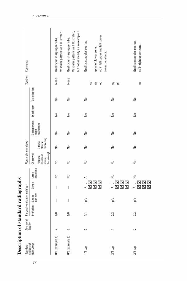
APPENDIX C
29
Des
crip
tion
of
stan
dard
rad
iogr
aphs
Stan
dard
Te
chni
cal
Pare
nchy
mal
abn
orm
aliti
es
Pl
eura
l abn
orm
aliti
es
Sy
mbo
ls
Com
men
tsra
diog
raph
Qu
ality
(ILO,
200
0)
Pr
ofus
ion
Shap
e
Zone
s La
rge
Ches
t wal
l
Cost
ophr
enic
Di
aphr
agm
Ca
lcifi
catio
n
and
size
opac
ities
an
gle
Plaq
ues
Diffu
se
oblit
erat
ion
(loca
lized
pl
eura
l
pl
eura
l th
icke
ning
thic
keni
ng)
0/0
(exa
mpl
e 1)
2
0/0
—
—
No
No
No
No
No
No
Non
e Qu
ality
: uns
harp
upp
er ri
bs.
Vasc
ular
pat
tern
wel
l illu
stra
ted.
0/0
(exa
mpl
e 2)
2
0/0
—
—
No
No
No
No
No
No
Non
e Qu
ality
: uns
harp
upp
er ri
bs.
Vasc
ular
pat
tern
wel
l illu
stra
ted,
bu
t not
as c
lear
ly as
in e
xam
ple
1.
1/1
p/p
2 1/
1 p/
p R
L
A N
o N
o N
o N
o N
o
Qual
ity: s
capu
lar o
verla
p.
ca
rp
rp in
left
low
er zo
ne.
od
od
in le
ft up
per a
nd le
ft lo
wer
zo
nes ;
eva
luat
e.
2/2
p/p
1 2/
2 p/
p R
L
No
No
No
No
No
No
cg
pi
3/3
p/p
2 3/
3 p/
p R
L
No
No
No
No
No
No
Qu
ality
: sca
pula
r ove
rlap.
ca
ca
in ri
ght u
pper
zone
.
GUIDELINES FOR THE USE OF RADIOGRAPHS OF PNEUMOCONIOSES
30
Des
crip
tion
of
stan
dard
rad
iogr
aphs
Stan
dard
Te
chni
cal
Pare
nchy
mal
abn
orm
aliti
es
Pl
eura
l abn
orm
aliti
es
Sy
mbo
ls
Com
men
tsra
diog
raph
Qu
ality
(ILO,
200
0)
Pr
ofus
ion
Shap
e
Zone
s La
rge
Ches
t wal
l
Cost
ophr
enic
Di
aphr
agm
Ca
lcifi
catio
n
and
size
opac
ities
an
gle
Plaq
ues
Diffu
se
oblit
erat
ion
(loca
lized
pl
eura
l
pl
eura
l th
icke
ning
thic
keni
ng)
1/1
q/q
2 1/
1 q/
q R
L
No
No
No
No
No
No
Non
e Qu
ality
: ove
rexp
osed
;
co
stop
hren
ic a
ngle
s ex
clud
ed.
2/2
q/q
1 2/
2 q/
q R
L
No
No
No
Yes
No
No
Non
e Ri
ght c
osto
phre
nic
angl
e
R
L
ap
pear
ance
due
to m
uscl
e sl
ip.
3/3
q/q
2 3/
3 q/
q R
L
No
No
No
No
No
No
Qu
ality
: und
erex
pose
d;
pi
co
stop
hren
ic a
ngle
exc
lude
d.
1/1
r/r
2 1/
1 r/r
R
L
No
No
No
Yes
No
No
Non
e Qu
ality
: sca
pula
r ove
rlap;
uns
harp
R
L
lo
wer
zone
s. P
rofu
sion
of s
mal
l
opac
ities
is m
ore
mar
ked
in ri
ght
lung
.
2/2
r/r
2 2/
2 r/r
R
L
No
No
No
No
No
No
Qu
ality
: con
trast
too
high
.
hi
hi in
righ
t par
atra
chea
l are
a;
eval
uate
.

APPENDIX C
31
Des
crip
tion
of
stan
dard
rad
iogr
aphs
Stan
dard
Te
chni
cal
Pare
nchy
mal
abn
orm
aliti
es
Pl
eura
l abn
orm
aliti
es
Sy
mbo
ls
Com
men
tsra
diog
raph
Qu
ality
(ILO,
200
0)
Pr
ofus
ion
Shap
e
Zone
s La
rge
Ches
t wal
l
Cost
ophr
enic
Di
aphr
agm
Ca
lcifi
catio
n
and
size
opac
ities
an
gle
Plaq
ues
Diffu
se
oblit
erat
ion
(loca
lized
pl
eura
l
pl
eura
l th
icke
ning
thic
keni
ng)
3/3
r/r
2 3/
3 r/r
R
L
No
No
No
No
No
No
Qu
ality
: con
trast
too
high
.
ax
ax in
righ
t upp
er zo
ne.
ih
1/1
s/t
2 1/
1 s/
t R
L
No
No
No
No
No
No
Non
e Qu
ality
: uns
harp
are
as;
cost
ophr
enic
ang
les
excl
uded
.
2/2
s/s
2 2/
2 s/
s R
L
No
No
No
No
No
No
Qu
ality
: slig
htly
und
erex
pose
d;
cost
ophr
enic
ang
les
excl
uded
.
em
em in
upp
er zo
nes.
pb
pb
in le
ft lo
wer
zone
.
3/3
s/s
2 3/
3 s/
s R
L
No
No
No
No
No
No
Qu
ality
: slig
htly
und
erex
pose
d;
scap
ular
ove
rlap.
ho
ho
at r
ight
cos
troph
enic
ang
le
ih
pi
Se
e fo
otno
te 1
4 on
pag
e 9.
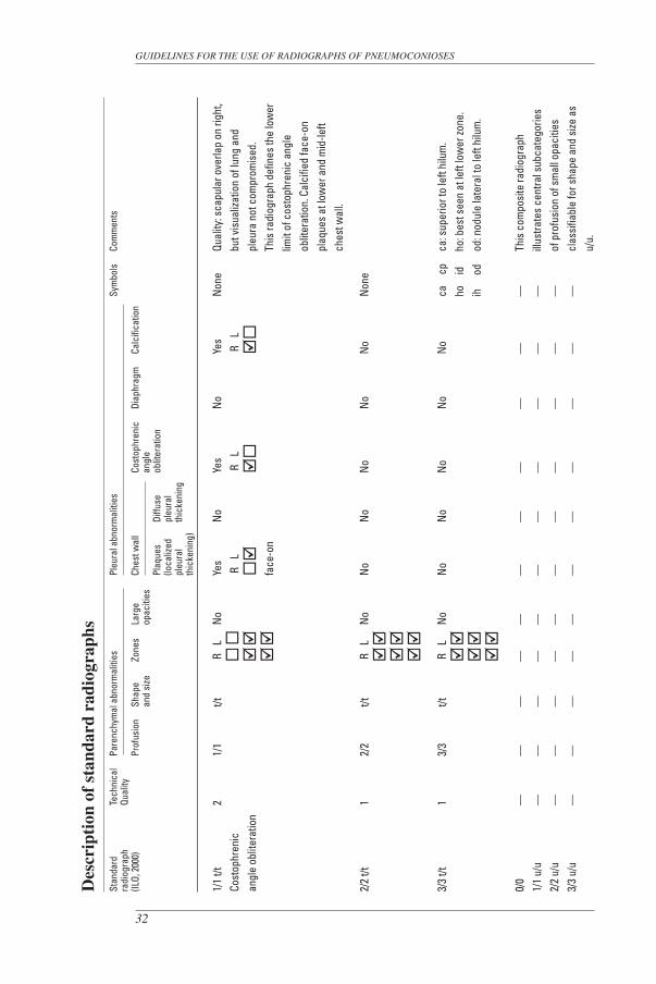
Des
crip
tion
of
stan
dard
rad
iogr
aphs
Stan
dard
Te
chni
cal
Pare
nchy
mal
abn
orm
aliti
es
Pl
eura
l abn
orm
aliti
es
Sy
mbo
ls
Com
men
tsra
diog
raph
Qu
ality
(ILO,
200
0)
Pr
ofus
ion
Shap
e
Zone
s La
rge
Ches
t wal
l
Cost
ophr
enic
Di
aphr
agm
Ca
lcifi
catio
n
and
size
opac
ities
an
gle
Plaq
ues
Diffu
se
oblit
erat
ion
(loca
lized
pl
eura
l
pl
eura
l th
icke
ning
thic
keni
ng)
1/1
t/t
2 1/
1 t/t
R
L
No
Yes
No
Yes
No
Yes
Non
e Qu
ality
: sca
pula
r ove
rlap
on ri
ght,
Cost
ophr
enic
R
L
R
L
R
L
but v
isua
lizat
ion
of lu
ng a
ndan
gle
oblit
erat
ion
pl
eura
not
com
prom
ised
.
fa
ce-o
n
This
radi
ogra
ph d
efin
es th
e lo
wer
lim
it of
cos
toph
reni
c an
gle
oblit
erat
ion.
Cal
cifie
d fa
ce-o
n
pl
aque
s at
low
er a
nd m
id-le
ft
ch
est w
all.
2/2
t/t
1 2/
2 t/t
R
L
No
No
No
No
No
No
Non
e
3/3
t/t
1 3/
3 t/t
R
L
No
No
No
No
No
No
ca
cp
ca: s
uper
ior t
o le
ft hi
lum
.
ho
id
ho: b
est s
een
at le
ft lo
wer
zone
.
ih
od
od: n
odul
e la
tera
l to
left
hilu
m.
0/0
—
—
—
—
—
—
—
—
—
—
—
This
com
posi
te ra
diog
raph
1/
1 u/
u —
—
—
—
—
—
—
—
—
—
—
ill
ustra
tes
cent
ral s
ubca
tego
ries
2/2
u/u
—
—
—
—
—
—
—
—
—
—
—
of p
rofu
sion
of s
mal
l opa
citie
s3/
3 u/
u —
—
—
—
—
—
—
—
—
—
—
cl
assi
fiabl
e fo
r sha
pe a
nd s
ize a
s
u/
u.
GUIDELINES FOR THE USE OF RADIOGRAPHS OF PNEUMOCONIOSES
32
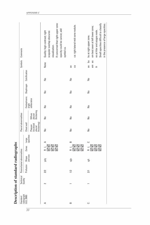
Des
crip
tion
of
stan
dard
rad
iogr
aphs
Stan
dard
Te
chni
cal
Pare
nchy
mal
abn
orm
aliti
es
Pl
eura
l abn
orm
aliti
es
Sy
mbo
ls
Com
men
tsra
diog
raph
Qu
ality
(ILO,
200
0)
Pr
ofus
ion
Shap
e
Zone
s La
rge
Ches
t wal
l
Cost
ophr
enic
Di
aphr
agm
Ca
lcifi
catio
n
and
size
opac
ities
an
gle
Plaq
ues
Diffu
se
oblit
erat
ion
(loca
lized
pl
eura
l
pl
eura
l th
icke
ning
thic
keni
ng)
A 2
2/2
p/q
R
L A
No
No
No
No
No
Non
e Qu
ality
: hig
h co
ntra
st; r
ight
sc
apul
ar o
verla
p ob
scur
es
visu
aliza
tion.
If co
ncer
ned
that
righ
t upp
er zo
ne
op
acity
cou
ld b
e ca
ncer
, add
sy
mbo
l ca.
B 1
1/2
q/p
R
L B
No
No
No
No
No
ax
ca
ca: r
ight
late
ral m
id-z
one
nodu
le.
C 1
2/1
q/t
R
L C
No
No
No
No
No
ax
bu
bu a
t rig
ht u
pper
zone
.
em
es
em b
est s
een
at le
ft lo
wer
zone
;
ih
es a
t hila
r and
azy
gos
node
.
Sm
all o
paci
ties
diffi
cult
to c
lass
ify
in th
e pr
esen
ce o
f lar
ge o
paci
ties.
APPENDIX C
33

GUIDELINES FOR THE USE OF RADIOGRAPHS OF PNEUMOCONIOSES
34
ILO (2000) Composite standard radiograph showing examples of pleural abnormalities
Upper-left section: Upper-right section: calcified plaques at diaphragm calcified in-profile and face-on plaques
Lower-left section: diffuse in-profile pleural thickening with the required costophrenic angle obliteration; also diffuse face-on pleural thickening Lower-right section: calcified and uncalcified face-on plaques
Appendix D – Diagrams
The diagrams on the following pages represent illustrations of radiographic features that are included in the Complete Classification. Those features are defined in the text of these Guidelines and by the appearances on the standard radiographs. The diagrams are intended to serve as pictorial reminders, but they are not a substitute for the standard radiographs or the written text.
Diagrams that represent symbols do not illustrate all the manifestations of the conditions defined by these symbols, for example ca (carcinoma), cg (calcified granu-loma), od (other disease). The two drawings of appearances classifiable as od in this appendix represent lobar pneumonia and aspergilloma, goiter and hiatal hernia.
35
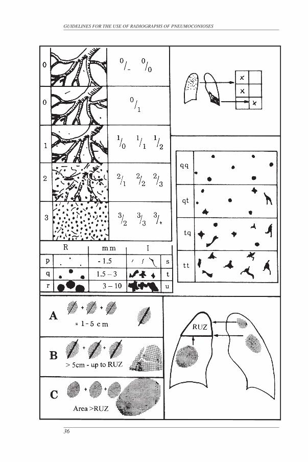
GUIDELINES FOR THE USE OF RADIOGRAPHS OF PNEUMOCONIOSES
36
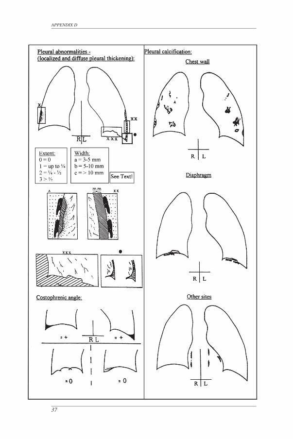
APPENDIX D
37
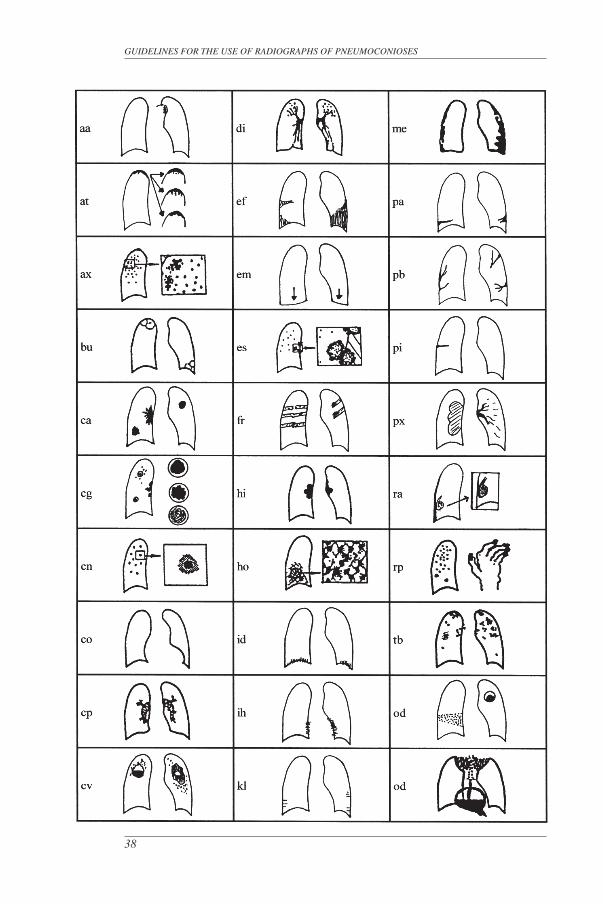
GUIDELINES FOR THE USE OF RADIOGRAPHS OF PNEUMOCONIOSES
38
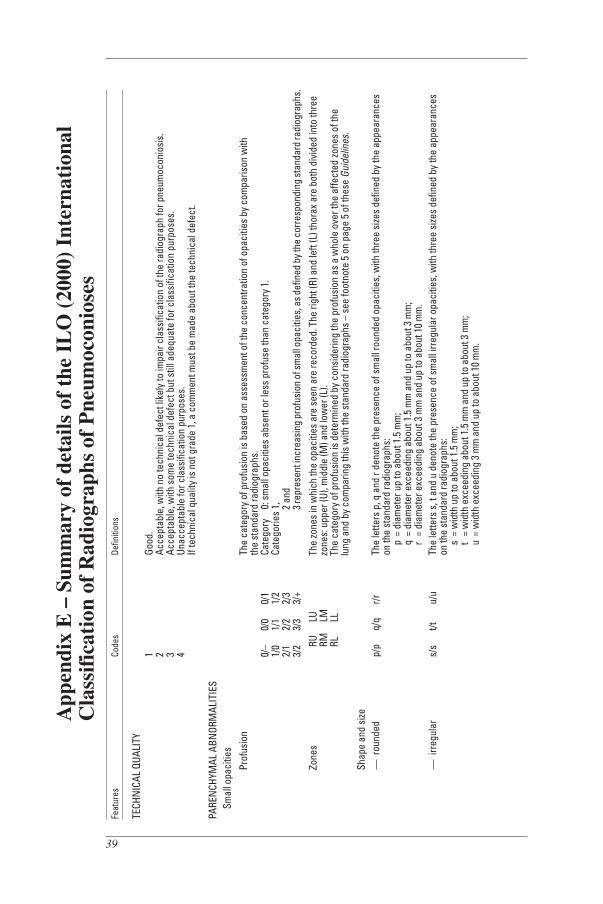
39
App
endi
x E
– S
umm
ary
of d
etai
ls o
f th
e IL
O (
2000
) In
tern
atio
nal
Cla
ssif
icat
ion
of R
adio
grap
hs o
f P
neum
ocon
iose
sFe
atur
es
Code
s De
finiti
ons
TECH
NIC
AL Q
UALI
TY
1 Go
od.
2
Acce
ptab
le, w
ith n
o te
chni
cal d
efec
t lik
ely
to im
pair
clas
sific
atio
n of
the
radi
ogra
ph fo
r pne
umoc
onio
sis.
3
Acce
ptab
le, w
ith s
ome
tech
nica
l def
ect b
ut s
till a
dequ
ate
for c
lass
ifica
tion
purp
oses
.
4 Un
acce
ptab
le fo
r cla
ssifi
catio
n pu
rpos
es.
If te
chni
cal q
ualit
y is
not
gra
de 1
, a c
omm
ent m
ust b
e m
ade
abou
t the
tech
nica
l def
ect.
PARE
NCH
YMAL
ABN
ORM
ALIT
IES
Sm
all o
paci
ties
Pr
ofus
ion
Th
e ca
tego
ry o
f pro
fusi
on is
bas
ed o
n as
sess
men
t of t
he c
once
ntra
tion
of o
paci
ties
by c
ompa
rison
with
th
e st
anda
rd ra
diog
raph
s.
0/–
0/0
0/1
Cate
gory
0
: sm
all o
paci
ties
abse
nt o
r les
s pr
ofus
e th
an c
ateg
ory
1.
1/0
1/
1 1/
2 Ca
tego
ries
1,
2/1
2/2
2/3
2 a
nd
3/2
3/3
3/+
3 re
pres
ent i
ncre
asin
g pr
ofus
ion
of sm
all o
paci
ties,
as d
efin
ed b
y the
cor
resp
ondi
ng st
anda
rd ra
diog
raph
s.
Zone
s
RU
LU
The
zone
s in
whi
ch th
e op
aciti
es a
re s
een
are
reco
rded
. The
righ
t (R)
and
left
(L) t
hora
x ar
e bo
th d
ivid
ed in
to th
ree
RM
LM
zo
nes:
upp
er (U
), m
iddl
e (M
) and
low
er (L
).
RL
LL
The
cate
gory
of p
rofu
sion
is d
eter
min
ed b
y co
nsid
erin
g th
e pr
ofus
ion
as a
who
le o
ver t
he a
ffect
ed zo
nes
of th
e
lu
ng a
nd b
y co
mpa
ring
this
with
the
stan
dard
radi
ogra
phs
– se
e fo
otno
te 5
on
page
5 o
f the
se G
uide
lines
.
Shap
e an
d si
ze
— r
ound
ed
p/p
q/q
r/r
The
lette
rs p
, q a
nd r
deno
te th
e pr
esen
ce o
f sm
all r
ound
ed o
paci
ties,
with
thre
e si
zes
defin
ed b
y th
e ap
pear
ance
s
on th
e st
anda
rd ra
diog
raph
s:
p
= di
amet
er u
p to
abo
ut 1
.5 m
m;
q =
diam
eter
exc
eedi
ng a
bout
1.5
mm
and
up
to a
bout
3 m
m;
r =
diam
eter
exc
eedi
ng a
bout
3 m
m a
nd u
p to
abo
ut 1
0 m
m.
—
irr
egul
ar
s/s
t/t
u/u
The
lette
rs s
, t a
nd u
den
ote
the
pres
ence
of s
mal
l irr
egul
ar o
paci
ties,
with
thre
e si
zes
defin
ed b
y th
e ap
pear
ance
s
on th
e st
anda
rd ra
diog
raph
s:
s
= w
idth
up
to a
bout
1.5
mm
;
t
= w
idth
exc
eedi
ng a
bout
1.5
mm
and
up
to a
bout
3 m
m;
u =
wid
th e
xcee
ding
3 m
m a
nd u
p to
abo
ut 1
0 m
m.
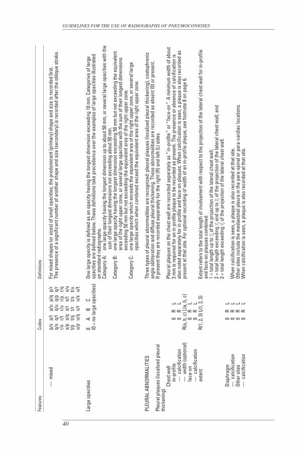
GUIDELINES FOR THE USE OF RADIOGRAPHS OF PNEUMOCONIOSES
40
Feat
ures
Co
des
Defin
ition
s
—
mix
ed
p/s
p/t
p/u
p/q
p/r
For m
ixed
sha
pes
(or s
izes)
of s
mal
l opa
citie
s, th
e pr
edom
inan
t (pr
imar
y) s
hape
and
size
is re
cord
ed fi
rst.
q/
s q/
t q/
u q/
p q/
r Th
e pr
esen
ce o
f a s
igni
fican
t num
ber o
f ano
ther
sha
pe a
nd s
ize (s
econ
dary
) is
reco
rded
afte
r the
obl
ique
stro
ke.
r/s
r/t
r/u
r/p
r/q
s/p
s/q
s/r
s/t
s/u
t/p
t/q
t/r
t/s
t/u
u/p
u/q
u/r
u/s
u/t
Larg
e op
aciti
es
0 A
B C
On
e la
rge
opac
ity is
def
ined
as
an o
paci
ty h
avin
g th
e lo
nges
t dim
ensi
on e
xcee
ding
10
mm
. Cat
egor
ies
of la
rge
(0
= n
o la
rge
opac
ities
) op
aciti
es a
re d
efin
ed b
elow
. The
se d
efin
ition
s ta
ke p
rece
denc
e ov
er th
e ex
ampl
es o
f lar
ge o
paci
ties
illus
trate
d
on
sta
ndar
d ra
diog
raph
s.
Cate
gory
A:
one
larg
e op
acity
hav
ing
the
long
est d
imen
sion
up
to a
bout
50
mm
, or s
ever
al la
rge
opac
ities
with
the
sum
of t
heir
long
est d
imen
sion
s no
t exc
eedi
ng a
bout
50
mm
.
Cate
gory
B:
one
larg
e op
acity
hav
ing
the
long
est d
imen
sion
exc
eedi
ng 5
0 m
m b
ut n
ot e
xcee
ding
the
equi
vale
nt
area
of t
he ri
ght u
pper
zone
, or s
ever
al la
rge
opac
ities
with
the
sum
of t
heir
long
est d
imen
sion
s
ex
ceed
ing
50 m
m b
ut n
ot e
xcee
ding
the
equi
vale
nt a
rea
of th
e rig
ht u
pper
zone
.
Cate
gory
C:
one
larg
e op
acity
whi
ch e
xcee
ds th
e eq
uiva
lent
are
a of
the
right
upp
er zo
ne, o
r sev
eral
larg
e
op
aciti
es w
hich
whe
n co
mbi
ned
exce
ed th
e eq
uiva
lent
are
a of
the
right
upp
er zo
ne.
PLEU
RAL
ABN
ORM
ALIT
IES
Th
ree
type
s of
ple
ural
abn
orm
aliti
es a
re re
cogn
ized:
ple
ural
pla
ques
(loc
alize
d pl
eura
l thi
cken
ing)
, cos
toph
reni
c
an
gle
oblit
erat
ion
and
diffu
se p
leur
al th
icke
ning
. The
se a
bnor
mal
ities
are
reco
rded
as
abse
nt (0
) or p
rese
nt.
Pleu
ral p
laqu
es (l
ocal
ized
pleu
ral
If
pres
ent t
hey
are
reco
rded
sep
arat
ely
for t
he ri
ght (
R) a
nd le
ft (L
) sid
es.
thic
keni
ng)
Ches
t wal
l
Pleu
ral p
laqu
es o
n th
e ch
est w
all a
re re
cord
ed s
epar
atel
y as
“ in
-pro
file ”
or “
face
-on ”
. A m
inim
um w
idth
of a
bout
in
-pro
file
0 R
L
3 m
m is
requ
ired
for a
n in
-pro
file
plaq
ue to
be
reco
rded
as
pres
ent.
The
pres
ence
or a
bsen
ce o
f cal
cific
atio
n is
—
cal
cific
atio
n
0
R
L al
so n
oted
sep
arat
ely
for i
n-pr
ofile
and
face
-on
plaq
ues.
Whe
n ca
lcifi
catio
n is
see
n, a
pla
que
is a
lso
reco
rded
as
— w
idth
(opt
iona
l) R(
a, b
, c) L
(a, b
, c)
pres
ent a
t tha
t site
. For
opt
iona
l rec
ordi
ng o
f wid
th o
f an
in-p
rofil
e pl
aque
, see
foot
note
8 o
n pa
ge 6
.
fa
ce-o
n
0
R
L
—
cal
cific
atio
n
0
R
L
ex
tent
R(
1, 2
, 3) L
(1, 2
, 3)
Exte
nt re
fers
to th
e to
tal l
engt
h of
invo
lvem
ent w
ith re
spec
t to
the
proj
ectio
n of
the
late
ral c
hest
wal
l for
in-p
rofil
e
and
face
-on
plaq
ues
com
bine
d:
1 =
tota
l len
gth
up to
1 ⁄ 4 of t
he p
roje
ctio
n of
the
late
ral c
hest
wal
l;
2 =
tota
l len
gth
exce
edin
g 1 ⁄ 4 a
nd u
p to
1 ⁄ 2 of t
he p
roje
ctio
n of
the
late
ral c
hest
wal
l; an
d
3 =
tota
l len
gth
exce
edin
g 1 ⁄ 2 o
f the
pro
ject
ion
of th
e la
tera
l che
st w
all.
Di
aphr
agm
0
R
L
— c
alci
ficat
ion
0 R
L
Whe
n ca
lcifi
catio
n is
see
n, a
pla
que
is a
lso
reco
rded
at t
hat s
ite.
Ot
her s
ites
0 R
L
Othe
r site
s in
clud
e th
e m
edia
stin
al p
leur
a in
the
para
-spi
nal o
r par
a-ca
rdia
c lo
catio
ns.
—
cal
cific
atio
n
0
R
L W
hen
calc
ifica
tion
is s
een,
a p
laqu
e is
als
o re
cord
ed a
t tha
t site
.
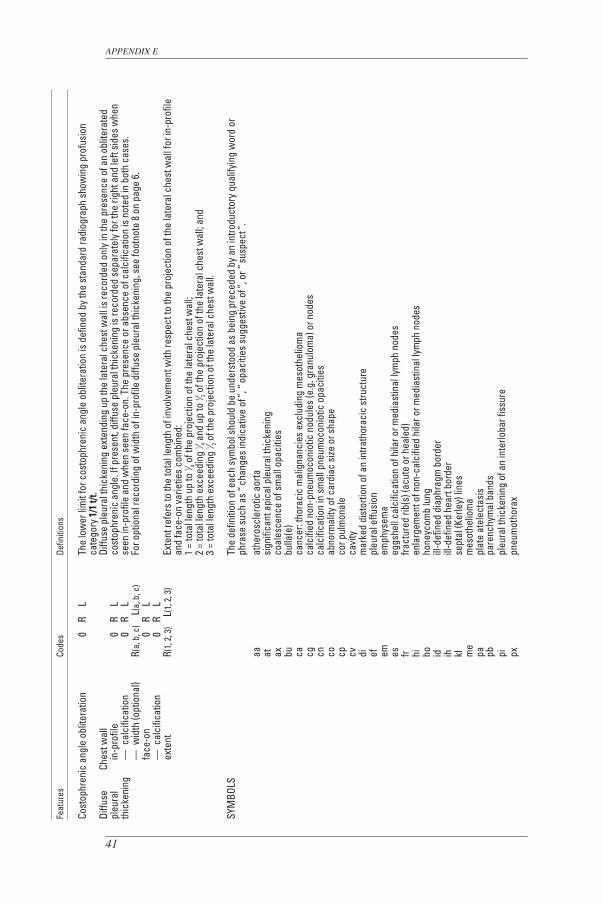
APPENDIX E
41
Feat
ures
Co
des
Defin
ition
s
Cost
ophr
enic
ang
le o
blite
ratio
n
0
R
L Th
e lo
wer
lim
it fo
r cos
toph
reni
c an
gle
oblit
erat
ion
is d
efin
ed b
y th
e st
anda
rd ra
diog
raph
sho
win
g pr
ofus
ion
ca
tego
ry 1
/1 t/
t.Di
ffuse
Ch
est w
all
Di
ffuse
ple
ural
thic
keni
ng e
xten
ding
up
the
late
ral c
hest
wal
l is
reco
rded
onl
y in
the
pres
ence
of a
n ob
liter
ated
pleu
ral
in
-pro
file
0 R
L
cost
ophr
enic
ang
le. I
f pre
sent
, diff
use
pleu
ral t
hick
enin
g is
reco
rded
sep
arat
ely
for t
he ri
ght a
nd le
ft si
des
whe
n th
icke
ning
— c
alci
ficat
ion
0 R
L
seen
in-p
rofil
e an
d w
hen
seen
face
-on.
The
pre
senc
e or
abs
ence
of c
alci
ficat
ion
is n
oted
in b
oth
case
s.
—
wid
th (o
ptio
nal)
R(a,
b, c
) L
(a, b
, c)
For o
ptio
nal r
ecor
ding
of w
idth
of i
n-pr
ofile
diff
use
pleu
ral t
hick
enin
g, s
ee fo
otno
te 8
on
page
6.
face
-on
0 R
L
— c
alci
ficat
ion
0 R
L
exte
nt
R(1,
2, 3
) L
(1, 2
, 3)
Exte
nt re
fers
to th
e to
tal l
engt
h of
invo
lvem
ent w
ith re
spec
t to
the
proj
ectio
n of
the
late
ral c
hest
wal
l for
in-p
rofil
e
and
face
-on
varie
ties
com
bine
d:
1
= to
tal l
engt
h up
to 1 ⁄ 4 o
f the
pro
ject
ion
of th
e la
tera
l che
st w
all;
2
= to
tal l
engt
h ex
ceed
ing
1 ⁄ 4 and
up
to 1 ⁄ 2 o
f the
pro
ject
ion
of th
e la
tera
l che
st w
all;
and
3
= to
tal l
engt
h ex
ceed
ing
1 ⁄ 2 of t
he p
roje
ctio
n of
the
late
ral c
hest
wal
l.
SYM
BOLS
The
defin
ition
of e
ach
sym
bol s
houl
d be
und
erst
ood
as b
eing
pre
cede
d by
an
intro
duct
ory
qual
ifyin
g w
ord
or
phra
se s
uch
as “
chan
ges
indi
cativ
e of
”, “
opac
ities
sug
gest
ive
of ”,
or “
susp
ect ”
.
aa
athe
rosc
lero
tic a
orta
at
si
gnifi
cant
api
cal p
leur
al th
icke
ning
ax
co
ales
cenc
e of
sm
all o
paci
ties
bu
bu
lla(e
)
ca
canc
er: t
hora
cic
mal
igna
ncie
s ex
clud
ing
mes
othe
liom
a
cg
calc
ified
non
-pne
umoc
onio
tic n
odul
es (e
.g. g
ranu
lom
a) o
r nod
es
cn
calc
ifica
tion
in s
mal
l pne
umoc
onio
tic o
paci
ties
co
ab
norm
ality
of c
ardi
ac s
ize o
r sha
pe
cp
cor p
ulm
onal
e
cv
cavi
ty
di
mar
ked
dist
ortio
n of
an
intra
thor
acic
stru
ctur
e
ef
pleu
ral e
ffusi
on
em
emph
ysem
a
es
eggs
hell
calc
ifica
tion
of h
ilar o
r med
iast
inal
lym
ph n
odes
fr
fract
ured
rib(
s) (a
cute
or h
eale
d)
hi
enla
rgem
ent o
f non
-cal
cifie
d hi
lar o
r med
iast
inal
lym
ph n
odes
ho
ho
neyc
omb
lung
id
ill
-def
ined
dia
phra
gm b
orde
r
ih
ill-d
efin
ed h
eart
bord
er
kl
sept
al (K
erle
y) li
nes
m
e m
esot
helio
ma
pa
pl
ate
atel
ecta
sis
pb
pa
renc
hym
al b
ands
pi
pl
eura
l thi
cken
ing
of a
n in
terlo
bar f
issu
re
px
pneu
mot
hora
x
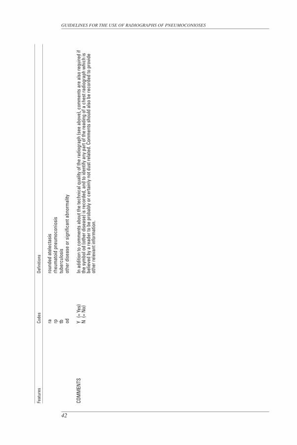
GUIDELINES FOR THE USE OF RADIOGRAPHS OF PNEUMOCONIOSES
42
Feat
ures
Co
des
Defin
ition
s
ra
ro
unde
d at
elec
tasi
s
rp
rheu
mat
oid
pneu
moc
onio
sis
tb
tu
berc
ulos
is
od
othe
r dis
ease
or s
igni
fican
t abn
orm
ality
COM
MEN
TS
Y (=
Yes
) In
add
ition
to c
omm
ents
abo
ut th
e te
chni
cal q
ualit
y of
the
radi
ogra
ph (s
ee a
bove
), co
mm
ents
are
als
o re
quire
d if
N
(=
No)
th
e sy
mbo
l od
(oth
er d
isea
se) i
s re
cord
ed, a
nd to
iden
tify
any
part
of th
e re
adin
g of
a c
hest
radi
ogra
ph w
hich
is
belie
ved
by a
read
er to
be
prob
ably
or c
erta
inly
not
dus
t rel
ated
. Com
men
ts s
houl
d al
so b
e re
cord
ed to
pro
vide
ot
her r
elev
ant i
nfor
mat
ion.
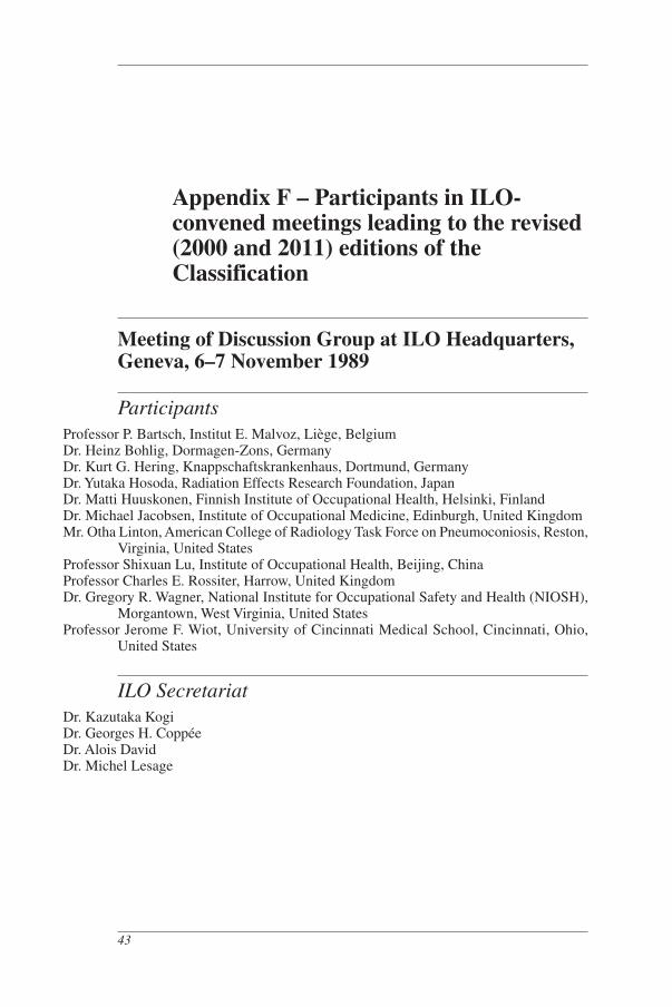
Appendix F – Participants in ILO-convened meetings leading to the revised (2000 and 2011) editions of the Classification
Meeting of Discussion Group at ILO Headquarters, Geneva, 6–7 November 1989
ParticipantsProfessor P. Bartsch, Institut E. Malvoz, Liège, BelgiumDr. Heinz Bohlig, Dormagen-Zons, GermanyDr. Kurt G. Hering, Knappschaftskrankenhaus, Dortmund, GermanyDr. Yutaka Hosoda, Radiation Effects Research Foundation, JapanDr. Matti Huuskonen, Finnish Institute of Occupational Health, Helsinki, FinlandDr. Michael Jacobsen, Institute of Occupational Medicine, Edinburgh, United KingdomMr. Otha Linton, American College of Radiology Task Force on Pneumoconiosis, Reston,
Virginia, United StatesProfessor Shixuan Lu, Institute of Occupational Health, Beijing, ChinaProfessor Charles E. Rossiter, Harrow, United KingdomDr. Gregory R. Wagner, National Institute for Occupational Safety and Health (NIOSH),
Morgantown, West Virginia, United StatesProfessor Jerome F. Wiot, University of Cincinnati Medical School, Cincinnati, Ohio,
United States
ILO SecretariatDr. Kazutaka KogiDr. Georges H. Coppée Dr. Alois DavidDr. Michel Lesage
43
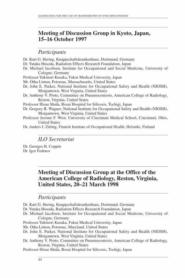
Meeting of Discussion Group in Kyoto, Japan, 15–16 October 1997
ParticipantsDr. Kurt G. Hering, Knappschaftskrankenhaus, Dortmund, GermanyDr. Yutaka Hosoda, Radiation Effects Research Foundation, JapanDr. Michael Jacobsen, Institute for Occupational and Social Medicine, University of
Cologne, GermanyProfessor Yukinori Kusaka, Fukui Medical University, JapanMr. Otha Linton, Potomac, Massachusetts, United States Dr. John E. Parker, National Institute for Occupational Safety and Health (NIOSH),
Morgantown, West Virginia, United States Dr. Anthony V. Proto, Committee on Pneumoconiosis, American College of Radiology,
Reston, Virginia, United StatesProfessor Hisao Shida, Rosai Hospital for Silicosis, Tochigi, JapanDr. Gregory R. Wagner, National Institute for Occupational Safety and Health (NIOSH),
Morgantown, West Virginia, United StatesProfessor Jerome F. Wiot, University of Cincinnati Medical School, Cincinnati, Ohio,
United StatesDr. Anders J. Zitting, Finnish Institute of Occupational Health, Helsinki, Finland
ILO SecretariatDr. Georges H. CoppéeDr. Igor Fedotov
Meeting of Discussion Group at the Office of the American College of Radiology, Reston, Virginia, United States, 20–21 March 1998
ParticipantsDr. Kurt G. Hering, Knappschaftskrankenhaus, Dortmund, GermanyDr. Yutaka Hosoda, Radiation Effects Research Foundation, JapanDr. Michael Jacobsen, Institute for Occupational and Social Medicine, University of
Cologne, GermanyProfessor Yukinori Kusaka, Fukui Medical University, JapanMr. Otha Linton, Potomac, Maryland, United StatesDr. John E. Parker, National Institute for Occupational Safety and Health (NIOSH),
Morgantown, West Virginia, United StatesDr. Anthony V. Proto, Committee on Pneumoconiosis, American College of Radiology,
Reston, Virginia, United StatesProfessor Hisao Shida, Rosai Hospital for Silicosis, Tochigi, Japan
GUIDELINES FOR THE USE OF RADIOGRAPHS OF PNEUMOCONIOSES
44
Dr. Gregory R. Wagner, National Institute for Occupational Safety and Health (NIOSH), Morgantown, West Virginia, United States
Professor Jerome F. Wiot, University of Cincinnati Medical School, Cincinnati, Ohio, United States
Dr. Anders J. Zitting, Finnish Institute of Occupational Health, Helsinki, Finland
ILO SecretariatDr. Igor Fedotov
Meeting of Discussion Group at the ILO Branch Office, Washington, DC, United States, 26 October 2000
ParticipantsDr. Kurt G. Hering, Knappschaftskrankenhaus, Dortmund, GermanyDr. Yutaka Hosoda, Radiation Effects Research Foundation, JapanProfessor Michael Jacobsen, Institute for Occupational and Social Medicine, University
of Cologne, GermanyProfessor Yukinori Kusaka, Fukui Medical University, JapanMr. Otha Linton, Potomac, Maryland, United StatesProfessor John E. Parker, Pulmonary and Critical Care Medicine, West Virginia Univer-
sity, Morgantown, West Virginia, United StatesDr. Anthony V. Proto, Committee on Pneumoconiosis, American College of Radiology,
Reston, Virginia, United StatesProfessor Hisao Shida, Rosai Hospital for Silicosis, Tochigi, JapanDr. Gregory R. Wagner, National Institute for Occupational Safety and Health (NIOSH),
Morgantown, West Virginia, United StatesDr. Anders J. Zitting, Helsinki, Finland
ILO SecretariatDr. Benjamin O. Alli
Meeting of Panel on revision of the ILO (2000) Classification of Radiographs of Pneumoconioses in Rockville, Maryland, United States, 13–14 March 2008
ParticipantsDr. Eduardo Algranti, Occupational Health Division, FUNDACENTRO, São Paulo,
Brazil
APPENDIX F
45

GUIDELINES FOR THE USE OF RADIOGRAPHS OF PNEUMOCONIOSES
46
Professor Jacques Ameille, AP-HP, Occupational Health Unit, Hôpital Raymond Poincaré, Garches, France
Dr. Michael Attfield, National Institute for Occupational Safety and Health (NIOSH), Morgantown, West Virginia, United States
Dr. Joseph E. Burkhart, National Institute for Occupational Safety and Health (NIOSH), Morgantown, West Virginia, United States
Dr. A. Franzblau, University of Michigan School of Public Health, Ann Arbor, Michigan, United States
Dr. Daniel A. Henry, Department of Radiology, Medical College of Virginia Hospitals, Virginia Commonwealth University, Richmond, Virginia, United States
Dr. Kurt G. Hering, Knappschaftskrankenhaus (Miners’ Hospital), Dortmund, GermanyProfessor Michael Jacobsen, Ulverston, United KingdomProfessor Yukinori Kusaka, Fukui Medical University, Fukui, Japan Professor John E. Parker, Pulmonary and Critical Care Medicine, West Virginia Univer-
sity, Morgantown, West Virginia, United StatesDr. Edward L. Petsonk, National Institute for Occupational Safety and Health (NIOSH),
Morgantown, West Virginia, United States Dr. Ralph T. Shipley, University of Cincinnati College of Medicine, Cincinnati, Ohio,
United States Professor Narufumi Suganuma, Department of Environmental Medicine, Kochi Medical
School, Kochi University, Kochi, Japan Dr. Gregory R. Wagner, National Institute for Occupational Safety and Health (NIOSH),
Washington, DC, United States Dr. David N. Weissman, National Institute for Occupational Safety and Health (NIOSH),
Morgantown, West Virginia, United States
ILO SecretariatDr. Igor Fedotov
Film readers who participated in the international film-reading trial of new composite standard radiographs (the “ Quad ” trial), 1992–95
CanadaDr. Raymond Bégin, Faculté de médecine, Université de Sherbrooke, QuébecDr. Marc Desmeules, Hôpital Laval Centre de pneumologie, Ste-Foy, QuébecDr. W. Keith C. Morgan, Chest Diseases Unit, University of Western Ontario, London,
OntarioDr. David C. F. Muir, Health Sciences Center, McMaster University, Hamilton, Ontario
ChinaDr. Guowei Li, Zhaoyang Red Cross Hospital, BeijingDr. Shunging Liu, Chendu Peoples’ Hospital, ChenduDr. Yulin Liu, Institute of Industrial Health, Anshan LiaoningProfessor Cuijuan Zhang, National Institute of Occupational Medicine, Beijing
Czech Republic 1
Professor Alois David, Postgraduate Medical School, PragueDr. Jirí Slepicka, Faculty Hospital, OstravaDr. Frantisek Staník, Department of Occupational Diseases, Miners’ Hospital, Karviná
FinlandDr. Marja-Liisa Kokko, Tampere City Hospital, TampereDr. Ossi Korhola, Helsinki University Central Hospital, HelsinkiDr. Kristina M. Virkola, Helsinki University Children’s Hospital, HelsinkiDr. Anders J. Zitting, Finnish Institute of Occupational Health, Helsinki
FranceProfessor Jacques Ameille, Université Paris V, Faculté de médecine Paris Ouest,
GarchesProfessor Patrick Brochard, Université Bordeaux II, Bordeaux Professor Dominique Choudat, Université Paris V, Faculté de médecine Cochin, ParisProfessor Marc Letourneux, Université de Caen
GermanyDr. Kurt G. Hering, Knappschaftskrankenhaus, DortmundDr. Peter Rathjen, Knappschaftskrankenhaus, DortmundDr. Klaus Siegmund, Institut für Arbeitsmedizin der Heinrich-Heine-Universität, Düssel-
dorfDr. Volkmar Wiebe, Berufgenossenschaftliche Krankenanstalten, Universitätsklinik,
Bochum
1 As of 1 January 1993. Prior to that date, Czechoslovakia.
APPENDIX F
47

GUIDELINES FOR THE USE OF RADIOGRAPHS OF PNEUMOCONIOSES
48
JapanDr. Keizo Chiyotani, Rosai Hospital for Silicosis, TochigiProfessor Yukinori Kusaka, Fukui Medical University, FukuiDr. Hiroshi Morikubo, Rosai Hospital for Silicosis, TochigiProfessor Hisao Shida, Rosai Hospital for Silicosis, Tochigi
PolandProfessor Aleksandra Kujawska, Institute of Occupational Medicine and Environmental
Health, SosnowiecProfessor Kazimierz Marek, Institute of Occupational Medicine and Environmental
Health, SosnowiecDr. Aleksander Stachura, Institute of Occupational Medicine and Environmental Health,
SosnowiecDr. Andrzej Stasiow, Hospital Ward and Outpatient Clinic for Occupational Diseases in
Coalminers, Katowice-Ochojec
Slovakia 1
Professor Ladislav Benicky, Medical Faculty, Kosice
United KingdomDr. Douglas Scarisbrick, British Coal Corporation Radiological Service, Mansfield
Woodhouse, NottinghamshireProfessor Anthony Seaton, Department of Environmental and Occupational Medicine,
Aberdeen University, AberdeenDr. Colin A. Soutar, Institute of Occupational Medicine, EdinburghDr. Paul Willdig, British Coal Corporation Radiological Service, Mansfield Woodhouse,
Nottinghamshire
United StatesProfessor N. LeRoy Lapp, Pulmonary and Critical Care Medicine, West Virginia Univer-
sity, Morgantown, West VirginiaDr. Steven Short, Manhattan, KansasDr. Mei-Lin Wang, Morgantown, West VirginiaDr. Susan Weber, Pulmonary and Critical Care Medicine, West Virginia University,
Morgantown, West Virginia
1 As of 1 January 1993. Prior to that date, Czechoslovakia.