Embed Size (px)
Citation preview
Idiopathic Intracranial Hypertension
Dr. Irfan A Shah
DM scholar Neurology
SKIMS ,Srinagar, Kashmir.
• Idiopathic intracranial hypertension (IIH) is a disorder characterized by increased intracranial pressure (ICP) of unknown cause, predominantly seen in women of childbearing age and associated with a history of recent weight gain .
terminology
• The first report of IIH was by the German physician Heinrich Quincke, who described it in 1893 under the name serous meningitis.
• The concept of raised intracranial pressure in the absence of a space occupying lesion was first introduced by Max Nonne as ‘pseudotumour cerebri’ (PTC) in 1904. ,
• the term otitic hydrocephalus reported by London neurologist Sir Charles Symonds(1931) may have resulted from venous sinus thrombosis caused by otitis media .
• Later the term ‘benign intracranial hypertension’ became popular and was often used interchangeably with PTC.
• The condition was considered ‘benign’ in comparison with cases of tumour but it has been argued that loss of visual function in up to 25% of cases and progression to blindness if untreated means that it should not be considered ‘benign’. Hence the term IIH is now generally used.
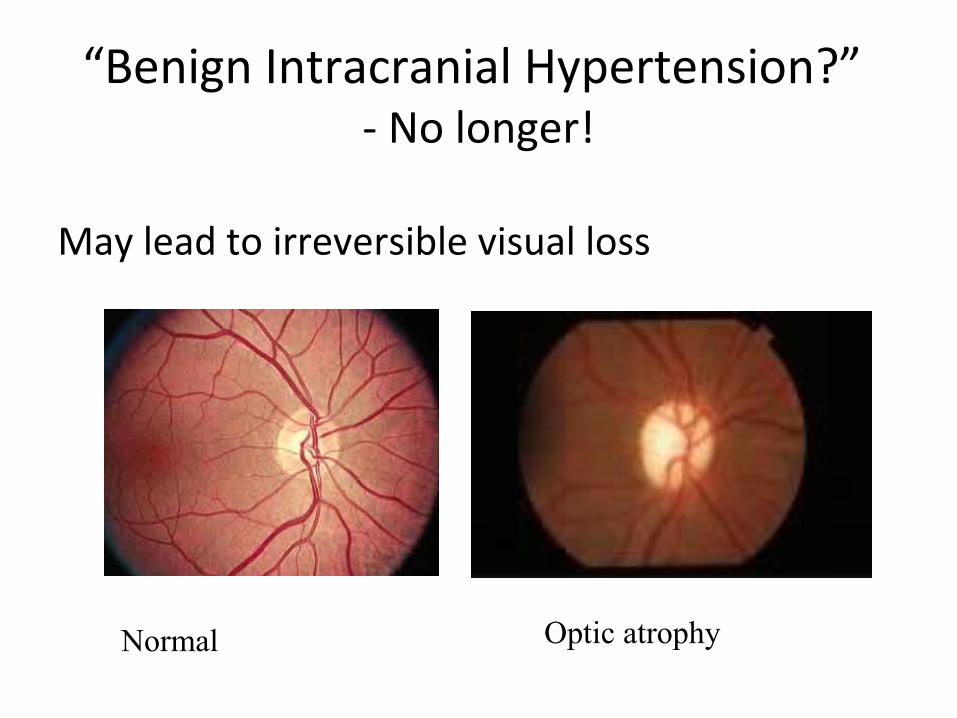
“Benign Intracranial Hypertension?” - No longer!
May lead to irreversible visual loss
Normal Optic atrophy
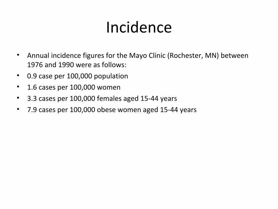
Incidence
• Annual incidence figures for the Mayo Clinic (Rochester, MN) between 1976 and 1990 were as follows:
• 0.9 case per 100,000 population• 1.6 cases per 100,000 women• 3.3 cases per 100,000 females aged 15-44 years• 7.9 cases per 100,000 obese women aged 15-44 years
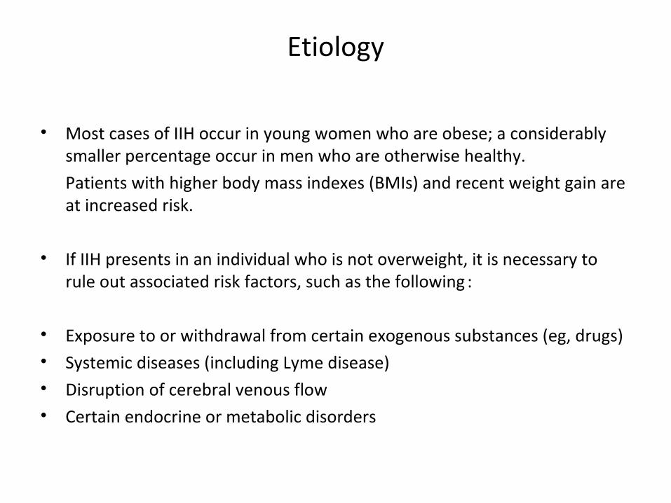
Etiology
• Most cases of IIH occur in young women who are obese; a considerably smaller percentage occur in men who are otherwise healthy.
Patients with higher body mass indexes (BMIs) and recent weight gain are at increased risk.
• If IIH presents in an individual who is not overweight, it is necessary to rule out associated risk factors, such as the following :
• Exposure to or withdrawal from certain exogenous substances (eg, drugs)• Systemic diseases (including Lyme disease)• Disruption of cerebral venous flow• Certain endocrine or metabolic disorders
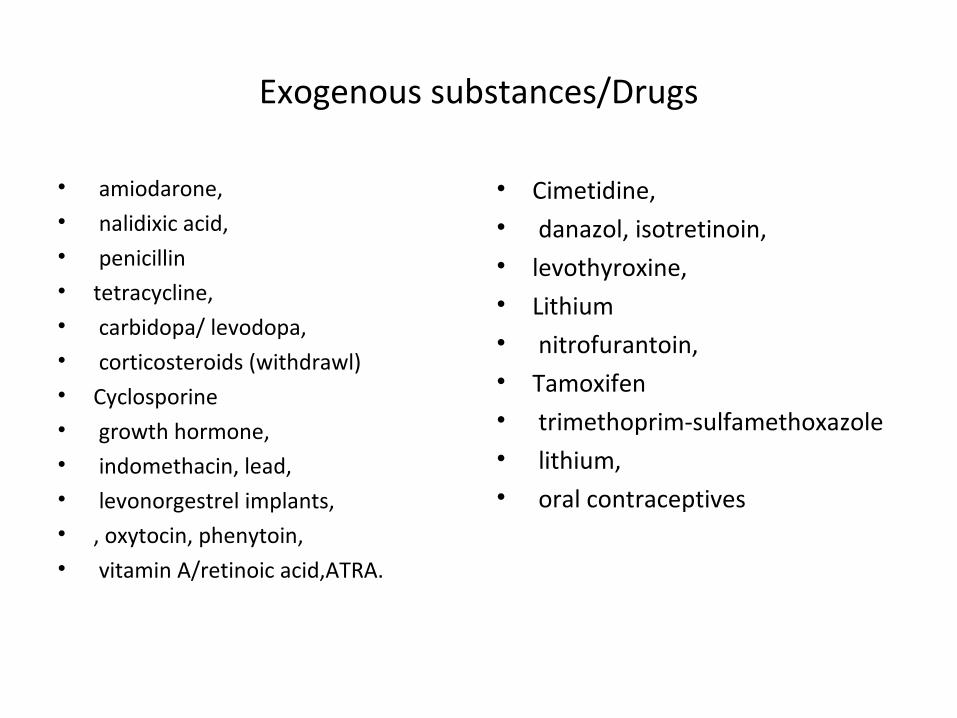
Exogenous substances/Drugs
• amiodarone, • nalidixic acid,• penicillin• tetracycline,• carbidopa/ levodopa,• corticosteroids (withdrawl)• Cyclosporine• growth hormone,• indomethacin, lead,• levonorgestrel implants,• , oxytocin, phenytoin,• vitamin A/retinoic acid,ATRA.
• Cimetidine,• danazol, isotretinoin,• levothyroxine,• Lithium• nitrofurantoin, • Tamoxifen• trimethoprim-sulfamethoxazole• lithium,• oral contraceptives
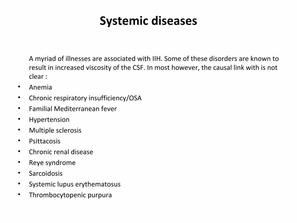
Systemic diseases
A myriad of illnesses are associated with IIH. Some of these disorders are known to result in increased viscosity of the CSF. In most however, the causal link with is not clear :
• Anemia• Chronic respiratory insufficiency/OSA• Familial Mediterranean fever• Hypertension• Multiple sclerosis• Psittacosis• Chronic renal disease• Reye syndrome• Sarcoidosis• Systemic lupus erythematosus• Thrombocytopenic purpura
Endocrine risk factors
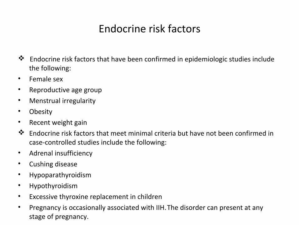
Endocrine risk factors that have been confirmed in epidemiologic studies include the following:
• Female sex• Reproductive age group• Menstrual irregularity• Obesity• Recent weight gain Endocrine risk factors that meet minimal criteria but have not been confirmed in
case-controlled studies include the following:• Adrenal insufficiency• Cushing disease• Hypoparathyroidism• Hypothyroidism• Excessive thyroxine replacement in children• Pregnancy is occasionally associated with IIH. The disorder can present at any
stage of pregnancy.
Criteria for including a drug or a disease as a cause of IIH:
• Radhakrishnan et al..– At least 2 cases should have been described– The reported cases should have met all the criteria for the
diagnosis of IIH.– Intracranial dural sinus thrombosis should have been ruled
out with reasonable certainty
Radhakrishnan K, Ahlskog JE, Garrity JA, Kurland LT. Idiopathic intracranial hypertension. Mayo Clin Proc. Feb 1994;69(2):169-80
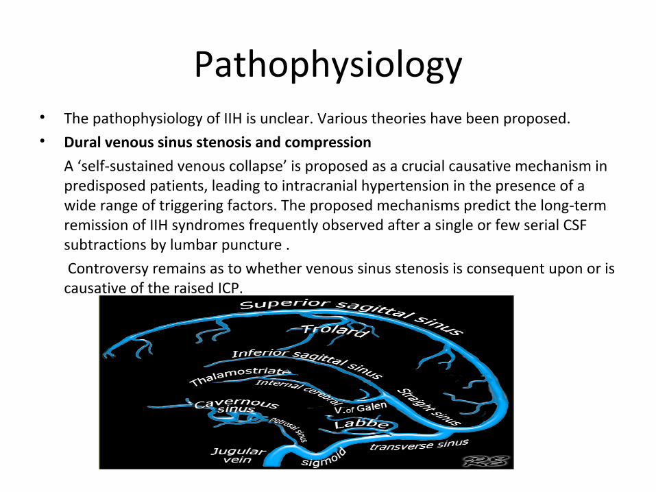
Pathophysiology• The pathophysiology of IIH is unclear. Various theories have been proposed.• Dural venous sinus stenosis and compression
A ‘self-sustained venous collapse’ is proposed as a crucial causative mechanism in predisposed patients, leading to intracranial hypertension in the presence of a wide range of triggering factors. The proposed mechanisms predict the long-term remission of IIH syndromes frequently observed after a single or few serial CSF subtractions by lumbar puncture .
Controversy remains as to whether venous sinus stenosis is consequent upon or is causative of the raised ICP.
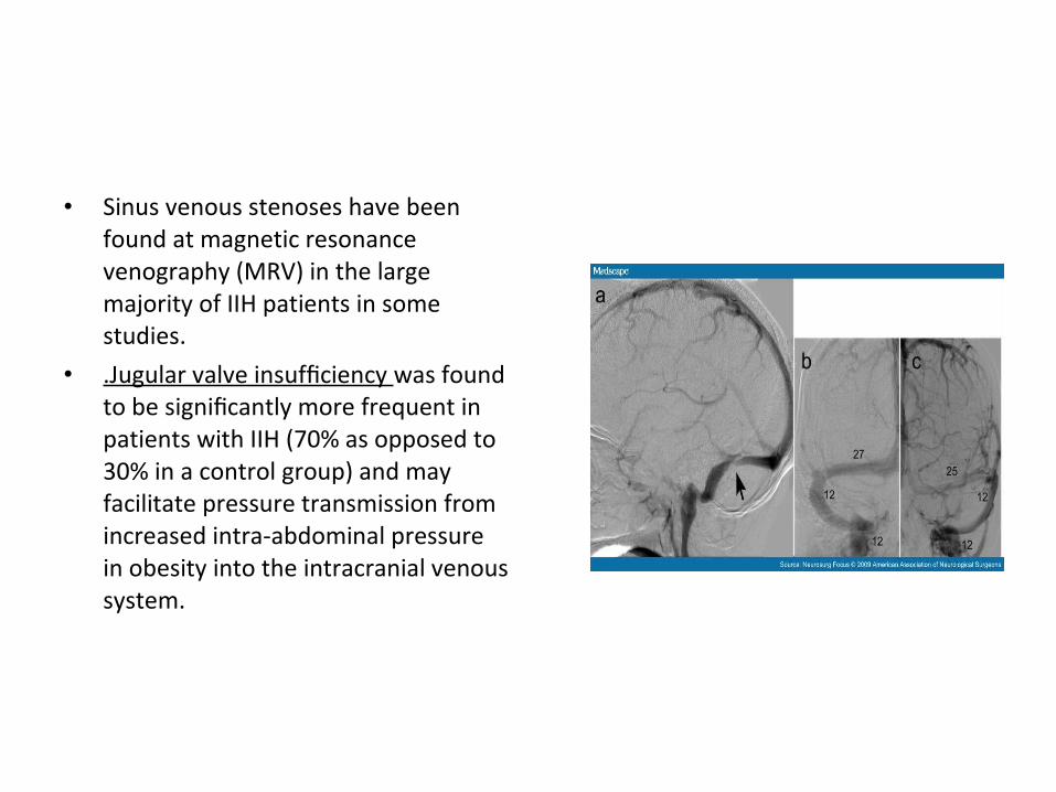
• Sinus venous stenoses have been found at magnetic resonance venography (MRV) in the large majority of IIH patients in some studies.
• .Jugular valve insufficiency was found to be significantly more frequent in patients with IIH (70% as opposed to 30% in a control group) and may facilitate pressure transmission from increased intra-abdominal pressure in obesity into the intracranial venous system.
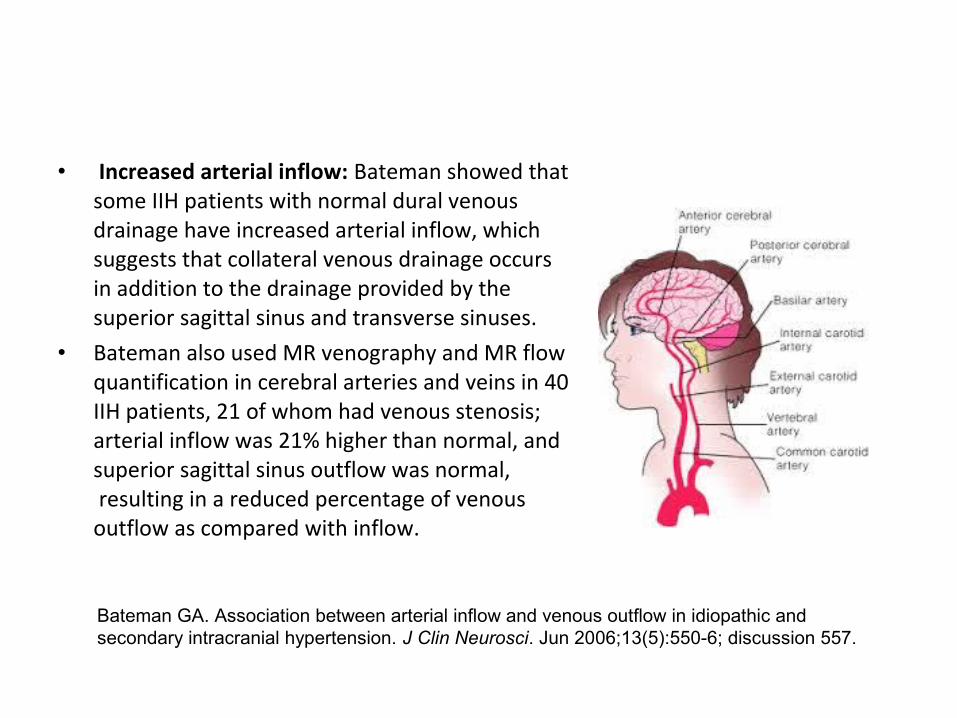
• Increased arterial inflow: Bateman showed that some IIH patients with normal dural venous drainage have increased arterial inflow, which suggests that collateral venous drainage occurs in addition to the drainage provided by the superior sagittal sinus and transverse sinuses.
• Bateman also used MR venography and MR flow quantification in cerebral arteries and veins in 40 IIH patients, 21 of whom had venous stenosis; arterial inflow was 21% higher than normal, and superior sagittal sinus outflow was normal, resulting in a reduced percentage of venous outflow as compared with inflow.
Bateman GA. Association between arterial inflow and venous outflow in idiopathic and secondary intracranial hypertension. J Clin Neurosci. Jun 2006;13(5):550-6; discussion 557.
IIH and Obesity
• IIH commonly occurs in women who are overweight; however, the role obesity plays in this disorder is unclear. In some instances, obesity and IIH may be familial.
• It has been proposed that obesity increases intra-abdominal pressure and thereby raises cardiac filling pressures. These rises in pressure lead to impeded venous return from the brain (due to the valveless venous system that exists from the brain to the heart) with a subsequent elevation in intracranial venous pressure.
• Waist-to-hip ratio (WHR) is a descriptive measure of body fat distribution approximately reflecting upper to lower body fat ratio. In a study of IIH patients compared with two obese control groups, WHR was 0.79 in IIH versus 0.84 and 0.91 in the controls. The authors concluded that in IIH, fat tends to preferentially accumulate in the lower body .
Signs and symptoms
• Symptoms of elevated intracranial pressure (ICP)• Headaches are recorded in almost all IIH patients. They are typically
nonspecific and vary in type, location, and frequency. The pain is generally described as being diffuse, worsening in the morning and being exacerbated by the Valsalva maneuver.
• Patients who present with double vision most frequently complain of horizontal displacement of the images. Horizontal diplopia is a symptom of a false-localizing CN VI palsy. Vertical diplopia is rare, but it has been reported.
• Pulsatile tinnitus may be reported. This is a rhythmic sound, heard in one or both ears, with a pulsing synchronous rhythm that may be exacerbated by the supine or bending position.
• Radicular pain (usually in the arms) is an uncommon symptom.
Symptoms of papilledema
• Transient visual obscurations occur in most patients. The disturbance can last up to 30 seconds and is described as a dimming or blackout of vision in one or both of the eyes. These obscurations may be predominantly or uniformly orthostatic (ie, developing with standing up or bending over).
• Progressive loss of peripheral vision in one or both of the eyes may be noted. Typically, the vision loss starts in the nasal inferior quadrant and is followed by loss of peripheral & later central visual field (possibly affecting visual acuity) and, finally, loss of color vision.
• Blurring and distortion (ie, metamorphopsia) of central vision is caused by macular wrinkling and subretinal fluid spreading from the swollen optic disc.
• Sudden visual loss is due to intraocular hemorrhage secondary to peripapillary subretinal neovascularization related to chronic papilledema.
• In a study among 353 patients studied, the prevalence of those without papilloedema was 5.7% .
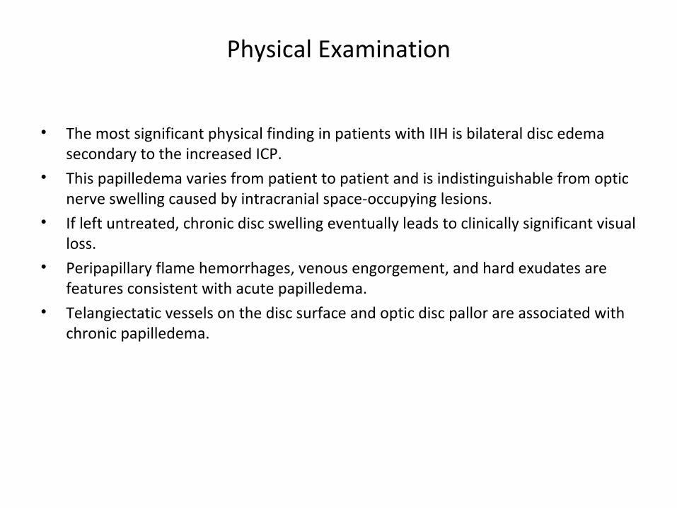
Physical Examination
• The most significant physical finding in patients with IIH is bilateral disc edema secondary to the increased ICP.
• This papilledema varies from patient to patient and is indistinguishable from optic nerve swelling caused by intracranial space-occupying lesions.
• If left untreated, chronic disc swelling eventually leads to clinically significant visual loss.
• Peripapillary flame hemorrhages, venous engorgement, and hard exudates are features consistent with acute papilledema.
• Telangiectatic vessels on the disc surface and optic disc pallor are associated with chronic papilledema.
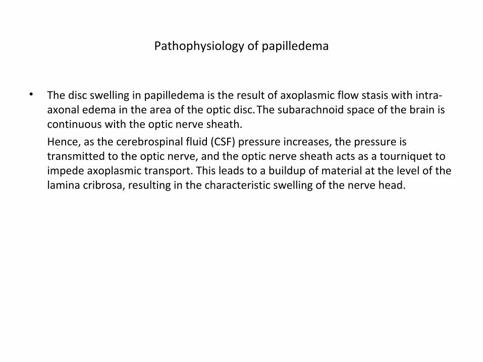
Pathophysiology of papilledema
• The disc swelling in papilledema is the result of axoplasmic flow stasis with intra-axonal edema in the area of the optic disc. The subarachnoid space of the brain is continuous with the optic nerve sheath.
Hence, as the cerebrospinal fluid (CSF) pressure increases, the pressure is transmitted to the optic nerve, and the optic nerve sheath acts as a tourniquet to impede axoplasmic transport. This leads to a buildup of material at the level of the lamina cribrosa, resulting in the characteristic swelling of the nerve head.
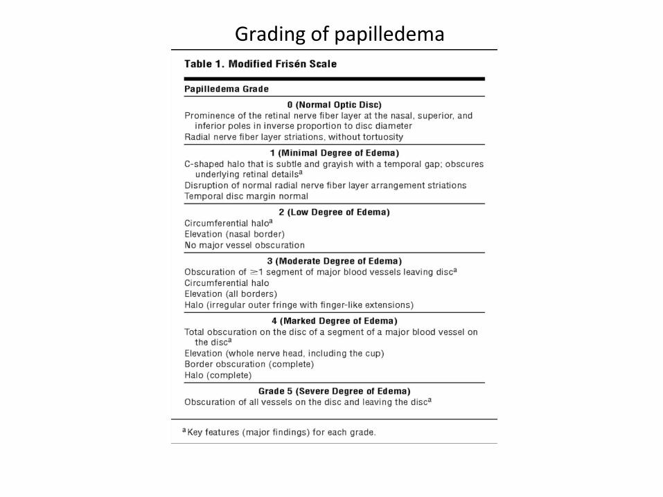
Grading of papilledema
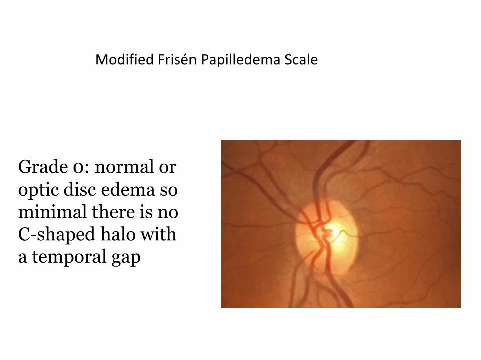
Modified Frisén Papilledema Scale
Grade 0: normal or optic disc edema sominimal there is noC-shaped halo witha temporal gap
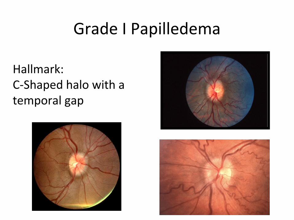
Grade I Papilledema
Hallmark: C-Shaped halo with a temporal gap
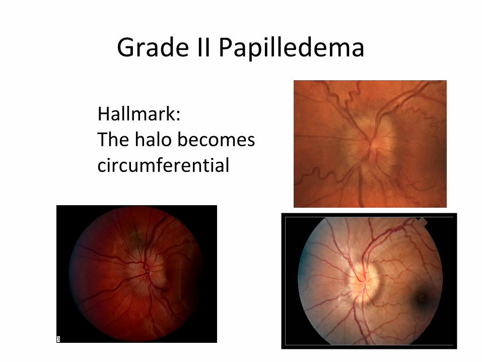
Grade II Papilledema
Hallmark: The halo becomescircumferential
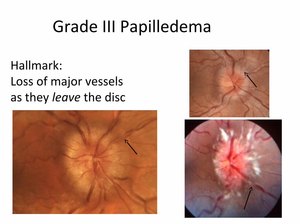
Grade III Papilledema
Hallmark: Loss of major vessels as they leave the disc
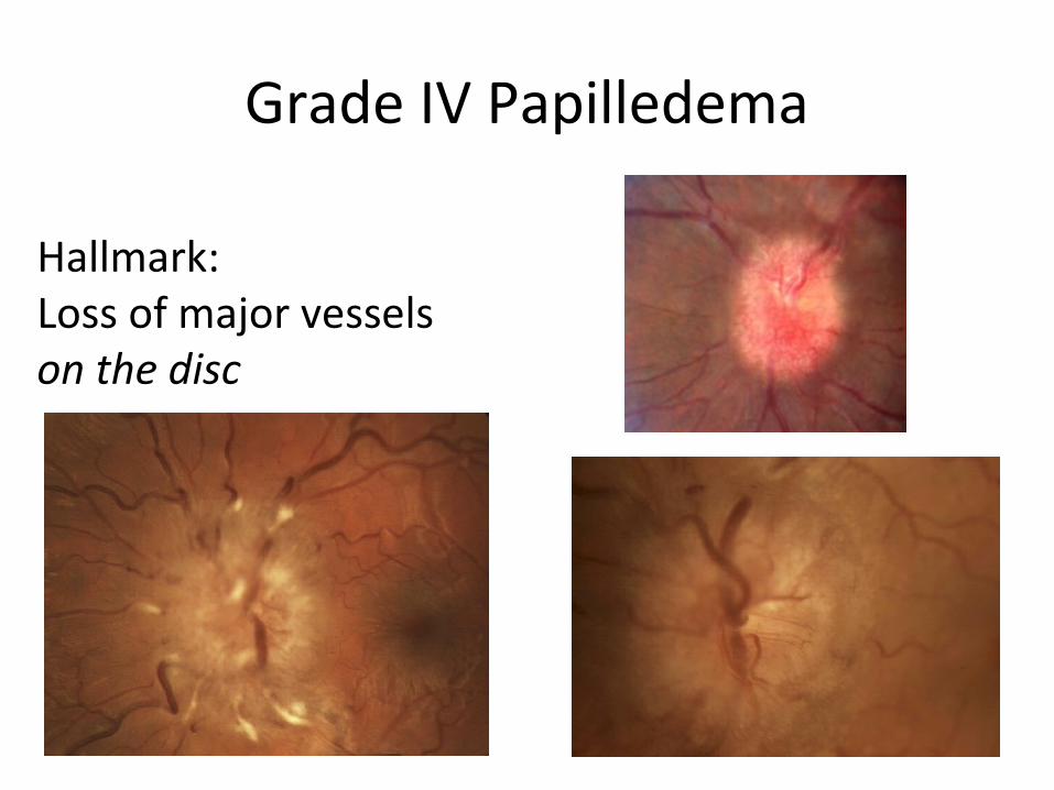
Grade IV Papilledema
Hallmark: Loss of major vessels on the disc
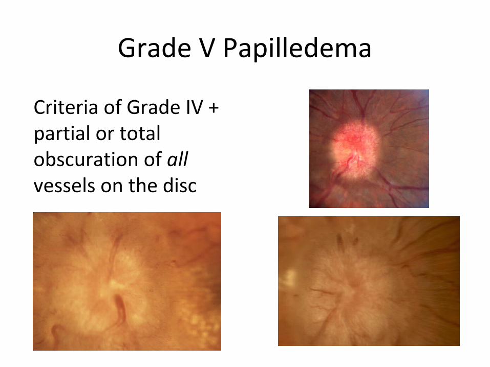
Grade V Papilledema
Criteria of Grade IV + partial or total obscuration of all vessels on the disc
Duration & papilledema
• Early manifestations– Disc hyperemia– obscured fine peripapillary vessels.– Small hemorrhages– Spontaneous venous pulsations lost.
• Late manifestations– obscured normal disc margins & disc becomes grossly elevated.– Venous congestion develops, and peripapillary hemorrhages become more
obvious, along with exudates and cotton-wool spots.– The peripapillary sensory retina may develop concentric or radial folds known
as Paton lines.• Chronic manifestations
– If the papilledema persists for months, the disc hyperemia slowly subsides, giving way to a gray or pale disc that loses its central cup.
– With time, the disc may develop small glistening crystalline deposits (disc pseudodrusen).
Evaluation of vision
• The most common visual finding in IIH is enlargement of blind spot.• The first sign of incipient post papilledema optic atrophy is
constriction of the inferior nasal quadrant of the visual field with a border reflecting the nasal horizontal midline (nasal step).
• colour testing is not sensitive in picking up early postpapilledema optic atrophy, because color perception is concentrated in the central visual field.
• Diplopia testing may be used to detect even a low-grade CN VI paresis.
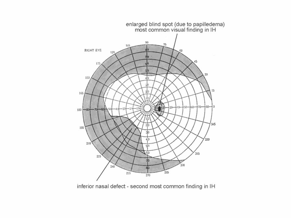
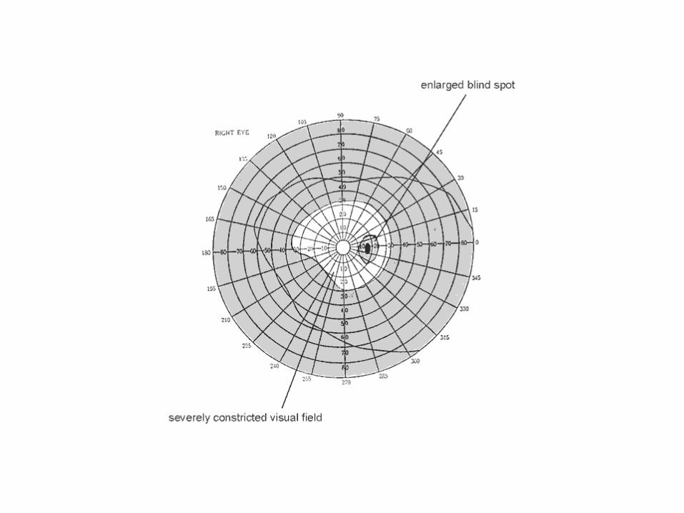
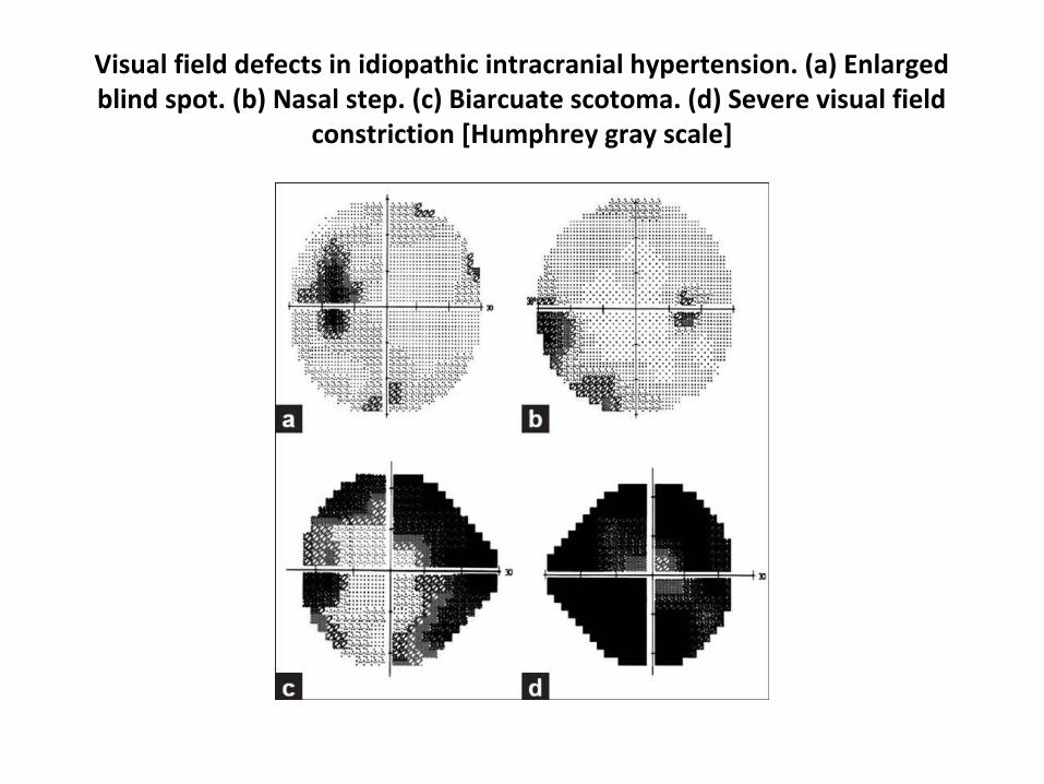
Visual field defects in idiopathic intracranial hypertension. (a) Enlarged blind spot. (b) Nasal step. (c) Biarcuate scotoma. (d) Severe visual field
constriction [Humphrey gray scale]
complications
• The only severe and permanent complication of IIH is progressive blindness from postpapilledema optic atrophy. As optic nerve axons die, the apparent degree of papilledema may diminish, giving a false sense of improvement. For this reason the patients must be monitored with frequent visual field examinations.
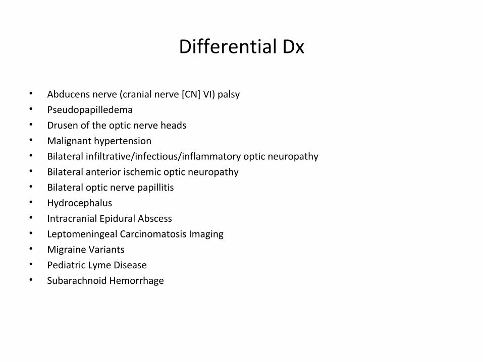
Differential Dx
• Abducens nerve (cranial nerve [CN] VI) palsy• Pseudopapilledema• Drusen of the optic nerve heads• Malignant hypertension• Bilateral infiltrative/infectious/inflammatory optic neuropathy• Bilateral anterior ischemic optic neuropathy• Bilateral optic nerve papillitis• Hydrocephalus• Intracranial Epidural Abscess• Leptomeningeal Carcinomatosis Imaging• Migraine Variants• Pediatric Lyme Disease• Subarachnoid Hemorrhage
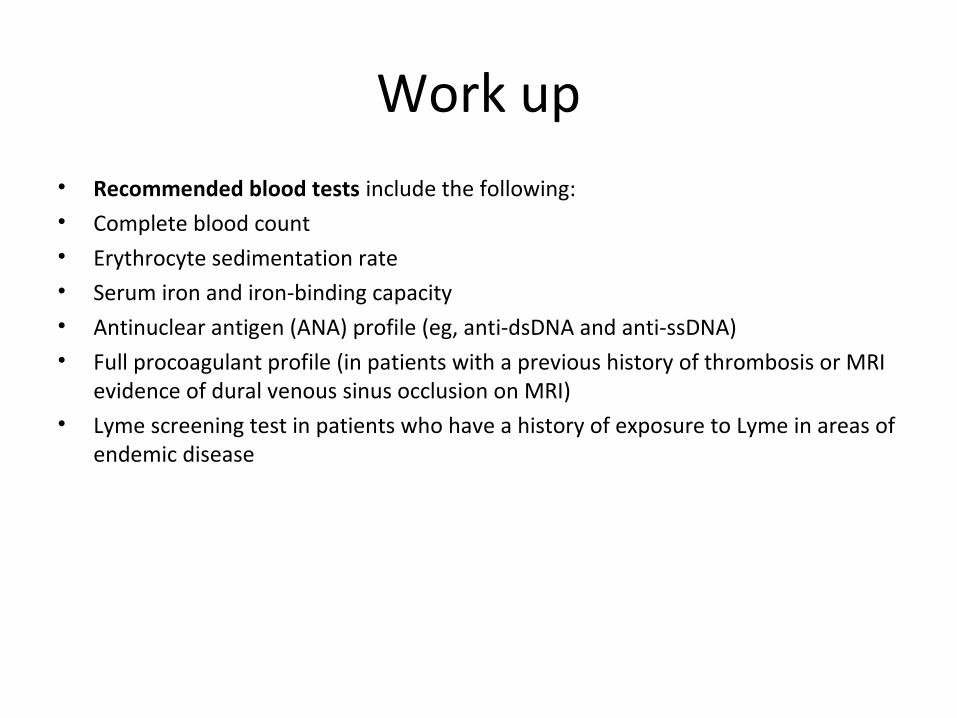
Work up• Recommended blood tests include the following:• Complete blood count• Erythrocyte sedimentation rate• Serum iron and iron-binding capacity• Antinuclear antigen (ANA) profile (eg, anti-dsDNA and anti-ssDNA)• Full procoagulant profile (in patients with a previous history of thrombosis or MRI
evidence of dural venous sinus occlusion on MRI)• Lyme screening test in patients who have a history of exposure to Lyme in areas of
endemic disease
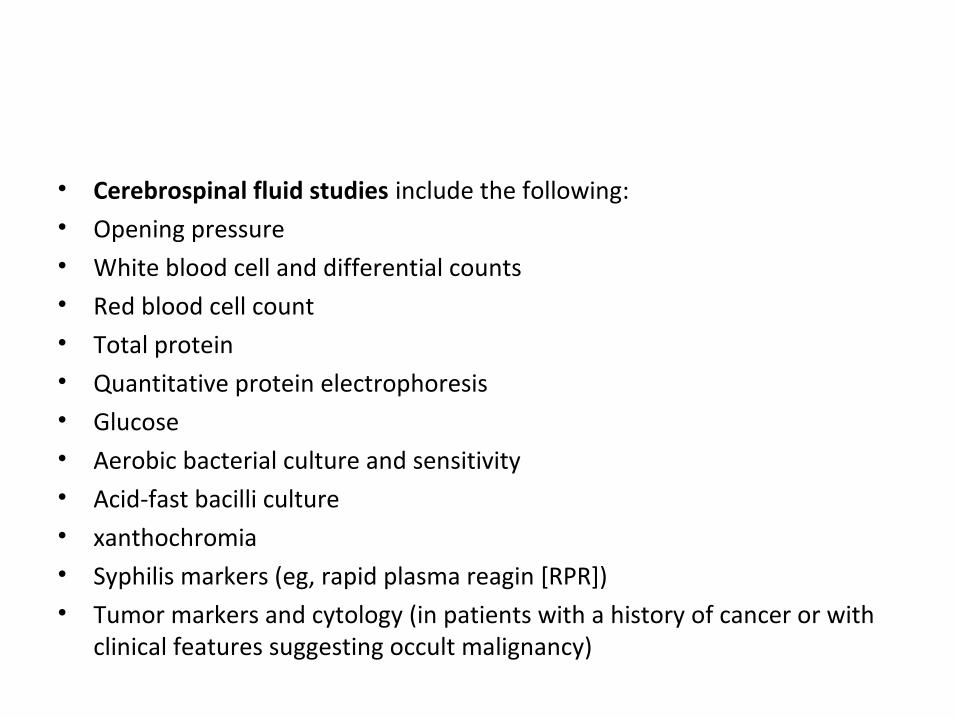
• Cerebrospinal fluid studies include the following:• Opening pressure• White blood cell and differential counts• Red blood cell count• Total protein• Quantitative protein electrophoresis• Glucose• Aerobic bacterial culture and sensitivity• Acid-fast bacilli culture• xanthochromia• Syphilis markers (eg, rapid plasma reagin [RPR])• Tumor markers and cytology (in patients with a history of cancer or with
clinical features suggesting occult malignancy)
Imaging
• MRI of the brain with gadolinium enhancement is probably the study of choice for all patients with IIH.
• MR venography is now increasingly accepted as a routine study for all patients with IIH.
• In a retrospective study of imaging features that have been suggested as typical for patients with IIH, only flattening of the posterior globe was found to be a reliable indicator of IIH, with a specificity of 100% and a sensitivity of 43.5%.
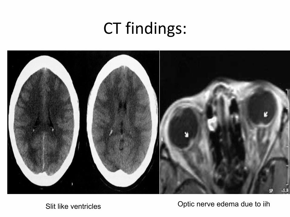
CT findings:
Optic nerve edema due to iihSlit like ventricles
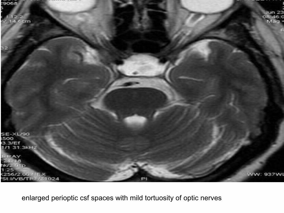
enlarged perioptic csf spaces with mild tortuosity of optic nerves
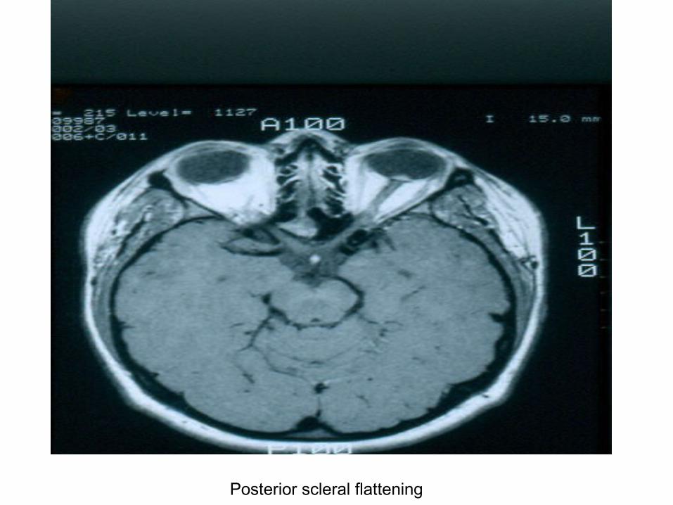
Posterior scleral flattening
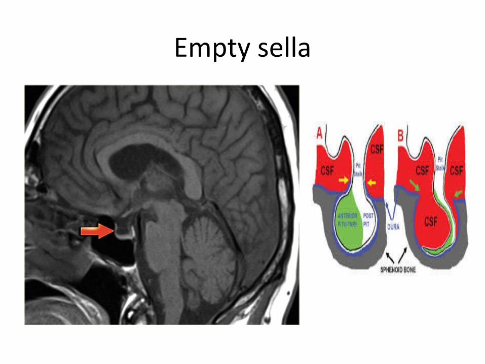
Empty sella
Treatment Approach
• The treatment goal for patients with idiopathic intracranial hypertension (IIH) is to preserve optic nerve function while managing increased intracranial pressure (ICP).
• Optic nerve function should be carefully monitored with an assessment of visual acuity, color vision, optic nerve head appearance, and perimetry.
• Weight control is recommended for obese patients.• Exogenous agents related to increased ICP should be discontinued.• Patients without visual loss most often are treated with a carbonic anhydrase
inhibitor (eg, acetazolamide) to lower the ICP.
• In patients with severe symptoms, early visual field loss, or poor response to standard medical therapy, some clinicians utilize a short course of high-dose corticosteroids .
• When new visual field loss is documented, medical management should be coupled with plans for emergency surgical intervention if the visual function continues to deteriorate .
Pharmacological
• Acetazolamide appears to be the most effective agent for lowering ICP. Most patients experience adequate relief of symptoms (typically, headache) with this first-line agent.
Most patients respond to a dosage of 1-2 g/day. In the event of intolerance to acetazolamide, furosemide may be used.
• In a new study of 165 patients with IIH and mild vision loss, researchers found that acetazolamide treatment for six months in conjunction with a low-sodium weight-reduction diet modestly improved vision, reduced intracranial pressure, improved quality of life, and reduced papilledema. Data show that the effect of the drug was independent of weight loss.
• {Wall M, McDermott MP, Kieburtz KD, Corbett JJ, Feldon SE, Friedman DI, et al. Effect of acetazolamide on visual function in patients with idiopathic intracranial hypertension and mild visual loss: the idiopathic intracranial hypertension treatment trial. JAMA. Apr 23-30 2014;311(16):1641-51.}
• Headache prophylaxis
For patients with stable visual function but inadequate headache relief with first-line pressure-lowering drugs, primary headache prophylaxis should be considered.
amitriptyline, propranolol, or other commonly prescribed migraine prophylaxis agents are used.
• Corticosteroids are effective in lowering ICP in patients whose IIH has an inflammatory etiology. In addition, they may be used as a supplement to acetazolamide to hasten recovery in patients who present with severe papilledema. Patients experiencing a progressive loss of visual field in one or both of the eyes should immediately be placed on high-dose (60-100 mg/day) oral prednisone or an equivalent
Acta Neurol Scand. 2007 Nov;116(5):322-7.Treatment of idiopathic intracranial hypertension: topiramate vs acetazolamide, an open-label
study.
• Topiramate is also an excellent choice, in that one of its side effects is weight loss , which can help put the disease in remission.
• A study of 44 pts assessed the efficacy of topiramate in the treatment of idiopathic intracranial hypertension (IIH) and to compare it with acetazolamide.
When the follow-up visual field grades were compared with the visual field grades at the beginning of the study in each group a statistically significant improvement was detected with both drugs. When the results of the two treatment groups were compared with each other no statistically significant difference was present. Prominent weight loss was recorded in the topiramate group.
Surgical management of IIH
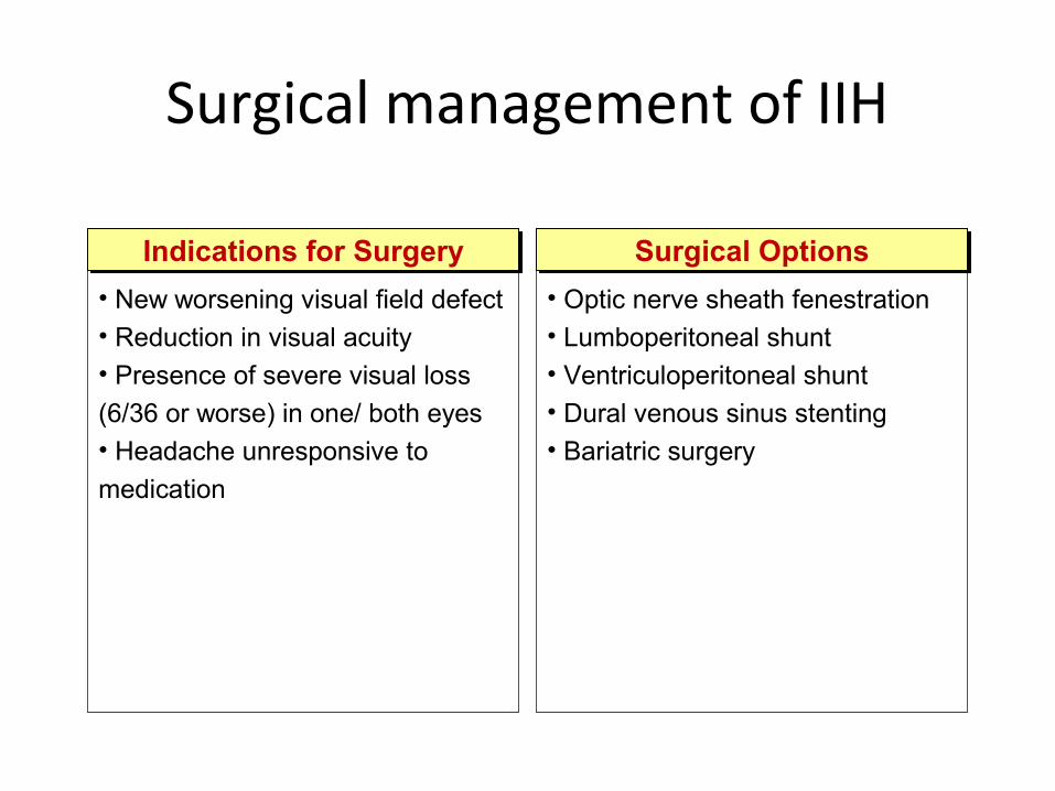
Indications for SurgeryIndications for Surgery
• New worsening visual field defect• Reduction in visual acuity• Presence of severe visual loss
(6/36 or worse) in one/ both eyes • Headache unresponsive to
medication
Surgical OptionsSurgical Options
• Optic nerve sheath fenestration• Lumboperitoneal shunt• Ventriculoperitoneal shunt• Dural venous sinus stenting• Bariatric surgery
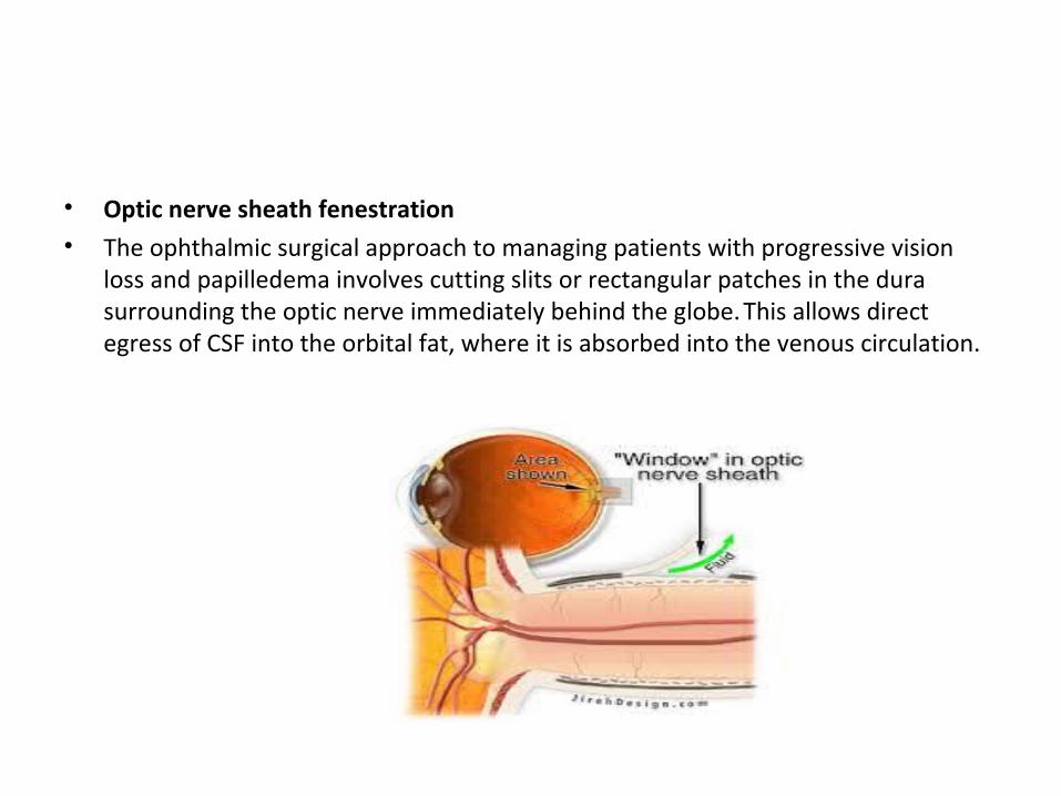
• Optic nerve sheath fenestration• The ophthalmic surgical approach to managing patients with progressive vision
loss and papilledema involves cutting slits or rectangular patches in the dura surrounding the optic nerve immediately behind the globe. This allows direct egress of CSF into the orbital fat, where it is absorbed into the venous circulation.
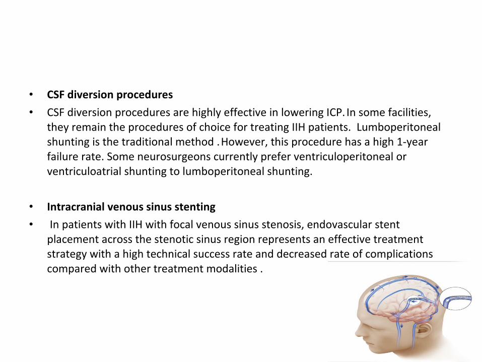
• CSF diversion procedures
• CSF diversion procedures are highly effective in lowering ICP. In some facilities, they remain the procedures of choice for treating IIH patients. Lumboperitoneal shunting is the traditional method . However, this procedure has a high 1-year failure rate. Some neurosurgeons currently prefer ventriculoperitoneal or ventriculoatrial shunting to lumboperitoneal shunting.
• Intracranial venous sinus stenting
• In patients with IIH with focal venous sinus stenosis, endovascular stent placement across the stenotic sinus region represents an effective treatment strategy with a high technical success rate and decreased rate of complications compared with other treatment modalities .
Bariatric surgery
• A review of the literature examining the use of bariatri surgery in IIH was published, reporting on a total of 62 patients. The Roux-en-Y gastric bypass was the most common bariatric procedure performed. Over 90% had resolution of their IIH symptoms, and 97% of those with papilloedema were found to have resolution. The average postoperative ICP decrease was 25 cmH2O. Prospective, controlled studies are necessary .
[Fridley J, Foroozan R, Sherman V, Brandt ML, Yoshor D. Bariatric surgery for the treatment of idiopathic intracranial hypertension. J Neurosurg. Jan 2011;114(1)]
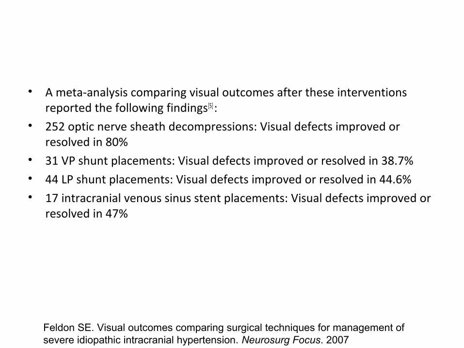
• A meta-analysis comparing visual outcomes after these interventions reported the following findings[5] :
• 252 optic nerve sheath decompressions: Visual defects improved or resolved in 80%
• 31 VP shunt placements: Visual defects improved or resolved in 38.7%• 44 LP shunt placements: Visual defects improved or resolved in 44.6%• 17 intracranial venous sinus stent placements: Visual defects improved or
resolved in 47%
Feldon SE. Visual outcomes comparing surgical techniques for management of severe idiopathic intracranial hypertension. Neurosurg Focus. 2007
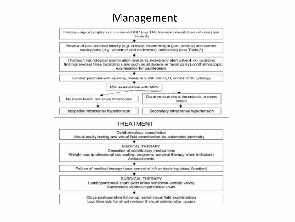
Management
COURSE & Monitoring
• The appearance of papilledema can be deceptive. The swelling may decrease among patients with papilledema due to progressive optic atrophy. Therefore, it is difficult to interpret the appearance of the optic disc in cases of chronic papilledema; optic nerve function is most important.
• Testing of the visual fields with finger confrontation is notoriously insensitive. The best method is automated perimetry(Humphrey perimetry),
• All patients with a suspected diagnosis of IIH should undergo regular testing of the visual fields with quantitative perimetry.

thankyou


![Idiopathic Intracranial Hypertension and the Evaluation of ...Papilledema is the swelling of the optic disc only in the setting of increased intracranial . pressure (ICP) [1]. Swelling](https://img.pdfslide.us/doc/110x75/5e638a517c188f5ba85f36a6/idiopathic-intracranial-hypertension-and-the-evaluation-of-papilledema-is-the.jpg)