Embed Size (px)
Citation preview

HYPERSENSITIVITY
Dr. Amit Makkar, Sudha Rustagi Dental College

• Hypersensitivity reactions are harmful antigen-specific immune
responses , occur when an individual who has been primed by an
innocuous antigen subsequently encounters the same antigen ,
produce tissue injury and dysfuntion.
• It is defined as a state of exagerrated immune response to an antigen.

Coombs & Gell Classification (1963)
Type I (Anaphylactic)
Type II (Cytotoxic )
Type III (Immune complex)
Type IV (Delayed or Cell mediated)

Sell’s Classification (1972)
Immediate (antibody mediated)
Arthus toxic complex associated with precipitating
IgG antibodiesArthus reactionSerum sickness
Atopic anaphylactic reaction associated with IgE antibodiesHay feverAsthmaUrticaria
Hematologic reaction associated with cytotoxic
effect, IgG IgM cause lysis by
complement
Early inflammatory
Antibodies causing
neutralization of biological molecules
HormonesClotting factor
Involving epithelial and
giant cells(granulomatous)TBFungal Inf
Involving perivascular round cell infiltration.
TBBacterialhypersensitivityContact dermatitis
Delayed (cell mediated)

Chase Classification IMMEDIATE REACTION DELAYED REACTION
Appears and recedes rapidly Appears slowly and lasts longer
Induced by antigen by any route Indued by infection, antigen injection, skin contact
Circulating antibodies present and responsible for reaction
Cell mediated reaction
Passive transfer possible with serum Transfer possible by lymphocytes or tranfer factors
Desensitisation is easy but short lived Desensitisation is difficult but long lasting
Lesions are acute exudation and fatty necrosis Mononuclear cell collection around blood vessels
Wheal and flare with maximum diameter in ^ hours
Erythema and induration with maximum diameter in 24-48 hours

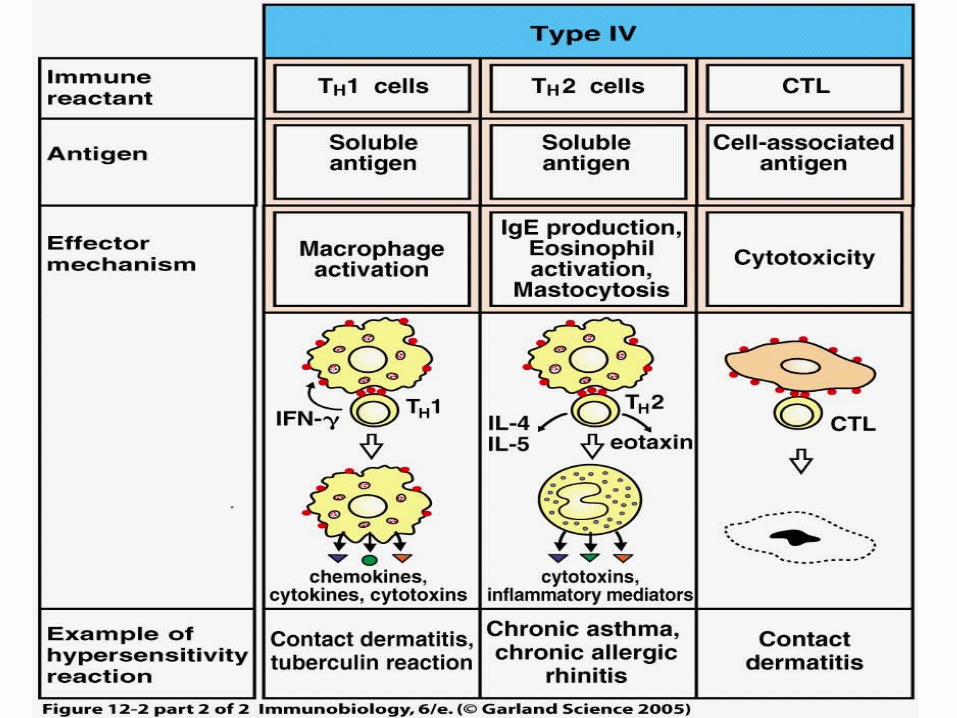

I)Type I Hypersensitivity(Anaphylactic, Atopic)
• It is defined as a state of rapidly developing immune response to an
antigen to which the individual is previously sensitised.
• The response is mediated by humoral antibodies of IgE type or reagin
antibodies.

Priming stage
Activating stage Effect stage
The process and mechanism of Type I hypersensitivity

1) Priming stage:last more than half a year
2) Activating stage:
Cross-linkage Enzyme reaction
De-granulation of mast cell , basophil

3) Effect stage
Immediate/early phase response
•Mediated by histamine
•Start within seconds
•Last several hours
Late-phase response
•Mediated by new-synthesized lipid mediators
•Take up 8-12hours to develop
•Last several days

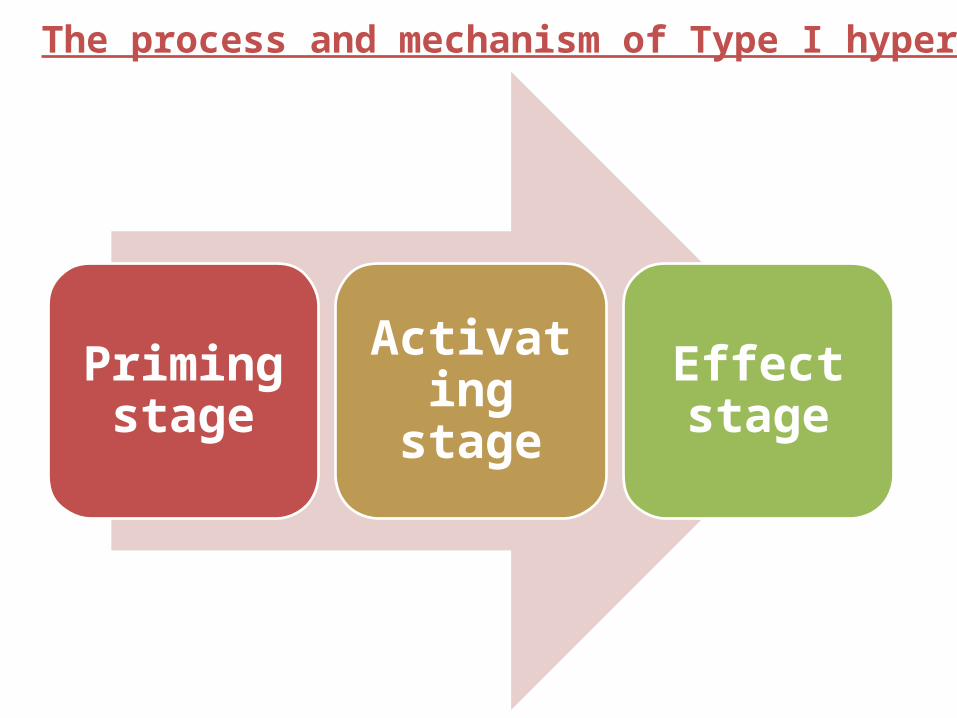
Allergen Primary
Individual Generation
IgE
Adhesion
IgE binds to the FceRI on mast cell and basophil Secondary
Allergen binds to the IgE on primed target cell
Crosslikage of FceRI
Degranulate and release the biological mediators
Preformed granule mediators New generated mediators
Histamine Bradykinin Leukotrienes PAF Prostaglandin D2
Dilate capillaries,increase permeability, increase mucus secretion, contract smooth muscle
Systemic anaphylaxis Skin Respiratory tract Degist tract
Mechanism of type I hypersensitivity

Allergen
Degranulation ,release and synthesis of biological mediators of primed target cells
LOXCOXAcetyl-transferases
Phosphoration of ITAM
MAPK
Lipid mediatiors
Endoplasmic reticulum
Degranulation
Myosin
Phosphoration of Light chain
Cell membrane
Activation of PTK
Phosphatidylcholine
Histamine
Arachidonic acid
Inactivated PKC
ActivatedPKC
Hydroxyl phosphalipid
Phosphalipid
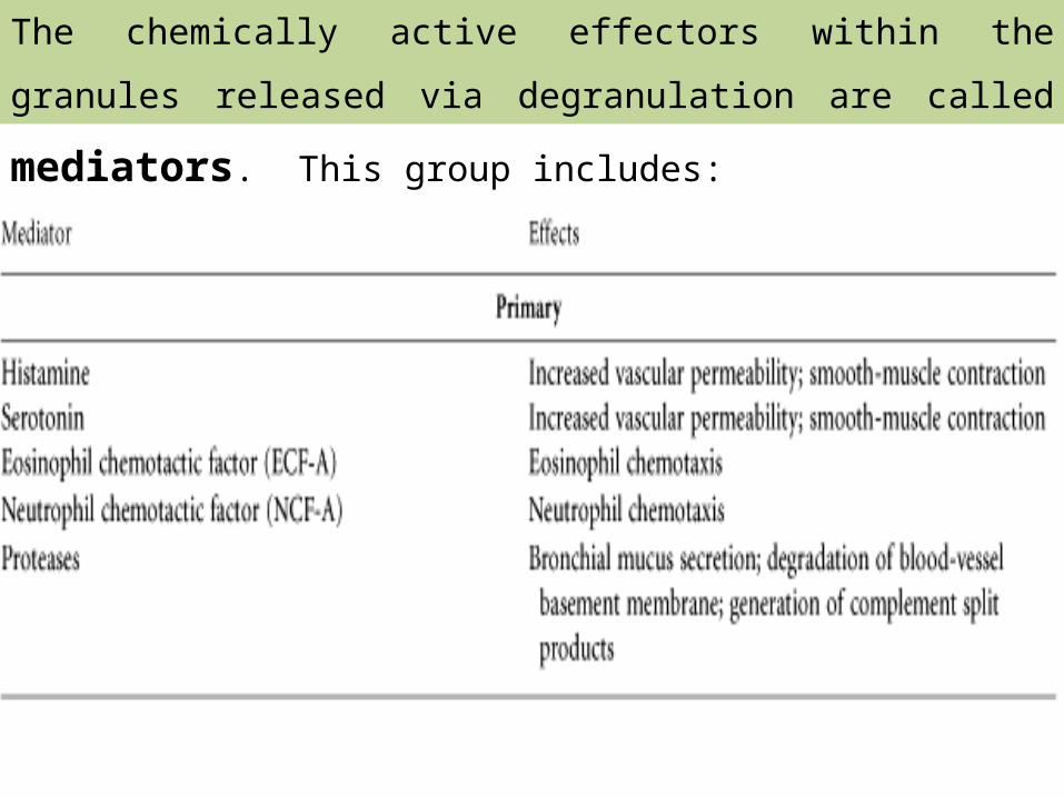
The chemically active effectors within the granules released via
degranulation are called mediators. This group includes:

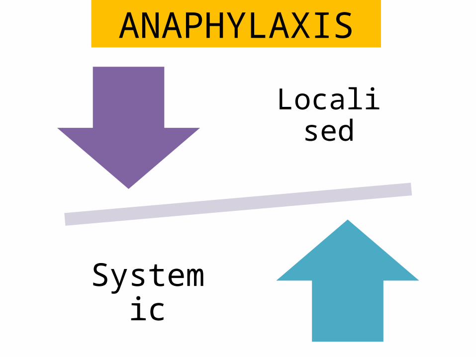
ANAPHYLAXIS
Localised
Systemic

The clinical features of systemic anaphylaxis include itching,
erythema, contraction of respiratory bronchioles, diarrhoea,
pulmonary oedema, pulmonary haemorrhage, shock and death.
Examples of systemic anaphylaxis :-
i) Administration of antisera e.g. anti-tetanus serum (ATS)
ii) Administration of drugs e.g. penicillin
iii) Sting by wasp or bee.
Systemic Anaphylaxis

Pathophysiology of Systemic Anaphylaxis
• Systemic vasodilation and smooth muscle contraction leading to
severe bronchiole constriction, edema, and shock.
• Similar to systemic inflammation.

wheal\ urticarial rash Thick lips and periorbital edema (angioedema)

Localised Anaphylaxis
• Local anaphylaxis is common, affecting about 10% of population.
About 50% of these conditions are familial with genetic
predisposition and therefore also called atopic reactions

HAY FEVER (seasonal allergic rhinitis) due to pollen sensitisation of conjunctiva and nasal passages
BRONCHIAL ASTHMA due to allergy to inhaled allergens like house dust
FOOD ALLERGY to ingested allergens like fish, cow's milk etc
CUTANEOUS ANAPHYLAXIS due to contact of antigen with skin characterised by urticaria, wheal and flare
ANGIOEDEMA, an autosomal dominant inherited disorder characterised by laryngeal oedema, oedema of eyelids, lips, tongue and trunk.


Therapy of type I hypersensitivity
The basic 4A’s in the management of anaphylactic reaction :-
•Antihistaminic agent (benedryl 20-50mg)
•Adrenaline 0.5 ml of 1:1000 i.m
•Aminophylline 0.5mg i.m
•Airway oxygen
•Other---
Adrenaline inhalents
Hydrocortisone sodium succinate 100mg i.m
Cricothyrotomy for airway maintainance if required

Therapy of type I hypersensitivity
1. Allergen avoidance : Atopy patch test
2. Desensitivity therapy / Hyposensitization :
i) Allogenic serum desensitivity therapy:
ii) Specific allergen desensitivity therapy
Repeated injection small amounts of allergen (allergy shots)
IgG+allergen Neutralizing antibody,
Blocking antibody

3. Drug therapy:i) Stabilization of triggering cells
Sodium cromoglycate stabilize the membrane,inhibit mast cell degranulation
ii) Mediator antagonism
Chlor-Trimeton Antihistamine
Acetylsalicylic acid Bradykinin antagonism
iii) Improve the responsibility of target organs
4. Allergen immunotherapy : Rehabilitates the immune system and involves administering increasing doses of allergens to accustom the body to substances that are generally harmless (pollen, house dust mites) and thereby induce specific long-term tolerance.Allergen immunotherapy can be administered under the tongue (sublingually with drops or tablets) or by injections under the skin (subcutaneous).

• Cytotoxic reactions are defined as those reactions which cause injury
to the cell by combining humoral antibodies with cell surface
antigens; blood cells being affected more commonly.
Characteristic features
Primed IgG or IgM +Antigen or hapten on membrane
Injury and dysfunction of target cells
I)Type II Hypersensitivity(Cytotoxic Reaction)

• Involves the antibody mediated destruction of cells.
• Can mediate cell destruction by activating the complement system to
create pores in the membrane of the foreign cell.
• Can also be mediated by Antibody-Dependent Cell-Mediated
Cytotoxicity (ADCC) where the Fc receptors bind to Fc receptor of
antibody on the target cell and promote killing.

Allergen
Stimulate
Antibody
A. Opsonic phagocytosis
D. ADCC of NK
C. Effect of complement
Combined opsonic activities
Cell injury ways of type II hypersensitivity
Cell

1. Surface antigen on target cells
Target cells: Normal tissue cell, changed or modified self tissue cells
Antigen : Blood group antigen, Common antigen,
Self-antigen modified by physical factors orinfection
Drug antigen, Antigen-antibody complex
Mechanism of type II hypersensitivity
Activate complement Lyse target cells
Opsonic phogacytosis Destroy target cells
ADCCMf 、 NK 、 T
Stimulating or blocking effect Promote /surpress the target cell funcion
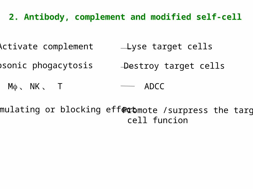
2. Antibody, complement and modified self-cell

Antigen or hapten on cell
Antibody (IgG, IgM)
Activate complement
Lyse target cell
Opsonic phagocytosis NK , phagocyte Stimulate / block
Destroy target cell ADCC
Target cell injury Change the function ofTarget cell
Mechanism of Type II hypersensitivity

Mechanisms involved in mediating cytotoxic reactions
Cytotoxic antibodies to blood cells
Cytotoxic antibodies to
tissue components Antibody-
dependent cell-mediated
cytotoxicity (ADCC).

– Involves direct cytolysis of blood cells (red blood cells, leucocytes
and platelets) by combining the cell surface antigen with IgG or
IgM class antibodies.
– Complement system is activated resulting in injury to the cell
membrane.
– Cell surface is made susceptible to phagocytosis due to coating or
opsonisation from serum factors or opsonins.
A. CYTOTOXIC ANTIBODIES TO BLOOD CELLS


Autoimmune hemolytic anemia and type II drug reaction
FOREIGN ANTIGEN OR HAPTEN
1. Penicillin RBC hemolytic anemia
2.Quinine Platelet thrombocytopenic purpura
3.Pyramidone Granulocyte agranulocytosis
SELF-ANTIGEN
Drug
conversion from a hapten to a full antigen
induce self antibody
autoimmune hemolytic anemia
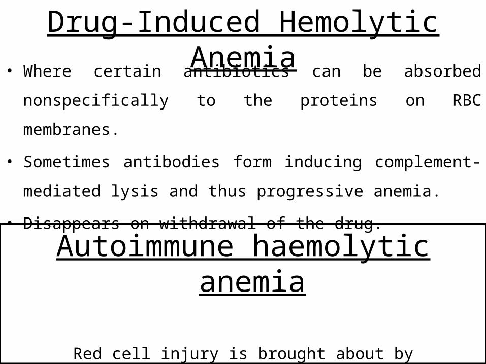
Drug-Induced Hemolytic Anemia
• Where certain antibiotics can be absorbed nonspecifically to the
proteins on RBC membranes.
• Sometimes antibodies form inducing complement-mediated lysis and
thus progressive anemia.
• Disappears on withdrawal of the drug.
Autoimmune haemolytic anemia
Red cell injury is brought about by autoantibodies reacting with antigens
present on red cell surface.

Transfusion reaction
• Mismatch of ABO blood group
• Severely destroy RBCHemolysis
• Repeat transfusion of allogenic HLA
• Drug anaphylactic shock:penicilline
Nonhemolysis

Transfusion reactions • Antibodies of the A,B, and O antigens are usually of the IgM class
(these antigens are call isohemagglutinins)
• For example an A individual produce isohemagglutinins to B-like
epitopes but not to A epitopes because they are self
• Person who are transfused with the wrong blood type will produce
anti-hemmagglutinins causing complement mediated lysis
• Antibodies are usually of the IgG class

• Transfusion reactions can be delayed or immediate but have different
Ig isohemagglutinins
• Immediate reactions has a complement-mediated lysis triggered by
IgM isohemagglutinins
• Delayed reactions induce clonal selection and the productions of IgG
which is less effective in activating the complement.
• This leads to incomplete complement-mediated lysis
• Cross-matching can detect antibodies in the sera to prevent this
Haemolytic disease of the newborn
• Fetal red cells are destroyed by maternal isoantibodies crossing
placenta
• This is where maternal IgG antibodies specific for fetal blood group
antigens cross the placenta and destroy fetal RBC’s
• Erythroblastosis fetalis - severe hemolytic disease of newborns
– Most commonly develops when an Rh+ fetus expresses an Rh
antigen on it’s blood that and Rh- mother doesn’t recognize


During the 1st pregnancy small amounts of fetal blood pass through the placenta but not enough to induce a response
During delivery larger amounts of fetal blood cross the placenta causing an activation of B-cells that are Rh specific thus leading
to memory B-cells (anti-Rh antibodies)
The IgM antibody clears the Rh+ cells from the mother
In subsequent pregnancies with an Rh+ fetus, the Rh+ RBC cross the placenta activating the memory B-cells
These in turn cross the placenta and damage the fetal RBC because they are seen as “foreign”

• This type of reaction can be prevented by administering antibodies
against the Rh antigen within 25-48 hours after the 1st delivery
• Rhogam - is the antibody that is injected
– it will bind to the fetal RBC that enter the mother’s circulation and
facilitate the clearance of them before B-cell activation
– In subsequent pregnancies the mother is unlikely to produce IgG
anti-Rh antibodies
– If the mother doesn’t receive this injection there are other ways to
treat this, depending on the severity
Treatment of Erythroblastosis fetalis

• Cell injury may be brought about by autoantibodies reacting, with
some components of tissue cells in certain diseases,
• Example –
In myasthenia gravis, antibody to acetylcholine receptors of skeletal
muscle is formed which blocks neuromuscular transmission at the
motor end-plate resulting in muscle weakness
B. CYTOTOXIC ANTIBODIES TO TISSUE COMPONENTS


• Mediated by leucocytes like monocytes, neutrophils, eosinophils &
NK cells.
• Antibodies involved - mostly IgG
• The cellular injury occurs by lysis of antibody-coated target cells
through Fc receptors on leucocytes.
• The examples of target cells killed by this mechanism are tumour
cells, parasites etc.
C. ANTIBODY-DEPENDENT CELL MEDIATED CYTOTOXICITY
(ADCC)


1.Anti -glomerular basement membrane nephritis
β-Hemolytic streptococcus and human glomerular basement membrane ---- cross reaction Common antigen ---nephrotoxic nephritis
2. Super acute rejection in allogenic organ transplantation
3. Goodpasture syndrome
4.Hyperthyroidism or hypothyroidism—receptor diseases
OTHER DISEASES

Type III reactions results from formation of immune complexes by
direct antigen-antibody (Ag-Ab) combination as a result of which the
complement system gets activated causing cell injury.
III) Type III Hypersensitivity(Immune Complex Reaction)
Antigens causes immune complex mediated tissue injury
Exogenous Antigens
Endogenous Antigens

Endogenous Antigens– Blood components
(Ig, tumour antigens)– Antigens in cells & tissues
(nuclear antigens in SLE)
Exogenous Antigens
– Infectious agents (bacteria, viruses, fungi, parasites)
– Certain drugs & chemicals

Depending upon the distribution & location of antigens, Type III are of 2 types
LOCAL
Arthus reactions
SYSTEMICCirculating immune complex disease or
Serum sickness

1. Local : Arthus Reaction
• Localised inflammatory reaction, usually an immune complex
vasculitis of skin of an individual with circulating antibody.
• Large immune complexes formed due to excess of antibodies, which
precipitate locally in the vessel wall causing fibrinoid necrosis.
1. Local : Arthus Reaction
• Localised inflammatory reaction, usually an immune complex
vasculitis of skin of an individual with circulating antibody.
• Large immune complexes formed due to excess of antibodies, which
precipitate locally in the vessel wall causing fibrinoid necrosis.
Injection of an Antigen:• Can lead to an acute Arthus reaction within 4-8 hours
• Localized tissue and vascular damage result from accumulation
of fluid (edema) and RBC (erythema)
• Severity can vary from mild swelling to redness to tissue necrosis
EXAMPLES :-
1. Injection of Antitetanus serum
2. Farmer`s lung (allergic alveolitis in response to bacterial antigen from
mouldy hay)
3. Insect bite:
• May first have a rapid type I reaction
• Some 4-8 hours later a typical Arthus reaction develops
4. Ulcer
5. Local Human Reaction :- Insulin Dependent Diabetes Mellitus

2. Systematic : Circulating immune complex disease or serum sickness
• Develops when antigen is intravenously administered resulting in
formation of large amountsvantigen-antibody complexes and their
deposition in the tissues.
• These circulating complexes can’t be cleared by phagocytosis and can
cause tissue damaging Type III reactions

• Ag-Ab complexes are deposited at different tissue sites containing
basement membrane exposed to circulating blood.
• Following this deposition, there is acute inflammatory reaction &
activation of complement system with elaboration of chemotactic
factors, vasoactive amines & anaphylatoxins.
• This all causes type III hypersensitivity reactions

• Eg of circulating immune complex diseases are:-– Skin diseases
– Various forms of Glomerulonephritis
• Other conditions caused by Type III-1. Infectious Diseases
• Meningitis
• Hepatitis
• Mononucleosis
2. Drug Reactions
• Allergies to penicillin and sulfonamides
3. Autoimmune Collagen Diseases
• Systematic lupus erythematosus
• Rheumatoid arthritis

Serum Sickness Systemic immune complex disease
Days after Antigen Injection
Large amounts of antigen such as injection of foreign serum.

Soluble antigen Body Antibody
Immune complex
Small molecular soluble Immune complex
intermediate molecular soluble Immune complex
Large molecular insoluble Immune complex
Deposit on the basement of capillaries
Combine and activate complement system
C3a,C5a,C3b
Infiltration of neutrophils
Phagocytose complex
Release the enzymes in lysosome
Tissue injury
Eliminate by phogacytosis
Platelets
Thrombus
Aggregation of platlets
Blood Clotting Mechanisms
Release of vasoactive amine
Increase vascular permeability
Bleeding Edema
Basophils and mast cells
Release of vasoactive amine
Increase vascular permeability
Edema
Local or systemic immune complex diseases
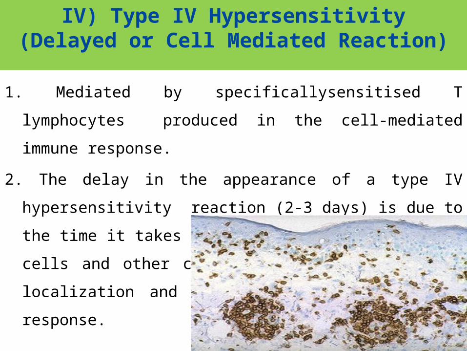
1. Mediated by specifically sensitised T lymphocytes produced in the
cell-mediated immune response.
2. The delay in the appearance of a type IV hypersensitivity reaction (2-
3 days) is due to the time it takes to recruit antigen-specific T cells
and other cells to the site of antigen localization and to develop the
inflammatory response.
IV) Type IV Hypersensitivity(Delayed or Cell Mediated Reaction)

Antigen introduced to the tissue modifies
extracellularand cell surface proteins
Macrophages process antigen, present to Th1
cells
Th1 effector cell recognizes the antigen
and
Releases cytokines which act on local
vascular endothelium and
Further activateion of macrophages (increase in size, microbicidal activity, & lysosome
content)
T cell recruitment (CD4 & CD8), fluid and
protein
Absorption by interstitum (edema

Classical delayed
hypersensitivity T Cell-mediated cytotoxicity
Types of Cell mediated Reactions

Classical delayed hypersensitivity
– Mediated by specifically
sensitised CD4+ T cell
subpopulation on contact with
antigen.
– These cells possess surface
receptors which bind to the
antigen, resulting in cell injury
characterised by slowly
developing inflammatory response

T Cell-mediated cytotoxicity
CD8+ subpopulation of T lymphocytes are the cytotoxic T cells are
generated in response to antigens like virus-infected cells, tumour
cells and incompatible transplanted tissue or cells.

Mechanism of type IV hypersensitivity
Formation of effector and memory T cells
Inflammation and cytotoxicity caused by effector T cells
1) Inflammation and tissue injury mediated by CD4+Th1
Release chemokines and cytokines
Immune injury mainly caused by infiltration of mononuclear cells and
lymphocytes
2) Cytotoxicity of CD8+CTL

Antigen T cell(CD4+,CD8+)
Secondary contact
Induce
Primed T cell
CD4+ T cell
CD8+ T cell
Release CytokinesIL-2
TNF-bINF-gMCFMIFSRF
Directly kill target cells
Infiltration of monocyte and Mf
Proliferation of T cell
Exudation and edema
Cytotoxicity
Inflammation characterized by infiltration of Mf , monocyte, And tissue injury
Mechanism of type IV hypersensitivity

Stages of a Type IV Hypersensitivity Reaction

Th1 derived cytokines and chemokines direct Type IV reactions
Delayed Type Hypersensitivity (DTH)
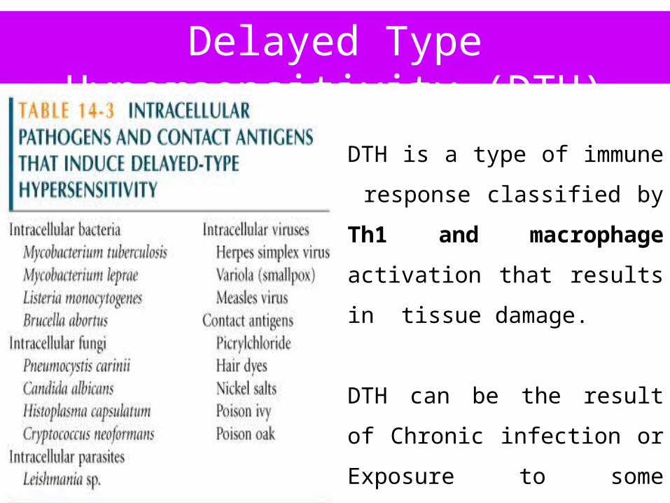
DTH is a type of immune
response classified by Th1 and
macrophage activation that results
in tissue damage.
DTH can be the result of Chronic
infection or Exposure to some
antigens.
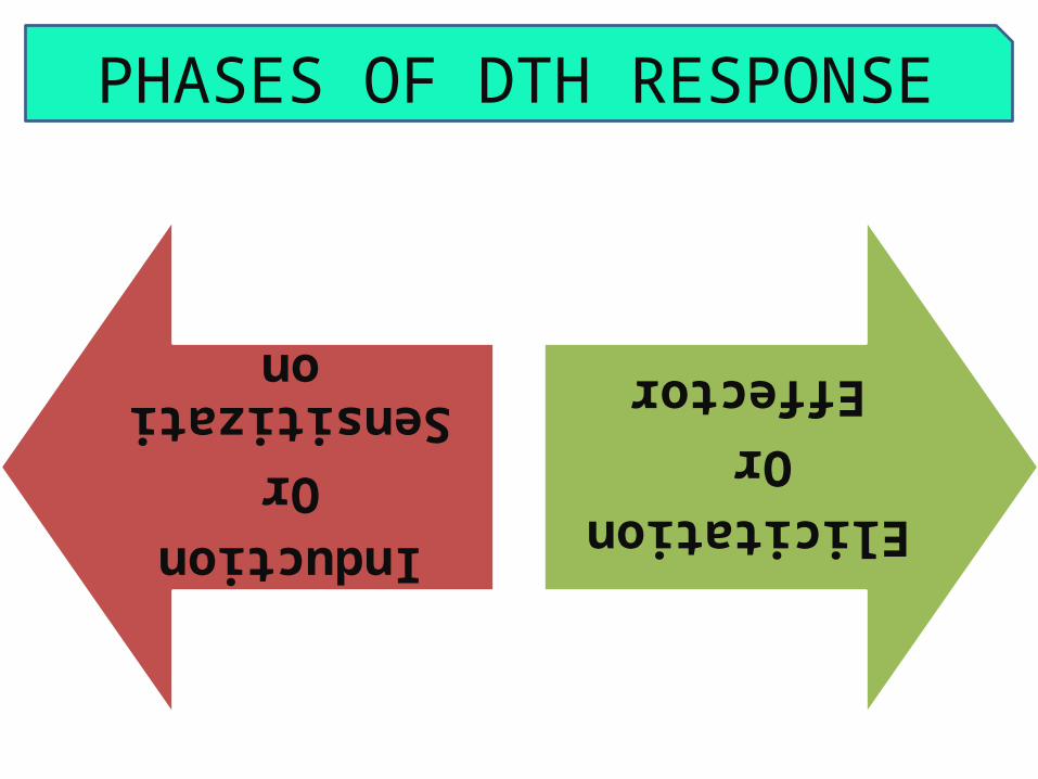
PHASES OF DTH RESPONSE
Induction
Or
Sensitization
Elicitation
Or
Effector

Occurs 1-2 weeks after primary contact with Ag
• TH cells are activated and clonally expanded by Ag presented together with class II MHC
on an appropriate APC, such as macrophages or Langerhan cell (dendritic epidermal cell)
• Generally CD4+ cells of the TH1 subtype are activated during sensitization and designated
as TDTH cells
Sensitization Phase

Occurs upon subsequent exposure to the Ag
• TDTH cells secrete a variety of cytokines and chemokines, which recruit and activate
macrophages
• Macrophage activation promotes phagocytic activity and increased concentration of lytic
enzymes for more effective killing
• Activated macrophages are also more effective in presenting Ag and function as the primary
effector cell
Effector Phase

Chemical Factors InvolvedIL-12: (macrophages). Drives differentiation of T cells, induces IFN- gamma secretionIFN-gamma (T cells). Further activates macrophages
IL-2: (T cells). Increases T cell proliferation within tissue
IL-3: (T cells). Stimulates monocyte production
TNF (T cells). Increase secretion of Nitric Oxide & Prostacyclins by endothelial cells, local tissue destruction, and increase expression of adhesion molecules on vessels
E- selectins: vascular adhesion molecule, increases mononuclear cell attachment

Prolongation of DTH Response
A granuloma develops…
• Continuous activation of
macrophages induces the
macrophages to adhere closely
to one another, assuming an
epithelioid shape and
sometimes fusing together to
form giant, multinucleated cells.

Detrimental Effects of DTH Response
• The initial response of the DTH is nonspecific and often results in
significant damage to healthy tissue.
• In some cases, a DTH response can cause such extensive tissue
damage that the response itself is pathogenic.
• Example: Mycobacterium tuberculosis – an accumulation of activated
macrophages whose lysosomal enzymes destroy healthy lung tissue.
In this case, tissue damage far outweighs any beneficial effects.
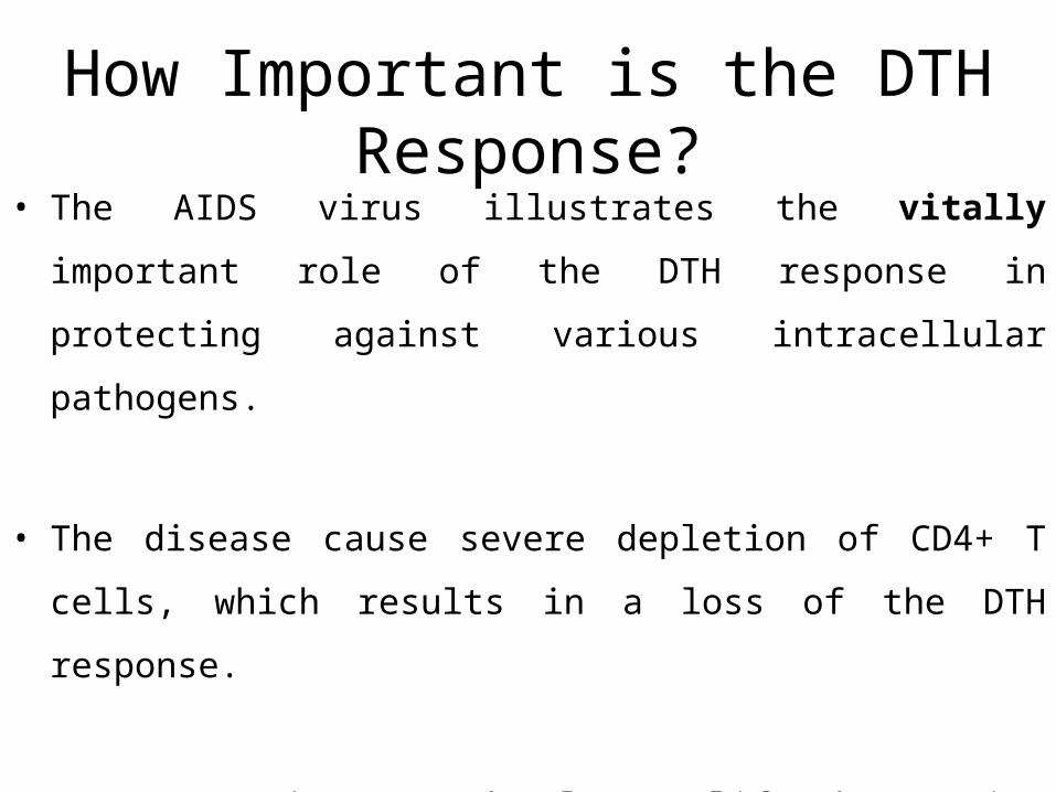
How Important is the DTH Response?
• The AIDS virus illustrates the vitally important role of the DTH
response in protecting against various intracellular pathogens.
• The disease cause severe depletion of CD4+ T cells, which results in a
loss of the DTH response.
• AIDS patients develop life-threatening infections from intracellular
pathogens that normally would not occur in individuals with intact
DTH responses.

1) Infectious delayed type hypersensitivity
OT( Old Tuberculin ) test
2) Contact dermatitis :
Paint, drug red rash, papula, water blister, dermatitis
3) Acute rejection of allogenic transplantation and
immune response in local tumor mass
Common disease of type IV hypersensitivity

TUBERCULIN REACTION
–On intradermal injection of tuberculoprotein (PPD), an unsensitised
individual develops no response (tuberculin negative).
– A person who has developed cell-mediated immunity to
tuberculoprotein as a result of BCG immunisation (exposed to
tuberculous infection) develops typical delayed inflammatory reaction,
reaching its peak in 48 hours (tuberculin positive), after which it
subsides slowly.

Contact Dermatitis

Allergic Contact Dermatitis: A type IV reaction

Blistering skin lesions on hand of patient with poison ivy
contact dermatitis (a type IV reaction)

Granulomatous inflammation is a consequence of chronic Type IV reactions

Type IV Hypersensitivity Reactions
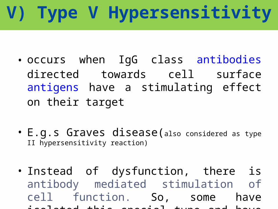
• occurs when IgG class antibodies directed towards cell surface antigens have a stimulating effect on their target
• E.g.s Graves disease(also considered as type II hypersensitivity reaction)
• Instead of dysfunction, there is antibody mediated stimulation of cell function. So, some have isolated this special type and have named it type V hypersensitivity reaction.
V) Type V Hypersensitivity

HYPERSENSITIVITY DUE TO VARIOUS DENTAL
MATERIALS
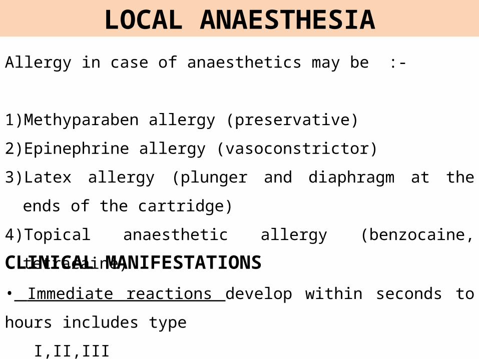
LOCAL ANAESTHESIA
Allergy in case of anaesthetics may be :-
1) Methyparaben allergy (preservative)
2) Epinephrine allergy (vasoconstrictor)
3) Latex allergy (plunger and diaphragm at the ends of the cartridge)
4) Topical anaesthetic allergy (benzocaine, tetracaine)
CLINICAL MANIFESTATIONS
• Immediate reactions develop within seconds to hours includes type
I,II,III
• Delayed reactins shows manifestations in hours to days.

SIGNS AND SYMPTOMS
1 Dermatological Reactions
Urticaria associated with wheals
Angioedema of face, hand, feet, genitalia
2 Respiratory Reactions
Bronchospasm(distres, dyspnoea, cyanosis, flushing,
tachycardia, perspiration)
Laryngeal edema
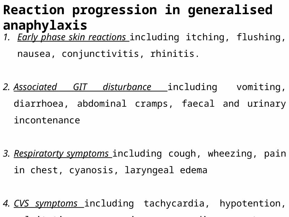
Reaction progression in generalised anaphylaxis
1. Early phase skin reactions including itching, flushing, nausea,
conjunctivitis, rhinitis.
2. Associated GIT disturbance including vomiting, diarrhoea, abdominal
cramps, faecal and urinary incontenance
3. Respiratorty symptoms including cough, wheezing, pain in chest,
cyanosis, laryngeal edema
4. CVS symptoms including tachycardia, hypotention, palpitation,
unconscoiusness, cardiac arrest

MANAGEMENT1. Delayed skin reactions : oral histamine blocker (50g
diphenhydramine or 10g chlorpheniramine)
2 Immediate skin reactions : Epinehrine 0.3mg i.m
oral histamine blocker
3. Respiratory reactions : oxygen administration
Epinehrine or bronchodilator
Histamine blocker i.m
4. Laryngeal edema : epinehrine
airway mainantance, emergency call
Histamine blocker i.m , i.v
Hydrocortisone 100mg
Cricothyrotomy

Ti can induce clinically relevant hypersensitivity and other immune
dysfunctions in certain patients chronically exposed to this reactive
metal. At the same time, no standard patch test for Ti has so far been
developed.
TITANIUM

Hypersensitivity reaction to a metal comes from the presence of ions
following ingestion, skin or mucosal contact, or from implant corrosion
processes. In their ionic form, metals can be bonded with native proteins
to form haptenic Antigens ,or can trigger the degranulation of mastocytes
and basophiles, being capable of developing type I or type IV
hypersensitive reactions according to Schramm and Pitto
Sensitivity to titanium is characterized by the local presence of abundant
macrophages and T lymphocytes and the absence of B lymphocytes,
indicating type IV hypersensitivity
J Indian Prosthodont Soc (Oct-Dec 2012) 12(4):201–207

Allergy to methyl methacrylate monomer in acrylic resin are less
common and usually are of the delayed or contact allergy. Residual
monomer left by incomplete polymerization is the allergen in contact
stomatitis caused by acrylic resin.
The patient may complain of a burning sensation, soreness, dryness, or
excessive salivation. Examination of the oral mucosa may show
punctuate or diffuse redness with or without erosions.
METHY METHACRYLATE

Contact type dermatitis - due to acrylic resin materials acting as
haptens via delayed hypersensitivity mechanism, which is observed in
several dentists and dental laboratory technicians.
Allergic Stomatitis - The term “sore mouth due to dentures” is applied
to any pathologic change of the oral mucosa due to dentures, whether the
cause is allergic, traumatic, or toxic.
Patch testing is a reliable method to check
for Allergy
Clinical Dentistry, Mumbai • September 2012
Dr. Suraj R Suvarna

EUGENOL
Eugenol is a material commonly used in dentistry but is not a bio-
friendly material when in contact with oral soft tissues
Intra-orally it causes destruction of the interdental col and surrounding gingivae..
Erythema and ulceration in left buccal mucosa, adjacent to UL7 (27) on closure of the mouth

1. Eugenol is generally cytotoxic at high concentrations and has an
adverse effect on fibroblasts and osteoblast-like cells. Thus at high
concentrations it produces necrosis and reduced healing.
This effect is dose related and will potentially affect all patients.
2. In lower concentrations, eugenol can act as a contact allergen
evoking a localised delayed hypersensitivity reaction.
3. Rarely, eugenol when placed in the mouth, can cause a more
significant generalised allergic response.

BRITISH DENTAL JOURNAL VOLUME 193 NO. 5 SEPTEMBER 14 2002N. Sarrami1 M. N. Pemberton2 M. H. Thornhill3 E. D. Theaker4
The patient was prescribed benzydamine hydrochloride mouthwash and
triamcinolone in orobase for relief of his acute symptoms
Erythema and ulceration on the innersurface of the upper lip and around thegingival margin ofUR1 (11)

AMALGAM
Hypersensitivity to the constituents of dental amalgam is
uncommon.
1. When hypersensitivity reactions occur, they most commonly take
the form of delayed type IV lichenoid reactions affecting oral
mucosa in direct contact with amalgam fillings.
2. Much more rarely a more acute generalised mucocutaneous response
can occur.
BRITISH DENTAL JOURNAL, VOLUME 188, NO. 2, JANUARY 22 2000 B.McGivern, M.Pemberton, E.D. Theaker, J.A.G.Buchanan, and M.H.Thornhill

Buccal lichenoid reaction to a large amalgam restoration of 7
Resolution of thelichenoid reactionfollowing crowningof 7


Red, itchy rash on the neck and arm following contact with mercury while performing amalgam restorations

PREVENTION AND TREATMENT
1. A detailed history of occupation, lifestyle, environment and prevention of exposure
to mercury is important.
2. Various barrier techniques like using a mask, gloves, hair caps and eye-shields are
advised while working.
3. Careful handling of silver amalgam waste
4. Air conditioners and proper ventilation of the operating room, intermittent use of
the rotary along with coolant to avoid excess heat, high vacuum suction, proper
cleaning and proper handling Of amalgam scraps in a covered container or under
sulphide solution is advocated to avoid vapour production.

Dermashield (dimethisone) is a silicone polymer. Pharmologically inert, it has
water repellant and surface tension. It adheres to skin and protects it and avoid contact
with mercury vapour on the skin. This helps in reducing the lesion’s development.
Clonate lotion contains 0.05% clobetasol propionate which is a glucocorticoid
used topically for a large variety of dermatological conditions due to their anti-
inflammatory, immunosuppressive, vasoconstrictor and antiproliferativev(for scaling
lesions) property.
BRITISH DENTAL JOURNAL VOLUME 205 NO. 7 OCT 11 2008V. K. Bains,1 K. Loomba,2 A. Loomba3 and R. Bains4
Strong positive skin patch test response, with vesiculation, spreading erythema and oedema, following 24 hours exposure to ammoniated mercury

Natural rubber latex, which is an extract from the sap of Hevea
brasiliensis trees, contains 256 proteins,2 including 11 potential
allergens.
Exposure to latex poses the risk of sensitizing both clinicians and their
patients.
Adverse reactions to latex range from mild irritant contact dermatitis to
potentially life threatening hypersensitivity and its risk increases with
prolonged and repeated exposure.
LATEX

Exposure to latex allergens occurs via mucous membranes, the
vascular system, inhalation and direct skin contact.
Adverse reactions to latex include nonallergic contact dermatitis,
delayed type IV
Immunoglobulin E (IgE) mediated type I responses to latex proteins
result in adverse reactions within minutes to hours of exposure ranging
from mild irritation to loss of life.
Symptoms include pruritis, erythema, edema, rhinoconjunctivitis,
urticaria, dyspnea, palpitations, dizziness, bronchospasm, vasodilation,
gastrointestinal cramping, vomiting, hypotension and even death
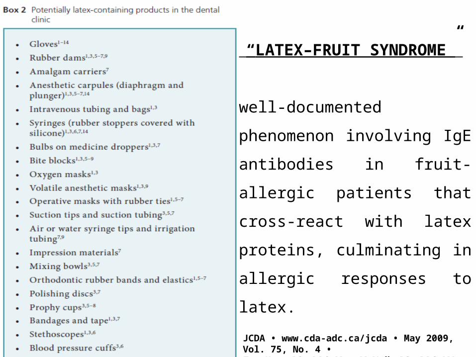
JCDA • www.cda-adc.ca/jcda • May 2009, Vol. 75, No. 4 •Tara Kean, BSc, DDS; Mary McNally, BSc, DDS, MA
“LATEX–FRUIT SYNDROME”
well-documented phenomenon
involving IgE antibodies in fruit-
allergic patients that cross-react
with latex proteins, culminating in
allergic responses to latex.

CDC recommendations to help minimize potential adverse
reactions to dental items.
1 Educate DHCP regarding the signs, symptoms and diagnosis of skin
reactions associated with frequent hand hygiene and glove use
2 Screen all patients for latex allergy (e.g., take a health history and refer for
medical consultation when latex allergy is suspected)
3 Ensure a latex-free environment for patients and DHCP with latex
allergy
4 Have emergency treatment kits with latex-free products available at
all times
5 Develop a written health program for DHCP that includes policies,
procedures and guidelines for contact dermatitiS and latex hypersensitivity

PREVENTION AND TREATMENT
1. Administering prophylactic antihistamines, such as diphenhydramine, or
corticosteroids,such as prednisone, before dental treatment to those at
known risk
2. Reduce the amount of latex allergens present in their products, which are
labeled as "low protein" products (Powder-free gloves)
3. Contact dermatitis and type IV allergy - topical corticosteroids.
4. Mild type I reactions without respiratory distress - topical steroids and
antihistamines (50 mg diphenhydramine 4 times a day until swelling
resolves). Cynthia A. Chillock, CDA, RDH, EF, and Charles John Palenik, MS, PhD, MBA; January 2006 RDH

5 Severe type I hypersensitivity with respiratory distress, swelling of the
tongue, larynx or pharynx and anaphylaxis - assessment of ABCs (airway,
breathing and circulation) and activation of emergency medical services
6 For anaphylaxis - latex-free resuscitation carts are used to administer high-
flow oxygen and deliver 0.3–0.5 mL intramuscular or subcutaneous doses
of 1:1000 epinephrine15 (0.1 mL/kg every 5 minutes for children).
6 Vitals and ABCs should be continually monitored and cardiopulmonary
resuscitation provided if necessary
7 Following stabilization, antihistamines, such as diphenhydramine and
corticosteroids, should be prescribed

ORTHODONTICS RELATED
The causes comprised the metal parts of fixed appliances, polymer-
based activators, retention appliances and brackets, and latex-based
elastics or gloves
Most of the reactions associated with the metallic parts of orthodontic
appliances were either presumed to be a nickel allergy or were
unexplained.

The adverse effects comprised intra-oral reactions such as marked
redness, swelling and soreness of the oral mucosa and palate and similar
symptoms of the gingiva and lips.
Occasionally reactions of a systemic nature, compatible with general
allergic symptoms are seen.
Ezema of the peri-oral area, the cheeks, chin, neck, scalp, earlobes and
skin elsewhere. Occasionally rashes and swelling were seen in the peri-
ocular region.

A 12-year-old girl, with a previous history of bronchial reaction and
contact dermatitis to sodium hypochlorite, was referred for root canal
treatment.
Complete immunologic evaluation revealed a mild hypersensitivity
condition, as it was assessed by the RAST(Radioallergosorbant test)
investigation to different allergens and the DTH reactivity expressed
though migration inhibition test.
Dandakis C, Lambrianidis T, Boura P. Immunologic evaluation ofdental patient with history of hypersensitivity reaction to sodium hypochlorite. Endod Dent Traumatol 2000; 16: 184–187.
SODIUM HYPOCHLORITE

This study describes a case of fatal anaphylaxis that appeared
immediately after the oral mucosa came into contact with an alginate
paste used for dental impressions.
The cadaveric examination and the postmortem toxicology
report confirmed that the cause of death was anaphylactic shock. The
patient was affected by both cardiovascular and lung diseases that
worsened the condition and forbade the use of epinephrine.
Int J Prosthodont 2009;22:33–34Sebastiano Gangemi, PhDa/Elvira Ventura Spagnolo, PhDb/Giulio Cardia, PhDc/Paola L. Minciullo, PhDad.
ALGINATE

Severe occlusion of the laryngeal ostium at the cadaveric
.
Edema of the tongue.
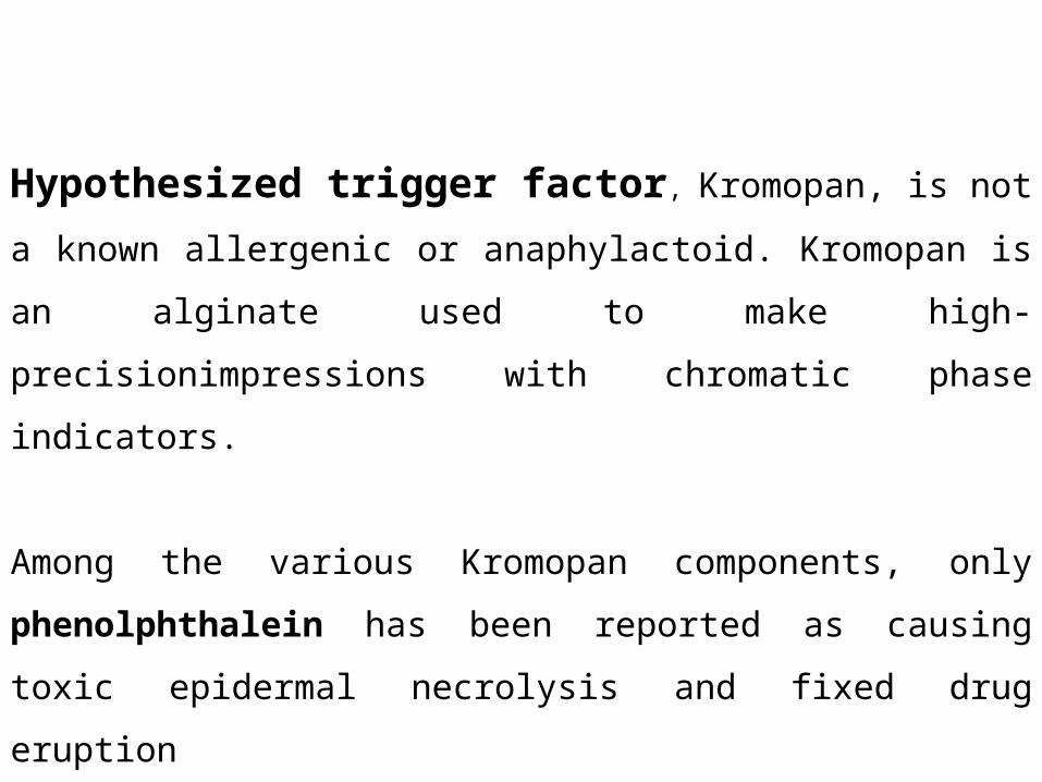
Hypothesized trigger factor, Kromopan, is not a known allergenic
or anaphylactoid. Kromopan is an alginate used to make high-
precisionimpressions with chromatic phase indicators.
Among the various Kromopan components, only phenolphthalein has
been reported as causing toxic epidermal necrolysis and fixed drug
eruption

THANK YOU!!!!