Embed Size (px)
Citation preview
International Commission on Radiation Units & Measurements (ICRU)
ICRU REPORT 62
Dr. Ayush Garg

INTERNATIONAL COMISSION ON RADIATION UNITS AND MEASUREMENTS( ICRU )
ICRU Report No - 29 (1978)Dose specification for reporting external beam therapy in
photons and electrons
ICRU Report – 50 (1993) Supercedes and updates Report 29 Prescribing, Recording, and Reporting photon beam therapy
ICRU Report – 62 (1999) Supplement to ICRU Report No: 50 (ICRU 50 still valid)

ICRU 29First report of ICRU 29 was published in 1978.Defined the target volume as the volume containing those tissues that
are to be irradiated to a specified absorbed dose according to a specified time–dose pattern .
Following parameters were considered when describing the target volume:
1. Expected movements (e.g., caused by breathing) of those tissues that contain the target volume relative to anatomic reference points (e.g., skin markings, suprasternal notch).
2. Expected variation in shape and size of the target volume during a course of treatment (e.g., urinary bladder, stomach).
3. Inaccuracies or variations in treatment setup during the course of treatment.

• ICRU Report 29 defined two other volumes: 1. Treatment volume - defined as the volume
enclosed by the isodose surface representing the minimal target dose.
2. Irradiated volume - defined as the volume that receives a dose considered significant in relation to normal tissue tolerance (e.g., 50% isodose surface).
These volumes were not based on anatomy, but instead were based on the dose distribution.
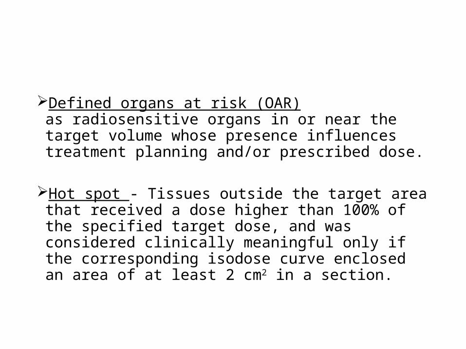
Defined organs at risk (OAR) as radiosensitive organs in or near the target volume whose presence influences treatment planning and/or prescribed dose.
Hot spot - Tissues outside the target area that received a dose higher than 100% of the specified target dose, and was considered clinically meaningful only if the corresponding isodose curve enclosed an area of at least 2 cm2 in a section.

DRAWBACK
• However, the report did not address the issues of coordinate systems (e.g., patient vs. treatment machine), and no attempt was made to define and explicitly separate the margins for the different types of uncertainties.
• ICRU Report 29 recommendations were well suited for the technology of the 1970s and 1980s, using a conventional simulator to generate a planning radiograph for designing beam portals based on bony and soft tissue landmarks.

Volumes defined prior to treatment planning :
- Gross Tumor Volume (GTV) - Clinical Target Volume (CTV)
Volumes defined during the treatment planning :
- Planning target Volume (PTV) - Organs at risk - Treated Volume - Irradiated Volume
ICRU 50

ICRU 50
Irradiated Volume
Treated Volume
Planning Target Volume (PTV)
Clinical Target Volume (CTV)
Gross Tumor Volume (GTV)
GROSS TUMOR VOLUME ( GTV )
Gross palpable or visible/demonstrable extent and location of the malignant growth.
It consists of : Primary tumor Metastatic lymphadenopathy Other metastasis
Corresponds to those parts of the malignant growth where the tumor density is largest. If the tumor has been removed prior to radiotherapy then no GTV can be defined.

GTV can be determined by using either clinical examination (inspection, palpation) and by various imaging techniques (X-rays, CT, MRI etc.)
Method used for determination of GTV should meet the requirements for staging the tumor according to the clinical TNM (AJCC).
Reasons for identification of GTV :
An adequate dose should be delivered to the whole of GTV to obtain local tumor control in radical treatments.
To allow for recording of tumor response in relation to dose and its variation, and to other relevant factors.
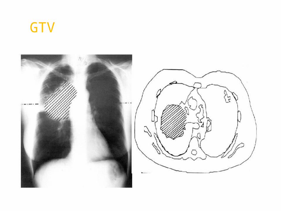
GTV

CLINICAL TARGET VOLUME ( CTV )
It is a tissue volume that contains a GTV and/or subclinical microscopic disease, which has to be eliminated.
This volume has to be treated adequately in order to achieve the aim of therapy : cure or palliation.
The delineation of this volume requires consideration of factors like local invasive capacity of the tumor and its potential to spread to different regions ( eg: regional lymph nodes).

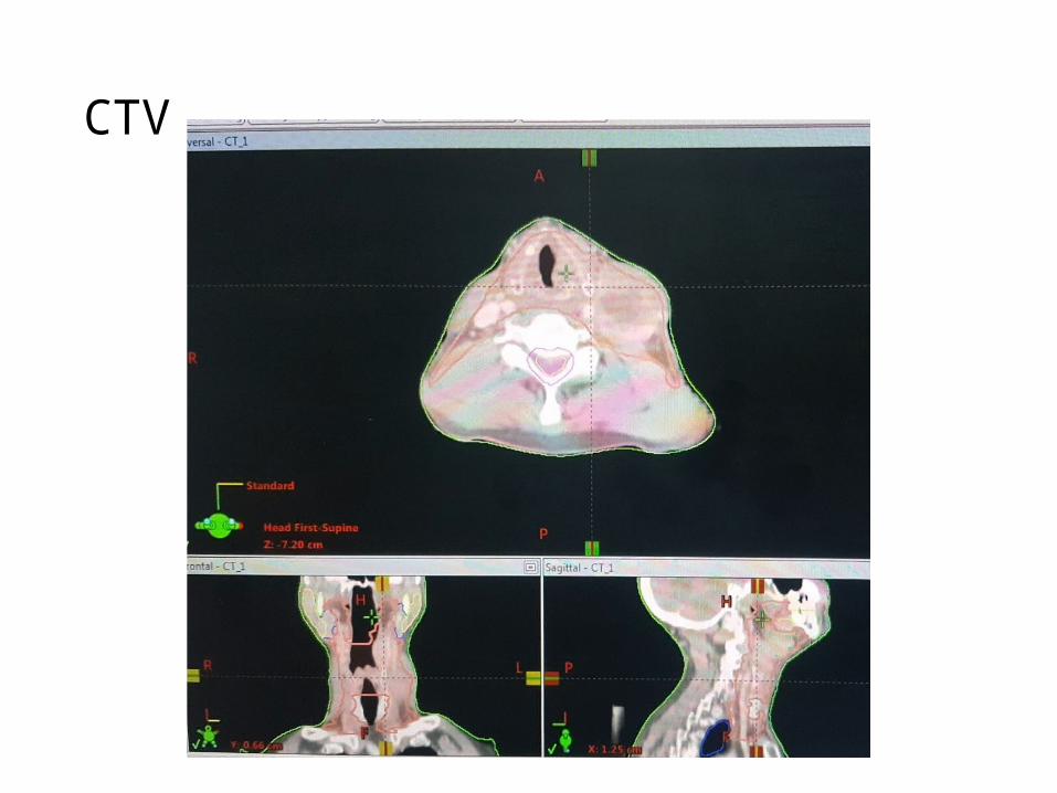
CTV

The delineation of GTV and CTV are based on purely anatomic-topographic and biological considerations without regard to technical factors of treatment.

PLANNING TARGET VOLUME ( PTV )
It is a geometrical concept, and is defined to select appropriate beam sizes and arrangements, taking into consideration the net effect of all possible geometrical variations, in order to ensure that the prescribed dose is actually absorbed in the CTV.
It is used for dose planning and for specification of dose.
It has to be clearly indicated on sections used for dose planning and the dose distribution to the PTV has to be considered to be representative of the dose to the CTV.
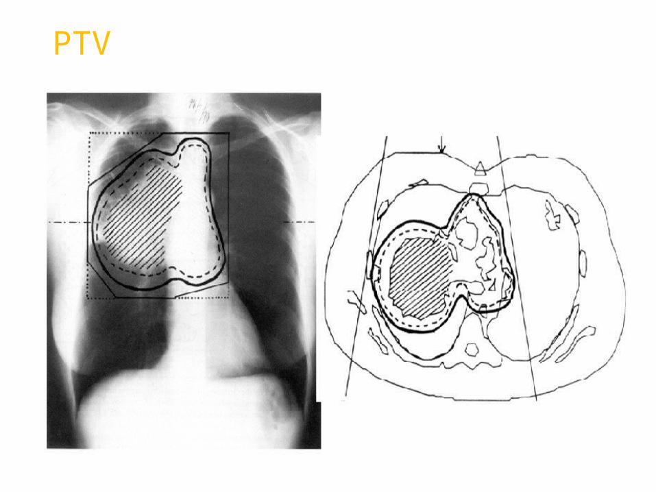
PTV

TREATED VOLUME
Definition:-
It is the volume enclosed by an isodose surface that is selected and specified by the radiation oncologist as being appropriate to achieve the purpose of treatment (palliation or cure).
Usually taken as the volume enclosed by the 95% isodose curve.
Ideally dose should be delivered only to the PTV but due to limitations in the radiation treatment technique.
ICRU-50
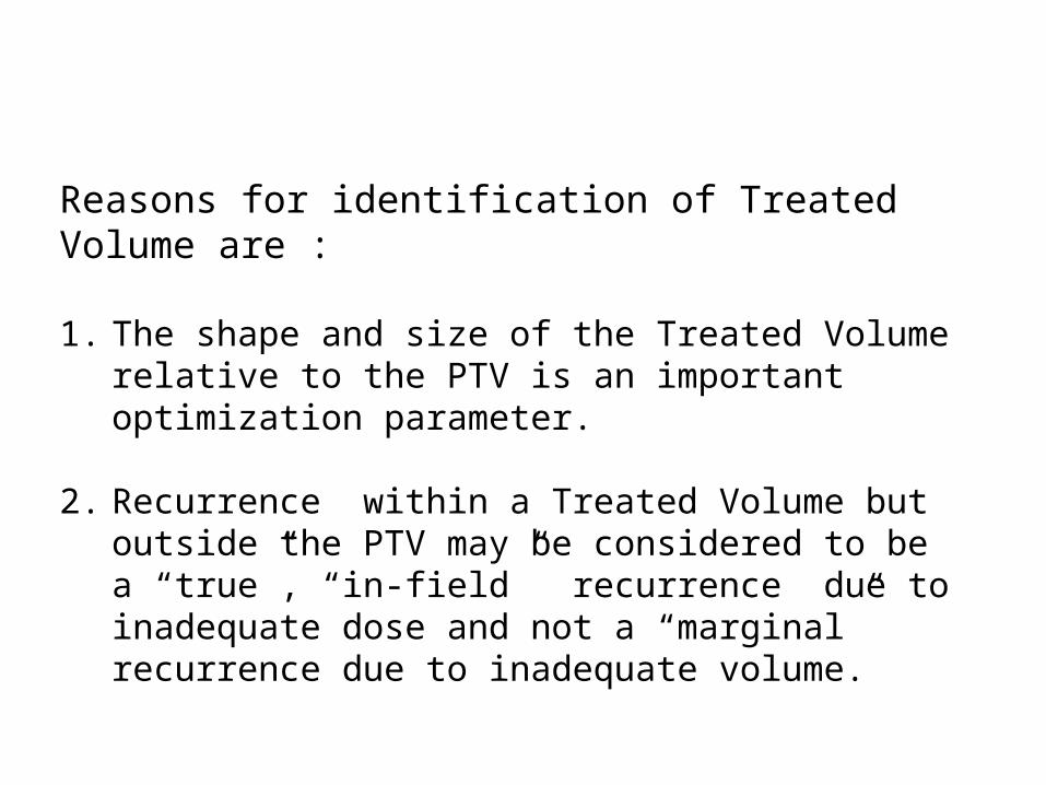
Reasons for identification of Treated Volume are :
1. The shape and size of the Treated Volume relative to the PTV is an important optimization parameter.
2. Recurrence within a Treated Volume but outside the PTV may be considered to be a “true”, “in-field” recurrence due to inadequate dose and not a “marginal” recurrence due to inadequate volume.

2 field (AP-PA) 3 field
4 field (box)

IRRADIATED VOLUME(IRV)Definition:-
It is the volume that receives a dose considered significant in relation to normal tissue tolerance
Usually taken as the volume enclosed by the 50% isodose curve.
It depends on the treatment technique used.
ICRU-50

ORGANS AT RISK ( OAR )• These are normal tissues whose radiation sensitivity may significantly influence the treatment planning and/or prescribed dose.
• They may be divided into 3 classes :
1. Class I : Radiation lesions are fatal or result in severe morbidity.
2. Class II : Radiation lesions result in mild to moderate morbidity.
3. Class III : Radiation lesions are mild, transient, and reversible, or result in no significant morbidity.


GTV
CTV

PTV


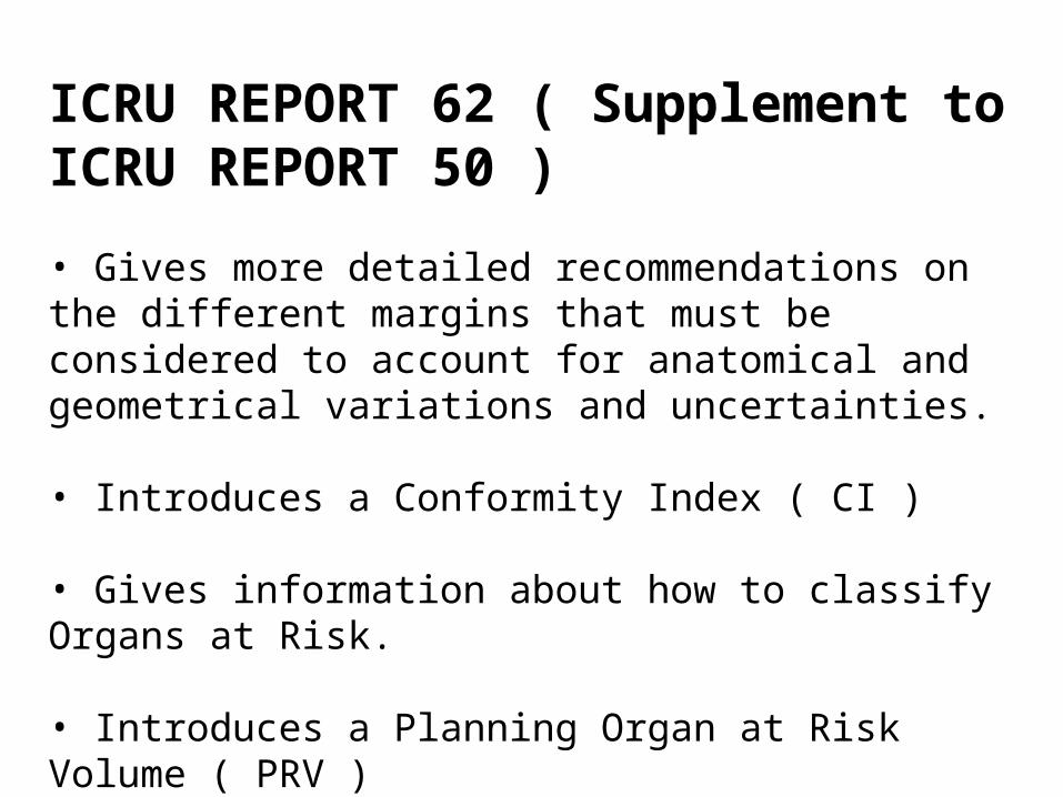
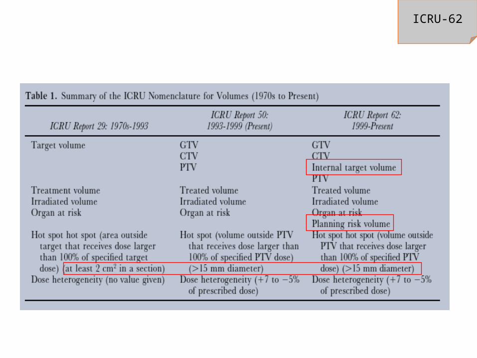
ICRU REPORT 62 ( Supplement to ICRU REPORT 50 ) • Gives more detailed recommendations on the different margins that must be considered to account for anatomical and geometrical variations and uncertainties.
• Introduces a Conformity Index ( CI )
• Gives information about how to classify Organs at Risk.
• Introduces a Planning Organ at Risk Volume ( PRV )
• Gives recommendations on graphics.

VOLUMES ICRU-62

Volumes defined prior to treatment planning :
- Gross Tumor Volume (GTV)- Clinical Target Volume (CTV)
Volumes defined during the treatment planning :
- Planning target Volume (PTV)- Treated Volume- Irradiated Volume- Planning Organ at Risk Volume (PRV)
ICRU 62
Same as ICRU 50
ICRU-62

INTERNAL MARGIN A margin that must be added to the CTV to compensate
for expected physiologic movements and the variations in size, shape and position of the CTV during therapy in relation to the Internal Reference Point and its corresponding Coordinate System. Motion is associated with adjacent respiratory and digestive organs.
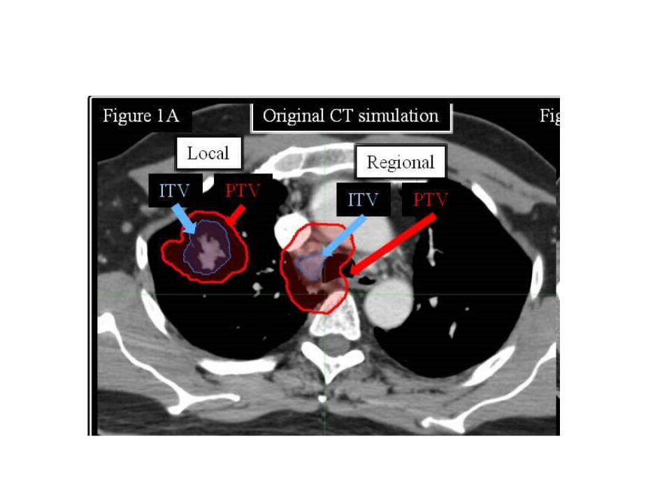
INTERNAL TARGET VOLUME (ITV) It is the margin given around the CTV to compensate for all
variations in the site, size and shapes of organs and tissues contained in or adjacent to CTV.
These may result from respiration, different fillings of the bladder and rectum, swallowing, heart beat, movements of bowel etc.

A “Normal” Treatment plan

The effect of motion

ITV Margin


SET-UP MARGIN ( SM )
•SET-UP MARGIN ( SM ) is the margin that must be added to account specifically for uncertainties (inacuracies and lack of reproducibility) in patient positioning and aligment of the therapeutic beams during treatment planning and through all treatment sessions.
•There can be many uncertainties ( inaccuracies and lack of reproducibility ) in patient positioning and alignment of the therapeutic beams during treatment planning and through all treatment sessions.

•These uncertainties depend on factors like :• Variations in patient positioning• Mechanical uncertainties of the equipment (sagging of gantry,
collimators, and couch)• Dosimetric uncertainties• Transfer set-up errors from CT & simulator to the treatment
unit• Human factors

ITV = CTV + IM
PTV = CTV + combined IM & SM

SYSTEMATIC AND RANDOM ERRORS
Systematic errors – treatment preparation errors (influence all fractions) like full rectum
Random errors – treatment execution errors (influence only the single fraction) like positioning
ICRU-62
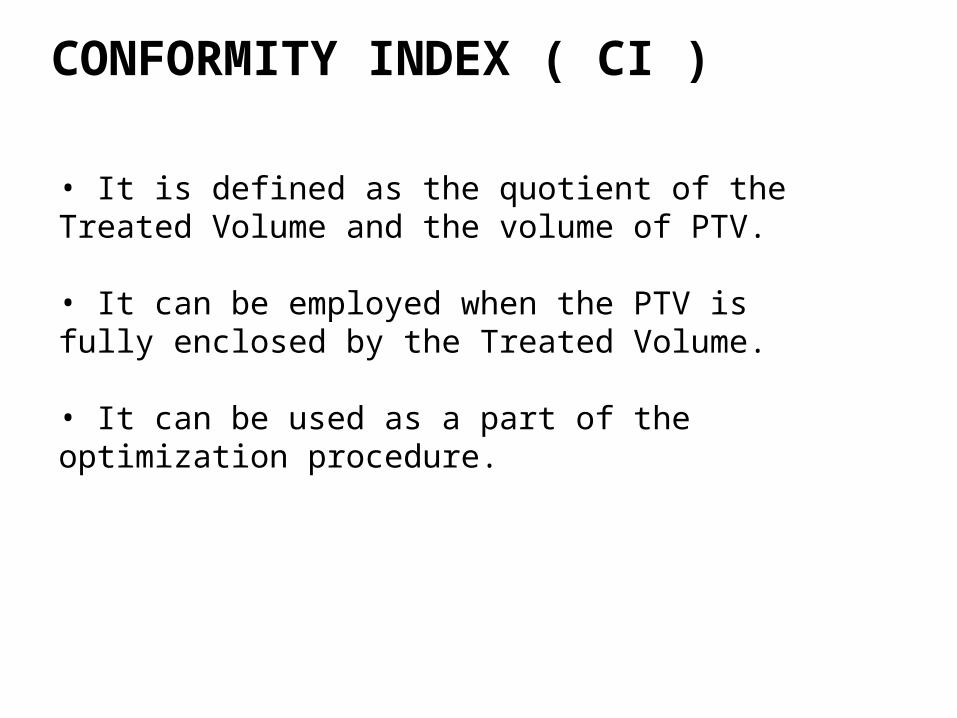
CONFORMITY INDEX ( CI )
• It is defined as the quotient of the Treated Volume and the volume of PTV.
• It can be employed when the PTV is fully enclosed by the Treated Volume.
• It can be used as a part of the optimization procedure.

CLASSIFICATION OF ORGANS AT RISK
• Classified as :Serial – whole organ is a continuous unit and damage at one point will cause
complete damage of the organ (spinal cord, digestive system). So even point dose is significant.
Parallel – organ consists of several functional units and if one part is damaged, the rest of the organ makes up for the loss (lung, bladder). Dose delivered to a given volume or average/mean dose is considered
Serial-parallel – kidney (glomerulus- parallel, tubules- serial), heart (myocardium- parallel, coronary arteries- serial).
ICRU-62

PLANNING ORGAN AT RISK VOLUME ( PRV )
• This is a volume which gives into consideration the movement of the Organs at Risk during the treatment.
• An integrated margin must be added to the Organ at Risk to compensate for the variations and uncertainties, using the same principle as PTV and is known as the Planning Organ at Risk volume ( PRV ).
• A PTV and PRV may occasionally overlap.

GRAPHICS
• These are used to delineate the different volumes and the other landmarks.
• These are in different colors for an easy and uniform interpretation.
• The convention recommended and used in ICRU 62 are:GTV - Dark RedCTV – Light RedITV – Dark BluePTV – Light BlueOR – Dark GreenPRV – Light GreenLandmarks - Black

Recommendations for reporting
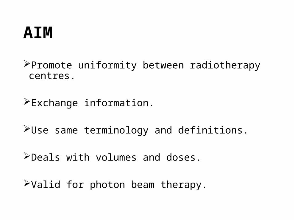
AIMPromote uniformity between radiotherapy centres.
Exchange information.
Use same terminology and definitions.
Deals with volumes and doses.
Valid for photon beam therapy.

DOSE REPORTING
Acceptable dose heterogeneity : +7% to - 5% of the prescribed dose.
Doses reported are :Minimum dose to PTVMaximum dose to PTVMean dose to PTVModal doseMedian doseDose at ICRU reference point
ICRU-50

MAXIMUM DOSE ( Dmax )• It is the maximum dose to the PTV and the Organ at Risk.
• The maximum dose to normal tissue is important for limiting and for evaluating the side-effects of treatment.
• Dose is reported as maximum only when a volume of tissue of diameter more than 15mm is involved (smaller volumes are considered for smaller organs like eye, optic nerve, larynx).
• When the maximum dose outside PTV exceeds the prescribed dose, then a “Hot Spot” can be identified.

MINIMUM DOSE ( Dmin )
It is the smallest dose in a defined volume.
In contrast to maximum adsorbed dose, no volume limit is recommended when reporting minimum dose.
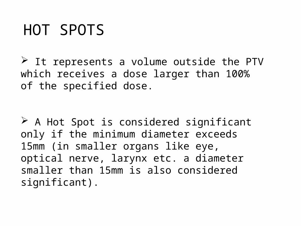
HOT SPOTS It represents a volume outside the PTV which receives a dose larger than 100% of the specified dose.
A Hot Spot is considered significant only if the minimum diameter exceeds 15mm (in smaller organs like eye, optical nerve, larynx etc. a diameter smaller than 15mm is also considered significant).

ICRU REFERENCE POINT• It has to be selected according to the following general criteria :
- the dose at the point should be clinically relevant.
- the point should be easy to define in a clear and unambiguous way.
- the point should be selected so that the dose should be accurately determined.
- the point should be in a region where there is no steep dose gradient.

The recommendations will be fulfilled if the ICRU reference point is located :
• Always at the centre ( or in the central part ) of PTV, and
• When possible, at the intersection of the beam axes.

ICRU REFERENCE DOSE
It is the dose at the ICRU Reference Point and should always be reported.
• According to the recommendations of ICRU, as a basic requirement, the following doses should always be reported :
the dose at ICRU reference point
the maximum dose to the PTV
the minimum dose to the PTV

CONCLUSIONS
• Proper identification and delineation of GTV is the most important factor in treatment.
• Other volumes like CTV, PTV, ITV should also be properly delineated.
• The errors like set-up error and human errors should be kept to a minimum.
• Dose prescription, fractionation and calculation should be done in the same way by all the different centers throughout the world for the proper exchange of information and reporting.

THANK YOU