Embed Size (px)
Citation preview
Cranial nerve III, IV and VITo MBBS 2nd year
05-04-2016
Dr. Laxman Khanal (Asst. Professor)
Department of Anatomy, BPKIHS
How to study the cranial nerves ??
• Know the position of nuclei and their functional components.
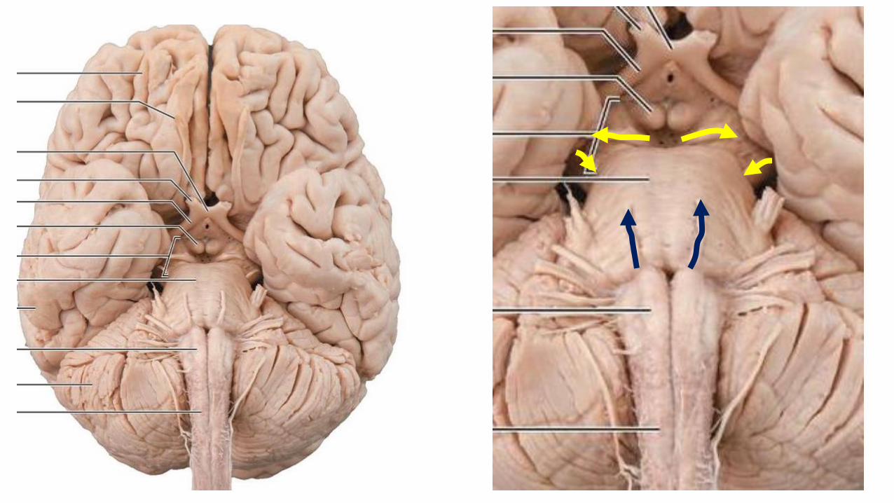
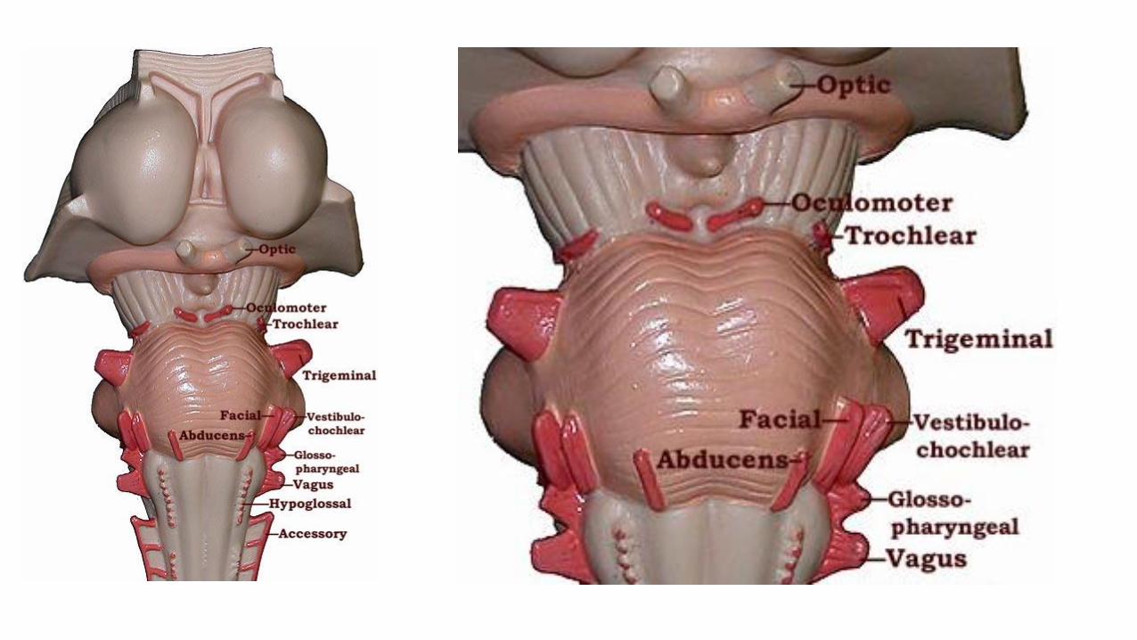
• Know the site of attachment of cranial nerve in brain stem.
• Course of cranial nerve.
• Functions of cranial nerves.
• Clinical correlation of nerve damage with the signs and symptoms.
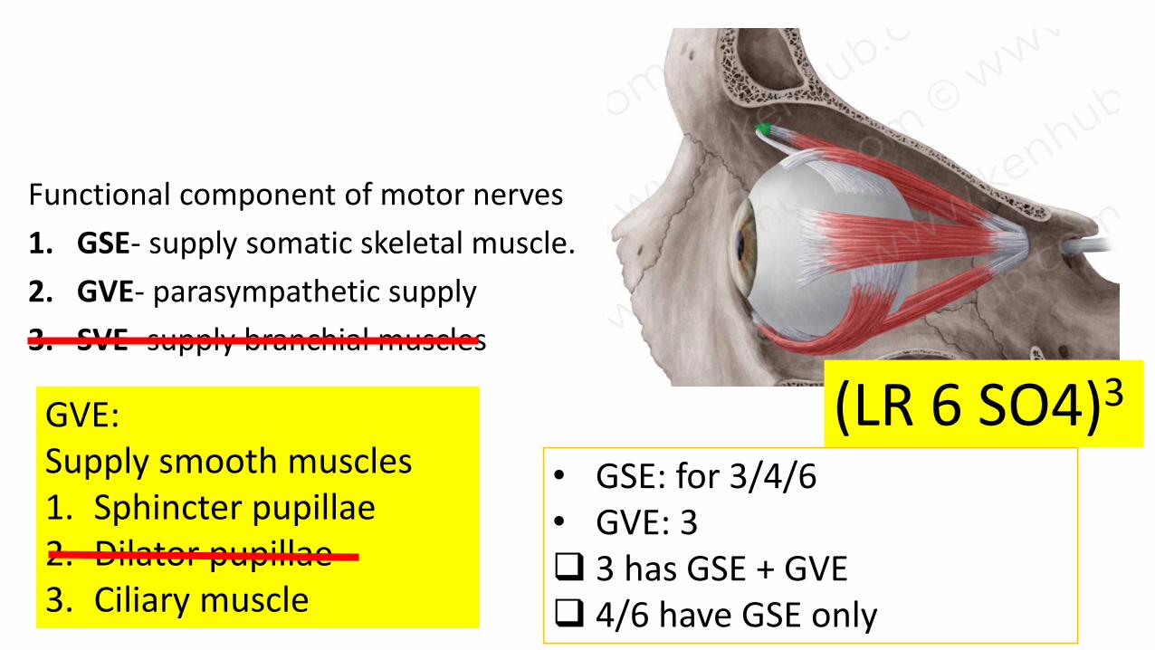
Functional component of motor nerves
1. GSE- supply somatic skeletal muscle.
2. GVE- parasympathetic supply
3. SVE- supply branchial muscles
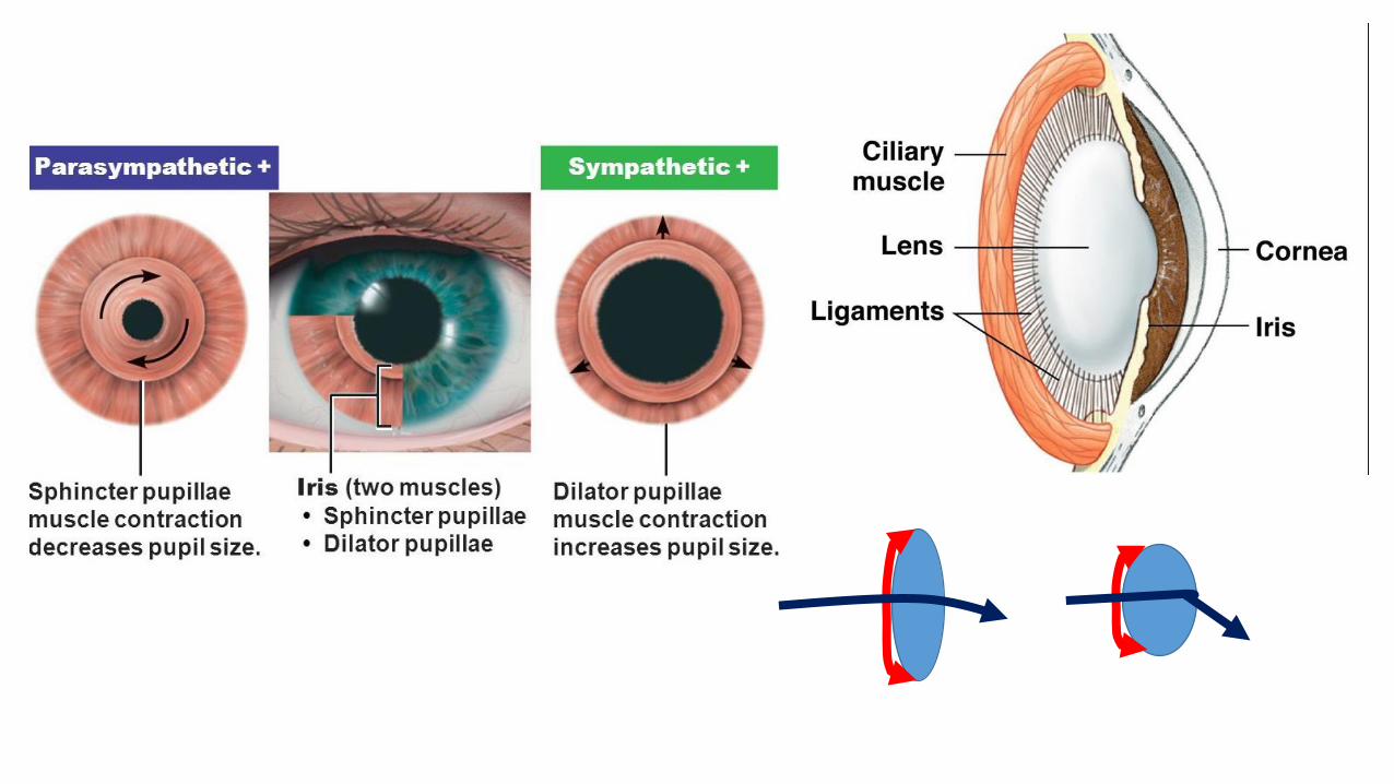
(LR 6 SO4)3GVE:Supply smooth muscles1. Sphincter pupillae2. Dilator pupillae3. Ciliary muscle
• GSE: for 3/4/6• GVE: 3 3 has GSE + GVE 4/6 have GSE only
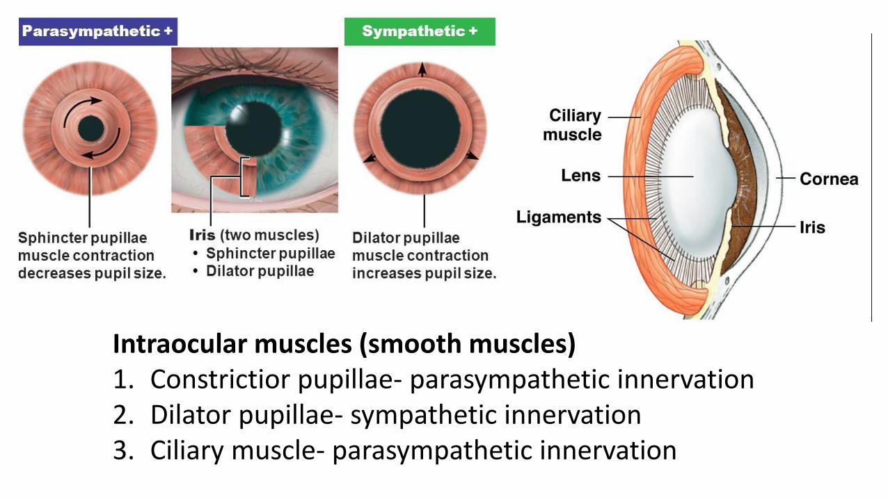
Intraocular muscles (smooth muscles)1. Constrictior pupillae- parasympathetic innervation 2. Dilator pupillae- sympathetic innervation 3. Ciliary muscle- parasympathetic innervation
Cranial nerve III
• Functional components: GSE and GVE
• Origin (nuclei): midbrain @ the level of superior colliculus
• Nuclei: two in number
1. Main motor nucleus- GSE
2. Parasympathetic nucleus (Edinger Westphal Nucleus)-GVE
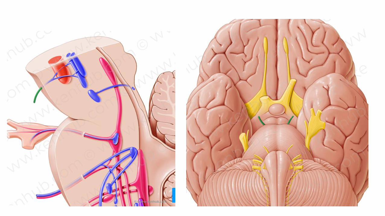
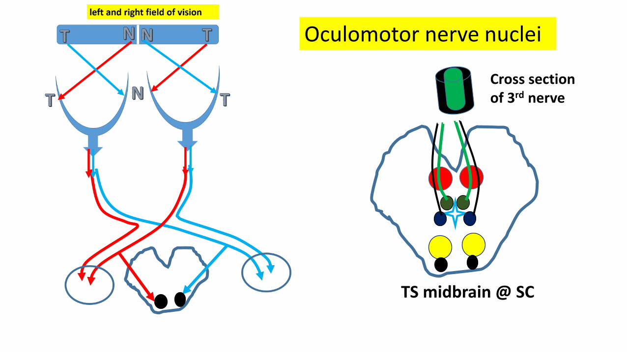
TS midbrain @ SC
Oculomotor nerve nuclei
Cross section of 3rd nerve
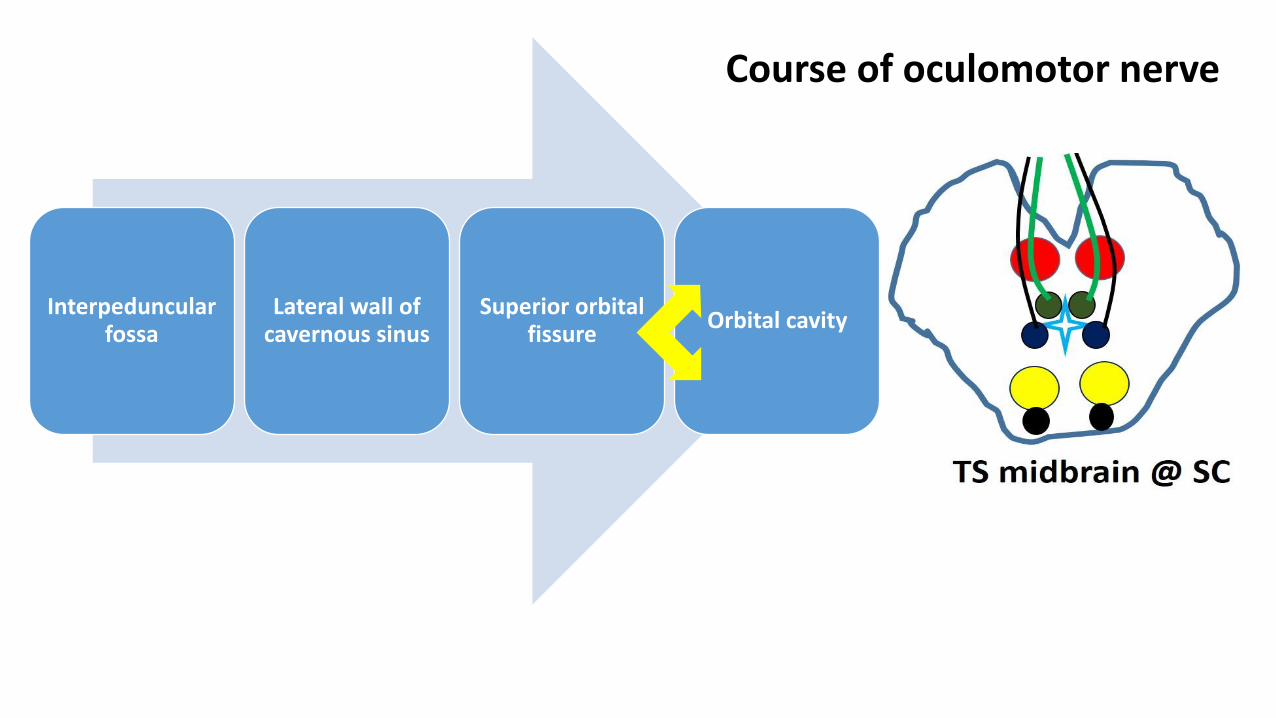
Interpeduncular fossa
Lateral wall of cavernous sinus
Superior orbital fissure
Orbital cavity
Course of oculomotor nerve
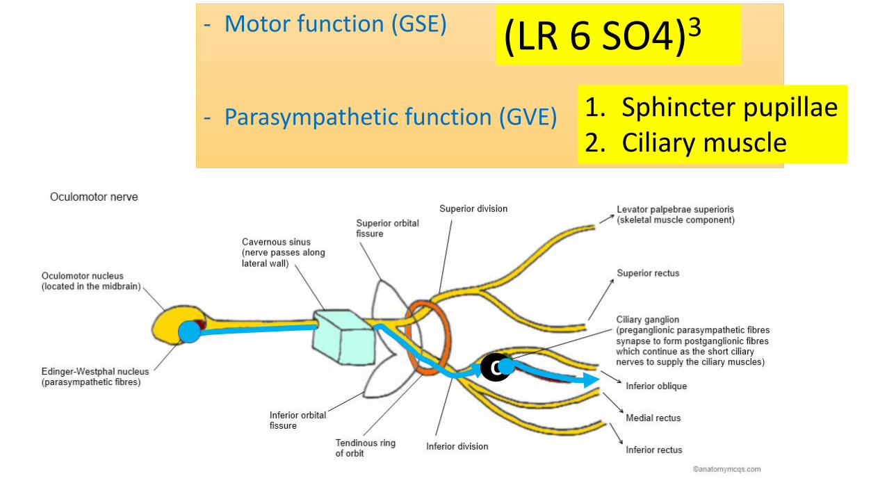
- Motor function (GSE)
- Parasympathetic function (GVE)
C
(LR 6 SO4)3
1. Sphincter pupillae2. Ciliary muscle
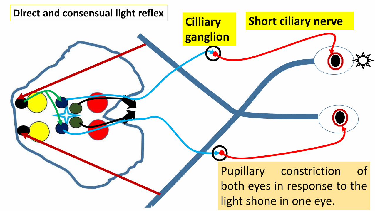
Direct and consensual light reflex
c
Cilliaryganglion
Short ciliary nerve
Pupillary constriction ofboth eyes in response to thelight shone in one eye.
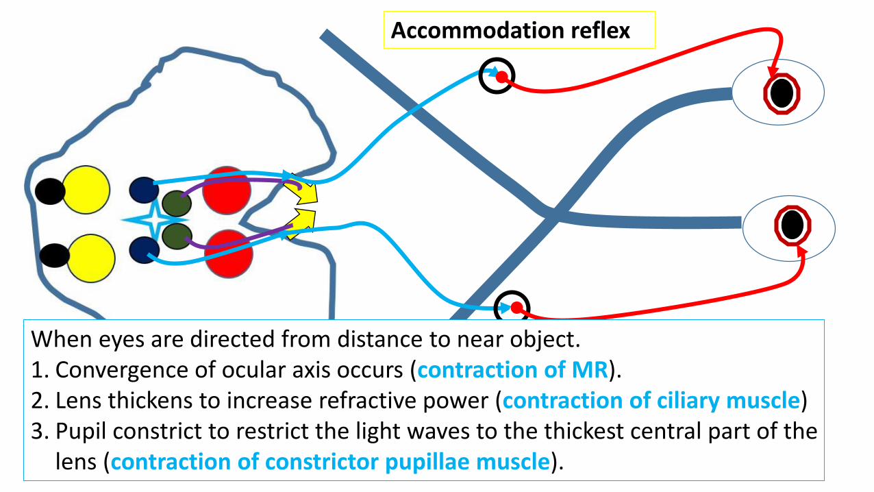
Accommodation reflex
c
When eyes are directed from distance to near object.1. Convergence of ocular axis occurs (contraction of MR).2. Lens thickens to increase refractive power (contraction of ciliary muscle)3. Pupil constrict to restrict the light waves to the thickest central part of the
lens (contraction of constrictor pupillae muscle).
c
c
c
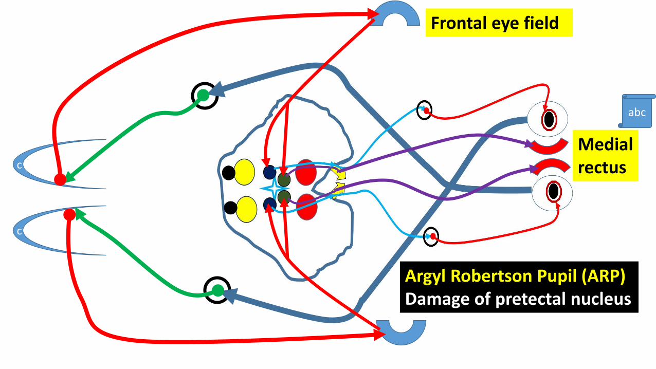
Frontal eye field
Medial rectus
Argyl Robertson Pupil (ARP)Damage of pretectal nucleus
abc
M
P
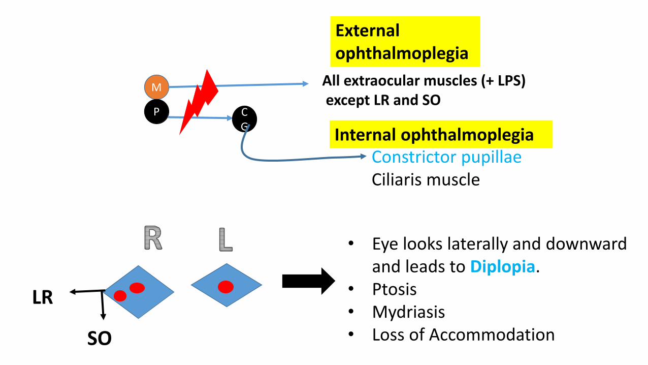
All extraocular muscles (+ LPS)except LR and SO
CG
Constrictor pupillaeCiliaris muscle
LR
SO
• Eye looks laterally and downward and leads to Diplopia.
• Ptosis• Mydriasis• Loss of Accommodation
External ophthalmoplegia
Internal ophthalmoplegia
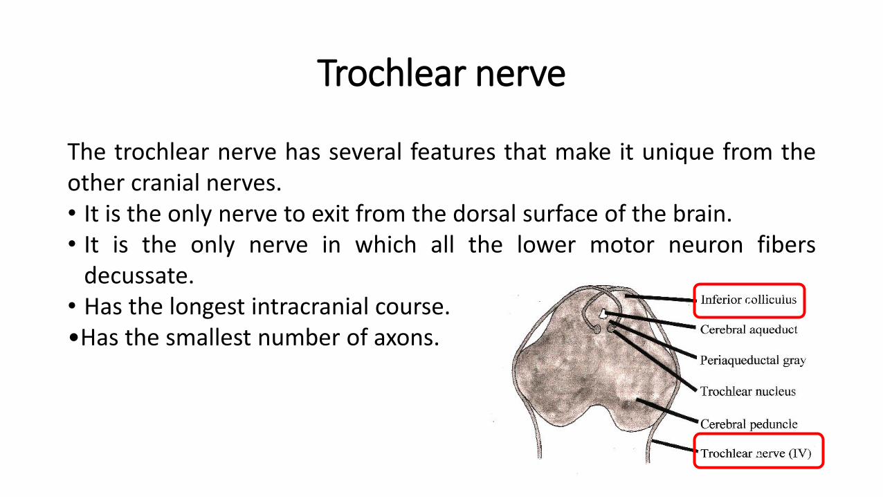
Trochlear nerve
The trochlear nerve has several features that make it unique from theother cranial nerves.• It is the only nerve to exit from the dorsal surface of the brain.• It is the only nerve in which all the lower motor neuron fibers
decussate.• Has the longest intracranial course.•Has the smallest number of axons.
c
c
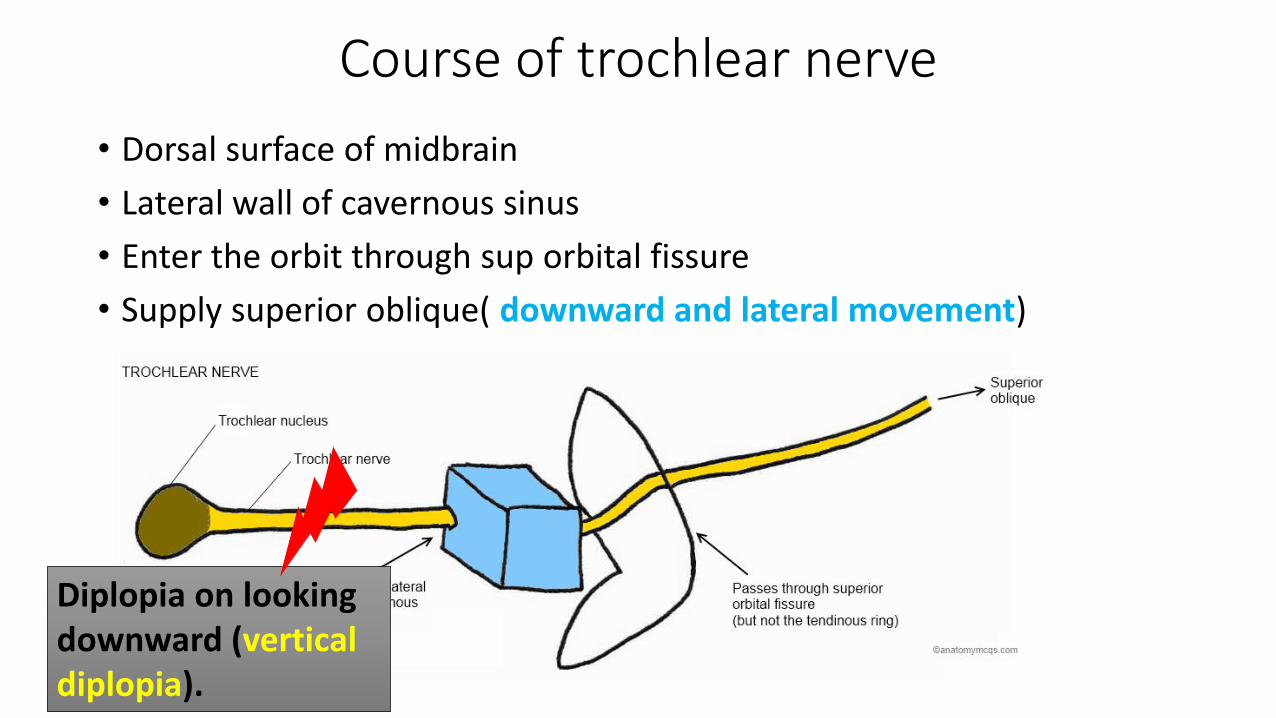
Course of trochlear nerve
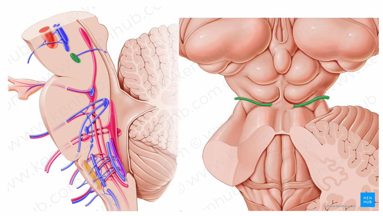
• Dorsal surface of midbrain
• Lateral wall of cavernous sinus
• Enter the orbit through sup orbital fissure
• Supply superior oblique( downward and lateral movement)
Diplopia on looking downward (vertical diplopia).
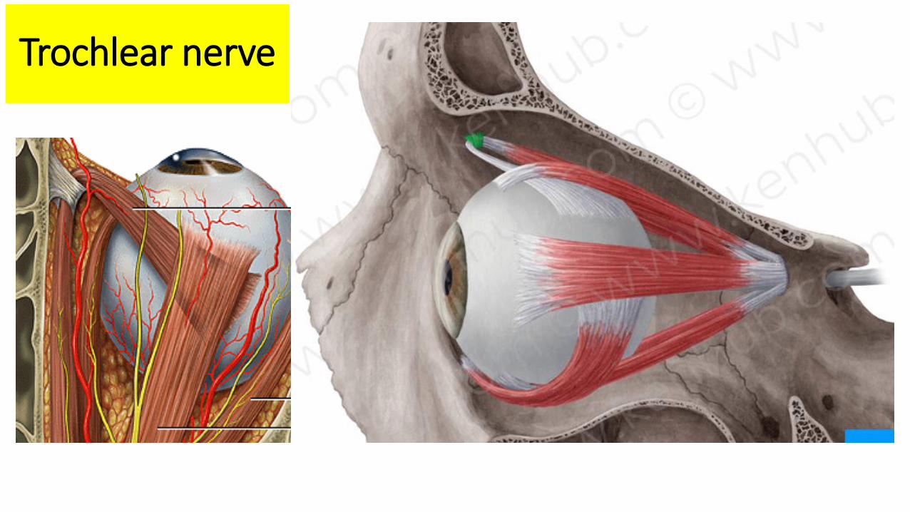
Trochlear nerve
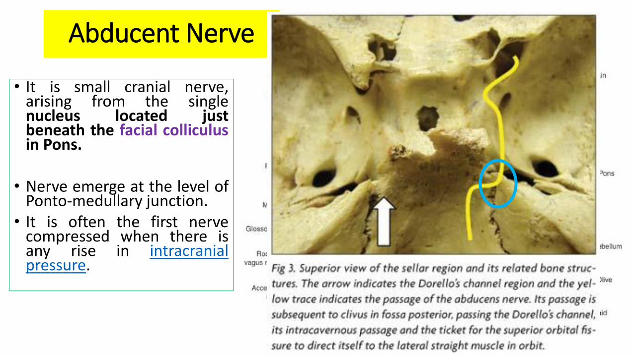
Abducent Nerve
• It is small cranial nerve,arising from the singlenucleus located justbeneath the facial colliculusin Pons.
• Nerve emerge at the level ofPonto-medullary junction.
• It is often the first nervecompressed when there isany rise in intracranialpressure.
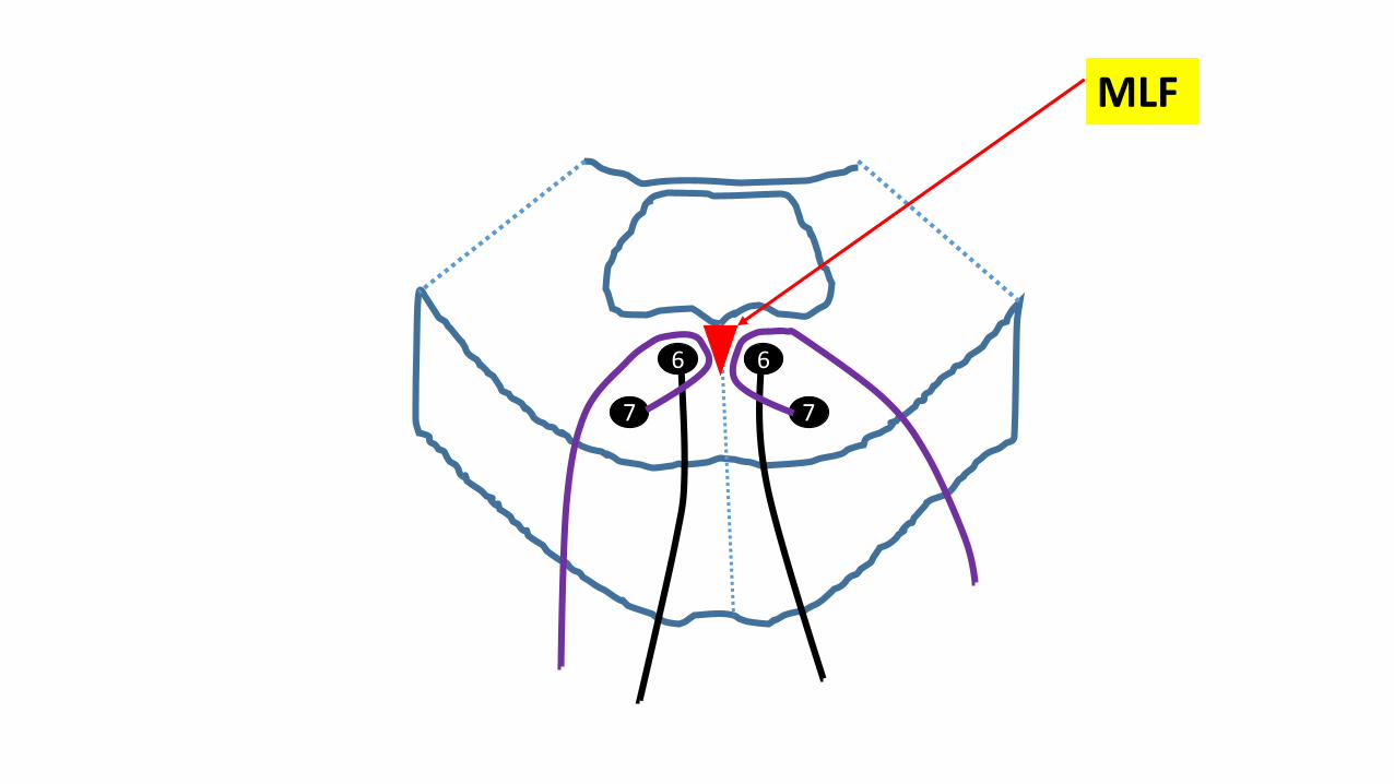
66
7 7
MLF

Course of Abducent nerve
Internal strabismus
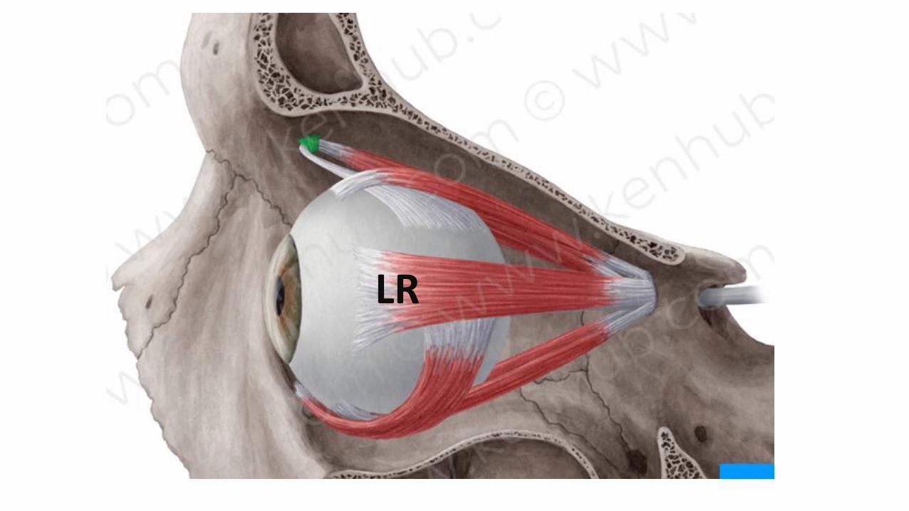
LR
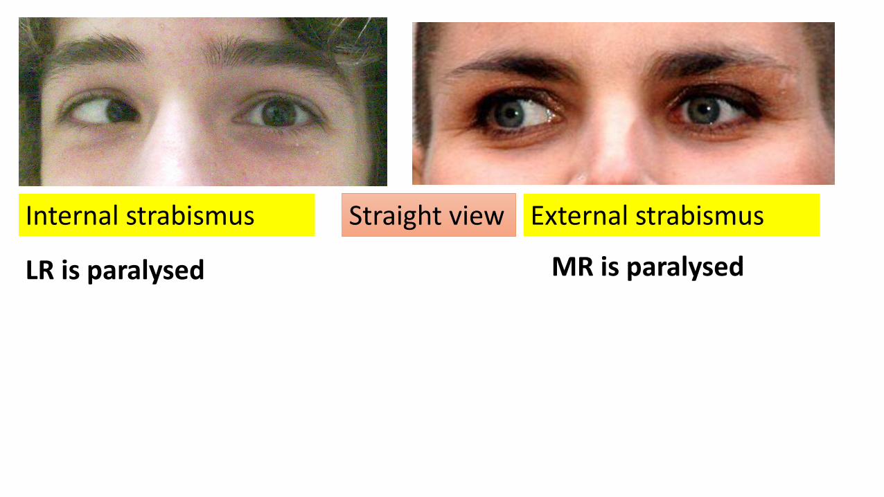
Internal strabismus External strabismusStraight view
LR is paralysed MR is paralysed
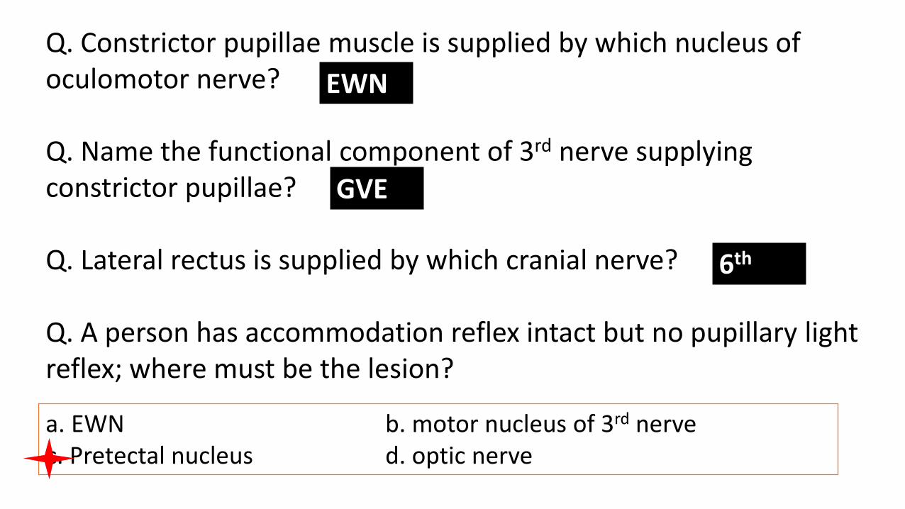
Q. Constrictor pupillae muscle is supplied by which nucleus of oculomotor nerve?
Q. Name the functional component of 3rd nerve supplying constrictor pupillae?
Q. Lateral rectus is supplied by which cranial nerve?
Q. A person has accommodation reflex intact but no pupillary light reflex; where must be the lesion?
EWN
GVE
6th
a. EWN b. motor nucleus of 3rd nervec. Pretectal nucleus d. optic nerve
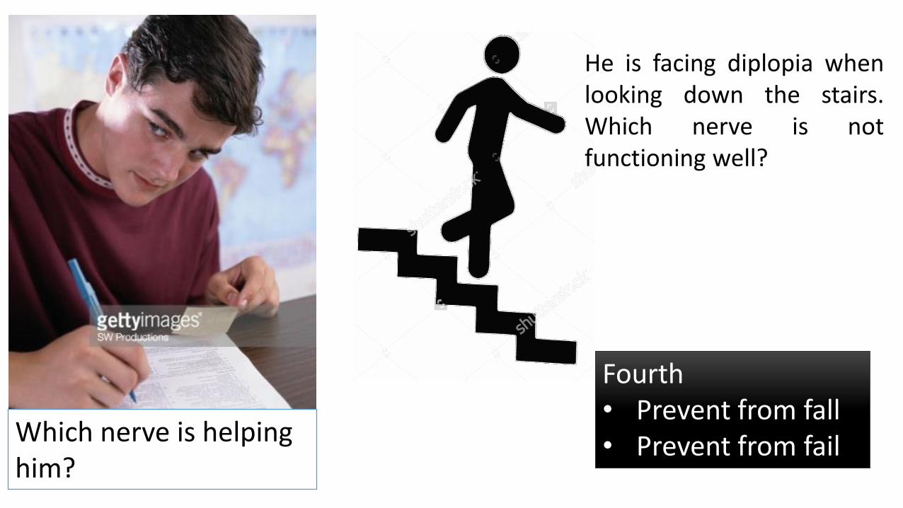
Which nerve is helping him?
He is facing diplopia whenlooking down the stairs.Which nerve is notfunctioning well?
Fourth • Prevent from fall • Prevent from fail